1. Introduction
Ergonomics plays an important role in preventing musculoskeletal disorders (MSDs) among healthcare professionals, particularly surgeons. Surgeons often perform repetitive movements and maintain static postures for extended periods, which contribute to MSDs, such as neck, back, and upper extremity pain [
1]. The cumulative overuse of muscles, tendons, and ligaments can lead to chronic discomfort and potentially debilitating conditions [
2]. Ophthalmologists, in particular, have a significantly higher risk for musculoskeletal pain compared to other medical specialties, as they frequently perform delicate procedures in and around the eye [
3]. The large percentage of surgeons who report chronic neck and back pain underscores the importance of ergonomic interventions to alleviate physical strain. Contemporary epidemiological studies indicate that 60–80% of ophthalmic surgeons experience career-limiting neck, back, or shoulder pain [
4].
One tool that has been both praised and criticized in the context of surgical ergonomics is the surgical loupe [
5]. Loupes are commonly used to enhance vision during intricate surgeries, but they may pose a negative impact on posture and musculoskeletal health. Some studies have shown that loupes can exacerbate musculoskeletal stress by forcing surgeons into awkward head and neck positions [
5]. While loupes can play a role in enhancing ergonomic practices, their frequent use may contribute to increased strain and biomechanical load on the cervical spine, necessitating the need for the development of more ergonomic and comfort-enhancing surgical loupes. Multiple ergonomically friendly loupes have since been developed, with studies utilizing electromyogram technology to demonstrate that their novel loupes can reduce neck muscle activity, discomfort, and head inclination during simulated surgical tasks compared to traditional “through the lens” (TTL) loupes, without affecting surgical performance [
6,
7,
8].
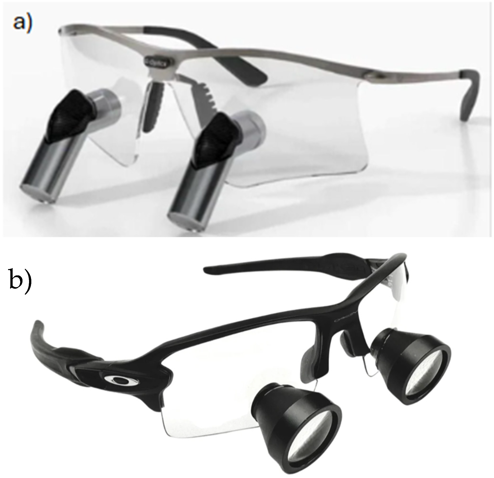
Prismatic deflection loupes (PDLs) represent a promising innovation in surgical loupes, as they utilize internal reflection optics to redirect the visual axis without compromising magnification or working distance. The Q-Optics Ergo-Pro
® (Q-Optics, Duncanville, TX, USA) surgical loupes provide an expanded field of view while using prismatic deflection at the angle of the loupes to ensure neutral neck and back posture in their users (
Figure 1). To further investigate the features of these loupes, this study compares the PDLs to TTL loupes and no surgical loupes on muscle strain, fatigue, and angle of inclination. Using electromyography (EMG), self-report surveys on ergonomics and comfort, and biomechanical modeling, this study assesses the effects of PDL ergonomics on medical student subjects during the performance of microsurgical tasks for ophthalmology procedures.
2. Methods
Institutional Review Board approval under the University of California, Irvine (UCI IRB-5177), was obtained to perform this prospective, randomized crossover study. All participants provided written informed consent before enrollment. A total of 10 fourth-year medical students were recruited for the study. Inclusion criteria required participants to have prior experience with surgical loupes, no history of cervical spine disorders, and the ability to perform simulated ophthalmic tasks for the duration of the study. All participants met these criteria and completed the study in full. Subjects performed the simulated ophthalmic surgery suturing activity for 10 min in three experimental conditions: (1) using no loupes, (2) using TTL loupes; Galilean design (3.5× magnification; 18″ working distance; 0° ocular deflection, weighing 50 g), and (3) using PDLs; Q-Optics Ergo-Pro
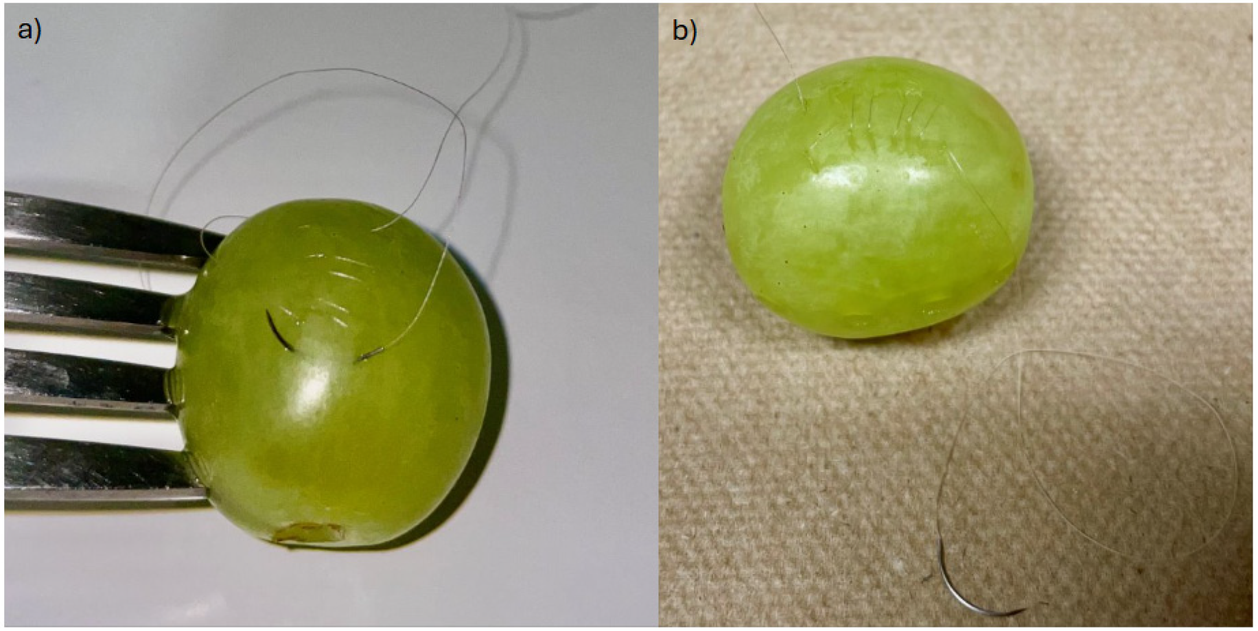
® (3.5× magnification; 18″ working distance; 45° ocular deflection, weighing 54 g). Each condition was performed in a randomized order with a 5 min break between each experimental condition to minimize fatigue carryover effects. Randomization was used to control for potential order effects, such as learning or cumulative muscle fatigue, that could bias the results if one condition consistently occurred before or after another. The suturing activity involved performing a running suture on a table grape using 7-0 Polysorb (Medtronic Covidien Inc., Minneapolis, MN, USA), curved needle holders (EN-04L, Epsilon Inc., Chino, CA, USA), and Castroviejo toothed suturing forceps (product 9534E, Ambler Inc., Exton, PA, USA) (
Figure 2) [
9]. Volunteers were seated in an office chair 6 inches away from the table, with the chair height adjusted so their forearms rested comfortably on the tabletop. The grape used for the simulated suturing task was placed 3 inches from the table’s edge. Standard classroom fluorescent lights were turned on in a windowless classroom for the experiment. Lighting was consistent between each subject. No additional training was provided before the activity; participants relied solely on their existing medical school surgical training, which included basic suturing skills taught during clinical rotations. Suturing accuracy was not measured.
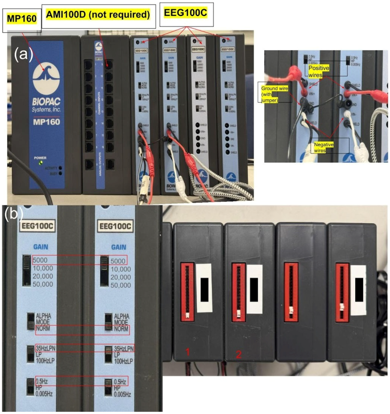
While suturing, surface EMG recordings of the bilateral upper trapezius muscles were performed to measure objective muscle strain. Signals were collected using a data acquisition system (BIOPAC Systems Inc., Goleta, CA, USA) configured with a gain of 5000, a low-pass filter at 35 Hz (LPN), a high-pass filter at 0.5 Hz, and a sample rate of 20,000. Data acquisition was performed using the MP160 system with an AMI100D amplifier interface (BIOPAC Systems Inc., Goleta, CA, USA) (
Figure 3).
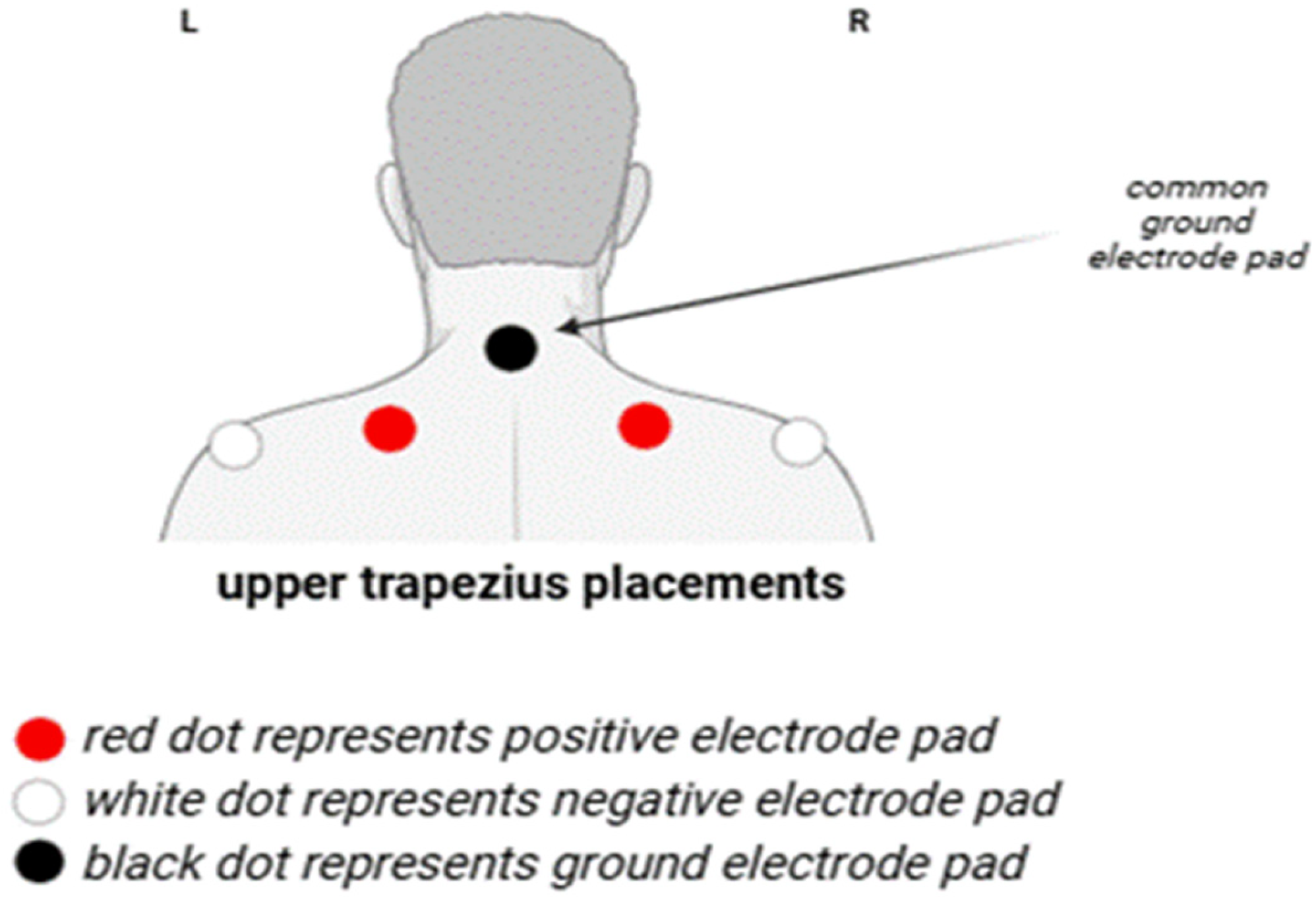
Positive leads were placed on the most prominent point of the upper trapezius, negative leads were placed at the trapezius insertion points near the posterior deltoid, and the ground was placed on the prominence of the C7 spinous process (
Figure 4).
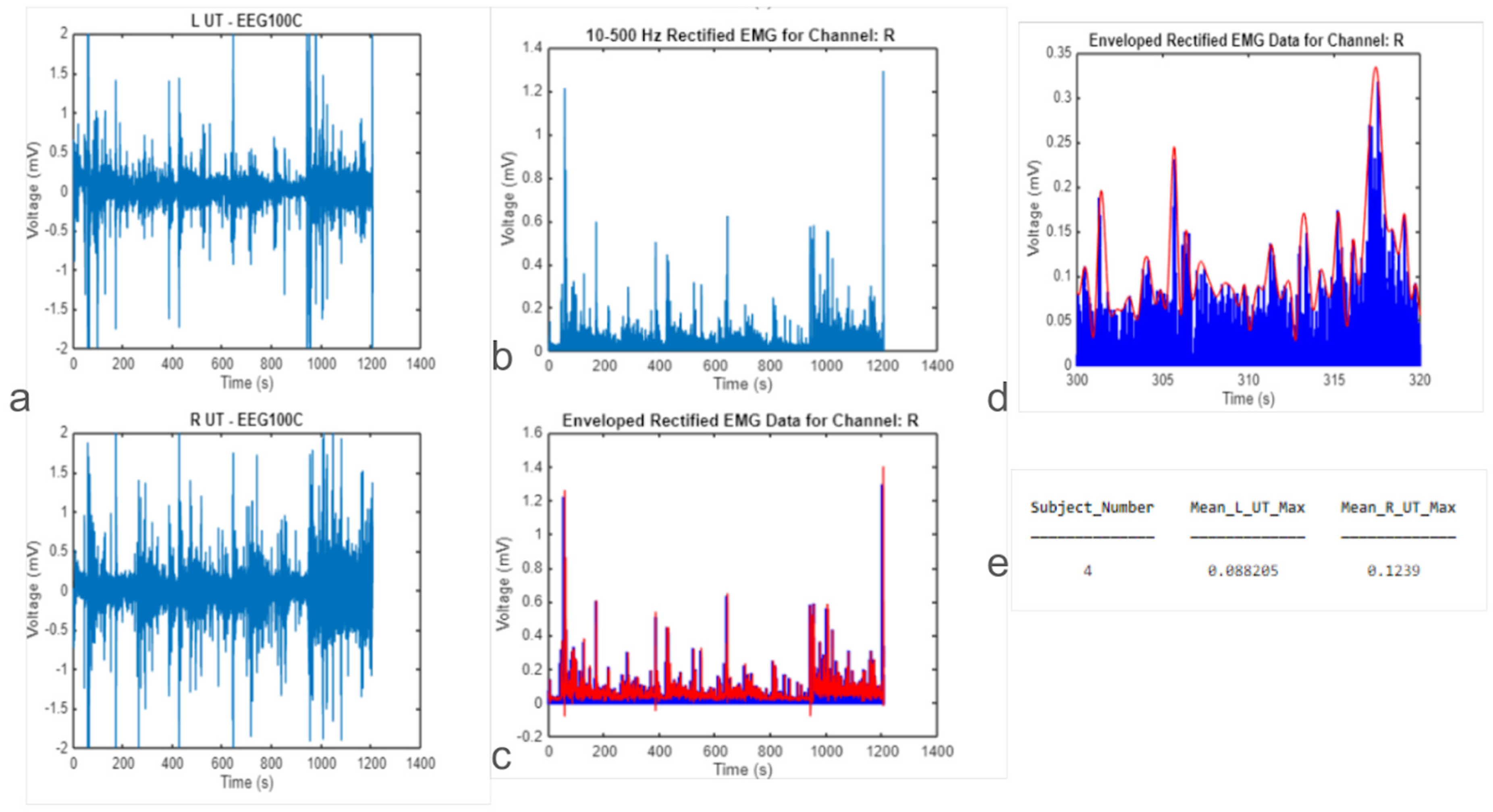
EMG data were processed in a mathematical computing software (MATLAB version R2023b, MathWorks, Natick, MA, USA) using a 4th-order Butterworth band-pass filter with a passband of 10–500 Hz to isolate muscle activity. An envelope function was applied using the Hilbert Transform to compute the EMG signal magnitude, from which a single root-mean-square (RMS) value was extracted for each condition (
Figure 5). To account for bilateral muscle activity, the RMS values from the left and right upper trapezius were averaged for each participant to produce a single representative value for statistical analysis
Statistical analysis was performed using a data spreadsheet editor (Excel, Microsoft, Redmond, WA, USA). For continuous measures such as EMG amplitude, a one-way repeated measures analysis of variance (ANOVA) was conducted to compare muscle activation across the three conditions (PDLs, TTL loupes, and no loupes). Where significant, post hoc pairwise comparisons were performed using paired t-tests, with Bonferroni correction applied to adjust for multiple comparisons.
For ordinal survey data (e.g., Likert scale ratings of comfort, strain, and fatigue), the Friedman test was used to detect overall differences across the three conditions [
10]. Where significant, Wilcoxon Signed-Rank tests were used for post hoc pairwise comparisons between conditions, with Bonferroni correction to maintain type I error control [
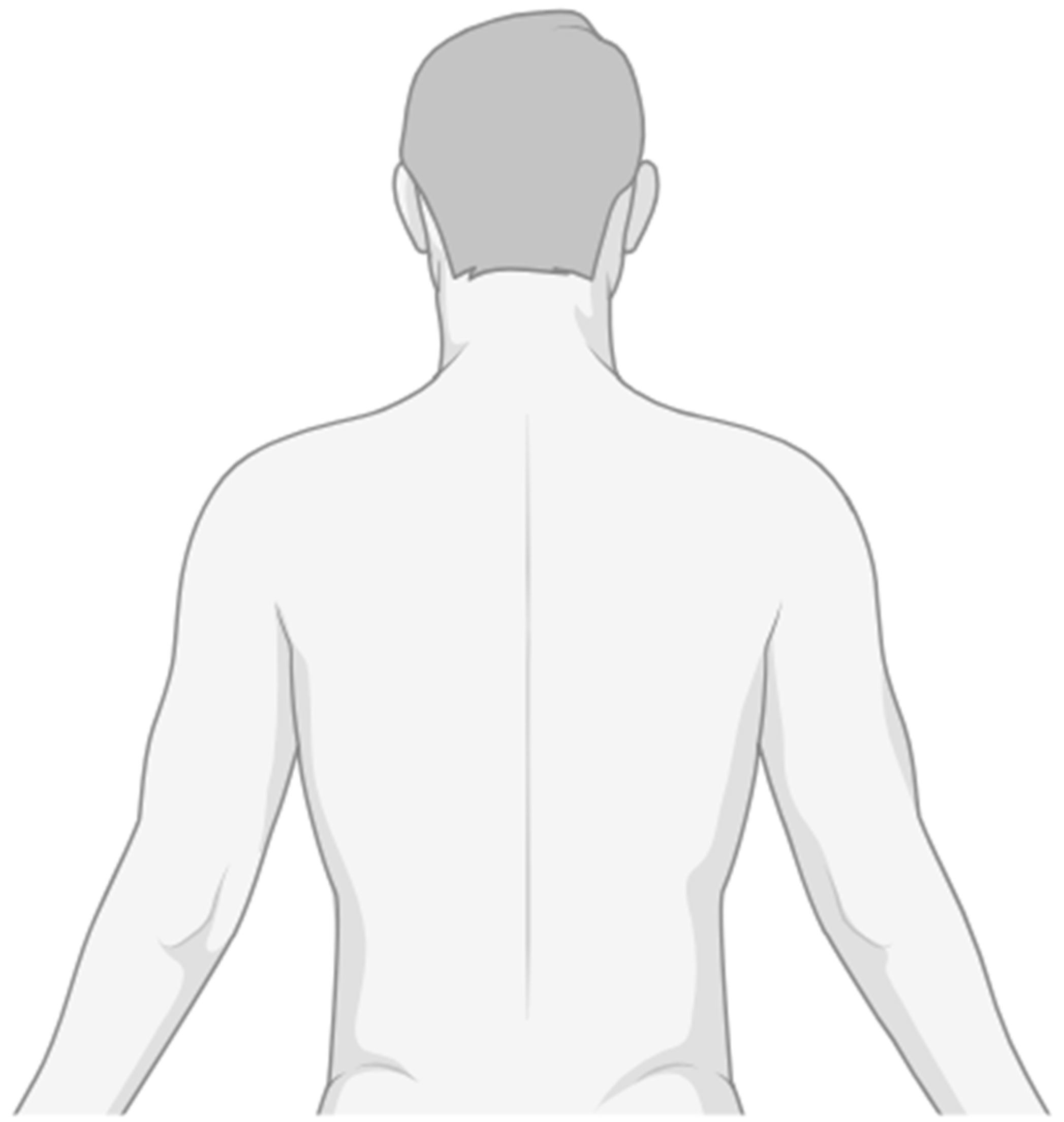
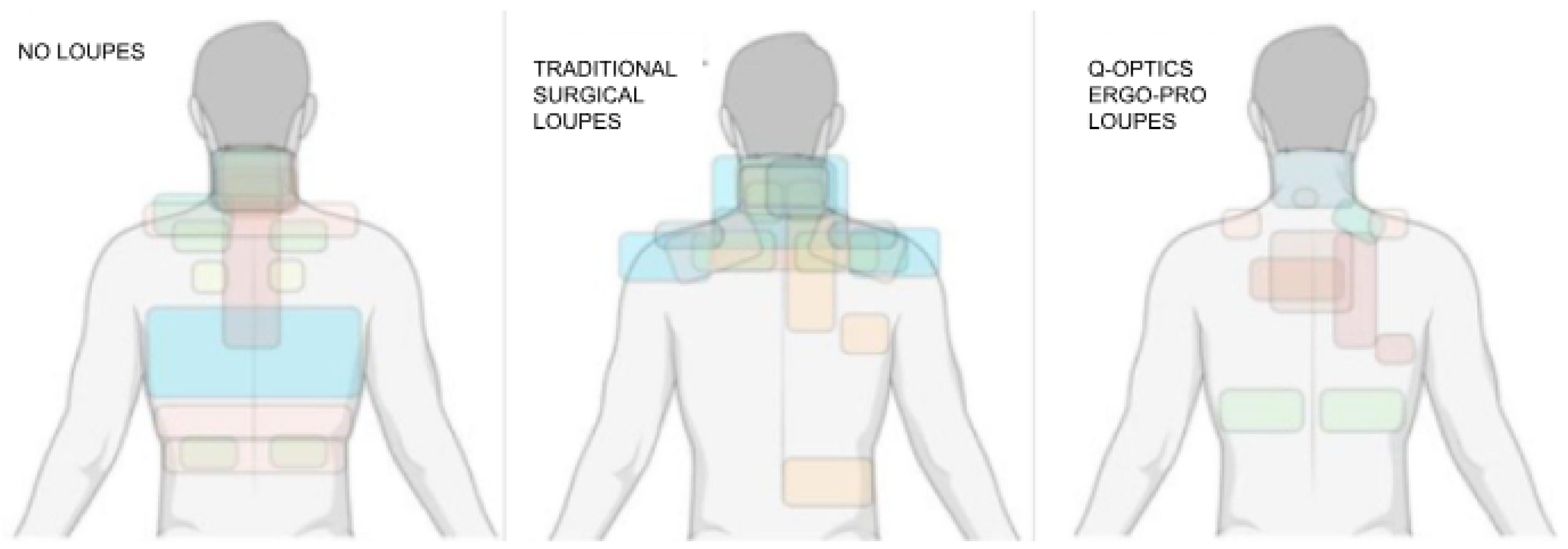
11]. To quantify participants’ subjective experiences, the survey employed a standard 1–10 Likert scale across all questions related to strain, fatigue, comfort, and likelihood of recommendation. For fatigue and strain-related questions, a score of 1–2 indicated “not at all”, 3–4 was “slightly”, 5–6 was “moderately”, 7–8 was “quite a bit”, and a score of 9–10 indicated “extremely”. Conversely, for comfort and recommendation questions, 1 represented the lowest level of comfort or likelihood to recommend, and 10 represented the highest. This scale allowed for consistent, comparable assessment of ergonomic performance across different loupe conditions. The survey also asked subjects to mark on a diagram of the human’s dorsal region where they felt the most strain (
Table 1,
Figure 6).
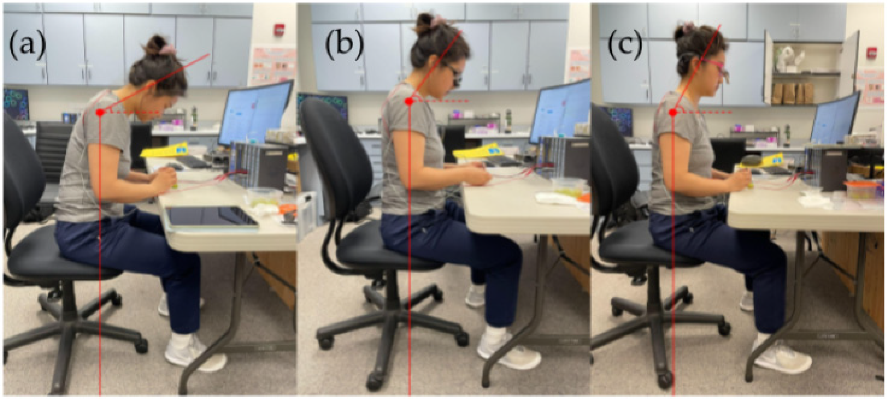
Side profile pictures were also taken of each subject in the three conditions to measure head posture via craniovertebral (CV) angle, the angle between a horizontal line through the spinous process of C7 and a line connecting the spinous process of C7 through the tragus of the ear [
12]. Lateral photographs were taken for each condition using an iPhone 12 (Apple, Cupertino, CA, USA) with the native Camera app (iOS 18.5) and its built-in gyroscopic leveling feature. Images were captured at a standardized height of 1.20 m using a tripod to maintain consistency in camera angle and perspective. CV angles were then measured via post-image analysis using an online protractor tool, with anatomical landmarks (tragus and C7 vertebra) visually estimated on-screen (
Figure 7). A smaller CV angle indicates a more pronounced forward head posture, which can increase spinal stress [
13]. The average length of the cervical spine (C1 to C7) is 60.9 mm (2.398 inches), which was used to calculate the mechanical load on the cervical spine (
Appendix A) [
14]. For every inch of forward head displacement, the force on the cervical spine increases by 10–12 pounds-force lbf [
15].
3. Results
All 10 medical student volunteers (6 female, 4 male; mean age 26.2 ± 1.1 years) completed the protocol without any reported adverse events. To assess for potential carryover effects, a mixed-design ANOVA was conducted with loupe condition (PDLs, TTL loupes, no loupes) as a within-subjects factor and presentation order (i.e., randomized sequence group) as a between-subjects factor. The absence of a significant interaction effect between condition and order (p > 0.05) indicated that no carryover effects were detected.
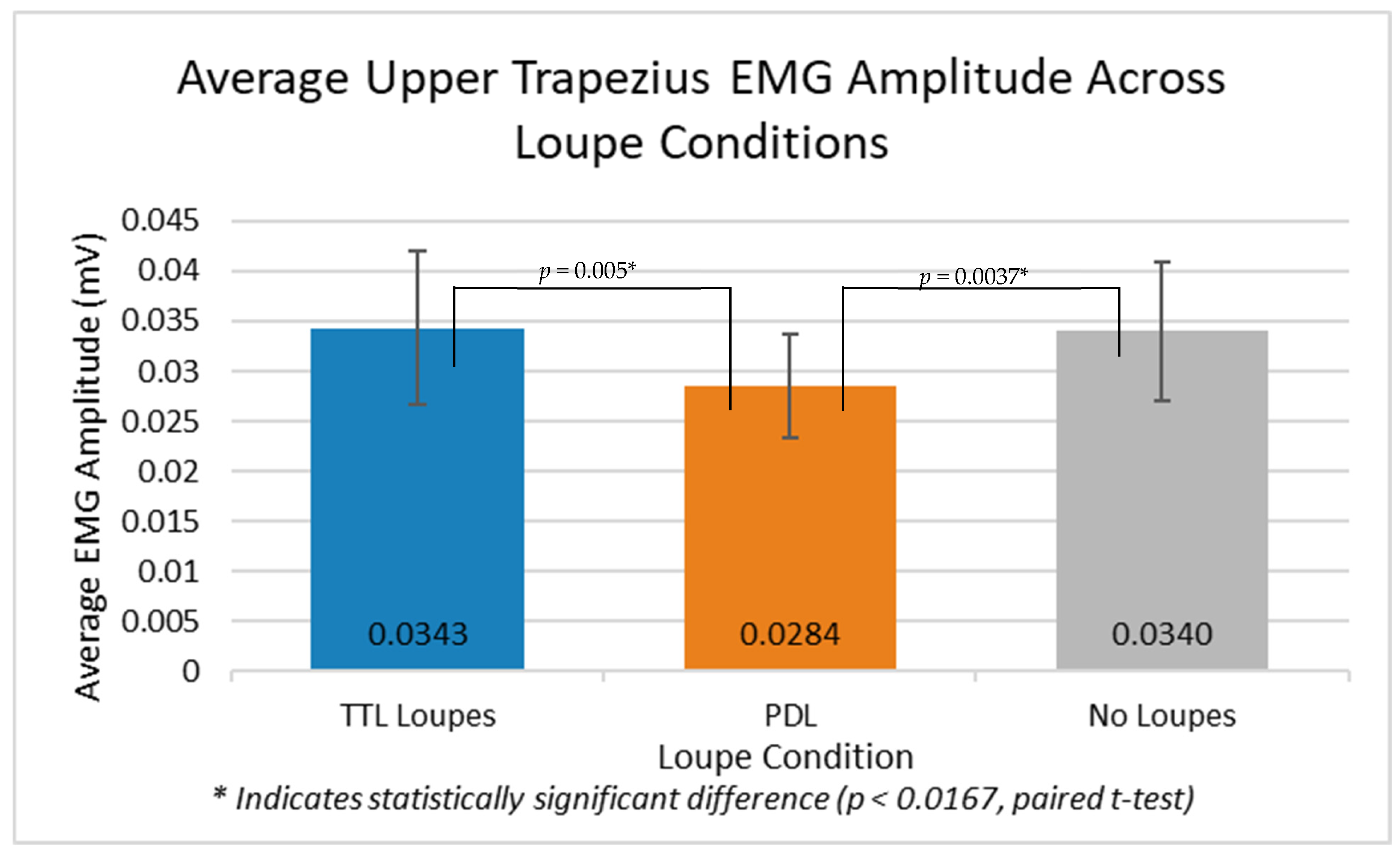
PDLs led to significantly less muscle activation (mean [M] = 0.0284 ± 0.0179 millivolts (mV)) compared to no loupes (M = 0.0340 ± 0.0243 mV) (p = 0.0037). Participants also experienced lower activation with PDLs (M = 0.0284 ± 0.0179 mV) compared to TTL loupes (M = 0.0343 ± 0.0251 mV) (p = 0.005). There was no significant difference in muscle activation between TTL loupes and no loupes (p = 0.42). PDLs reduced trapezius activation by 17.2% vs. TTL loupes (95% CI: 12.8–21.6%, p = 0.009).
A one-way repeated measures ANOVA revealed no statistically significant differences in upper trapezius EMG amplitude across the three loupe conditions (
p = 0.65), likely due to the limited sample size and high inter-subject variability. However, exploratory post hoc paired t-tests with Bonferroni correction were performed to identify potential ergonomic trends. These preliminary comparisons suggested that PDLs reduced muscle activation compared to both TTL loupes (
p = 0.005) and no loupes (
p = 0.0037), though these findings should be interpreted with caution, given the non-significant ANOVA There was no significant difference between TTL loupes and no loupes (
p = 0.42) (
Figure 8).
Survey responses revealed statistically significant differences in perceived strain, comfort, and likelihood to recommend across the three loupe conditions (
p < 0.01, Friedman test). Post hoc Wilcoxon Signed-Rank tests with Bonferroni correction confirmed that PDLs were rated significantly more comfortable and less straining than both TTL loupes (
p = 0.008) and no loupes (
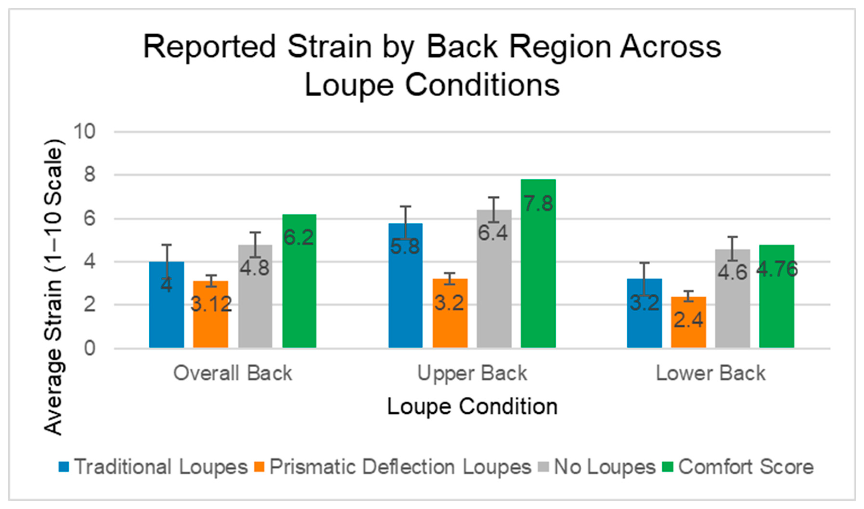
p = 0.004). Participants using PDLs reported the lowest average strain across all regions, with back strain averaging 3.12 out of 10 and upper back strain 3.20 (on a 1–10 Likert scale) (
Figure 9). These users also reported the highest comfort (7.80) and the highest likelihood of recommending the PDL to others (8.80). In contrast, those using TTL loupes experienced slightly higher strain, especially in the upper back (5.80) and back overall (4.00), and comfort and recommendation scores averaged 6.20 and 6.00, respectively. Participants working with no loupes reported the highest levels of back (4.80) and upper back (6.40) strain, along with the lowest comfort (4.76) and recommendation (4.00) scores. These findings suggest that the PDL not only reduces physical strain but is also preferred by users for its ergonomic support and comfort while performing surgical tasks. Measured muscle reduction assessed through the EMG recording when using the PDL strongly predicted subjective comfort scores (Pearson correlation coefficients [r] = -0.81,
p < 0.001), and comfort scores directly correlated with CV angle (r = 0.79,
p < 0.001). Median comfort scores increased by 1.6 points compared to TTL loupes (
p < 0.01, Wilcoxon Signed-Rank test with Bonferroni correction), representing a 26% increase on the 10-point scale.
The second part of the survey, in which participants circled areas of strain, revealed that subjects felt strain mostly in the upper back region across all three conditions. However, three participants reported no strain at all using PDLs, while all subjects indicated some areas of strain while wearing TTL loupes and no loupes (
Figure 10).
Head posture, measured by CV angles, differed substantially across the three conditions. The data were presented as mean cervical flexion angle ± standard deviation. The average cervical flexion angle was 38.6° ± 14.4° without loupes, 47.2° ± 9.7° with TTL loupes, and 66.3° ± 7.0° with PDLs. Compared to PDLs, both TTL loupes (p = 0.00013) and no loupes (p = 0.00061) exhibited significantly lower CV angles. The difference between traditional loupes and no loupe was not statistically significant (p = 0.18).
To estimate cervical spinal loading, we assumed an average cervical spine length (C1–C7) of 2.398 inches, based on published cadaveric data [
14]. This value, however, was derived from a small sample of Turkish cadavers (n = 5, 4 male) and may not generalize to our cohort, which was 60% female. Because spinal load is directly proportional to cervical spine length, surgeons with longer cervical spines may experience even greater forces than estimated. Given this limitation, our force calculations should be interpreted as approximations which may overestimate the lbf, as female cervical lengths are typically shorter, grounded in typical anatomical values, but variable by individual.
By utilizing biomechanical modeling, notable differences in cervical spine loading were observed across the three conditions. The free body diagram using the calculated CV revealed that 18.7 lbf was applied to the cervical spine in the subject’s position when not using any loupes [
14]. Conversely, TTL loupes required 16.3 lbf over the cervical spine, and the PDL required 9.6 lbf over the cervical spine to maintain the static position during the procedure (
Appendix A). Without loupes, an estimated 18.7 lbf were applied to the cervical spine. This load was reduced to 16.3 lbf (a 12.8% reduction) with TTL loupes, and further decreased to 9.6 lbf (a 48.6% reduction) when using prismatic deflection loupes (PDLs). When comparing TTL loupe usage to PDLs, the latter still demonstrated a 6.67 lbf reduction. These differences highlight the ergonomic advantage of the prismatic deflection design, which consistently resulted in the lowest spinal load. Every 1° increase in CV angle reduced cervical load by 0.31 lbf (R
2 = 0.94,
p < 0.001). PDLs decreased spinal force by 41.1% compared to TTL loupes (Δ = 6.7 lbf,
p < 0.001). Notably, this trend aligned with the EMG findings, in which trapezius muscle activation was lowest when using PDLs, moderate with TTL loupes, and highest without loupes, mirroring the progressive decrease in cervical spine loading.
4. Discussion
Our investigation provides evidence that the use of PDLs significantly mitigates biomechanical strain during ophthalmic microsurgeries. Findings from the EMG analysis demonstrated statistically significant reductions in trapezius muscle activation with PDL compared to no loupes (M = 0.0284 mV vs. 0.0340 mV, p = 0.0037) and TTL loupes (M = 0.0284 mV vs. 0.0343 mV, p = 0.005), indicating a marked biomechanical advantage. Our multimodal analysis demonstrates three key advances: (1) PDL use reduces trapezius muscle activation by 17.2% versus TTL loupes (p = 0.009), (2) cervical spine loading is decreased by 41.1% (16.3 lbf vs. 9.6 lbf, p < 0.001), and (3) subjective comfort was also significantly improved with PDL. Critically, we establish the first quantitative relationship between CV angle and spinal load (0.31 lbf/°, R2 = 0.94, p < 0.001), providing an evidence-based framework for ergonomic intervention design.
Complementing these objective findings, subjective assessments revealed markedly enhanced user experiences with prismatic deflection technology, as participants noted substantially less musculoskeletal strain while simultaneously reporting superior comfort ratings and recommendation likelihood. Furthermore, cervical vertebral angle analysis revealed progressive postural optimization, with PDL achieving a 66˚ cervical angle compared to 47˚ with TTL loupes and 39˚ without magnification, translating to an estimated 6.7 lbf reduction in cervical spine loading relative to TTL designs. The 6.7 lbf reduction in cervical force with PDL represents more than an incremental improvement; it fundamentally alters spine biomechanics. Cadaveric studies indicate compressive forces >10 lbf accelerate disc degeneration through nucleus pulposus dehydration [
16]. In our biomechanical model, estimated loads for TTL loupes (16.3 lbf) and no magnification (18.7 lbf) were higher than those estimated for PDL (9.6 lbf). While these values are approximations based on typical anatomical averages, the trend suggests that PDL use may help keep cervical loading within a more favorable physiological range. These convergent objective and subjective findings establish that PDL may confer ergonomic benefits that address the primary limitations of surgical loupes in ophthalmic surgery. Over time, such reductions in cervical loading could translate to a meaningful decrease in cumulative spinal stress and musculoskeletal strain for clinicians.
Assuming 2 h/week, PDL would spare surgeons 555 lbf/year versus TTL loupes and 950 lbf/year versus non-loupes-assisted surgery, equivalent to removing a grand piano’s weight from the cervical spine annually. A predominant determinant of career longevity in any manual profession is the cumulative physiological stress that accrues over a career. This phenomenon is particularly pronounced in the surgical disciplines, where decades of musculoskeletal strain affecting the vertebral column, cervical region, and peripheral appendages significantly influence both a surgeon’s intraoperative efficacy and quality of life beyond clinical settings [
1,
17]. Today, heightened awareness of these occupational hazards and a contemporary emphasis on clinicians’ physical and psychological well-being have generated a focus on ergonomic optimization throughout surgical specialties [
2]. The field of ophthalmology is no exception, and our findings present a compelling opportunity to enhance surgical comfort and somatic resilience for practitioners in this field.
While ocular surgery avoids certain major contributors to physiological deterioration through shorter procedural durations and the option to maintain a seated position during interventions, the specialty nevertheless presents distinctive sources of bodily strain. A majority of ophthalmologists exhibited manifestations of musculoskeletal pathologies, with notable correlations to operative durations [
18]. Microsurgical interventions, specifically, including those within ophthalmology, have been documented to pose elevated occupational health risks due to the utilization of conventional high-magnification surgical loupes, operating microscopes, and recurrent periods of compromised postural alignment of the neck and torso [
19].
Loupe utilization particularly offers an attractive target for ergonomic innovation, given its application across various dental and medical specialties. Although conventional loupes have demonstrated modest improvements in postural alignment and musculoskeletal discomfort in professions with high implementation rates, such as dentistry, their usage still necessitates substantial cervical flexion, which exacerbates musculoskeletal strain [
7,
8]. A survey examining oculoplastic surgeons revealed that approximately half of the respondents utilized surgical loupes, with non-adopters citing restricted visual field and musculoskeletal discomfort as primary deterrents [
20]. This limitation of TTL optical aids aligns with the EMG findings in our investigation, where no significant reduction in cervical muscle activation was observed with conventional loupe implementation (
p = 0.42). Furthermore, the absence of a perceived benefit in our subjective assessments underscores TTL loupes’ inadequacy in addressing the ergonomic improvements sought by ophthalmologists.
Conversely, our data provides robust evidence that the PDL successfully addresses many deficiencies inherent in TTL designs. The statistically significant differentiation in EMG measurements compared to both unassisted vision (p = 0.004) and conventional loupes (p = 0.005) demonstrates that PDL innovative architecture confers a biomechanical advantage over current ocular surgical methodologies. While the primary EMG analysis did not reach global statistical significance, the observed trends in our pairwise comparisons align with our significant biomechanical modeling and subjective comfort data. This suggests that, while this pilot study was underpowered to detect subtle changes in muscle activation via ANOVA, the biomechanical advantage of PDL warrants further investigation in a larger, more diverse surgical cohort. This reduction in cervical biomechanical stress could potentially mitigate cumulative axial forces experienced throughout an ophthalmologist’s operative career by an estimated 555 lbf annually (comparing prismatic deflection to TTL loupes) to 950 lbf annually (comparing prismatic deflection to no loupes). These projections are based on a very conservative assumption of 2 operative hours per week, which represents a promising ergonomic advantage with potential implications for long-term musculoskeletal health and professional longevity. Moreover, our subjective assessments establish that this mechanical superiority translates to a perceptible enhancement in comfort, potentially allowing for longer and more comfortable lifetime careers among ophthalmologists.
The biomechanical and user-comfort improvements position these innovative PDL as a readily implementable strategy for enhancing the physiological welfare of ocular surgeons. However, this advanced design must be complemented by heightened awareness regarding the long-term consequences of suboptimal ergonomic practices and the potential benefits these PDL may offer for professional longevity. Surgical practitioners are notoriously resistant to methodological modifications, but we hope that our investigation provides definitive evidence to stimulate exploration of novel optical assistance technologies.
Several methodological limitations merit acknowledgment. First, our participant cohort consisted of only 10 medical students, a relatively small sample size that limits statistical power and generalizability. Additionally, the handedness of participants was not recorded. While bilateral EMG data were collected, the lack of dominant-hand documentation limits the ability to assess potential asymmetries in muscle activation during the microsuturing task. Furthermore, while participants did not utilize personal prescription spectacles or supplemental loupe lighting, the medical student population is younger and may have greater muscular endurance than attending surgeons with 10+ years of cumulative strain and who perform longer, more complex surgeries under stress. Our experimental protocol employed simulated ophthalmologic procedures on grapes, which, despite utilizing identical instrumentation and techniques, may not perfectly recapitulate the physiological demands of authentic ophthalmic interventions, where not only frontal axis flexion is induced, but also sagittal and vertical (i.e., rotational) axis movements, which could even further compound the strain simulated in this study. Additionally, this study only quantitatively investigated upper trapezius activation by EMG; however, our participants noted strain in other muscle groups as well. Further, increased muscle activation is only a proxy for ultimate occupational musculoskeletal injury, which can occur or be mitigated by multiple factors. While our model estimated significant load reductions, it is important to note that increased muscle activation and calculated mechanical forces are proxies for musculoskeletal strain and do not directly correlate with definitive clinical outcomes or injury rates in this study’s timeframe. Finally, while surface EMG recordings allowed for noninvasive and practical assessment of muscle activity, this approach is inherently limited by potential signal noise, cross-talk from adjacent muscles, and reduced specificity compared to intramuscular EMG. Post-processing choices, including filtering range, signal normalization, and envelope extraction, may influence the sensitivity and interpretability of the results [
21]. Future studies should incorporate standard EMG testing to achieve superior measurement fidelity and facilitate data acquisition from deeper musculature inaccessible via surface EMG recordings. Notably, deeper muscle groups such as the semispinalis capitis, splenius cervicis, and levator scapulae, which are located beneath the trapezius and play a critical role in maintaining neck posture, are likely more significantly affected by surgical ergonomics. Targeting these muscles may have yielded results of greater statistical and clinical significance. However, due to the limitations of surface EMG and IRB constraints, we were unable to use invasive techniques. The use of monopolar needle electrodes, if performed by a licensed physician, could have provided more accurate measurements by isolating activity in these deeper muscles. Such procedures were outside the scope of our current study and qualifications.




 ,
,