Two-Stage Alignment of FIB-SEM Images of Rock Samples


Abstract
1. Introduction
2. Existing Approaches
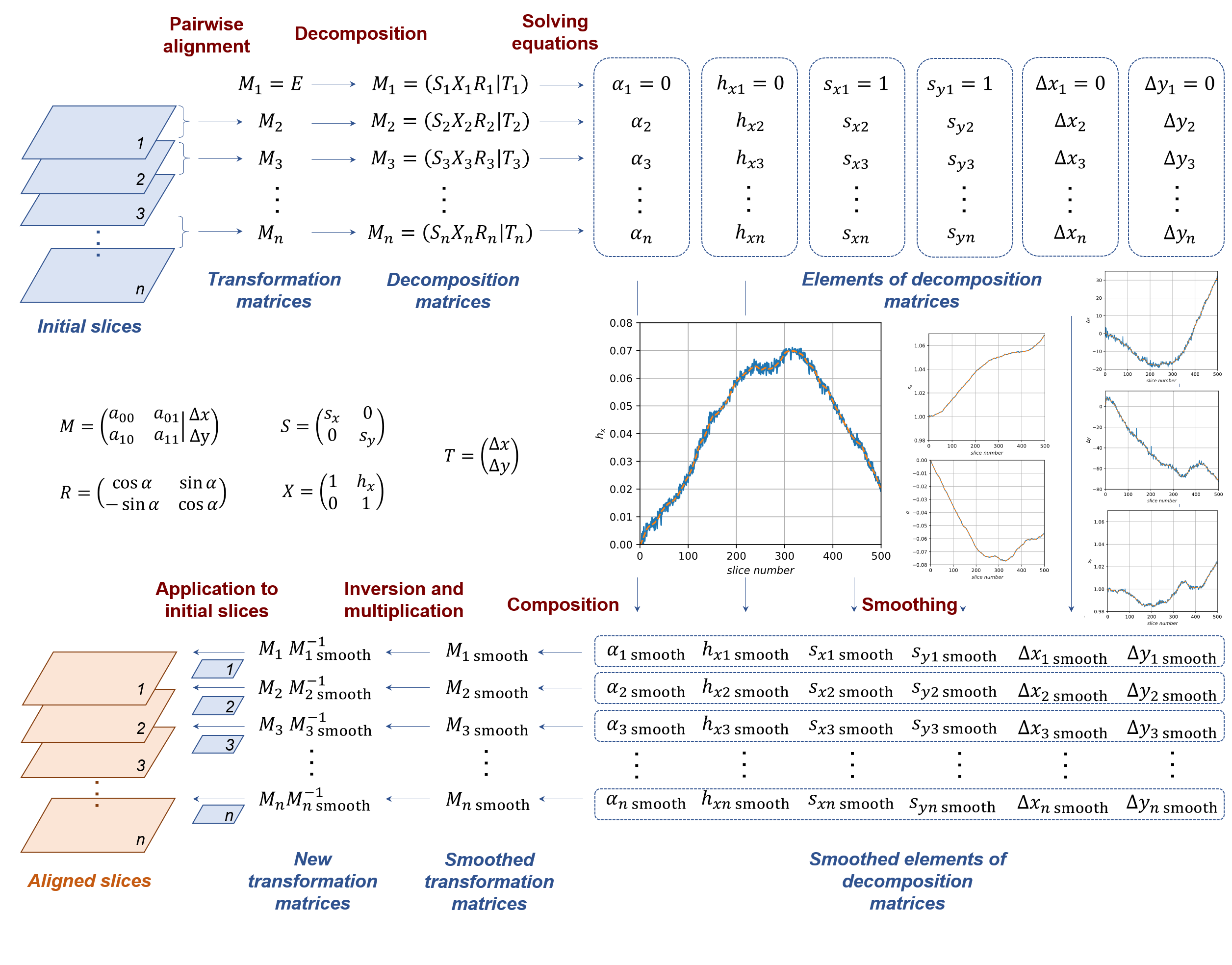
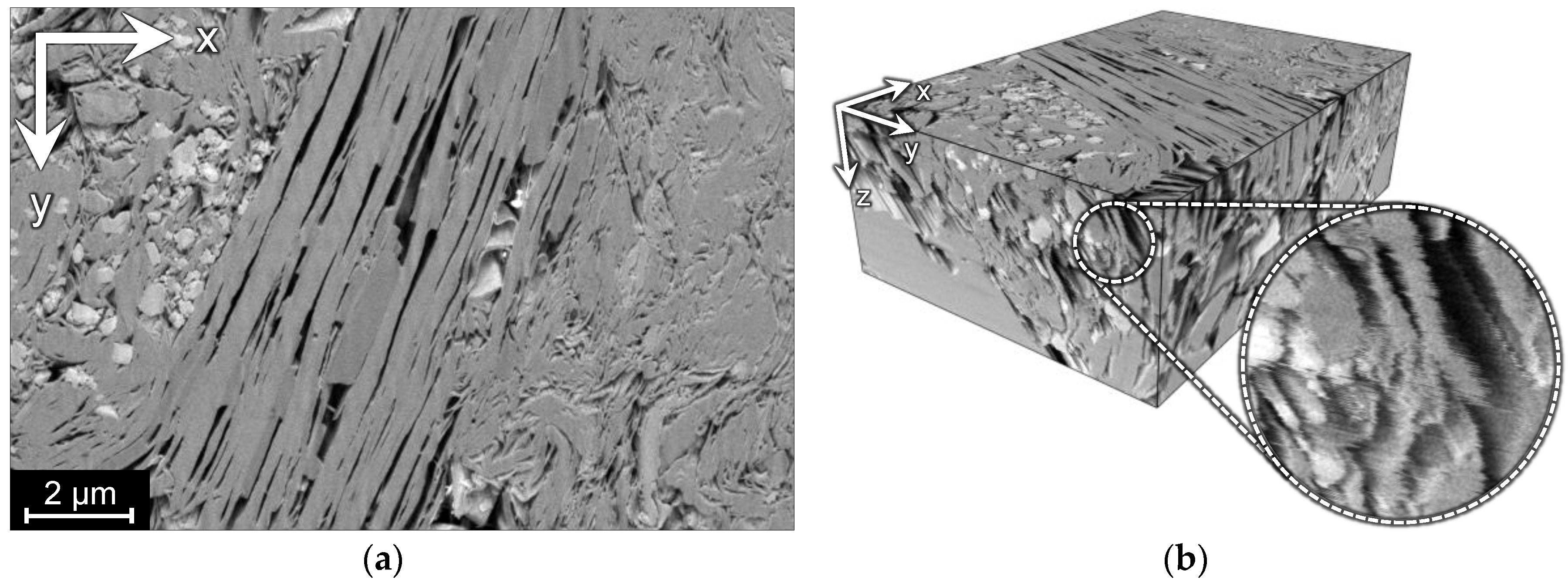
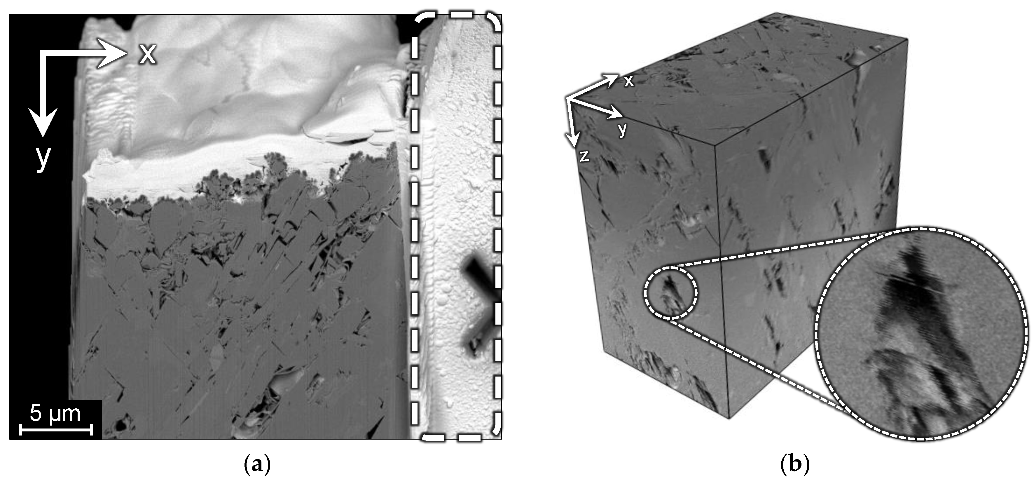
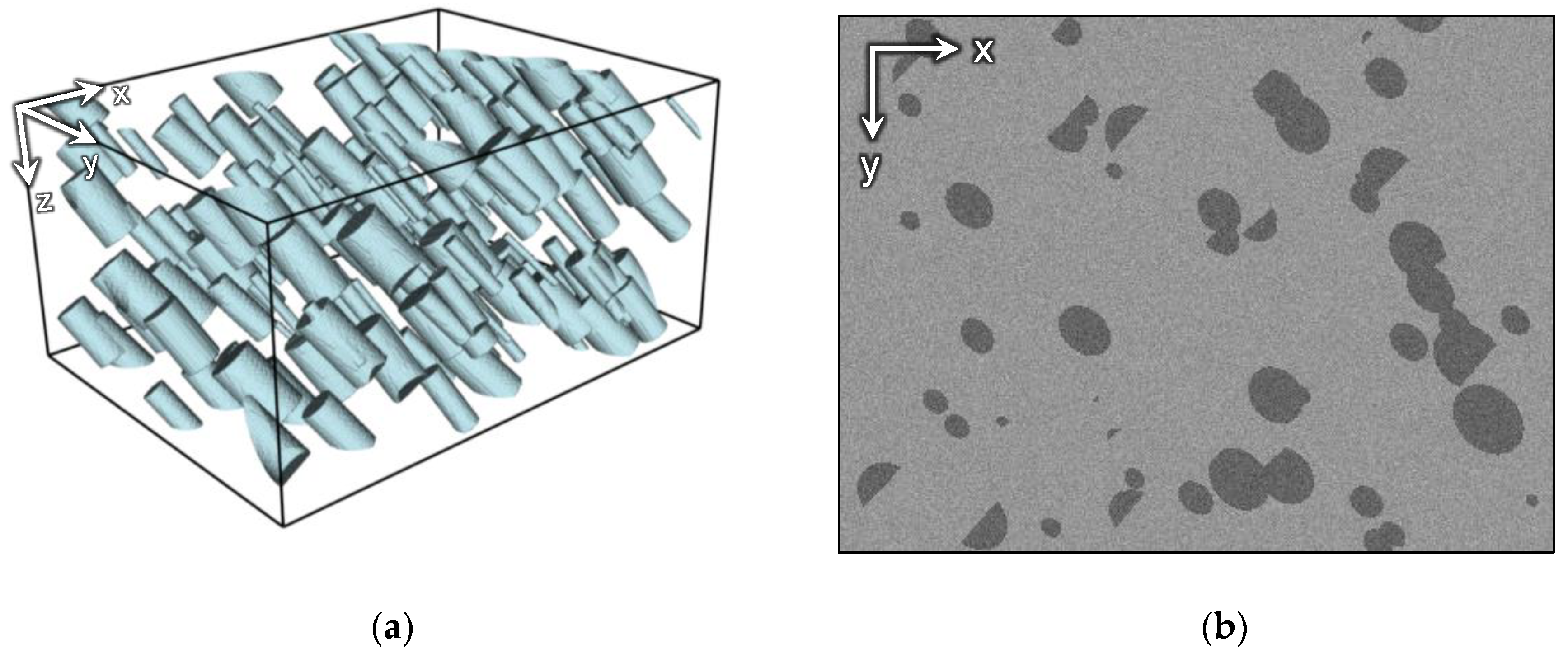
3. Datasets
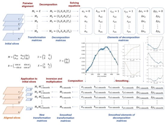
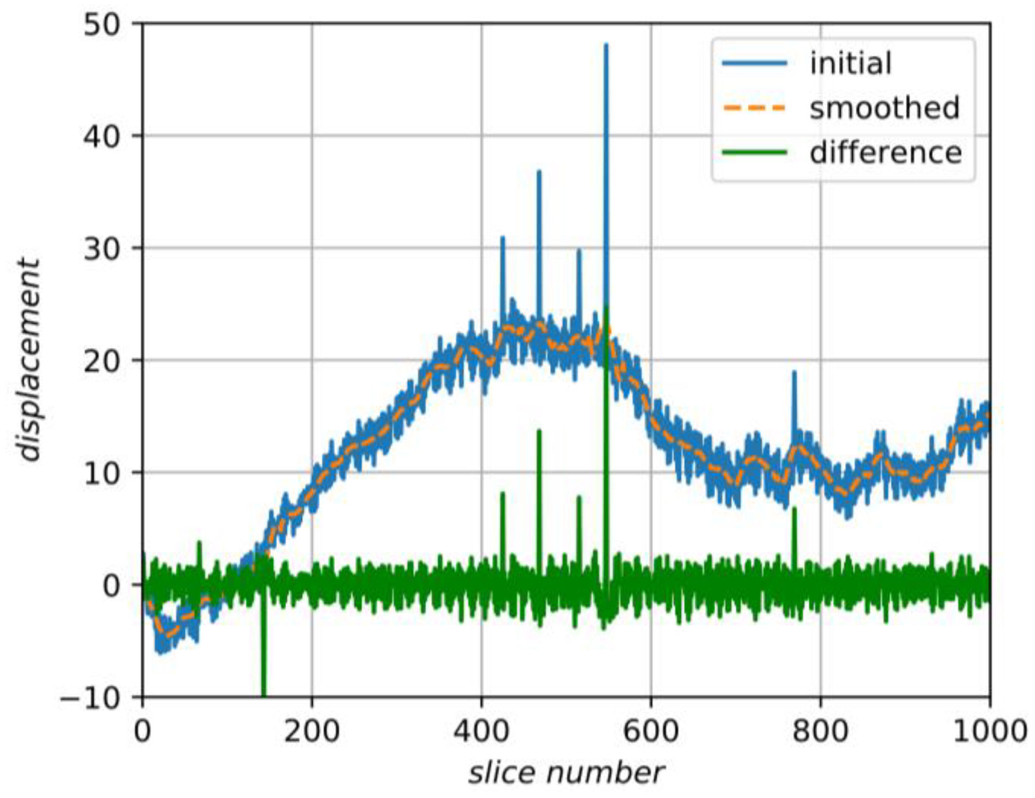
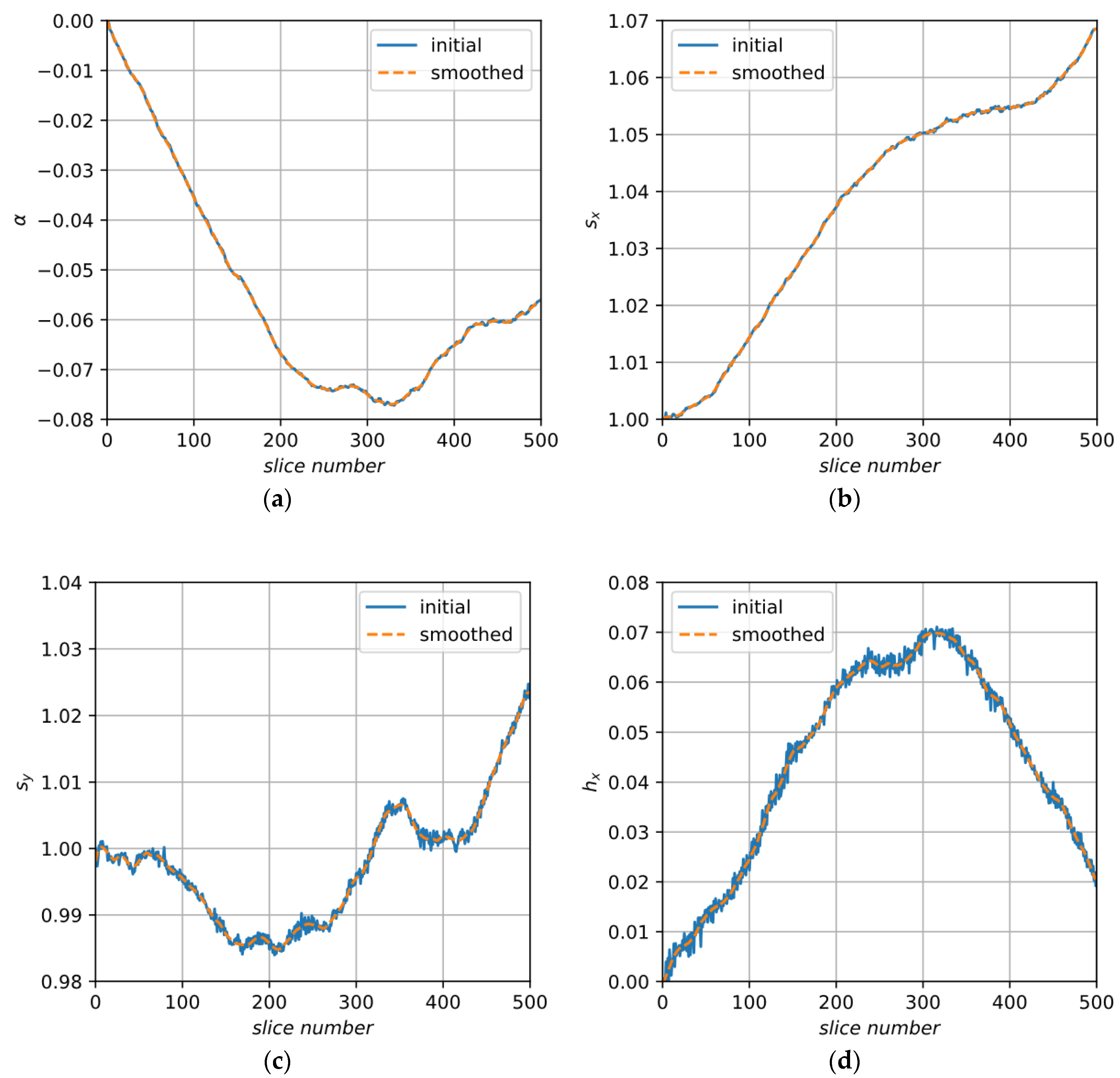
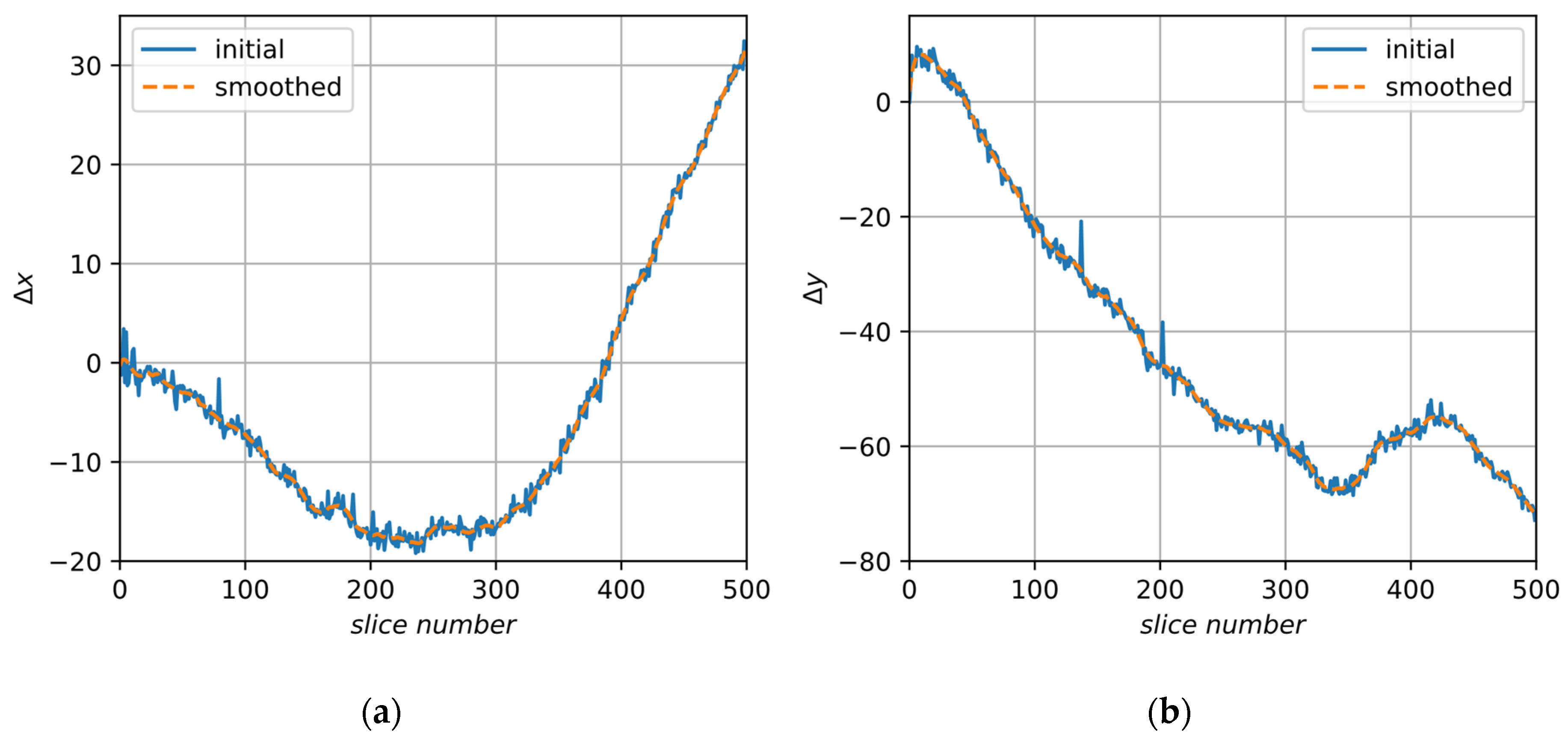
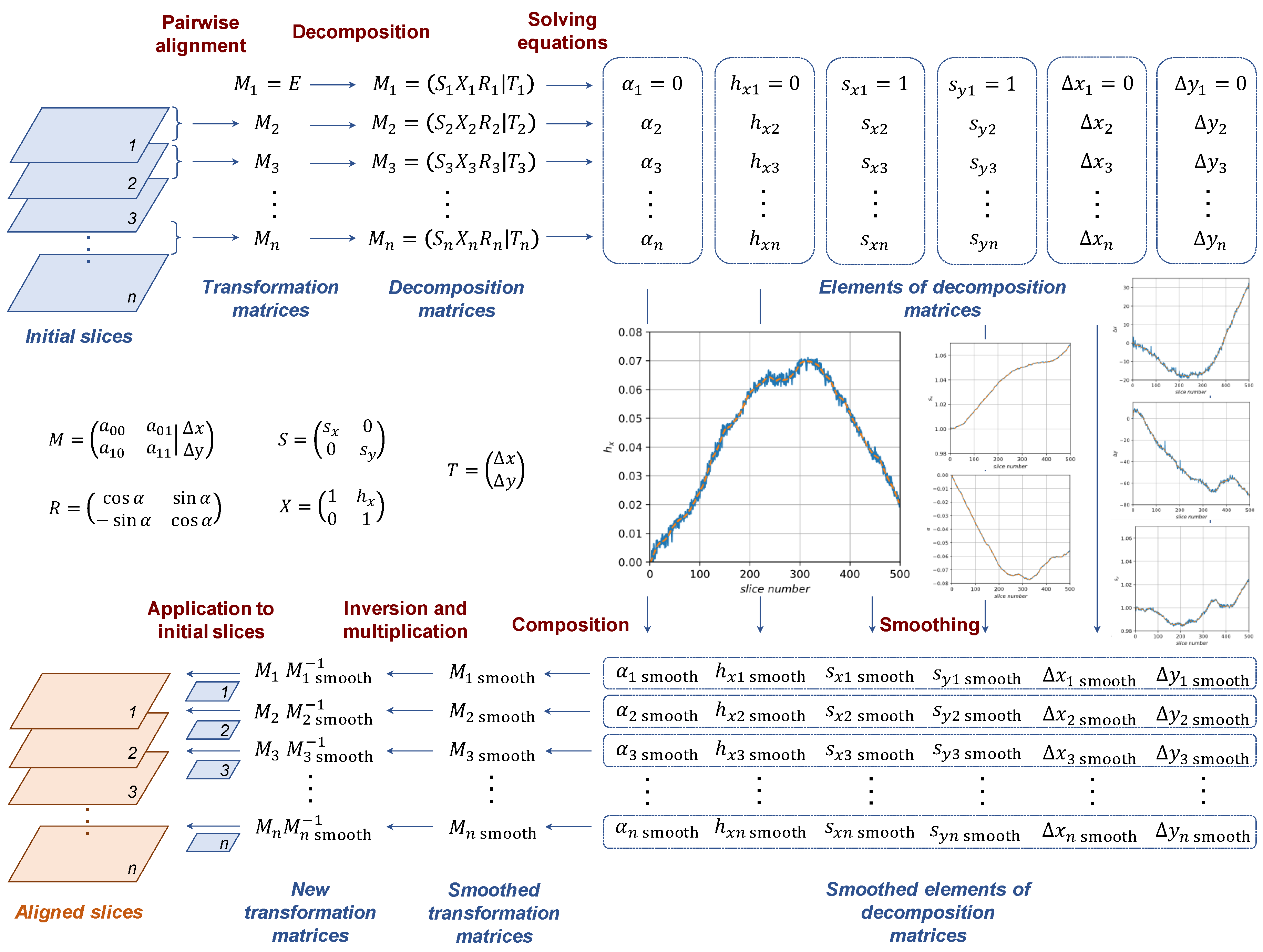
4. Proposed Method
5. Results and Discussion
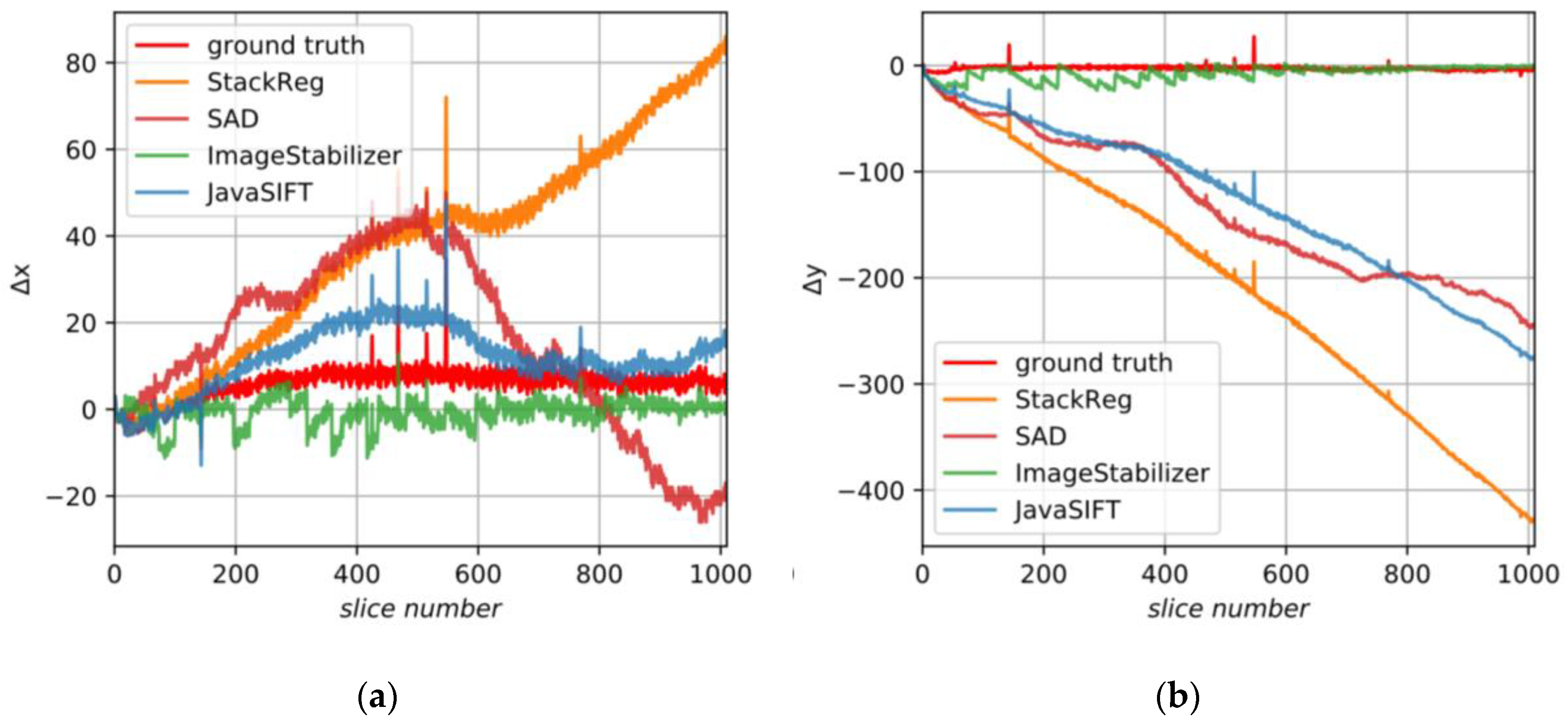
5.1. Quality Metrics for Alignment
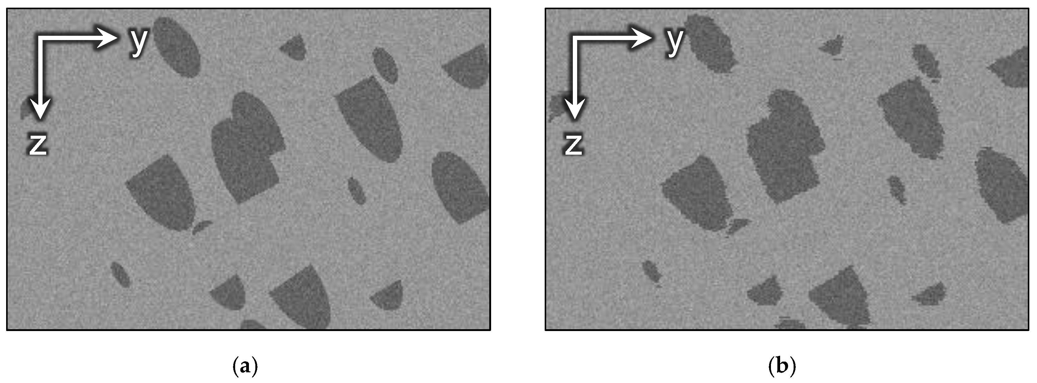
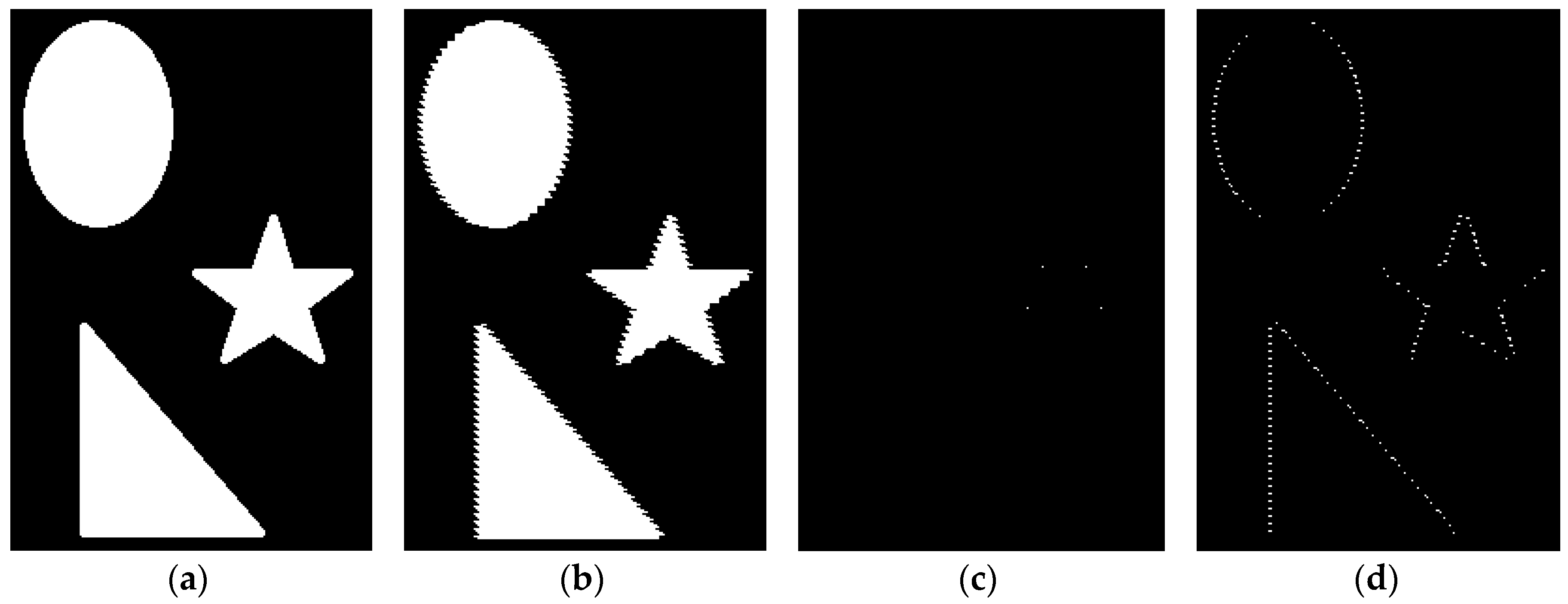
5.2. Alignment of the Synthetic Image
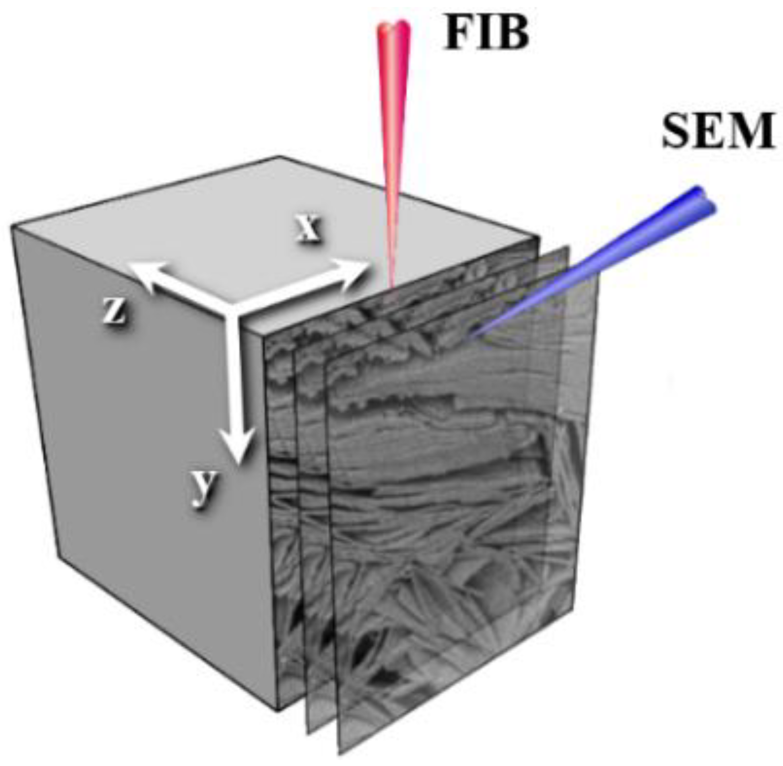
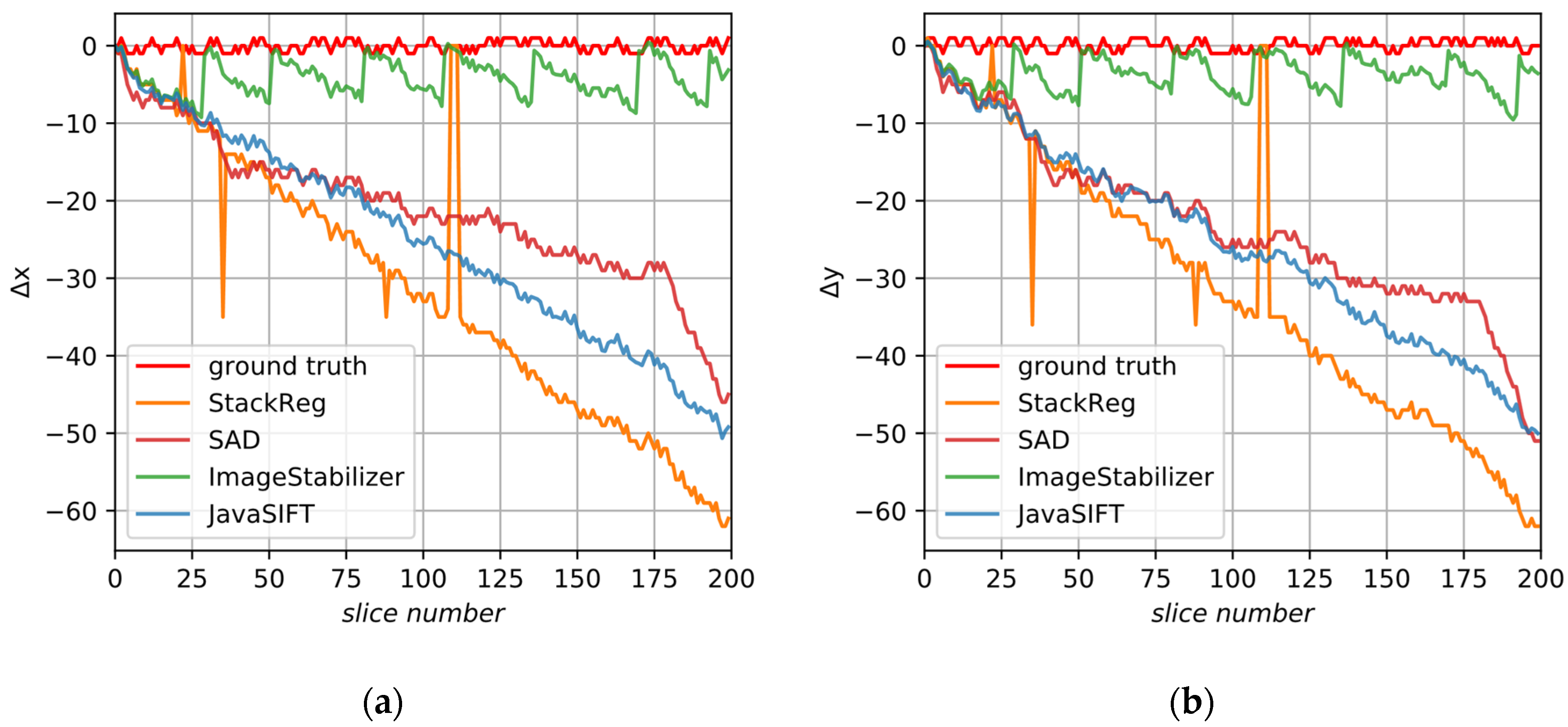
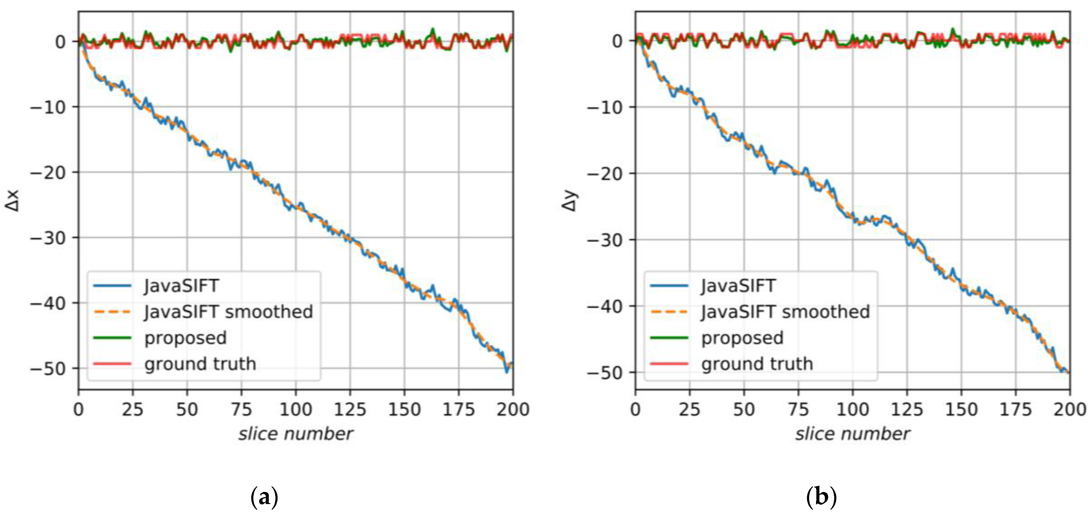
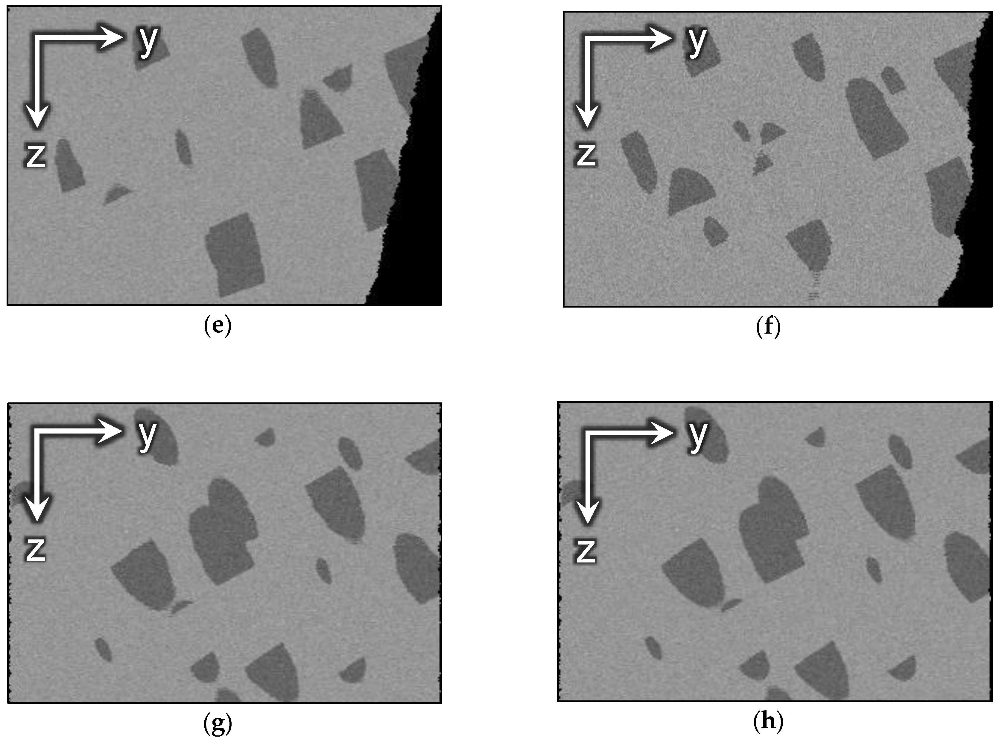
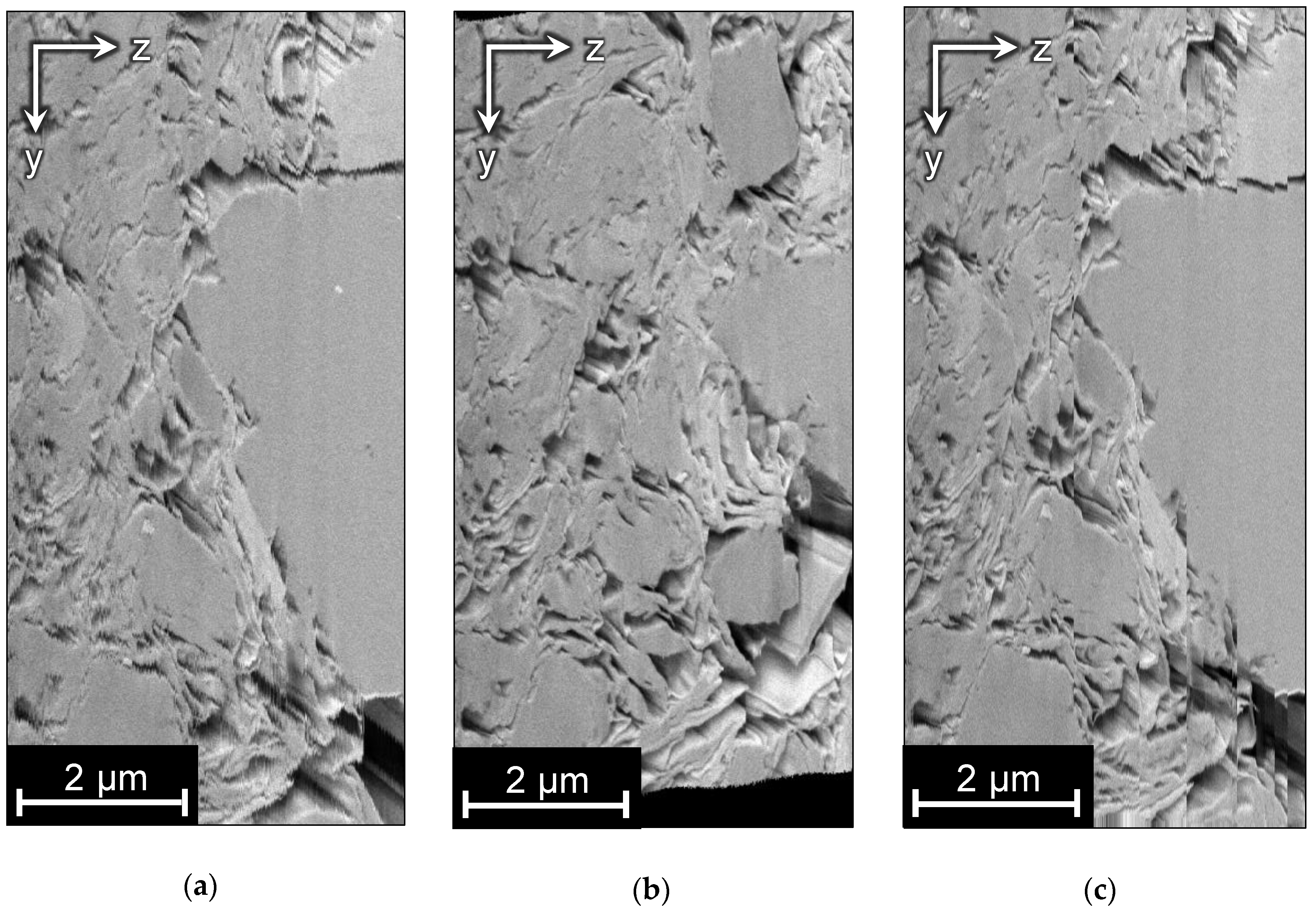
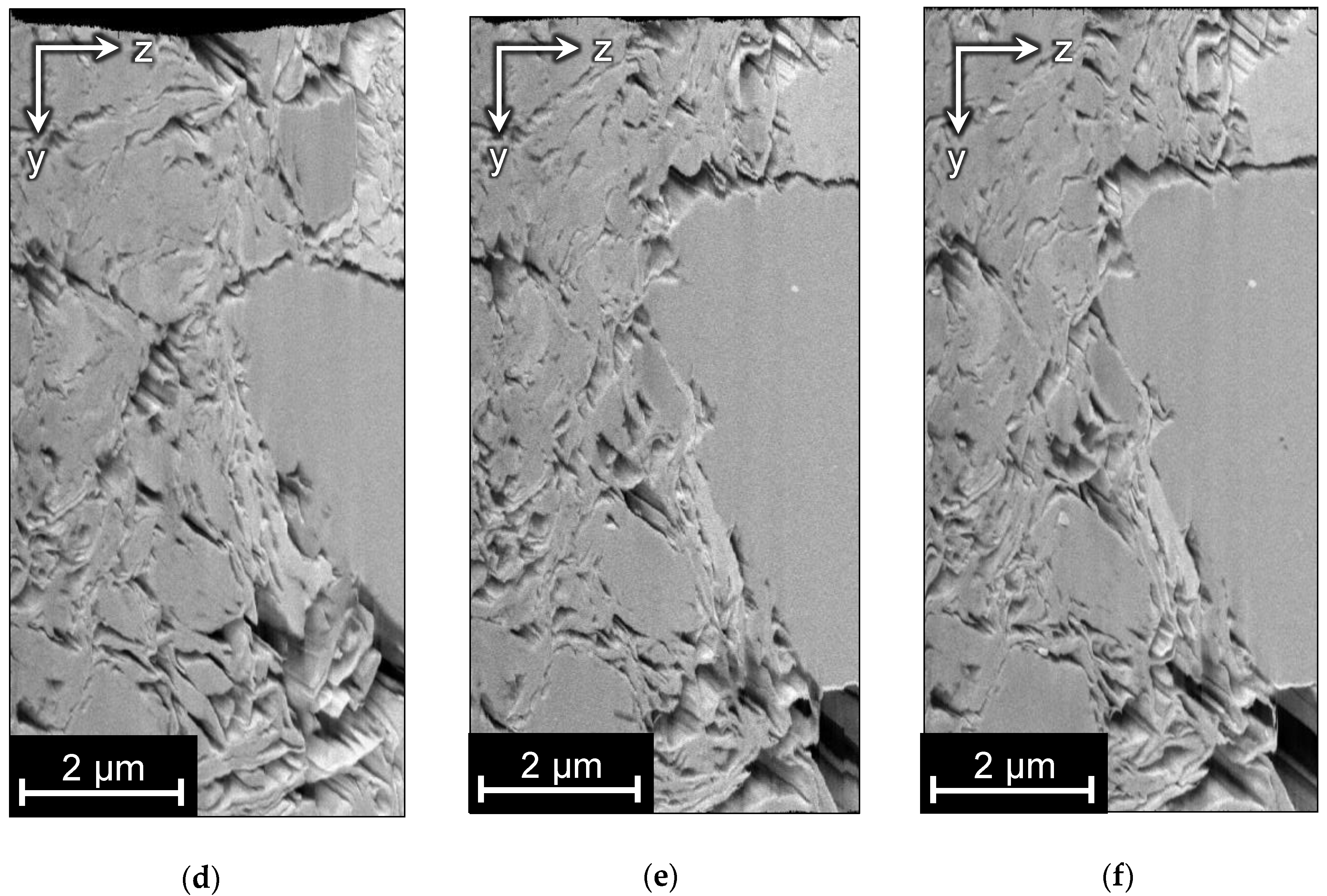
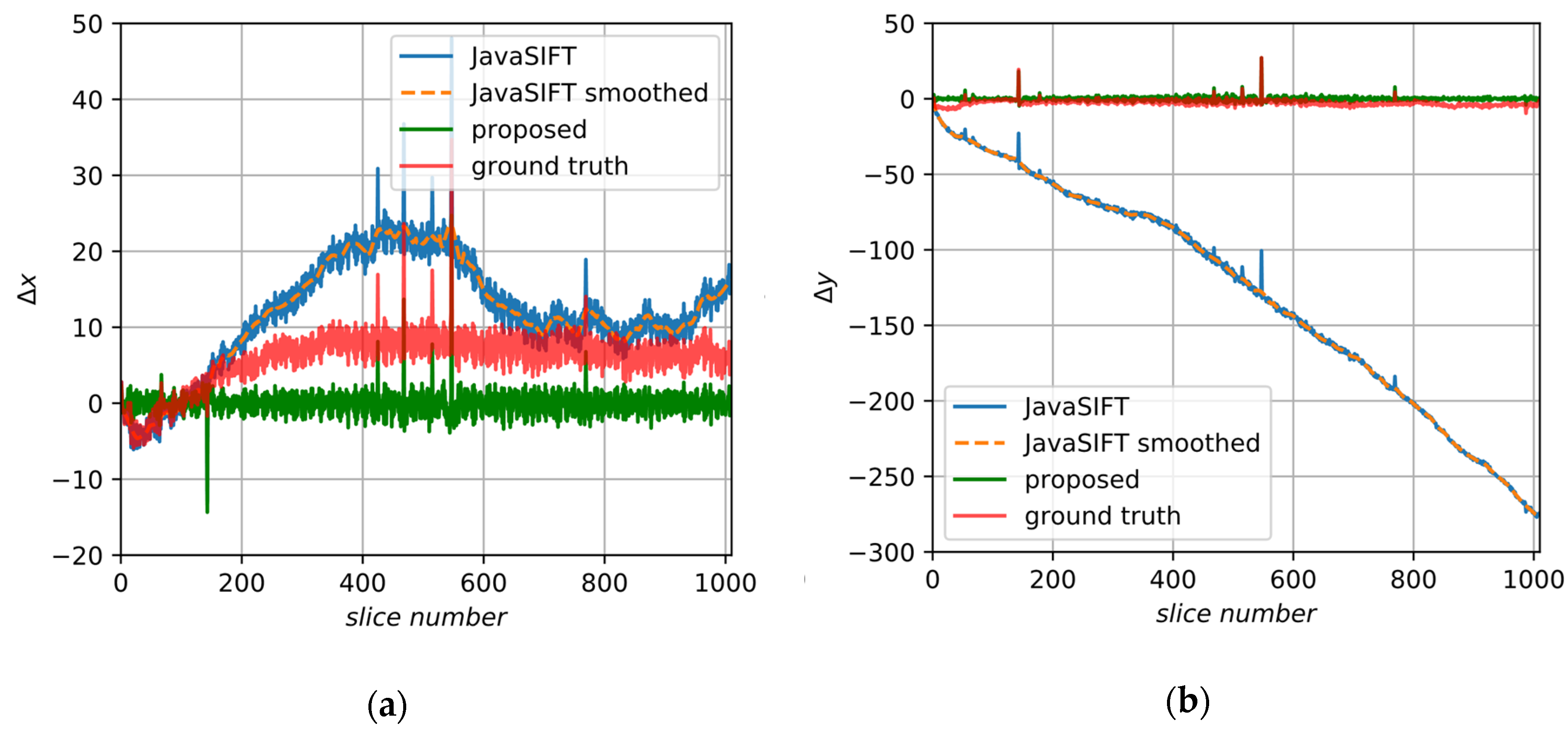
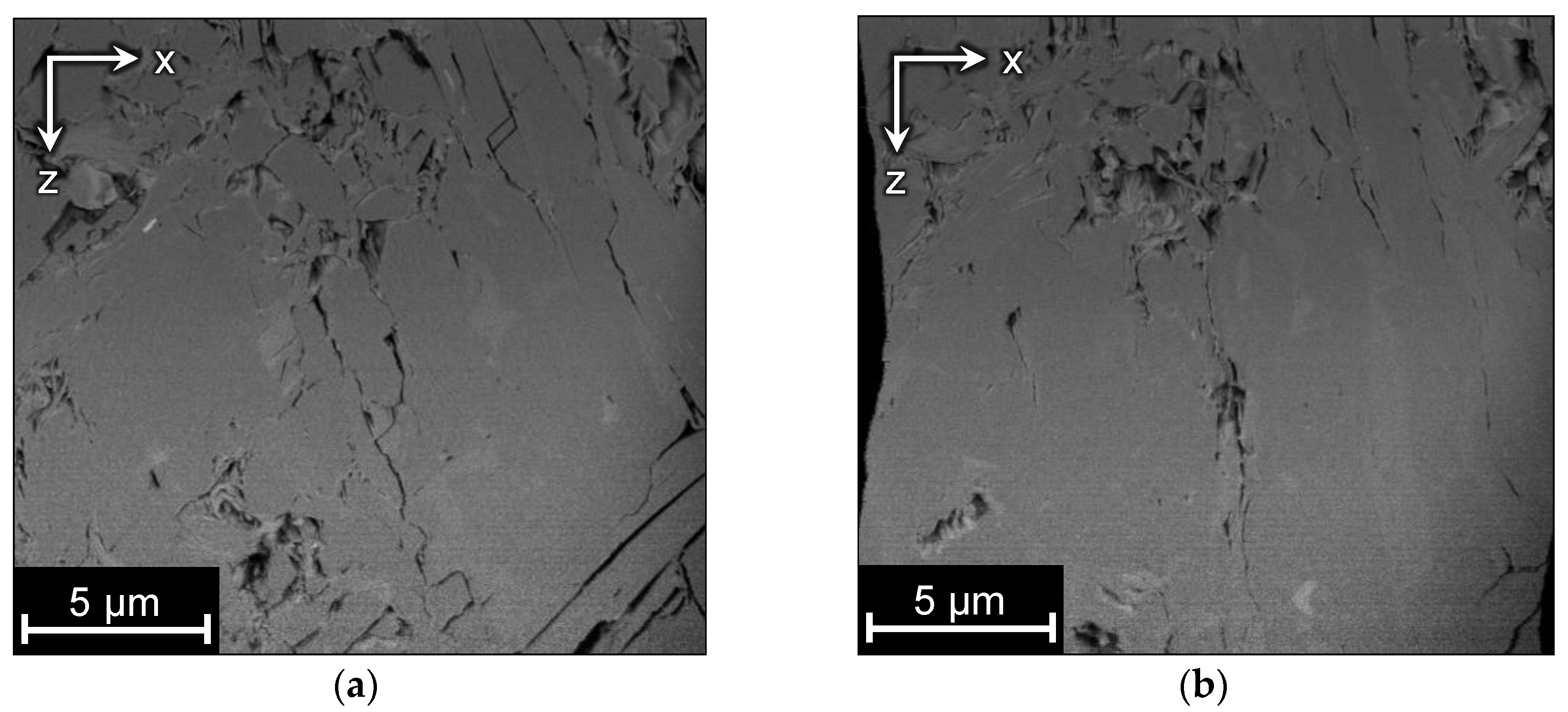
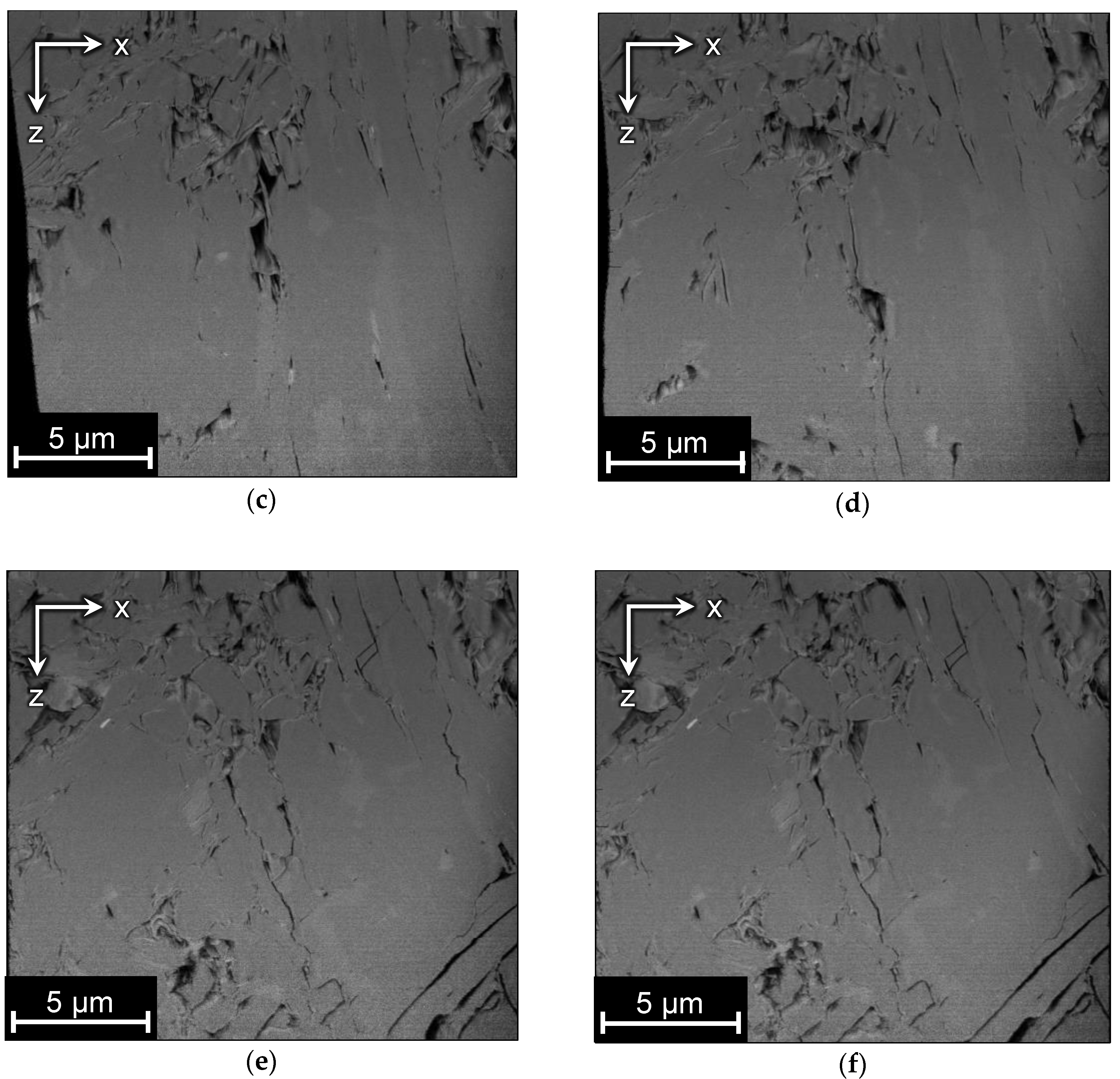
5.3. Alignment of Image A
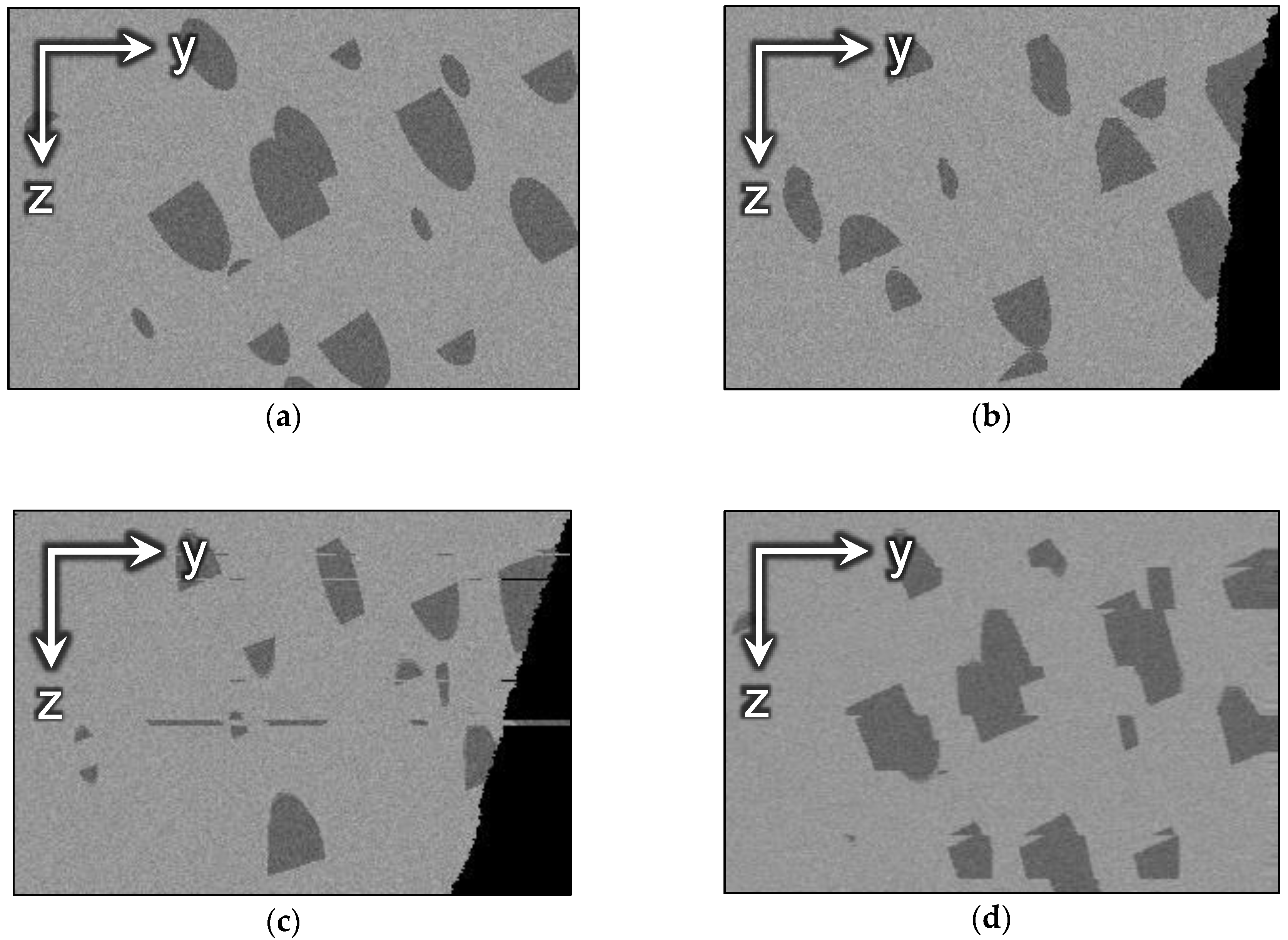
5.4. Alignment of Image B
6. Conclusions
Author Contributions
Funding
Acknowledgments
Conflicts of Interest
References
- Holzer, L.; Cantoni, M. Review of FIB-tomography. In Nanofabrication Using Focused Ion and Electron Beams: Principles and Applications; Oxford University Press: Oxford, UK, 2012; pp. 410–435. [Google Scholar]
- Liu, Y.; King, H.; van Huis, M.; Drury, M.; Plümper, O. Nano-Tomography of Porous Geological Materials Using Focused Ion Beam-Scanning Electron Microscopy. Minerals 2016, 6, 104. [Google Scholar] [CrossRef]
- Joos, J.; Carraro, T.; Weber, A.; Ivers-Tiffée, E. Reconstruction of porous electrodes by FIB/SEM for detailed microstructure modeling. J. Power Sources 2011, 196, 7302–7307. [Google Scholar] [CrossRef]
- Bosch, C.; Martínez, A.; Masachs, N.; Teixeira, C.M.; Fernaud, I.; Ulloa, F.; Pérez-Martínez, E.; Lois, C.; Comella, J.X.; DeFelipe, J. FIB/SEM technology and high-throughput 3D reconstruction of dendritic spines and synapses in GFP-labeled adult-generated neurons. Front. Neuroanat. 2015, 9, 60. [Google Scholar] [CrossRef] [PubMed]
- Lepinay, K.; Lorut, F. Three-Dimensional Semiconductor Device Investigation Using Focused Ion Beam and Scanning Electron Microscopy Imaging (FIB/SEM Tomography). Microsc. Microanal. 2013, 19, 85–92. [Google Scholar] [CrossRef]
- Ghosh, S.; Bhandari, Y.; Groeber, M. CAD-based reconstruction of 3D polycrystalline alloy microstructures from FIB generated serial sections. Comput. Aided Des. 2008, 40, 293–310. [Google Scholar] [CrossRef]
- Röding, M.; Fager, C.; Olsson, A.; Corswant, C.V.; Olsson, E.; Lorén, N. Three-dimensional reconstruction of porous polymer films from FIB-SEM nanotomography data using random forests. J. Microsc. 2020. [Google Scholar] [CrossRef]
- Berg, C.F.; Lopez, O.; Berland, H. Industrial applications of digital rock technology. J. Pet. Sci. Eng. 2017, 157, 131–147. [Google Scholar] [CrossRef]
- Koroteev, D.; Dinariev, O.; Evseev, N.; Klemin, D.; Nadeev, A.; Safonov, S.; Gurpinar, O.; Berg, S.; van Kruijsdijk, C.; Armstrong, R.; et al. Direct Hydrodynamic Simulation of Multiphase Flow in Porous Rock. Petrophysics 2014, 55, 294–303. [Google Scholar]
- Klemin, D.; Serebryanskaya, A.; Savelev, O.; Melnikov, S. Digital Rock Technology Accelerates Carbonate Rock Laboratory Analysis. In Proceedings of the SPE Gas & Oil Technology Showcase and Conference, Dubai, UAE, 21–23 October 2019. [Google Scholar]
- Dinariev, O.Y.; Evseev, N.V. Modeling of nanoscale liquid mixture transport by density functional hydrodynamics. Phys. Rev. E 2017, 95, 063307. [Google Scholar] [CrossRef]
- Goshtasby, A.A. 2-D and 3-D Image Registration: For Medical, Remote Sensing, and Industrial Applications; John Wiley & Sons: Hoboken, NJ, USA, 2005; ISBN 0-471-72426-2. [Google Scholar]
- Modersitzki, J. Numerical Methods for Image Registration; Oxford University Press on Demand: Oxford, UK, 2004; ISBN 0-19-852841-8. [Google Scholar]
- Brown, L.G. A survey of image registration techniques. ACM Comput. Surv. 1992, 24, 325–376. [Google Scholar] [CrossRef]
- Zitová, B.; Flusser, J. Image registration methods a survey. Image Vision Comput. 2003, 21, 977–1000. [Google Scholar] [CrossRef]
- Salzer, M.; Thiele, S.; Zengerle, R.; Schmidt, V. On the importance of FIB-SEM specific segmentation algorithms for porous media. Mater. Charact. 2014, 95, 36–43. [Google Scholar] [CrossRef]
- Prill, T.; Schladitz, K.; Jeulin, D.; Faessel, M.; Wieser, C. Morphological segmentation of FIB-SEM data of highly porous media. J. Microsc. 2013, 250, 77–87. [Google Scholar] [CrossRef]
- Moroni, R.; Thiele, S. FIB/SEM tomography segmentation by optical flow estimation. Ultramicroscopy 2020, 219, 113090. [Google Scholar] [CrossRef]
- Schindelin, J.; Arganda-Carreras, I.; Frise, E.; Kaynig, V.; Longair, M.; Pietzsch, T.; Preibisch, S.; Rueden, C.; Saalfeld, S.; Schmid, B.; et al. Fiji: An open-source platform for biological-image analysis. Nat. Methods 2012, 9, 676–682. [Google Scholar] [CrossRef]
- ImageJ Website. Available online: http://imagej.net (accessed on 24 June 2020).
- Avizo Website. Available online: https://www.thermofisher.com/uk/en/home/industrial/electron-microscopy/electron-microscopy-instruments-workflow-solutions/3d-visualization-analysis-software.html (accessed on 24 June 2020).
- Viola, P.; Wells III, W.M. Alignment by maximization of mutual information. Int. J. Comput. Vis. 1997, 24, 137–154. [Google Scholar] [CrossRef]
- Maes, F.; Vandermeulen, D.; Suetens, P. Medical image registration using mutual information. Proc. IEEE 2003, 91, 1699–1722. [Google Scholar] [CrossRef]
- IMOD Website. Available online: http://bio3d.colorado.edu/imod (accessed on 26 June 2020).
- Elastix Website. Available online: https://elastix.lumc.nl/index.php (accessed on 26 June 2020).
- Klein, S.; Staring, M.; Murphy, K.; Viergever, M.A.; Pluim, J. elastix: A Toolbox for Intensity-Based Medical Image Registration. IEEE Trans. Med. Imaging 2010, 29, 196–205. [Google Scholar] [CrossRef]
- Dewers, T.A.; Heath, J.; Ewy, R.; Duranti, L. Three-dimensional pore networks and transport properties of a shale gas formation determined from focused ion beam serial imaging. Int. J. Oil Gas Coal Technol. 2012, 5, 229–248. [Google Scholar] [CrossRef]
- Gaboreau, S.; Robinet, J.-C.; Prêt, D. Optimization of pore-network characterization of a compacted clay material by TEM and FIB/SEM imaging. Microporous Mesoporous Mater. 2016, 224, 116–128. [Google Scholar] [CrossRef]
- Saalfeld, S. ImageJ Plugin—Linear Stack Alignment with SIFT. Available online: https://imagej.net/Linear_Stack_Alignment_with_SIFT (accessed on 24 June 2020).
- Thévenaz, P. ImageJ Plugin—StackReg. Available online: http://bigwww.epfl.ch/thevenaz/stackreg (accessed on 24 June 2020).
- Thevenaz, P.; Ruttimann, U.E.; Unser, M. A pyramid approach to subpixel registration based on intensity. IEEE Trans. Image Process. 1998, 7, 27–41. [Google Scholar] [CrossRef] [PubMed]
- Lowe, D.G. Distinctive Image Features from Scale-Invariant Keypoints. Int. J. Comput. Vis. 2004, 60, 91–110. [Google Scholar] [CrossRef]
- Fischler, M.A.; Bolles, R.C. Random sample consensus: A paradigm for model fitting with applications to image analysis and automated cartography. Commun. ACM 1981, 24, 381–395. [Google Scholar] [CrossRef]
- Li, K. ImageJ Plugin—Image Stabilizer. Available online: http://www.cs.cmu.edu/~kangli/code/Image_Stabilizer.html (accessed on 24 June 2020).
- Lucas, B.; Kanade, T. An iterative Image Registration Technique with an Application to Stereo Vision. In Proceedings of the Imaging Understanding Workshop, Vancouver, BC, Canada, 24–28 August 1981; pp. 121–130. [Google Scholar]
- Saalfeld, S. ImageJ Plugin—Elastic Alignment and Montage. Available online: https://imagej.net/Elastic_Alignment_and_Montage (accessed on 26 June 2020).
- Saalfeld, S.; Fetter, R.; Cardona, A.; Tomancak, P. Elastic volume reconstruction from series of ultra-thin microscopy sections. Nat. Methods 2012, 9, 717–720. [Google Scholar] [CrossRef] [PubMed]
- Hennies, J.; Lleti, J.M.S.; Schieber, N.L.; Templin, R.M.; Steyer, A.M.; Schwab, Y. AMST—Alignment to Median Smoothed Template for Focused Ion Beam Scanning Electron Microscopy Image Stacks. Sci Rep. 2020, 10, 2004. [Google Scholar] [CrossRef]
- Stephensen, H.J.T.; Darkner, S.; Sporring, J. A Highly Accurate Model Based Registration Method for FIB-SEM Images of Neurons. arXiv 2018, arXiv:1810.01159. [Google Scholar] [CrossRef]
- Putyatin, E.P.; Yakovleva, E.V.; Lubchenko, V.A. Decomposition of the centro-affine transformation matrix for normalization of images. Radioelektron. Inform. 1998, 4, 91–94. (In Russian) [Google Scholar]
- Savitzky, A.; Golay, M.J.E. Smoothing and Differentiation of Data by Simplified Least Squares Procedures. Anal. Chem. 1964, 36, 1627–1639. [Google Scholar] [CrossRef]
- Reimers, I.; Safonov, I.; Yakimchuk, I. Construction of 3D Digital Model of a Rock Sample Based on FIB-SEM Data. In Proceedings of the 2019 24th Conference of Open Innovations Association (FRUCT), Moscow, Russia, 9–10 April 2019; pp. 351–359. [Google Scholar]
- Wang, Z.; Bovik, A.C.; Sheikh, H.R.; Simoncelli, E.P. Image quality assessment: From error visibility to structural similarity. IEEE Trans. Image Process. 2004, 13, 600–612. [Google Scholar] [CrossRef]
- Vogel, H.-J.; Weller, U.; Schlüter, S. Quantification of soil structure based on Minkowski functions. Comput. Geosci. 2010, 36, 1236–1245. [Google Scholar] [CrossRef]
- Blasquez, I.; Poiraudeau, J.-F. Efficient processing of Minkowski functionals on a 3D binary image using binary decision diagrams. J. WSCG 2003, 11, 1–8. [Google Scholar]


| Method | ||||||||
|---|---|---|---|---|---|---|---|---|
| No alignment | 1 | 0.51 | 0.55 | 0.53 | 0.60 | 0.51 | 0.56 | 0.43 |
| SAD | 0.19 | 21.1 | 23.3 | 22.2 | 0.23 | 0.26 | 0.24 | 0.28 |
| StackReg | 0.79 | 31.1 | 30.1 | 31.0 | 1.05 | 1.08 | 1.06 | 0.32 |
| ImageStabilizer | 0.30 | 4.08 | 3.82 | 3.95 | 0.53 | 0.51 | 0.52 | 0.53 |
| JavaSIFT | 0.08 | 25.0 | 25.4 | 25.2 | 0.28 | 0.28 | 0.28 | 0.43 |
| Elastic | 0.25 | 0.29 | ||||||
| Proposed algorithm (SAD) | 0.13 | 0.38 | 0.43 | 0.41 | 0.22 | 0.24 | 0.23 | 0.62 |
| Proposed algorithm (subpixel JavaSIFT) | 0.10 | 0.33 | 0.39 | 0.36 | 0.17 | 0.17 | 0.17 | 0.63 |
| Image | ||||
|---|---|---|---|---|
| GT | 3,104,667 | 949,114 | 8933 | −11 |
| With displaced slices | 3,104,667 | 1,077,930 | 10,159 | −176 |
| Aligned by JavaSIFT | 3,104,667 | 986,102 | 9074 | −62 |
| Aligned by the proposed method | 3,104,667 | 974,658 | 9368 | −52 |
| Method | |
|---|---|
| No alignment | 1.00 |
| SAD | 0.18 |
| StackReg | 0.05 |
| ImageStabilizer | 0.26 |
| JavaSIFT | 0.02 |
| Elastic | 0.10 |
| Proposed algorithm | 0.02 |
| Method | ||||||||
|---|---|---|---|---|---|---|---|---|
| No alignment | 1.00 | 6.30 | 2.87 | 4.59 | 2.69 | 1.57 | 2.13 | 0.31 |
| SAD | 0.77 | 17.9 | 130.4 | 74.2 | 0.42 | 0.65 | 0.54 | 0.27 |
| StackReg | 0.73 | 32.1 | 204.2 | 118.2 | 0.42 | 0.64 | 0.53 | 0.28 |
| ImageStabilizer | 0.62 | 6.88 | 5.83 | 6.35 | 0.45 | 0.78 | 0.62 | 0.34 |
| JavaSIFT | 0.64 | 7.98 | 59.4 | 33.7 | 0.51 | 0.74 | 0.62 | 0.31 |
| Elastic | 0.78 | 0.27 | ||||||
| Proposed algorithm | 0.64 | 6.27 | 2.74 | 4.51 | 0.38 | 0.48 | 0.43 | 0.35 |
© 2020 by the authors. Licensee MDPI, Basel, Switzerland. This article is an open access article distributed under the terms and conditions of the Creative Commons Attribution (CC BY) license (http://creativecommons.org/licenses/by/4.0/).
Share and Cite
Reimers, I.; Safonov, I.; Kornilov, A.; Yakimchuk, I. Two-Stage Alignment of FIB-SEM Images of Rock Samples. J. Imaging 2020, 6, 107. https://doi.org/10.3390/jimaging6100107
Reimers I, Safonov I, Kornilov A, Yakimchuk I. Two-Stage Alignment of FIB-SEM Images of Rock Samples. Journal of Imaging. 2020; 6(10):107. https://doi.org/10.3390/jimaging6100107
Chicago/Turabian StyleReimers, Iryna, Ilia Safonov, Anton Kornilov, and Ivan Yakimchuk. 2020. "Two-Stage Alignment of FIB-SEM Images of Rock Samples" Journal of Imaging 6, no. 10: 107. https://doi.org/10.3390/jimaging6100107
APA StyleReimers, I., Safonov, I., Kornilov, A., & Yakimchuk, I. (2020). Two-Stage Alignment of FIB-SEM Images of Rock Samples. Journal of Imaging, 6(10), 107. https://doi.org/10.3390/jimaging6100107