Article

In Vitro Propagation and Genetic Stability Assessment Using the ISSR Markers of Stachys byzantina K. Koch, a Promising Ornamental Species

by
Stefanos Hatzilazarou
1,
Chara Kantere
1,
Aikaterini-Angeliki Kotoula
1,
Athanasios Economou
1,
Konstantinos Bertsouklis
2,
Anastasios Darras
3 and
Stefanos Kostas
1,*
1
Laboratory of Floriculture, School of Agriculture, Aristotle University, 54124 Thessaloniki, Greece
2
Laboratory of Floriculture and Landscape Architecture, School of Plant Sciences, Agricultural University of Athens, 11855 Athens, Greece
3
Laboratory of Floriculture and Landscape Architecture, Department of Agriculture, University of Peloponnese, 24100 Kalamata, Greece
*
Author to whom correspondence should be addressed.
Horticulturae 2025, 11(5), 530; https://doi.org/10.3390/horticulturae11050530
Submission received: 27 April 2025 / Revised: 11 May 2025 / Accepted: 13 May 2025 / Published: 14 May 2025
(This article belongs to the Special Issue Innovative Micropropagation of Horticultural and Medicinal Plants)

Abstract

:
In this study, a reliable and efficient micropropagation protocol was developed for Stachys byzantina, a valuable and promising ornamental species. For the initial in vitro cultures on the Murashige and Skoog (MS) medium, shoot tips were used as explants. The addition of 5 μM of kinetin (KIN) resulted in the production of multiple (6.0 shoots/explant) and elongated (3.6 cm) shoots. The MS medium supplemented with 10 μM of a-Naphthaleneacetic acid (NAA) proved efficient for the in vitro rooting (73.3%) of the microshoots. For the ex vitro rooting of the microshoots, the treatment with 0.5 g L−1 of Indole-3-butyric acid potassium salt (K-IBA), before planting in 1:1 (v/v) peat and perlite substrate and placed in a fog system, led to 86.7% rooting. The acclimatization stage was successful, and 96.7% survival was recorded for the ex vitro-rooted plantlets. Inter Simple Sequence Repeat (ISSR) markers were employed to examine the genetic uniformity of the in vitro-derived plantlets with the mother S. byzantina plants. The monomorphic banding pattern in the micropropagated plants and the mother plant confirmed the genetic uniformity of the in vitro-derived plantlets and revealed the reliability of the proposed in vitro protocol for S. byzantina. As far as we know, this is the first study on a combined micropropagation and genetic uniformity assessment of the species, the findings of which could be further used to apply new in vitro cultivation techniques or to produce elite genotypes of S. byzantina.

1. Introduction

Modern ornamental horticulture supports the need to explore and introduce new plant species, adapted to climate change. However, the selection of suitable species is a challenge due to the diversity of plants and their varied morphological traits. Plants with specific ornamental and morphological characteristics, such as colorful foliage or naturally compact growth, can be effectively used in the landscape architecture and floriculture industry, either as potted or ground cover plants [1].
Stachys byzantina K. Koch (syn. S. lanata) is an evergreen, herbaceous perennial species of the cosmopolitan genus Stachys (f. Lamiaceae). The plant is one of approximately 54 species and subspecies of Stachys, which can be found in Greece [2]. It is also called lamb’s ear, because of the curved shape of its leaves and its white, soft, fur-like hair coating. S. byzantina is native to Armenia, Iran, and Mediterranean regions, and is found in semi-arid grasslands and phrygana ecosystems [3]. The plant blooms in late spring to early summer, forming spike-like stems, 50–60 cm in height, bearing small, pink–purple/lavender flowers [4]. Various Stachys species are considered of great ornamental value, due to their grey-colored foliage, and are recommended for use in Botanical Gardens [5]. The plant can be used as a unique species in urban landscapes as well, due to its extraordinary foliage, which makes it suitable for green roof establishment, contributing to storm water management [6]. Additionally, according to the literature, S. byzantina has anti-inflammatory, antitumor, and anti-cancer properties [3,7,8]. It contains valuable chemical constituents, such as fatty acids, alkaloids, and other bioactive compounds [9].
S. byzantina is characterized by bushy forms, with numerous shoots, and its closely spaced internodes, which make rooting and propagation challenging [10]. In vitro propagation methods could reinforce the development of optimized in vitro protocols for the exploitation of new biotypes, in large quantities [11,12,13,14,15]. The main advantages of micropropagation are that it enables the mass production of better new plants, which are free from pests and pathogens, in less time and space, and all year round [16]. It is estimated that the global annual in vitro production of ornamental plants has increased from EUR 800 million to EUR 2 billion over the past decade [17]. A recent study states that the micropropagation market size will reach EUR 2.5 billion by 2030 [15]. Europe plays a significant role in the global micropropagation market, with approximately 140 commercial in vitro production laboratories [17].
There is a number of studies on the in vitro propagation of some Stachys species [18,19,20,21]. However, to the best of our knowledge, there is no specific protocol for the in vitro propagation of S. byzantina. Legkobit and Khadeeva reported preliminary data on the in vitro culture of S. byzantina, using B5 Gamborg’s nutrient medium, with 1 to 20 mg/L of Benzylaminopurine, but limited data for the explant growth was provided and no information on the acclimatization performance was presented [10].
Stressful conditions during in vitro culture may lead to somaclonal variation in new plants with or without ornamental value [22,23]. Thus, it is crucial to assess the genetic uniformity of in vitro-derived individuals and the mother plants and evaluate the cultural potential of these new ornamentals for commercial utilization [24]. Various molecular markers have been used to assess genetic variation, such as the ISSR (Inter Simple Sequence Repeat). The technique is cost efficient, requiring small amounts of DNA [25]. Moreover, ISSRs have been used successfully in regard to a number of species, such as Ardisia crenata, Ficus religiosa, Lycium chinense, Nepeta asterotricha, Prunus salicina, Sideritis gulendamii, Solanum khasianum, and Gardenia jasminoides [26,27,28,29,30,31,32,33].
The present research aims to investigate the potential for in vitro establishment, multiple shoot production, and in vitro and ex vitro rooting of S. byzantina microshoots, as well as the acclimatization of rooted plantlets. Additionally, the genetic uniformity of the produced plantlets was assessed in comparison to the mother plants. Therefore, the goal of this research was to develop an efficient and reliable protocol for the quick and large-scale production of healthy, standardized propagating material. This approach could support the broader use of S. byzantina in ornamental horticulture and in landscape architecture.

2. Materials and Methods

2.1. Plant Material and Explant Disinfestation

Shoot tips (1–1.5 cm long, 3–4 nodes each), excised from one-year-old S. byzantina plants, were used as explants. The mother plants were grown in 5 L pots, containing a substrate of 3:1 (v/v) peat (TS2, Klasmann-Deilmann, Geeste, Germany) and perlite (Isocon, Athens, Greece), and maintained in a glass greenhouse as part of the Floriculture Laboratory at Aristotle University, Thessaloniki. For explant disinfestation, they were first washed under running tap water for 20 min. Then, the explants were transferred to a thread-flow bank and dipped in a dilute solution of 0.08% HgCl2 for 5 min, where the immersion and simultaneous agitation of the explants was performed. This step was followed by two rinses with sterilized–distilled water, 4 min each. Finally, a 1% NaOCl solution was used, followed by three rinses with sterilized–distilled water, 4 min each. The above process was carried out in spring and summer and 200 explants were used during each period.

2.2. Establishment of In Vitro Cultures

The initial culture of the explants was carried out in MS nutrient medium [34], supplemented with 2% (w/v) sucrose and 0.8% (w/v) agar (Technobiochem, Athens, Greece). Glass test tubes (10 × 3 cm, 10 mL medium each) were used for the cultures. The pH of the nutrient media was adjusted to 5.8, prior to agar inclusion. The sterilization of the media involved autoclaving for 20 min at 121 °C and 122 kPa, prior to explant placement. All the in vitro cultures were maintained in a plant growth chamber, with a photoperiod of 16 h, provided by cool white fluorescent lamps, with a photosynthetic photon flux density (PPFD) of 46 μmol m−2 s−1 at the glass tube level and a temperature of 22 ± 0.1 °C. After a 3-week period, the explants’ survival rate was assessed. For each treatment, four replications of twenty-five explants each were used.

2.3. Shoot Multiplication

2.3.1. Effect of Growth Regulators

Following the initial culture, a subculture was conducted on solid MS media supplemented with four different concentrations (0, 5, 10, 20 μΜ) of 6-Benzylaminopurine (BAP; Sigma-Aldrich, St. Louis, MI, USA) and kinetin (KIN; Sigma-Aldrich, St. Louis, MI, USA). The in vitro cultures were maintained in a plant growth chamber with the same environmental conditions as described previously. In each treatment, thirty explants (three replications of ten explants each) were used. At the end of the 4-week culture period, shoot formation (%), as well as the number and the length (cm) of the new shoots, were measured.

2.3.2. Effect of Nutrient Medium

To find out the most efficient nutrient medium for the production of multiple shoots on the explants, two media, namely WPM [35] and MS, with different strengths (1:1, 1:2, 1:4), were tested. All the nutrient media contained 5 μΜ of KIN. The explants were derived from in vitro cultures from the previous stage and all the cultures were maintained in a growth chamber, as described previously. For each treatment, three replications of ten explants each were used. Measurements were taken 4 weeks after establishment and concerned the frequency of shoot formation, as well as the number and the length (cm) of the new shoots.

2.4. Rooting of Microshoots

2.4.1. In Vitro Rooting

Axillary shoots (1.5–2 cm in length, with 2–4 leaves), produced from cultures from the previous multiplication stage, were excised and transferred for rooting on a solid MS medium. The medium was supplemented with 0, 1, 5, or 10 μM of Indole-3-butyric acid (IBA; Sigma-Aldrich, St. Louis, MI, USA) or α-Naphthaleneacetic acid (NAA; Sigma-Aldrich, St. Louis, MI, USA). The cultures were maintained in a growth chamber, according to the environmental conditions mentioned previously. Ten shoots in three replications were used for each auxin and concentration. After 5 weeks, the rooting rate and the number and length of the roots (cm) were calculated.

2.4.2. Ex Vitro Rooting

Axillary shoots (1.5–2 cm in length, with 2–4 leaves), derived from in vitro cultures, were first treated as microcuttings, with or without Indole-3-butyric acid potassium salt (K-IBA; Sigma-Aldrich, St. Louis, MI, USA) and, then, were transferred for rooting in multi-cell propagation trays (cell volume 34 cc), filled with peat or peat–perlite (1:1, v/v). When K-IBA was used, the basal portion of the shoots was dipped, for 10 s, into a water solution containing 0.5 g L−1 of K-IBA. The trays were then placed into a fog rooting system. The temperature at the root zone was adjusted to 19 ± 1 °C, while the ambient temperature in the fog system was 21 ± 1 °C and the relative humidity (RH) was 96 ± 2%. A total of 40% shading was provided over the trays by a cloth net (photosynthetic photon flux density approximately 175 μmol m−2 s−1). After 5 weeks, the shoot rooting frequency (%) and the number and length (cm) of the roots were measured. Thirty shoots in three replications were used for each substrate and K-IBA treatment.

2.5. Acclimatization of Young Plantlets

The rooted plantlets from both the in vitro and ex vitro rooting conditions were transplanted into nursery trays (8.5 × 8.5 × 9 cm), filled with a 3 peat/1 perlite (v/v) mixture, and placed in a greenhouse bed, with a natural photoperiod of 12–14 h and an ambient temperature of 24 ± 2 °C, under a polyethylene cover for shading, equipped with a fogging system. The initial RH of 95% under the polyethylene cover decreased gradually to 75%, while the light irradiance gradually increased, with the aim of approaching the conditions of the natural environment in a 3-week period. Three replications of ten plantlets each were used for each rooting condition. After three weeks, the plantlet survival rate was calculated.

2.6. Genetic Stability Assessment Using ISSR Markers

For the genetic assessment, genomic DNA (gDNA) was extracted from the leaves of 12 randomly selected S. byzantina plantlets, 5 weeks after acclimatization, and from the mother S. byzantina control plant, using the ammonium bromide (CTAB) method, as described by Tsaktsira et al. [36]. The quality and quantity of gDNA was verified using NanoDrop 2000 (Thermo Electron Corporation, Waltham, MA, USA) and the samples were diluted with TE buffer to 20 ng/μL (10 mM Tris: 0.1 mM EDTA; pH 8.0) and stored at −18 °C. Nine ISSR primers were used with the codes ISSR UBC 807, 809, 810, 811, 812, 815, 816, 818, and 821 (University of British Columbia, Vancouver, BC, Canada), with the most intense and sharpest zones, selected following tests conducted on a total of 20 primers.
The ISSR amplifications were carried out in solution with a volume of 20 μL, containing 40 ng total DNA, 0.16 μL Kapa Taq DNA Polymerase, 20 ng dNTPs, 2 μL Kapa PCR buffer A, and 20 ng from each primer, and ultra-pure water. The amplifications were performed using a chain of processes: an initial denaturation step at 94 °C for 5 min, 35 cycles comprising of a denaturation cycle at 94 °C for 1 min, 1 min of annealing at a specific temperature for each primer, and annealing at 72 °C for 2 min. The final extension step was carried out at 72 °C for 10 min. For the PCR amplifications, the SimpliAmpTM Thermal Cycler (Life Technologies, Thermo Fischer Scientific, Waltham, MA, USA) was used. The DNA amplification products were separated on 1.5% agarose gel, using 1 × TAE buffer (Tris, acetic acid and EDTA), and stained with ethidium bromide. The gels were photographed under UV light and the size of the amplification products (zones) was estimated using a 1 kb DNA ladder. Two independent PCR amplifications were performed for each sample. The ISSR zones were compared with each other for all the DNA samples to ascertain the genetic similarity of the plant material.

2.7. Statistical Analysis

The micropropagation experiments were conducted using complete randomized designs. The statistical analysis of the data was based on an analysis of variance (ANOVA). Prior to the statistical analysis, the data expressed as percentages were subjected to arcsine transformation in terms of their proportions and were then transformed back to percentages in order to be presented in tables. Mean separation was performed using Duncan’s multiple range test, at p ≤ 0.05. All the statistical analysis results were generated using the SPSS 27 software (IMB, Statistical Package for the Social Sciences, Chicago, IL, USA).

3. Results and Discussion

3.1. Establishment of In Vitro Cultures

The disinfestation treatment, with 1% NaOCl for 3 min, followed by 0.08% HgCl2 for 5 min, proved effective, and the highest percentage of healthy S. byzantina explants of 70% was observed during the summer period (Table 1). On the other hand, the explants collected in the spring showed a lower survival rate of 61%. In a previous study, 0.1% mercury bichloride was employed for the disinfestation of explants grown on B5 Gamborg’s nutrient medium, with no reported success rates [10]. The variable response of the explants to disinfestation could be due to the seasonal effect of explant collection, which has been reported for many woody species [37,38,39]. The application of a fungicide solution in conjunction with sodium hypochlorite could improve the overall efficacy of the disinfestation process [40,41].

3.2. Shoot Multiplication

3.2.1. Effect of Growth Regulators

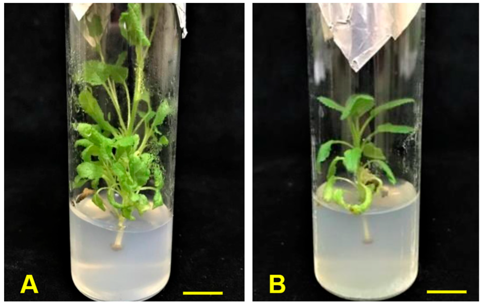
The S. byzantina explants fully responded to (100%) and produced new shoots during the multiplication stage. The highest shoot number was observed on the MS nutrient medium containing 5 µM of KIN, resulting in an average of six shoots/explant (Figure 1B). The lowest number of new shoots was observed on the media supplemented with BAP (1.7–2.9 shoots/explant) (Table 2, Figure 1A). In contrast, the use of 1–20 mg L−1 of BAP (~4.6–90 μΜ) in other Stachys species, like S. sieboldii and S. ocymastrum, led to the production of multiple shoots [10]. The same trend was reported by Mantovska et al. [42] in a study carried out on Stachys leucoglossa (a Balkan endemic species), which revealed that the most effective medium for in vitro multiplication was the MS medium containing 0.5 mg L−1 (~2.3 μΜ) of BAP. The application of KIN resulted in more than a two-fold increase in the number of new shoots compared to BAP (Figure 1A,B). Τhe shoot elongation was greater (5.7 cm), as expected, on the control media, while on the KIN-containing media, the shoot length was greater than the BAP-containing media (3.6–4.3 cm and 2.4–2.4 cm, respectively) (Table 2).
The proliferation rate observed in the present study was more than twice that reported by Legkobit and Khadeeva [10] on the B5 medium, which yielded 3.1 shoots per explant, with an average shoot length of 2.7 cm. Notably, even in the absence of growth regulators (control), both the shoot formation and proliferation rates were satisfactory, aligning with the findings by Panayotova et al. [43], who reported an excellent regeneration rate for Stachys maritima, with vigorous growth on a hormone-free MS medium. Nevertheless, an improvement in the shoot formation, and a sufficient length as well, was noticed when 5 μΜ of KIN was added to the nutrient medium. Hence, KIN in a low concentration (5 μΜ) was found to be the most efficient cytokinin for S. byzantina explant in vitro multiplication.

3.2.2. Effect of Nutrient Medium

All the explants cultured in either the MS media (Figure 2A) or WPM (Figure 2B) fully responded and formed new shoots. However, shoots formed in the MS media were more vigorous, while those cultured in WPM showed chlorosis after five weeks. The shoot proliferation rate was higher in the MS media compared to WPM, with the best results observed at a strength of 1:1 and 1:4 (2.9 shoots/explant) (Table 3). On the other hand, in WPM at a strength of 1:4, only 1.8 shoots/explant were formed. Legkobit and Khadeeva reported that the B5 medium was effective in regard to the in vitro establishment and multiplication of S. byzantina explants [10]. The present study did not evaluate the B5 medium due to the fact that the MS medium was shown to be effective in regard to in vitro shoot multiplication. Furthermore, the MS medium has also been reported as the most suitable medium for both the initial and multiplication stages for other Stachys species, i.e., S. leucoglossa [42] and S. maritima [43]. The strength of the nutrient medium was not a crucial factor for shoot multiplication in vitro. However, the MS medium proved to be superior to WPM in terms of shoot production (Table 3). The measurements of the shoot length indicated that MS strengths of 1:1 and 1:2 resulted in longer shoots (2.9–3.2 cm) compared to those of WPM (2.2–2.5 cm). At a strength of 1:4, the shoot lengths were not statistically different between the MS and WPM media (2.9 cm and 2.8 cm, respectively) (Table 3).
The effect of the nutrient medium strength may vary among different species and depends on the type and the physiological condition of the explants. Thus, the in vitro multiplication and growth of Typhonium flagelliforme was improved when the MS strength was decreased [44]. The same trend was also noticed in Delphinium cardinale, in which the best multiplication rate was assessed when the strength of the MS nutrient medium was reduced to 1:3 [45], as well as in Swertia chirata Buch, wherein the reduction in the strength of the medium resulted in a decline in the number and length of the shoots [46]. On the contrary, in Pogostemon cablin, more shoots were produced in full-strength MS compared to ½ strength MS [47]. In this study, the influence of various nutrient media strengths on the shoot multiplication of S. byzantina explants was rather limited in the MS medium, while a ¼ strength of WPM produced longer shoots than ½ or full-strength WPM.

3.3. Rooting of Microshoots

3.3.1. In Vitro Rooting

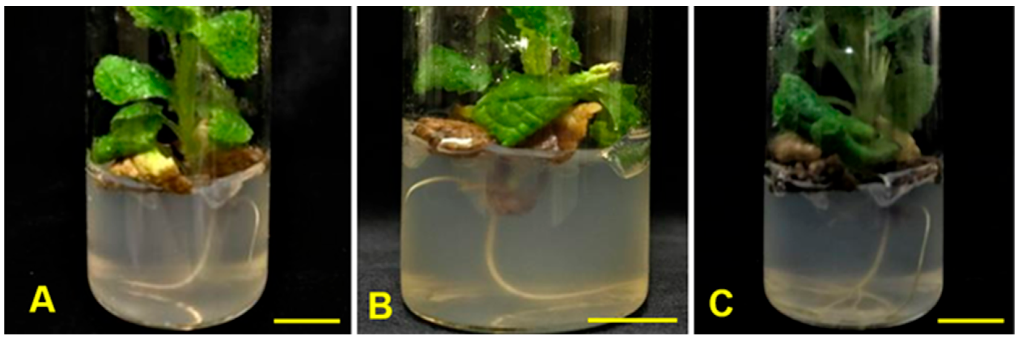
The rooting of shoots achieved high rates (73.3%), in a 5-week period, with the application of 10 μΜ of NAA, while no rooting was observed either in the control medium or in the medium containing 1 μΜ of IBA (Table 4). The rooting rate in the other two IBA treatments did not exceed 17%. On the other hand, the addition of NAA was crucial for the in vitro rooting of the shoots. As the concentration of NAA increased, higher rooting rates were achieved. Thus, a lower rooting rate was noticed in the treatment with 1 μΜ of NAA (10%); the application of 5 μΜ of NAA led to a rooting rate of 46.7%, while when 10 μΜ of NAA was applied, a rooting rate of 73.3% was recorded (Table 4, Figure 3B). The same trend was also noticed in regard to the number and length of the roots formed. Hence, when 1 μΜ of NAA was applied, two roots, 0.7 cm in length, were formed, while the addition of 10 μΜ of NAA, led to the development of more and longer roots (2.9 roots, 1.6 cm in length) (Table 4). Overall, the application of 5 or 10 μΜ of NAA led to the production of nearly twice the number and length of the roots compared to those formed with IBA (Figure 3A,C).
The shoots of S. byzantina formed roots promptly in high concentrations of NAA, exhibiting an adequate rooting ability and a well-formed rooting system. In contrast, Legkobit and Khadeeva [10] reported that bushy forms, with numerous shoots, whose internodes were closer to each other, were usually difficult to root or propagate via cuttings. Thus, they proposed the division of the bush into two or three parts and the supply of the nutrient medium with 0.5–1.0 mg L−1 of IBA and Gibberellin (~2.5–5 μΜ ΙΒA and 1.5–3 μΜ Gibberellin). The bushy form of the plants is usually not desired by nurseries, which prefer single-stemmed plants, so that they can shape them in any way they wish. Thus, in our study, from each in vitro culture, 2–3 rooted plantlets were produced, ready for transplanting.
The lack of rooting regulators in the nutrient medium resulted in the inability of S. byzantina shoots to form roots, which is in line with the findings by Kevers et al. [48], who stated that during the rooting induction phase, the exogenous application of auxins, even at low concentrations, promotes rooting. The formation of adventitious roots is generally a complex genetic trait, regulated by the interaction of environmental and endogenous factors, and auxin is a key regulator in this process [49]. It is evidence that some woody species, like Viburnum dentatum [50], root rather easily in vitro without auxin application, while other species, like Pyrus elaeagrifolia ‘Pallas’, require various auxins for in vitro rooting induction [51]. Mantovska et al. [42] reported that the rooting rate of Stachys leucoglossa in WPM media supplemented with 2.5 μΜ of IBA and 3 μΜ of NAA was 60% and 40%, respectively. As Legkobit and Khadeeva [10] stated, in vitro cultures of different Stachys species, growing under the same conditions, differed in regard to their behavior. Thus, the media and the concentrations of rooting regulators should be selected individually, for each case. In the present study, NAA enhanced the rooting of the shoots in terms of the root formation rate (%), root number, and root length. However, a high auxin concentration can be toxic when inducing ethylene production, which, in turn, can result in growth inhibition [52].

3.3.2. Ex Vitro Rooting

The ex vitro shoot rooting was affected by both the K-IBA application and the substrate used (Table 5). The application of 0.5 g L−1 of K-IBA was crucial for the rooting success and led to a 4–6-fold increase in the shoot rooting rates compared to untreated shoots. Thus, when K-IBA was applied in peat substrate, the rooting rate of the shoots increased from 16.7 (Figure 4C) to 73.3% (Figure 4A). Similarly, in the mixture of peat and perlite, the application of K-IBA led to an increase in the rooting rate from 13.3 to 86.7%. Furthermore, the mixture of peat/perlite (1:1) was more effective in regard to the rooting of S. byzantina shoots treated with K-IBA (Figure 4B) than those in peat (Figure 4A), whereas no statistically significant differences were found in the rooting of untreated shoots between the two substrates (Table 5). The number of roots was 4–5 times higher in substrates with K-IBA (Figure 4A,B) compared to substrates without K-IBA (Figure 4C,D). In the peat–perlite mixture, 6.2 roots/shoot were formed following the application of K-IBA (Table 5). The same trend was also noticed in regard to the length of the roots. Thus, longer roots were recorded in the peat–perlite mixture with the use of K-IBA (5.1 cm long), followed by peat with K-IBA (4.2 cm long) (Table 5).
A successful micropropagation system presumes that the formation of healthy roots has occurred for the subsequent establishment of the plantlets in the soil and the production of well-growing plants. Many studies report higher rooting and survival rates, as well as greater plant uniformity, as reasons for proposing the use of the in vitro rooting method for plant production, involving species such as Spiraea betulifolia [53], Pseudostellaria heterophylla [54], Siraitia grosvenorii [55], and Acer rubrum L. ‘Red Sunset’, Betula nigra L., and Malus × domestica Borkh ‘McIntosh’ [56]. This study highlighted the high rooting rate of microshoots treated ex vitro with K-IBA, due to their ability to adapt from a sterile and controlled in vitro environment (temperature, humidity, light intensity) to the natural environment of the greenhouse. The same trends were also reported for other plant species like Ceropegia bulbosa Roxb [57] and Siraitia grosvenorii [55].
The mixture of peat–perlite (1:1 v/v) was more efficient than peat in regard to promoting the rooting rate of S. byzantina shoots. Peat has low porosity and retains more moisture than perlite. The addition of perlite to a substrate aims to increase the porosity of the substrate, as well as to efficiently drain water, and, at the same time, allows oxygen to penetrate the soil [58].
The water-holding capacity and air in the substrate determine the amount of water to be added. A desirable substrate is one that is moistened immediately after irrigation and retains at least 10% or more of the volume of air afterwards [59]. An aerated substrate facilitates rooting and the development of a better root system. Conversely, peat retains a higher water potential than the peat–perlite mixture and has a lower aeration potential, with the possibility of retardation of rhizogenesis in the shoots. Furthermore, the fog system is used to maintain a high relative humidity in the atmosphere, which leads to low evaporation rates and water losses from the shoots through reduced transpiration, thus avoiding substantial tissue water deficits [60]. Thus, under these substrate and environmental conditions, the stress in the shoots, especially during the first weeks of rooting, is much less, exhibiting rapid recovery and, in the following weeks, a functional root system is formed [61]. The formation of more and longer roots in a mixture of peat–perlite (1:1), as compared with peat, bearing root hairs, has also been reported for Nerium oleander [62].

3.4. Acclimatization of Young Plantlets

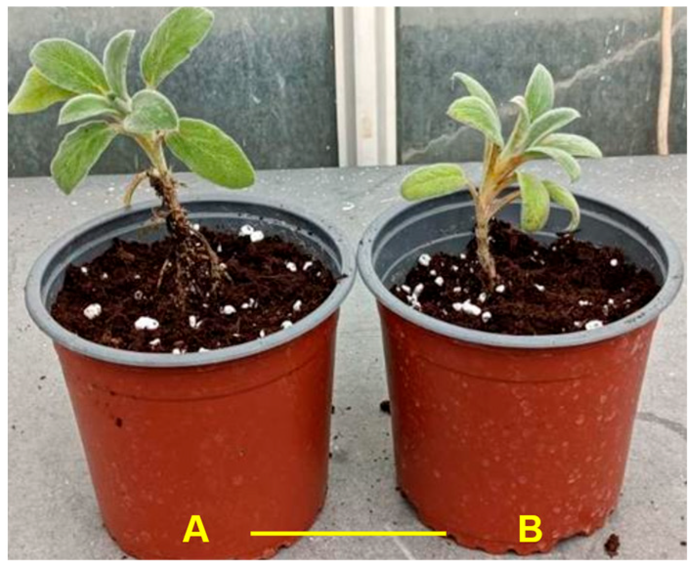
The acclimatization of the ex vitro-rooted plantlets was successful, with a high survival rate of 96.7%, compared to in vitro-rooted plantlets with a rate of 73.3% (Table 6; Figure 5A,B). The lower survival rate achieved by in vitro-rooted plantlets could be due to the structural characteristics of the rooting system. Kataoka [63] reported that the roots of Carica papaya growing in an agar medium showed structural abnormalities and lacked root hairs, which had adverse effects on their growth in field soil. In addition, McClelland et al. [56] stated that the availability of nutrients, water, and oxygen in an in vitro culture could negatively affect the development of a secondary phloem and xylem. However, long-term cultivation in the soil substrate eliminates these differences and the anatomy of the regenerated roots corresponds to that of normal plants.
The acclimatization of in vitro-produced plantlets requires their gradual adaptation to external environmental conditions and the transition of the root system from a non-functional structure to a functional one [61]. In the present study, the ex vitro-rooted plantlets formed a greater number of longer roots, with root hairs. On the other hand, the in vitro-rooted plantlets did not bear root hairs. This could be a crucial factor, influencing the survival rate of the plantlets at the acclimatization stage.
The physiology of the leaves of the plantlets derived from in vitro or ex vitro culture plays an important role in their successful acclimatization. In Pseudostellaria heterophylla plantlets produced in vitro, the stomata on the leaves were contiguous and open, the stomatal density was high and the stomatal function was abnormal, whereas plantlets derived from ex vitro cultures developed leaves with closed stomata and a low stomatal density [54]. The second case was attributed to the development of more functional leaves during rooting in inert substrates, facilitating the acclimatization of the plantlets to the external environment. Usually, a gradual reduction of the ambient relative humidity, coupled with a gradual increase in the light irradiance, greatly contributes to plantlet hardening towards successful acclimatization [61].
In the present study, the plantlets of S. byzantina that were rooted ex vitro easily acclimatized to the new environment, while those that were rooted in vitro were much more stressed during the first days of their adaptation to natural environmental conditions, resulting in a reduction in the survival rate by more than 26%. Nevertheless, all surviving plantlets, regardless of their rooting in vitro or ex vitro, subsequently had healthy and robust growth, with normal morphological characteristics (Figure 5A,B).

3.5. Genetic Stability

One of the main goals in the mass propagation of plants through tissue culture is the production of uniform plantlets of assured genetic fidelity. Hence, it is important to ensure that there is no genetic variation between the mother plant (the donor of the explants) and the produced plants. The genetic stability of plantlets produced by in vitro propagation should be examined prior to any commercial exploitation [64,65].
In this study, the screening involving nine ISSR primers (Table 7) generated 69 scorable and reproducible zones and a total of 897 zones (number of zones X number of gDNA samples), ranging in size from 200 to 1700 bp. The number of scorable zones for each primer varied from five (UBC 821) to nine (UBC 807 and 815) (Figure 6), with an average of 7.67 zones per primer. The ISSR-based zones were monomorphic and no genetic variation was detected among the 12 randomly selected plantlets, which were produced from the in vitro cultures and the S. byzantina mother plant. The results indicated that S. byzantina plantlets derived from in vitro cultures exhibited genetic stability after five weeks of acclimatization and resembled the mother plant based on their ISSR profiles.
These results confirm previous reports, wherein genetic stability using molecular markers has been identified in many other micropropagated ornamental plants, such as cold-stored artificial seeds of the woody species, Rauvolfia tetraphylla [66] and Viburnum dentatum [67]. ISSR markers, due to their simplicity and cost effectiveness, have been successfully employed to assess the genetic stability of many woody species [68,69,70,71,72].
The present study demonstrated that the in vitro-produced plantlets showed genetic uniformity both among themselves and compared to the mother plant (the donor of the explants), without any morphological differences being observed, underlining the reliability of the in vitro production protocol used.

4. Conclusions

The results of this study showed that in vitro cultures of S. byzantina can be successfully established in summer, with a protocol involving explant disinfestation, using a combination of NaOCl (1%, 3 min) and HgCl2 (0.08%, 5 min). For shoot multiplication, the addition of 5 μΜ of KIN to an MS medium results in a high proliferation rate (six shoots per explant). Furthermore, the ex vitro rooting of microshoots, using K-IBA (0.5 g L−1), in a mixture of peat–perlite (1:1), ensures a very high rooting rate (86.7%), which is also followed by a high survival rate (96.7%) of the plantlets after the acclimatization stage. The acclimatized plantlets showed genetic uniformity with the mother plant, as well as with each other, and, therefore, this protocol would be valuable in terms of its exploitation by the floriculture industry.

Author Contributions

Conceptualization, S.H. and A.E.; methodology, S.H., A.E., A.-A.K., K.B. and A.D.; software, S.K., C.K. and A.-A.K.; validation, S.H., C.K. and A.-A.K.; formal analysis, A.E. and C.K.; investigation, S.H., C.K., and, A.-A.K.; resources, S.K. and C.K.; data curation, S.H., C.K. and A.-A.K.; writing—original draft preparation, S.H., S.K., A.-A.K., A.E., K.B. and A.D.; writing—review and editing, S.H., S.K., A.E., K.B. and A.D.; visualization, S.K., A.E. and A.-A.K.; supervision, S.H., S.K. and A.E.; project administration, S.H. and S.K.; funding acquisition, S.H. and S.K. All authors have read and agreed to the published version of the manuscript.

Funding

This research received no external funding.

Data Availability Statement

All the data are presented in the article. Further inquiries can be directed to the corresponding author.

Acknowledgments

We would like to thank the technical staff at the Floriculture Laboratory as part of Aristotle University for their contribution to this publication.

Conflicts of Interest

The authors declare that there are no conflicts of interest.

References

  1. Seaton, K.; Bettin, A.; Grüneberg, H. New ornamental plants for horticulture. In Horticulture: Plants for People and Places, Volume 1; Dixon, G.R., Aldous, D.E., Eds.; Springer: Dordrecht, The Netherlands, 2014; pp. 435–463. ISBN 978-94-017-8577-8. [Google Scholar]
  2. Vascular Flora of Greece Web (Vascular Plants of Greece-An Annotated Checklist). Available online: https://portal.cybertaxonomy.org/flora-greece/intro (accessed on 1 August 2023).
  3. Asnaashari, S.; Delazar, A.; Alipour, S.S.; Nahar, L.; Williams, A.S.; Pasdaran, A.; Mojarab, M.; Azad, F.F.; Sarker, S. Chemical composition, free-radical-scavenging and insecticidal activities of the aerial parts of Stachys byzantina. Arch. Biol. Sci. 2010, 62, 653–662. [Google Scholar] [CrossRef]
  4. North Carolina Plant Toolbox, Stachys byzantina—Plant Information. Available online: https://plants.ces.ncsu.edu/plants/stachys-byzantina/?utm_source=chatgpt.com (accessed on 5 April 2025).
  5. Devecchi, M. The use of Labiatae of ornamental interest in the design of parks and gardens. Acta Hortic. 2006, 723, 51–58. [Google Scholar] [CrossRef]
  6. Kemp, S.; Hadley, P.; Blanuša, T. The Influence of plant type on green roof rainfall retention. Urban Ecosyst. 2019, 22, 355–366. [Google Scholar] [CrossRef]
  7. Naghibi, F.; Mosadegh, M.; Mohammadi, M.S.; Ghorbani, A. Labiatae family in folk medicine in Iran: From ethnobotany to pharmacology. Iran. J. Pharm. Res. 2005, 2, 2. [Google Scholar]
  8. Aminfar, P.; Abtahi, M.; Parastar, H. Gas chromatographic fingerprint analysis of secondary metabolites of Stachys lanata (Stachys byzantine C. Koch) Combined with Antioxidant Activity Modelling Using Multivariate Chemometric Methods. J. Chromat. A 2019, 1602, 432–440. [Google Scholar] [CrossRef]
  9. Tomou, E.-M.; Barda, C.; Skaltsa, H. Genus Stachys: A review of traditional uses, phytochemistry and bioactivity. Medicines 2020, 7, 63. [Google Scholar] [CrossRef]
  10. Legkobit, M.P.; Khadeeva, N.V. Variation and morphogenetic characteristics of different Stachys species during microclonal propagation. Russ. J. Genet. 2004, 40, 743–750. [Google Scholar] [CrossRef]
  11. Grigoriadou, K.; Krigas, N.; Sarropoulou, V.; Papanastasi, K.; Tsoktouridis, G.; Maloupa, E. In vitro propagation of medicinal and aromatic plants: The case of selected Greek species with conservation priority. Vitro Cell. Dev. Biol.-Plant 2019, 55, 635–646. [Google Scholar] [CrossRef]
  12. Katsanou, C.E.; Kostas, S.; Bantis, F.; Bertsouklis, K.; Hatzilazarou, S. Optimizing in vitro propagation of “Ladania” (Cistus creticus L.) through interaction of light spectra and plant growth regulators. Agronomy 2025, 15, 774. [Google Scholar] [CrossRef]
  13. Rai, A.C.; Kumar, A.; Modi, A.; Singh, M. Advances in Plant Tissue Culture: Current Developments and Future Trends; Academic Press: Cambridge, MA, USA, 2022; ISBN 0-323-99819-4. [Google Scholar]
  14. Ahmad, N.; Faisal, M.; Ahmad, A.; Alatar, A.A.; Qahtan, A.A.; Alok, A. Thidiazuron induced in vitro clonal propagation of Lagerstroemia speciosa (L.) Pers.—An important avenue Tree. Horticulturae 2022, 8, 359. [Google Scholar] [CrossRef]
  15. Yasemin, S.; Beruto, M. A Review on flower bulb micropropagation: Challenges and opportunities. Horticulturae 2024, 10, 284. [Google Scholar] [CrossRef]
  16. Bhojwani, S.S.; Dantu, P.K. Micropropagation. In Plant Tissue Culture: An Introductory Text; Springer: New Delhi, India, 2013; pp. 245–274. ISBN 978-81-322-1025-2. [Google Scholar]
  17. Podwyszyńska, M.; Orlikowska, T.; Trojak-Goluch, A.; Wojtania, A. Application and improvement of in vitro culture systems for commercial production of ornamental, fruit, and industrial plants in Poland. Acta Soc. Bot. Pol. 2022, 91, 914. [Google Scholar] [CrossRef]
  18. Bairu, M.W.; Fennell, C.W.; van Staden, J. The effect of plant growth regulators on somaclonal variation in Cavendish banana (Musa AAA Cv.‘Zelig’). Sci. Hortic. 2006, 108, 347–351. [Google Scholar] [CrossRef]
  19. Haisel, D.; Hofman, P.; Vágner, M.; Lipavska, H.; Tichá, I.; Schäfer, C.A.; Čapková, V. Ex vitro phenotype stability is affected by in vitro cultivation. Biol. Plant. 2001, 44, 321–324. [Google Scholar] [CrossRef]
  20. Zietkiewicz, E.; Rafalski, A.; Labuda, D. Genome fingerprinting by Simple Sequence Repeat (SSR)-Anchored polymerase chain reaction amplification. Genomics 1994, 20, 176–183. [Google Scholar] [CrossRef]
  21. Ai, X.; Wen, Y.; Wang, B. Assessment of genetic stability on in vitro propagation of Ardisia crenata var. bicolor using ISSR markers. Int. J. Plant Biol. 2023, 14, 218–227. [Google Scholar] [CrossRef]
  22. Kader, A.; Sinha, S.N.; Ghosh, P. A standardized protocol for genetically stable artificial seed production of Ficus religiosa L. using ISSR fingerprinting. J. Appl. Genet. 2023, 64, 275–287. [Google Scholar] [CrossRef]
  23. Jung, W.-S.; Chung, I.-M.; Kim, S.-H.; Chi, H.-Y.; Yu, C.Y.; Ghimire, B.K. Direct shoot organogenesis from Lycium chinense Miller leaf explants and assessment of genetic stability using ISSR markers. Agronomy 2021, 11, 503. [Google Scholar] [CrossRef]
  24. Zamani, M.; Sonboli, A.; Goldansaz, M.; Mirjalili, M.H. In vitro micropropagation and conservation of endangered medicinal plant Nepeta asterotricha Rech. f. (Lamiaceae): Genetic fidelity, phytochemical and biological assessment. Physiol. Mol. Biol. Plants 2024, 30, 67–80. [Google Scholar] [CrossRef]
  25. Thakur, M.; Sharma, V.; Luharch, R. Propagation of plum (Prunus salicina L.) cultivar frontier in vitro through control of shoot tip necrosis (stn) and validation of genetic integrity using ISSR markers. Plant Physiol. Rep. 2021, 26, 238–246. [Google Scholar] [CrossRef]
  26. Yıldırım, M.; Tuğ, G.N.; Yaprak, A.E. Analyses of genetic diversity and population structure of endemic and endangered species Sideritis gulendamii (Lamiaceae) and implications for its conservation. Genet. Resour. Crop Evol. 2024, 71, 4331–4345. [Google Scholar] [CrossRef]
  27. Chirumamilla, P.; Gopu, C.; Jogam, P.; Taduri, S. Highly efficient rapid micropropagation and assessment of genetic fidelity of regenerants by ISSR and SCoT markers of Solanum khasianum Clarke. Plant Cell Tiss. Org. Cult. 2021, 144, 397–407. [Google Scholar] [CrossRef]
  28. Hatzilazarou, S.; Kostas, S.; Nendou, T.; Economou, A. Conservation, regeneration and genetic stability of regenerants from alginate-encapsulated shoot explants of Gardenia jasminoides Ellis. Polymers 2021, 13, 1666. [Google Scholar] [CrossRef]
  29. Akin, B.; Bingöl, N.A.; Bulman, G.C. A first approach for the micropropagation of threatened endemic subspecies of Stachys cretica subsp. kutahyensis. Biologia 2024, 79, 1653–1661. [Google Scholar] [CrossRef]
  30. Cüce, M.; Bekircan, T.; Laghari, A.H.; Sökmen, M.; Sökmen, A.; Uçar, E.Ö.; Kılıç, A.O. Antioxidant phenolic constituents, antimicrobial and cytotoxic properties of Stachys annua L. from both natural resources and micropropagated plantlets. Indian J. Tradit. Knowl. 2017, 16, 407–416. [Google Scholar]
  31. Hadi, S.; Ahmadabadi, M.; Valizadeh Kamran, R. Efficient in vitro callus induction, regeneration and shoot multiplication protocols for Stachys inflata subsp. caucasica (Stschegl.); A rare medicinal plant. J. Med. Plants -Prod. 2024, 13, 570–576. [Google Scholar]
  32. Mantovska, D.I.; Zhiponova, M.K.; Petrova, D.; Alipieva, K.; Bonchev, G.; Boycheva, I.; Evstatieva, Y.; Nikolova, D.; Tsacheva, I.; Simova, S.; et al. Exploring the phytochemical composition and biological potential of Balkan endemic species Stachys scardica Griseb. Plants 2023, 13, 30. [Google Scholar] [CrossRef]
  33. Gamborg, O.; Murashige, T.; Thorpe, T.; Vasil, I. Plant tissue culture media. In Vitro 1976, 12, 473–478. [Google Scholar] [CrossRef]
  34. Murashige, T.; Skoog, F. A revised medium for rapid growth and bio assays with tobacco tissue cultures. Physiol. Plant. 1962, 15, 473–497. [Google Scholar] [CrossRef]
  35. McCown, B. Woody plant medium (WPM)-a mineral nutrient formulation for microculture for woody plant species. Hort. Sci. 1981, 16, 453. [Google Scholar]
  36. Tsaktsira, M.; Chavale, E.; Kostas, S.; Pipinis, E.; Tsoulpha, P.; Hatzilazarou, S.; Ziogou, F.-T.; Nianiou-Obeidat, I.; Iliev, I.; Economou, A. Vegetative propagation and ISSR-based genetic identification of genotypes of Ilex aquifolium ‘Agrifoglio commune’. Sustainability 2021, 13, 10345. [Google Scholar] [CrossRef]
  37. Kartsonas, E.; Papafotiou, M. Mother plant age and seasonal influence on in vitro propagation of Quercus euboica Pap., an endemic, rare and endangered oak species of Greece. Plant Cell Tiss. Org. Cult. 2007, 90, 111–116. [Google Scholar] [CrossRef]
  38. Wang, Y.; Yao, R. Plantlet regeneration of adult Pinus massoniana Lamb. Trees using explants collected in March and thidiazuron in culture medium. J. For. Res. 2017, 28, 1169–1175. [Google Scholar] [CrossRef]
  39. Bertsouklis, K.; Paraskevopoulou, A.T.; Petraki, E. In vitro regeneration from adult node explants of Juniperus oxycedrus. Not. Bot. Horti Agrobot. 2023, 51, 13062. [Google Scholar] [CrossRef]
  40. Loureiro, J.; Capelo, A.; Brito, G.; Rodriguez, E.; Silva, S.; Pinto, G.; Santos, C. Micropropagation of Juniperus phoenicea from adult plant explants and analysis of ploidy stability using flow cytometry. Biol. Plant. 2007, 51, 7–14. [Google Scholar] [CrossRef]
  41. Babu, G.A.; Mosa Christas, K.; Kowsalya, E.; Ramesh, M.; Sohn, S.-I.; Pandian, S. Improved sterilization techniques for successful in vitro micropropagation. In Commercial Scale Tissue Culture for Horticulture and Plantation Crops; Gupta, S., Chaturvedi, P., Eds.; Springer Nature: Singapore, 2022; pp. 1–21. ISBN 978-981-19-0054-9. [Google Scholar]
  42. Mantovska, D.I.; Kapchina, V.M.; Yordanova, Z.P. In vitro propagation of the Balkan endemic species Stachys leucoglossa Griseb. Bulg. J. Agric. Sci. 2019, 25, 1211–1215. [Google Scholar]
  43. Panayotova, L.G.; Ivanova, T.A.; Bogdanova, Y.Y.; Gussev, C.V.; Stanilova, M.I.; Bosseva, Y.Z.; Stoeva, T.D. In vitro cultivation of plant species from sandy dunes along the Bulgarian Black Sea coast. Phytol. Balc. 2008, 14, 119–123. [Google Scholar]
  44. Rezali, N.I.; Sidik, N.J.; Saleh, A.; Osman, N.I.; Adam, N.A.M. The effects of different strength of MS media in solid and liquid media on in vitro growth of Typhonium flagelliforme. Asian Pac. J. Trop. Biomed. 2017, 7, 151–156. [Google Scholar] [CrossRef]
  45. Ohki, S.; Sawaki, S. The effects of inorganic salts and growth regulators on in vitro shoot proliferation and leaf chlorophyll content of Delphinium cardinale. Sci. Hortic. 1999, 81, 149–158. [Google Scholar] [CrossRef]
  46. Pant, M.; Bisht, P.; Gusain, M.P. Effect of sucrose and media strength on in vitro multiplication in Swertia shirata Buch.-Ham ex wall: An endangered medicinal herb. Int. J. Pharm. Pharm. Sci. 2018, 10, 40–42. [Google Scholar] [CrossRef]
  47. Hida, W.N.; Norrizah, J.; Aminah, S.S.; Ruzaina, S.S.; Faezah, P. Effect of medium strength and hormones concentration on regeneration of Pogostemon cablin using nodes explant. Asian J. Biotechnol. 2012, 4, 46–52. [Google Scholar] [CrossRef]
  48. Kevers, C.; Hausman, J.-F.; Faivre-Rampant, O.; Evers, D.; Gaspar, T. Hormonal control of adventitious rooting: Progress and questions. Angew. Bot. 1997, 71, 71–79. [Google Scholar]
  49. Pacurar, D.I.; Perrone, I.; Bellini, C. Auxin is a central player in the hormone cross-talks that control adventitious rooting. Physiol. Plant. 2014, 151, 83–96. [Google Scholar] [CrossRef] [PubMed]
  50. Hatzilazarou, S.; Rifaki, N.; Patsou, M.; Kostas, S.; Economou, A.S. In vitro propagation of Viburnum dentatum L. ‘Lucidum Aiton’. Prop. Ornam. Plants 2009, 9, 39–42. [Google Scholar]
  51. Aygun, A.; Dumanoglu, H. In vitro shoot proliferation and in vitro and ex vitro root formation of Pyrus elaeagrifolia Pallas. Front. Plant Sci. 2015, 6, 225. [Google Scholar] [CrossRef]
  52. Gaba, V.P. Plant growth regulators in plant tissue culture and development. In Plant Development and Biotechnology; CRC Press: Boca Raton, FL, USA, 2005; pp. 87–99. [Google Scholar]
  53. Muraseva, D.; Kostikova, V. In vitro and ex vitro rooting of Spiraea betulifolia subsp. aemiliana (Rosaceae), an ornamental shrub. EDP Sci. 2020, 24, 00058. [Google Scholar] [CrossRef]
  54. Wang, F.; Xin, X.; Wei, H.; Qiu, X.; Liu, B. In vitro regeneration, ex vitro rooting and foliar stoma studies of Pseudostellaria heterophylla (Miq.) Pax. Agronomy 2020, 10, 949. [Google Scholar] [CrossRef]
  55. Yan, H.; Liang, C.; Yang, L.; Li, Y. In vitro and ex vitro rooting of Siratia grosvenorii, a traditional medicinal plant. Acta Physiol. Plant. 2010, 32, 115–120. [Google Scholar] [CrossRef]
  56. McClelland, M.; Smith, M.; Carothers, Z. The effects of in vitro and ex vitro root initiation on subsequent microcutting root quality in three woody plants. Plant Cell Tiss. Org. Cult. 1990, 23, 115–123. [Google Scholar] [CrossRef]
  57. Phulwaria, M.; Shekhawat, N.; Rathore, J.; Singh, R. An efficient in vitro regeneration and ex vitro rooting of Ceropegia bulbosa Roxb.—A threatened and pharmaceutical important plant of Indian Thar Desert. Ind. Crops Prod. 2013, 42, 25–29. [Google Scholar] [CrossRef]
  58. Kormanek, M.; Małek, S.; Banach, J.; Durło, G.; Jagiełło-Leńczuk, K.; Dudek, K. Seasonal changes of perlite–peat substrate properties in seedlings grown in different sized container trays. New For. 2021, 52, 271–283. [Google Scholar] [CrossRef]
  59. Cabrera, P.I.; Johnson, J.R. Fundamentals of Container Media Management: Part 1. Greenhouse and Nursery Crops Fact Sheets & Bulletins; Rutgers Fact Sheet FS 812; The State University of New Jersey: New Brunswick, NJ, USA, 2014; p. 3. [Google Scholar]
  60. Mateja, Š.; Dominik, V.; Franci, Š.; Gregor, O. The effects of a fogging system on the physiological status and rooting capacity of leafy cuttings of woody species. Trees 2007, 21, 491–496. [Google Scholar] [CrossRef]
  61. Economou, A.S. From microcutting rooting to microplant establishment: Key points to consider for maximum success in woody plants. Acta Hortic. 2013, 988, 43–56. [Google Scholar] [CrossRef]
  62. Hatzilazarou, S.; Kostas, S.; Economou, A.; Scaltsoyiannes, A. Efficient propagation of Nerium oleander L. through tissue culture. Prop. Ornam. Plants 2017, 17, 64–74. [Google Scholar]
  63. Kataoka, I. Influence of rooting substrates on the morphology of papaya root formed in vitro. Jap. J. Trop. Agric. 1994, 38, 251–257. [Google Scholar]
  64. Biswas, P.; Kumar, N. Application of molecular markers for the assessment of genetic fidelity of in vitro raised plants: Current status and future prospects. In Molecular Marker Techniques; Kumar, N., Ed.; Springer Nature: Singapore, 2023; pp. 233–256. ISBN 978-981-99-1611-5. [Google Scholar]
  65. Martínez, O. Selection of molecular markers for the estimation of somaclonal variation. In Plant Cell Culture Protocols; Loyola-Vargas, V.M., Ochoa-Alejo, N., Eds.; Methods in Molecular Biology; Springer: New York, NY, USA, 2018; Volume 1815, pp. 103–129. ISBN 978-1-4939-8593-7. [Google Scholar]
  66. Alatar, A.; Faisal, M. Encapsulation of Rauvolfia tetraphylla microshoots as artificial seeds and evaluation of genetic fidelity using RAPD and ISSR markers. J. Med. Plants Res. 2012, 6, 1367–1374. [Google Scholar] [CrossRef]
  67. Hatzilazarou, S.; Kostas, S.; Joachim, M.; Economou, A. Regeneration of Viburnum dentatum L. from alginate-encapsulated shoot explants after short-term cold storage and assessment of genetic stability using ISSR analysis. Agronomy 2020, 10, 1660. [Google Scholar] [CrossRef]
  68. Bramhanapalli, M.; Thogatabalija, L.; Gudipalli, P. Efficient in vitro plant regeneration from seedling-derived explants and genetic stability analysis of regenerated plants of Simarouba glauca DC. by RAPD and ISSR markers. Vitro Cell. Dev. Biol.-Plant 2017, 53, 50–63. [Google Scholar] [CrossRef]
  69. Ilczuk, A.; Jacygrad, E. In vitro propagation and assessment of genetic stability of acclimated plantlets of Cornus alba L. using RAPD and ISSR markers. Vitro Cell. Dev. Biol.-Plant 2016, 52, 379–390. [Google Scholar] [CrossRef]
  70. Soares, D.M.M.; Sattler, M.C.; Ferreira, M.F.d.S.; Praça-Fontes, M.M. Assessment of genetic stability in three generations of in vitro propagated Jatropha curcas L. plantlets using ISSR markers. Trop. Plant Biol. 2016, 9, 229–238. [Google Scholar] [CrossRef]
  71. Amin, S.; Wani, T.A.; Kaloo, Z.A.; Singh, S.; John, R.; Majeed, U.; Shapoo, G.A. Genetic stability using RAPD and ISSR markers in efficiently in vitro regenerated plants of Inula royleana DC. Meta Gene 2018, 18, 100–106. [Google Scholar] [CrossRef]
  72. Ali, H.; Musa, I.F.; Abu Bakar, N.A.; Karsani, S.A.; Yaacob, J.S. In vitro regeneration and ISSR-based genetic fidelity analysis of Orthosiphon stamineus Benth. Agronomy 2019, 9, 778. [Google Scholar] [CrossRef]
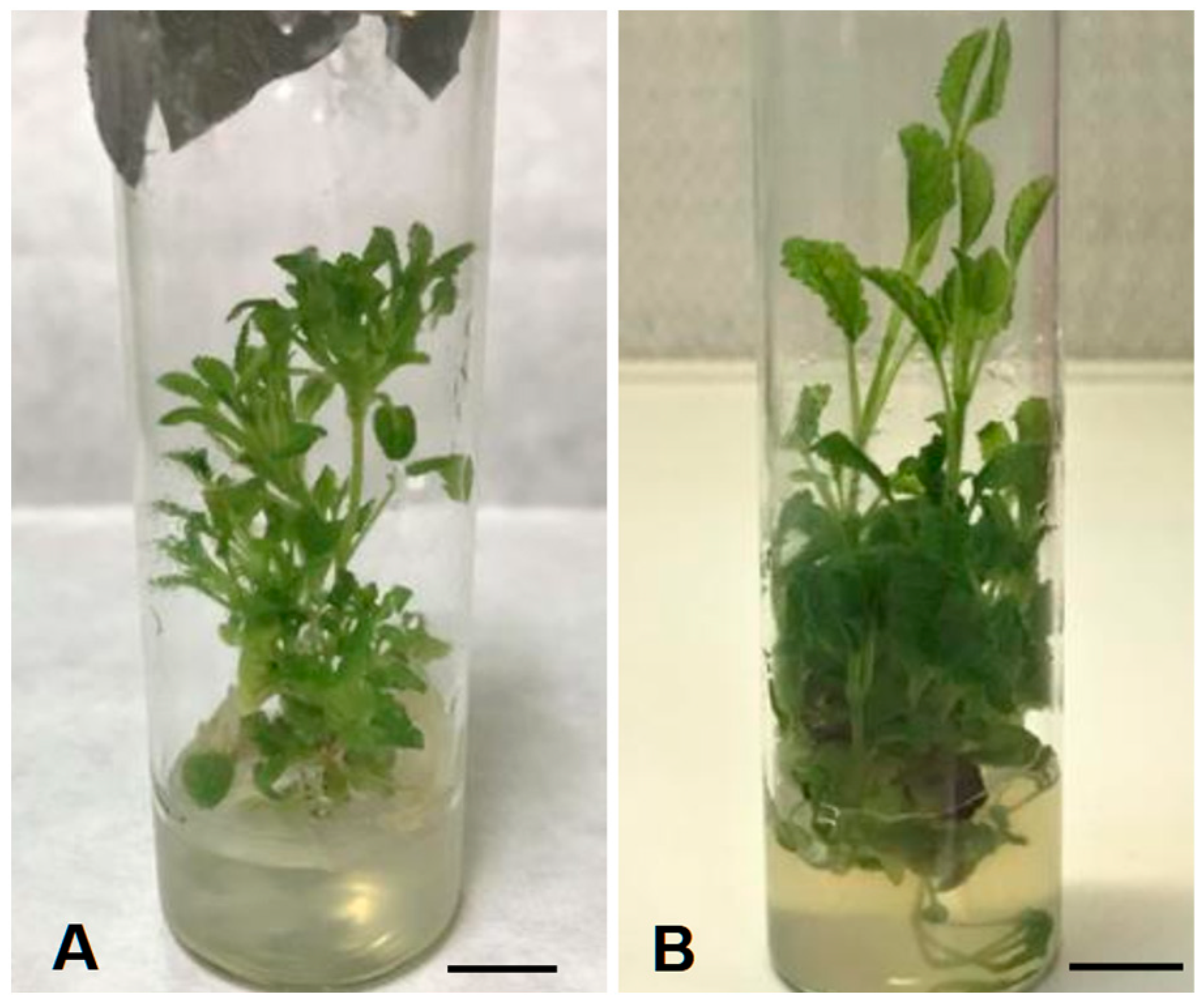
Figure 1. In vitro response of S. byzantina explants during multiplication stage in terms of shoot formation, after 5 weeks of culture on MS media, containing: (A) 5 μΜ BAP; and (B) 5 μΜ KIN. The black bar in the images corresponds to a size of 1 cm.
Horticulturae 11 00530 g001
Figure 2. Multiple shoot production by S. byzantina explants in: (A) the full-strength MS medium; and (B) the WPM medium. The yellow bar in the images corresponds to a size of 1 cm.
Horticulturae 11 00530 g002
Figure 3. In vitro rooting of S. byzantina shoots in MS nutrient medium, supplemented with: (A) 5 μΜ IBA; (B) 5 μΜ NAA; and (C) 10 μΜ IBA. The yellow bar in the images corresponds to a size of 1 cm.
Horticulturae 11 00530 g003
Figure 4. Ex vitro rooting of S. byzantina shoots following: (A) immersion of their base in 0.5 g L−1 K-IBA and planting in peat; (B) immersion of their base in 0.5 g L−1 K-IBA and planting in the peat–perlite mixture (1:1); (C) planting without K-IBA in peat; and (D) planting without K-IBA in the peat–perlite (1:1) mixture. The yellow bar in the images corresponds to a size of 1 cm.
Horticulturae 11 00530 g004
Figure 5. Plantlets of S. byzantina, which have been rooted ex vitro or in vitro (A,B), six weeks after transplanting into a mixture of three parts peat and one part perlite (v/v). The yellow bar in the image corresponds to a size of 10 cm.
Horticulturae 11 00530 g005
Figure 6. Amplification profiles of eight plantlets (P1–P8), derived from in vitro cultures and mother plant (MP) of S. byzantina, obtained using primers ISSR UBC 810 (A) and ISSR UBC 815 (B). The size of the amplified bands was calculated using a 1 kb DNA ladder (200–3000 bp). The image has been converted to a negative and to black and white.
Horticulturae 11 00530 g006
Table 1. Survival rates (%) following disinfestation of S. byzantina explants, observed during the spring and summer period (mean ± standard deviation).
Explant Collection PeriodSurvival Rate (%)
Spring61.0 ± 3.3 b *
Summer70.0 ± 3.3 a
* The means with different letters are significantly different, according to Duncan’s multiple range test, at p ≤ 0.05.
Table 2. Effect of different concentrations (0.5, 10, 20 μM) of BAP and KIN on shoot number and shoot length of S. byzantina explants in vitro (mean ± standard deviation).
Concentration
(μΜ)
Number of ShootsLength of Shoots (cm)
BAPKINBAPKIN
0 (control)3.7 ± 0.25 c *3.7 ± 0.25 c5.7 ± 0.35 a *5.7 ± 0.35 a
52.9 ± 0.28 d6.0 ± 0.50 a2.7 ± 0.28 d3.6 ± 0.50 c
101.9 ± 0.28 e4.9 ± 0.28 b2.9 ± 0.38 d4.3 ± 0.30 b
201.7 ± 0.28 e3.0 ± 0.50 d2.4 ± 0.15 d3.7 ± 0.20 c
* The means, among the eight treatments, with different letters are significantly different, according to Duncan’s multiple range test, at p ≤ 0.05.
Table 3. Effect of different strengths (1:1, 1:2, 1:4) of WPM or MS nutrient media on the number and length of shoots of S. byzantina explants produced in vitro, after five weeks of culture (mean ± standard deviation).
Nutrient Medium
Strength
Number of ShootsLength of Shoots (cm)
WPMMSWPMMS
1:12.2 ± 0.20 cd *2.9 ± 0.10 a2.2 ± 0.11 c *3.2 ± 0.15 a
1:22.1 ± 0.23 cd2.5 ± 0.10 bc2.5 ± 0.10 bc3.0 ± 0.11 a
1:41.8 ± 0.20 d2.9 ± 0.50 a2.8 ± 0.47 ab2.9 ± 0.10 ab
* The means, among the six treatments, with different letters are significantly different, according to Duncan’s multiple range test, at p ≤ 0.05.
Table 4. In vitro rooting (%), number and length of roots of S. byzantina shoots, in MS media, with the addition of different concentrations (0, 1, 5, or 10 μM) of IBA and NAA (mean ± standard deviation).
Concentration
(μΜ)
Rooting (%)Number of RootsLength of Roots (cm)
IBANAAIBANAAIBANAA
0 (control) 000000
1010.0 ± 2.70 c *02.0 ± 1.0 c *00.7 ± 0.2 cd *
513.3 ± 8.82 c46.7 ± 3.35 b1.3 ± 0.57 c2.6 ± 0.2 b0.5 ± 0.1 d1.1 ± 0.1 b
1016.7 ± 3.35 c73.3 ± 6.65 a1.7 ± 0.57 c2.9 ± 0.2 a0.8 ± 0.2 c1.6 ± 0.2 a
* The means, among the eight treatments, of each rooting regulator with different letters are significantly different, according to Duncan’s multiple range test, at p ≤ 0.05.
Table 5. Ex vitro rooting rate (%), number, and length of roots of S. byzantina shoots in different substrates, with or without the use of K-IBA (0.5 g L−1) (mean ± standard deviation).
SubstrateRooting (%)Number of RootsLength of Roots (cm)
+K-IBA−K-IBA+K-IBA−K-IBA+K-IBA−K-IBA
Peat73.3 ± 3.3 b *16.7 ± 3.35 c4.8 ± 0.2 b *1.0 ± 0.1 d4.2 ± 0.26 b *1.4 ± 0.3 c
Peat–Perlite (1:1)86.7 ± 6.65 a13.3 ± 3.35 c6.2 ± 0.26 a1.3 ± 0.2 c5.1 ± 0.26 a0.9 ± 0.2 d
* The means, among the four treatments, with different letters are significantly different, according to Duncan’s multiple range test, at p ≤ 0.05.
Table 6. Survival rate (%) of plantlets of S. byzantina, rooted in vitro or ex vitro, five weeks after transplanting (mean ± standard deviation).
RootingSurvival (%)
In vitro73.3 ± 3.35 b *
Ex vitro96.7 ± 3.86 a
* The means with different letters are significantly different, according to Duncan’s multiple range test, at p ≤ 0.05.
Table 7. Sequence, annealing temperature, and number of zones of nine ISSR primers, used to determine the genetic stability of S. byzantina plantlets produced from in vitro cultures.
PrimerPrimer Sequence (5′-3′)Annealing
Temperature (°C)
Number of Bands
ISSR UBC 807AGA GAG AGA GAG AGA GT569
ISSR UBC 809AGA GAG AGA GAG AGA GG588
ISSR UBC 810GAG AGA GAG AGA GAG AT527
ISSR UBC 811GAG AGA GAG AGA GAG AC547
ISSR UBC 812GAG AGA GAG AGA GAG AA548
ISSR UBC 815CTC TCT CTC TCT CTC TG509
ISSR UBC 816CAC ACA CAC ACA CAC AT547
ISSR UBC 818CAC ACA CAC ACA CAC AG569
ISSR UBC 821GTG TGT GTG TGT GTG TT545
Disclaimer/Publisher’s Note: The statements, opinions and data contained in all publications are solely those of the individual author(s) and contributor(s) and not of MDPI and/or the editor(s). MDPI and/or the editor(s) disclaim responsibility for any injury to people or property resulting from any ideas, methods, instructions or products referred to in the content.