
Application of Alginate-Based Hydrogels in Hemostasis


Abstract
:1. Introduction
2. Alginate Hydrogels
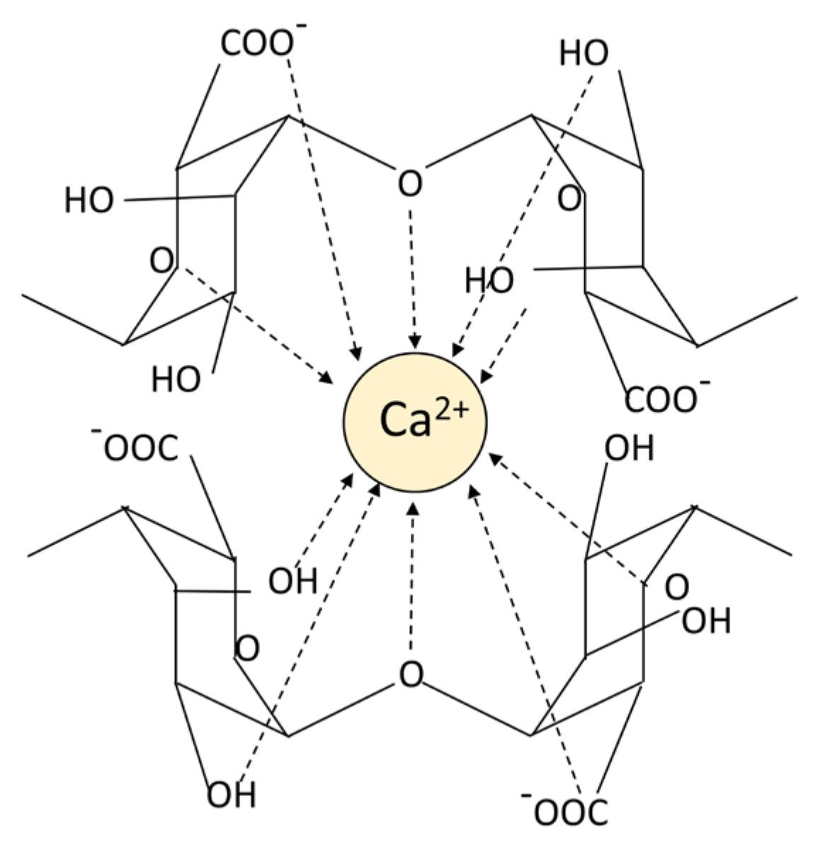
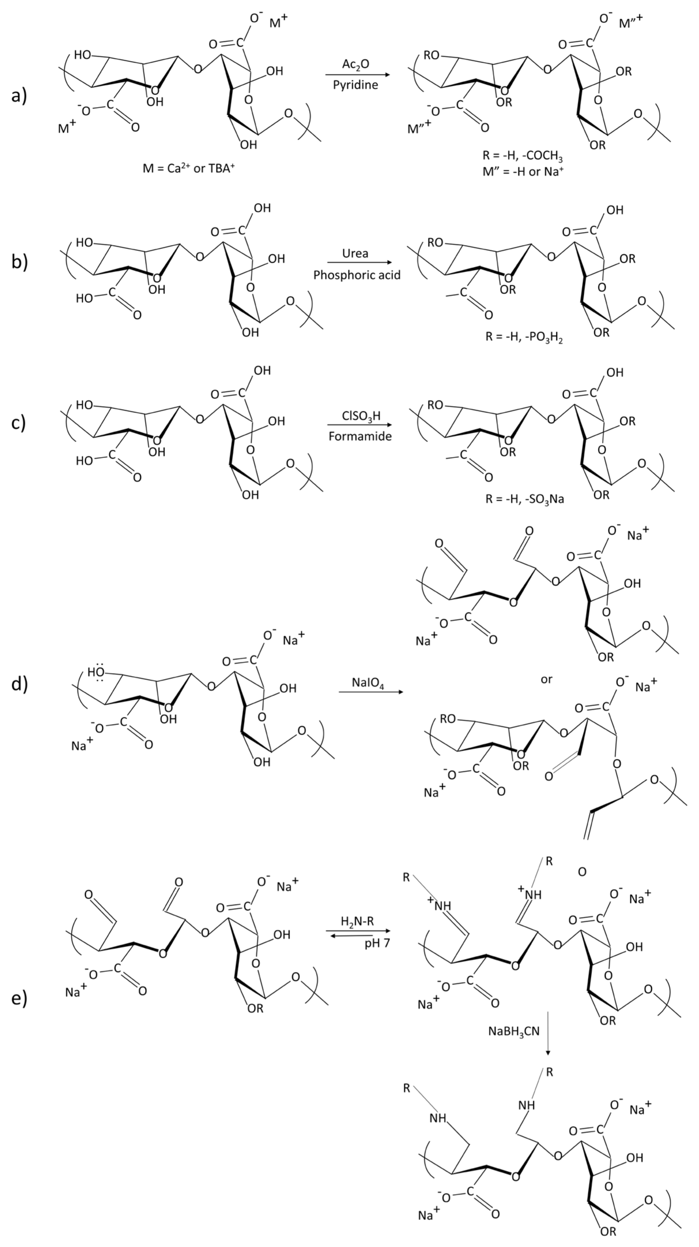
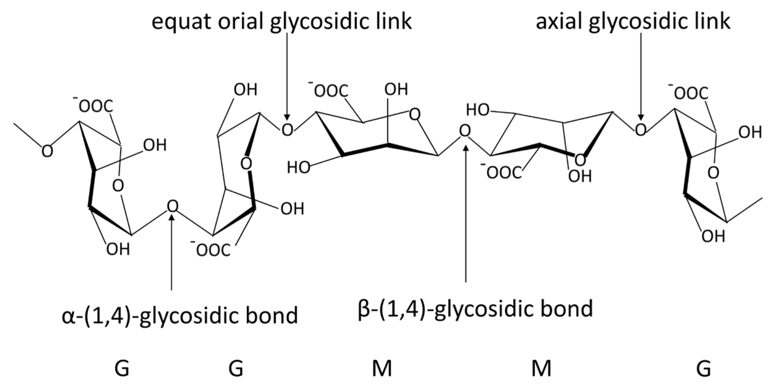
2.1. Structure, Properties and History
2.2. Preparation of Alginate Hydrogels
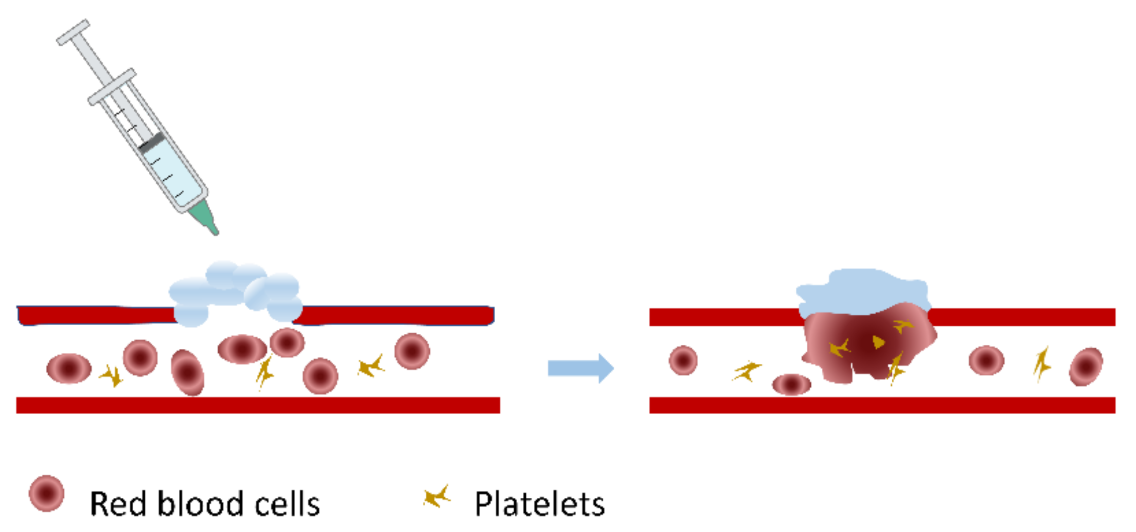
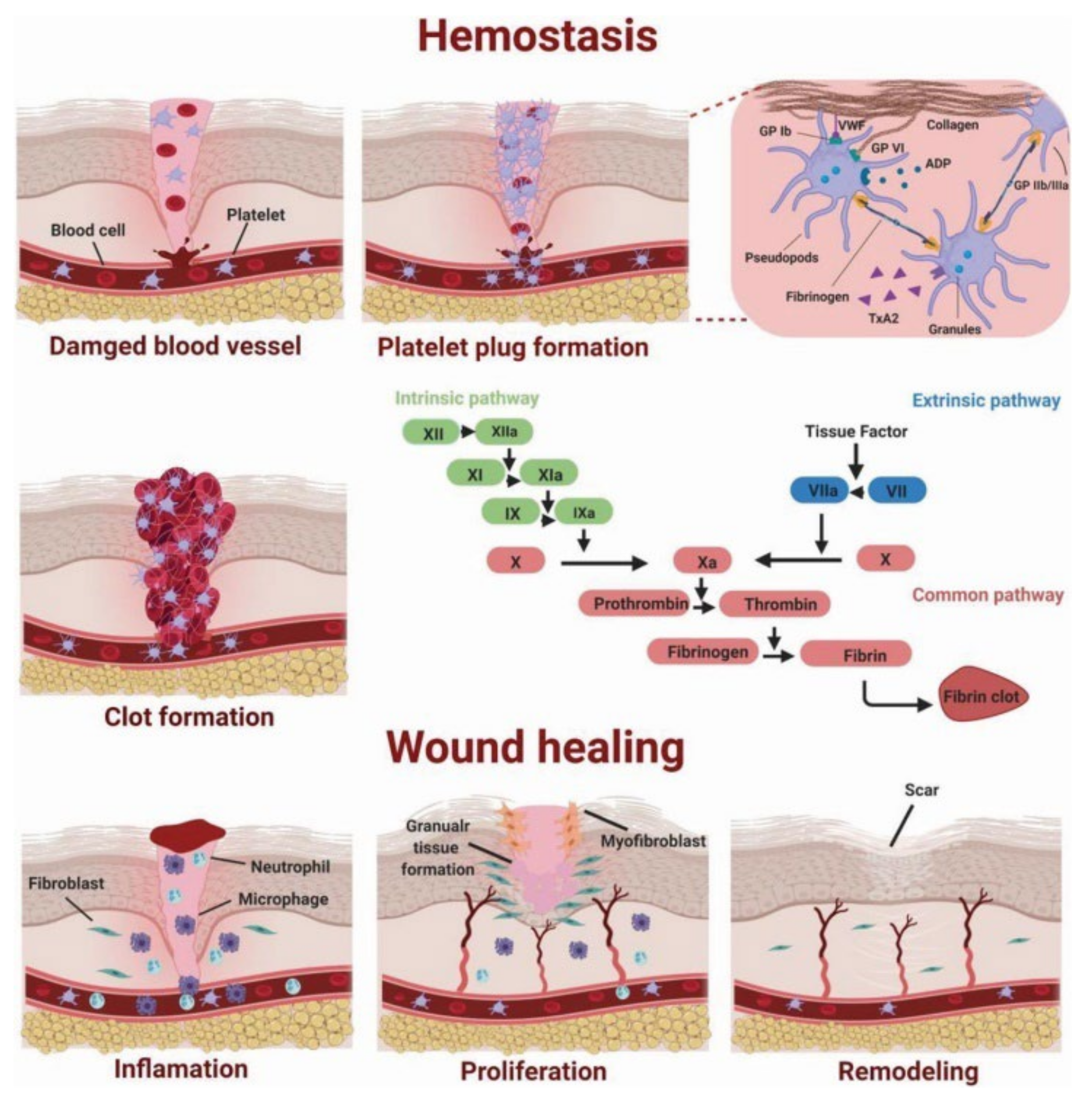
2.3. Hemostatic Mechanisms and Advantages of Alginate Hydrogels
3. Hemostasis of Superficial Wounds
3.1. Fibrous Dressings
3.2. Films and Membranes
3.3. Hemostatic Sponges
4. Hemostasis of Vessel and Viscera
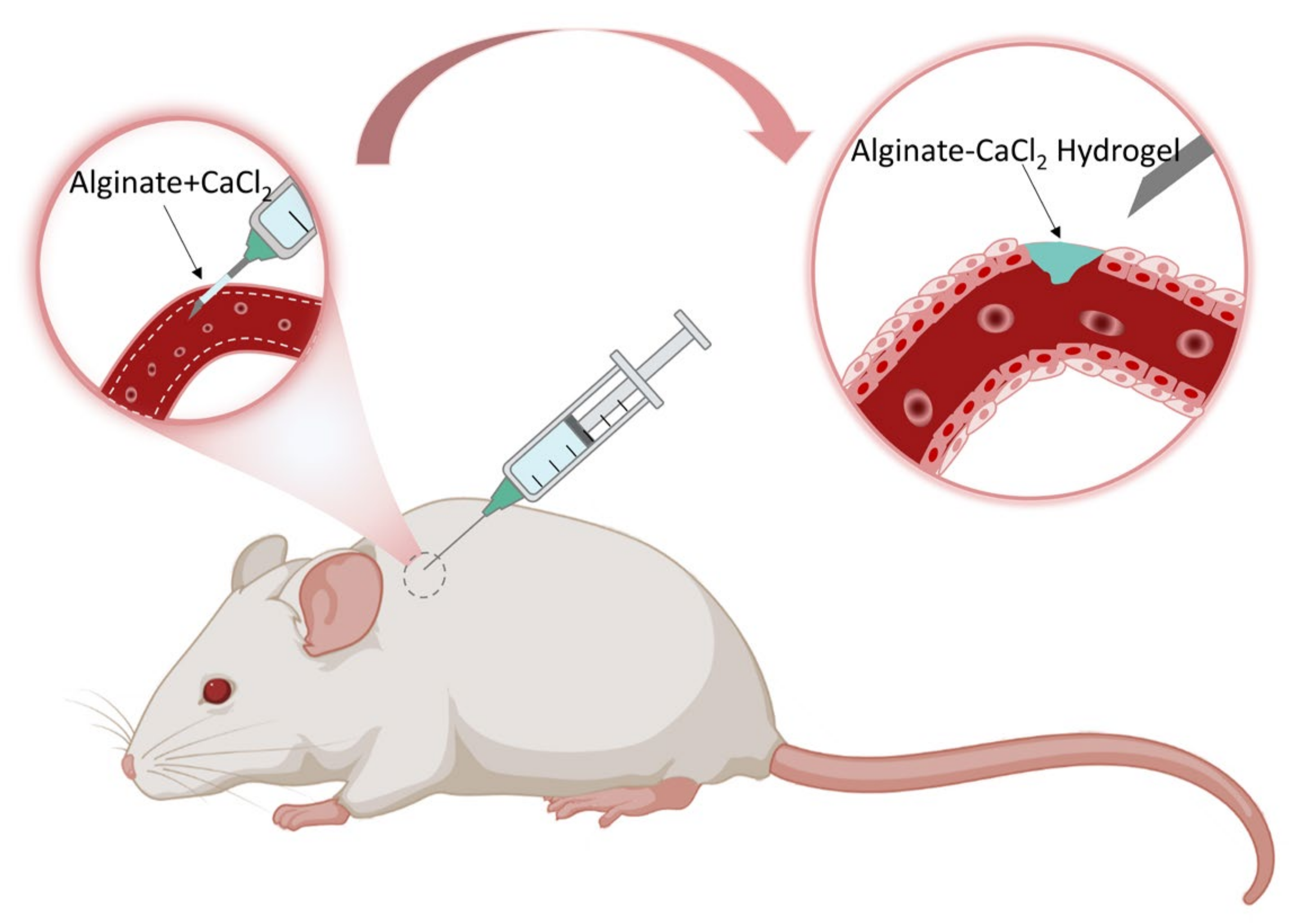
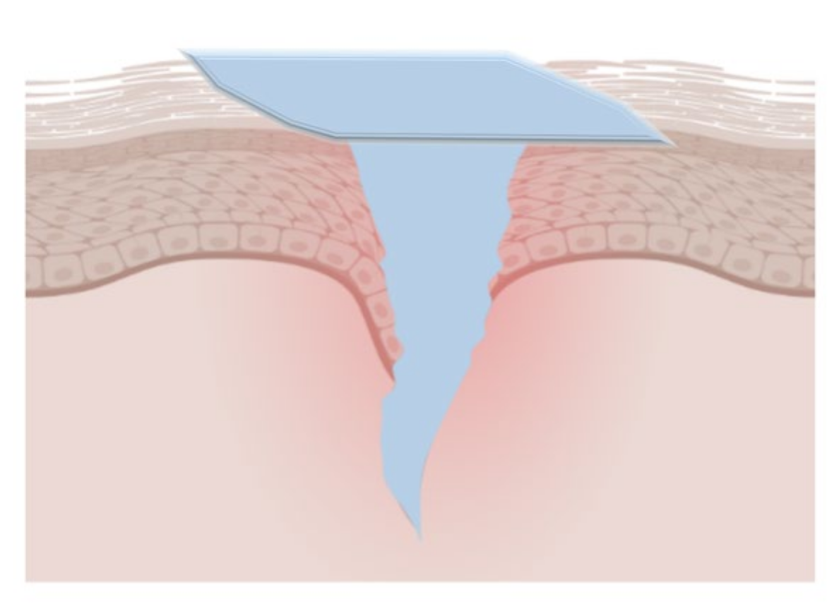
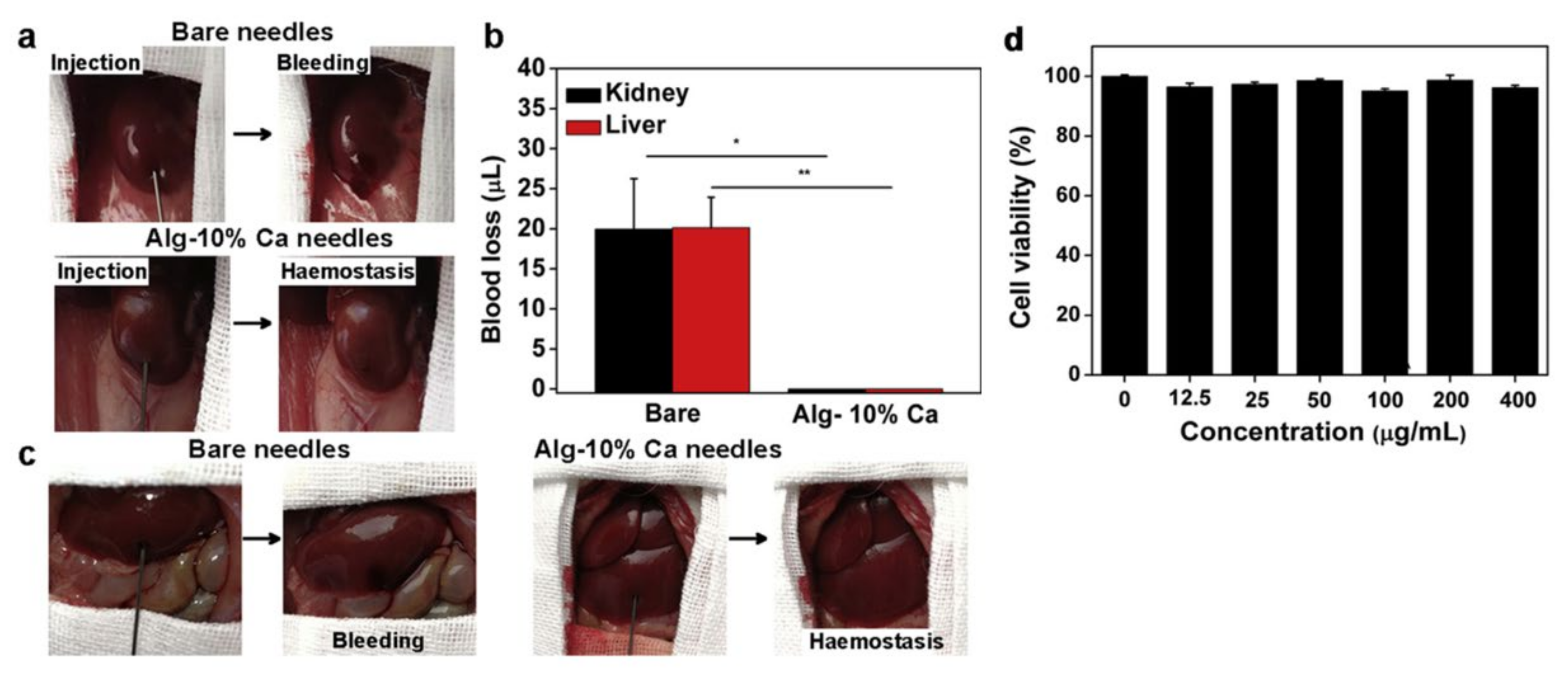
4.1. Hemostatic Needles
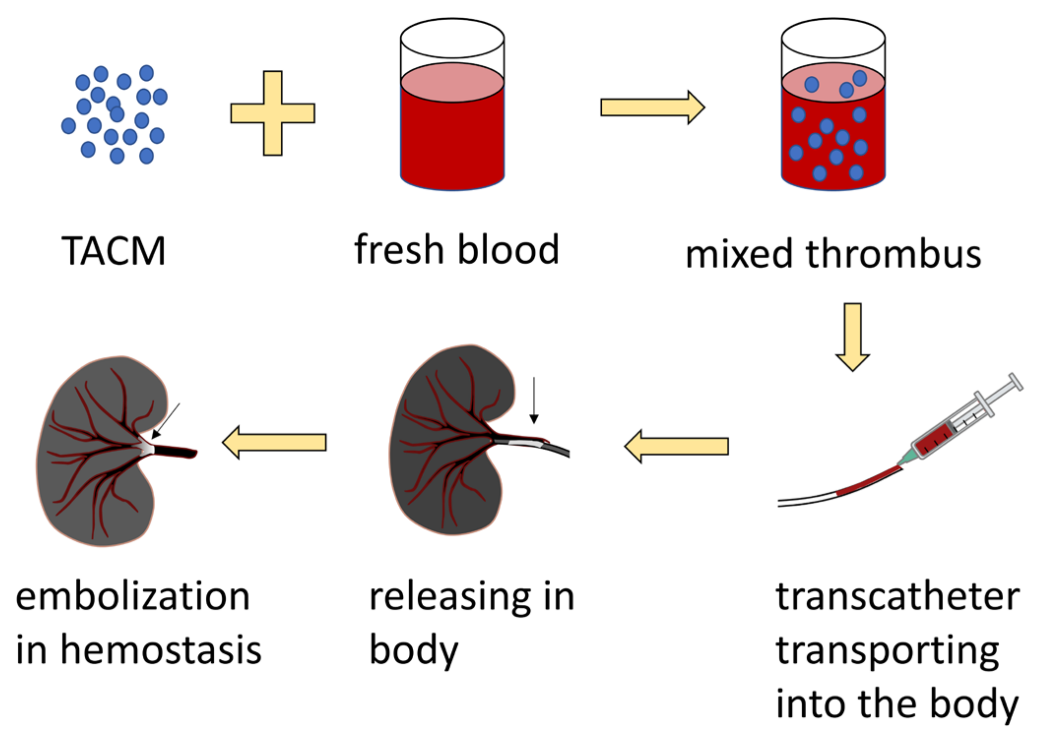
4.2. Embolic Materials
5. Hemostasis of Deep and Irregular Wounds
5.1. Injectable Hydrogels
5.2. Microspheres
5.3. Hydrogel Beads
5.4. Microneedles
6. Conclusions and Future Perspectives
Author Contributions
Funding
Institutional Review Board Statement
Informed Consent Statement
Data Availability Statement
Conflicts of Interest
References
- Naghavi, M.; Abajobir, A.A.; Abbafati, C.; Abbas, K.M.; Abd-Allah, F.; Abera, S.F.; Aboyans, V.; Adetokunboh, O.; Afshin, A.; Agrawal, A.; et al. Global, Regional, and National Age-Sex Specific Mortality for 264 Causes of Death, 1980–2016: A Systematic Analysis for the Global Burden of Disease Study 2016. Lancet 2017, 390, 1151–1210. [Google Scholar] [CrossRef] [Green Version]
- Kalkwarf, K.J.; Drake, S.A.; Yang, Y.; Thetford, C.; Myers, L.; Brock, M.; Wolf, D.A.; Persse, D.; Wade, C.E.; Holcomb, J.B. Bleeding to Death in a Big City: An Analysis of All Trauma Deaths from Hemorrhage in a Metropolitan Area during 1 Year. J. Trauma Acute Care Surg. 2020, 89, 716–722. [Google Scholar] [CrossRef] [PubMed]
- Gruen, R.L.; Brohi, K.; Schreiber, M.; Balogh, Z.J.; Pitt, V.; Narayan, M.; Maier, R.V. Haemorrhage Control in Severely Injured Patients. Lancet 2012, 380, 1099–1108. [Google Scholar] [CrossRef]
- Holcomb, J.B.; Moore, E.E.; Sperry, J.L.; Jansen, J.O.; Schreiber, M.A.; del Junco, D.J.; Spinella, P.C.; Sauaia, A.; Brohi, K.; Bulger, E.M.; et al. Evidence-Based and Clinically Relevant Outcomes for Hemorrhage Control Trauma Trials. Ann. Surg. 2021, 273, 395. [Google Scholar] [CrossRef] [PubMed]
- Boyum, J.H.; Atwell, T.D.; Schmit, G.D.; Poterucha, J.J.; Schleck, C.D.; Harmsen, W.S.; Kamath, P.S. Incidence and Risk Factors for Adverse Events Related to Image-Guided Liver Biopsy. Mayo Clin. Proc. 2016, 91, 329–335. [Google Scholar] [CrossRef] [PubMed]
- Corapi, K.M.; Chen, J.L.T.; Balk, E.M.; Gordon, C.E. Bleeding Complications of Native Kidney Biopsy: A Systematic Review and Meta-Analysis. Am. J. Kidney Dis. 2012, 60, 62–73. [Google Scholar] [CrossRef] [PubMed] [Green Version]
- Palm, M.D.; Altman, J.S. Topical Hemostatic Agents: A Review. Dermatol. Surg. 2008, 34, 431–445. [Google Scholar] [CrossRef]
- Gabay, M.; Boucher, B.A. An Essential Primer for Understanding the Role of Topical Hemostats, Surgical Sealants, and Adhesives for Maintaining Hemostasis. Pharmacother. J. Hum. Pharmacol. Drug Ther. 2013, 33, 935–955. [Google Scholar] [CrossRef] [PubMed]
- Tricco, A.C.; Cogo, E.; Isaranuwatchai, W.; Khan, P.A.; Sanmugalingham, G.; Antony, J.; Hoch, J.S.; Straus, S.E. A Systematic Review of Cost-Effectiveness Analyses of Complex Wound Interventions Reveals Optimal Treatments for Specific Wound Types. BMC Med. 2015, 13, 90. [Google Scholar] [CrossRef] [PubMed] [Green Version]
- Zhang, S.; Li, J.; Chen, S.; Zhang, X.; Ma, J.; He, J. Oxidized Cellulose-Based Hemostatic Materials. Carbohydr. Polym. 2020, 230, 115585. [Google Scholar] [CrossRef] [PubMed]
- Abbott, W.M.; Austen, W.G. The Effectiveness and Mechanism of Collagen-Induced Topical Hemostasis. Surgery 1975, 78, 723–729. [Google Scholar] [PubMed]
- Huang, L.; Liu, G.L.; Kaye, A.D.; Liu, H. Advances in Topical Hemostatic Agent Therapies: A Comprehensive Update. Adv. Ther. 2020, 37, 4132–4148. [Google Scholar] [CrossRef] [PubMed]
- Rhee, P.; Brown, C.; Martin, M.; Salim, A.; Plurad, D.; Green, D.; Chambers, L.; Demetriades, D.; Velmahos, G.; Alam, H. QuikClot Use in Trauma for Hemorrhage Control: Case Series of 103 Documented Uses. J. Trauma Acute Care Surg. 2008, 64, 1093–1099. [Google Scholar] [CrossRef] [PubMed] [Green Version]
- Carraway, J.W.; Kent, D.; Young, K.; Cole, A.; Friedman, R.; Ward, K.R. Comparison of a New Mineral Based Hemostatic Agent to a Commercially Available Granular Zeolite Agent for Hemostasis in a Swine Model of Lethal Extremity Arterial Hemorrhage. Resuscitation 2008, 78, 230–235. [Google Scholar] [CrossRef] [PubMed]
- Pourshahrestani, S.; Zeimaran, E.; Djordjevic, I.; Kadri, N.A.; Towler, M.R. Inorganic Hemostats: The State-of-the-Art and Recent Advances. Mater. Sci. Eng. C 2016, 58, 1255–1268. [Google Scholar] [CrossRef] [PubMed]
- Kheirabadi, B.S.; Mace, J.E.; Terrazas, I.B.; Fedyk, C.G.; Estep, J.S.; Dubick, M.A.; Blackbourne, L.H. Safety Evaluation of New Hemostatic Agents, Smectite Granules, and Kaolin-Coated Gauze in a Vascular Injury Wound Model in Swine. J. Trauma Inj. Infect. Crit. Care 2010, 68, 269–278. [Google Scholar] [CrossRef] [PubMed] [Green Version]
- Molinski, T.F.; Dalisay, D.S.; Lievens, S.L.; Saludes, J.P. Drug Development from Marine Natural Products. Nat. Rev. Drug Discov. 2009, 8, 69–85. [Google Scholar] [CrossRef] [PubMed]
- Snelgrove, P.V.R. An Ocean of Discovery: Biodiversity Beyond the Census of Marine Life. Planta Med. 2016, 82, 790–799. [Google Scholar] [CrossRef] [PubMed] [Green Version]
- Hu, Z.; Zhang, D.-Y.; Lu, S.-T.; Li, P.-W.; Li, S.-D. Chitosan-Based Composite Materials for Prospective Hemostatic Applications. Mar. Drugs 2018, 16, 273. [Google Scholar] [CrossRef] [PubMed] [Green Version]
- Rastogi, P.; Kandasubramanian, B. Review of Alginate-Based Hydrogel Bioprinting for Application in Tissue Engineering. Biofabrication 2019, 11, 042001. [Google Scholar] [CrossRef] [PubMed]
- Passi, A.; Vigetti, D. Hyaluronan as Tunable Drug Delivery System. Adv. Drug Deliv. Rev. 2019, 146, 83–96. [Google Scholar] [CrossRef]
- Chen, Y.; Wu, L.; Li, P.; Hao, X.; Yang, X.; Xi, G.; Liu, W.; Feng, Y.; He, H.; Shi, C. Polysaccharide Based Hemostatic Strategy for Ultrarapid Hemostasis. Macromol. Biosci. 2020, 20, 1900370. [Google Scholar] [CrossRef] [PubMed]
- Orive, G.; Ponce, S.; Hernández, R.M.; Gascón, A.R.; Igartua, M.; Pedraz, J.L. Biocompatibility of Microcapsules for Cell Immobilization Elaborated with Different Type of Alginates. Biomaterials 2002, 23, 3825–3831. [Google Scholar] [CrossRef]
- Zhong, H.; Gao, X.; Cheng, C.; Liu, C.; Wang, Q.; Han, X. The Structural Characteristics of Seaweed Polysaccharides and Their Application in Gel Drug Delivery Systems. Mar. Drugs 2020, 18, 658. [Google Scholar] [CrossRef] [PubMed]
- Lee, K.Y.; Mooney, D.J. Alginate: Properties and Biomedical Applications. Prog. Polym. Sci. 2012, 37, 106–126. [Google Scholar] [CrossRef] [PubMed] [Green Version]
- Clementi, F. Alginate Production by Azotobacter Vinelandii. Crit. Rev. Biotechnol. 1997, 17, 327–361. [Google Scholar] [CrossRef]
- Hernández-González, A.C.; Téllez-Jurado, L.; Rodríguez-Lorenzo, L.M. Alginate Hydrogels for Bone Tissue Engineering, from Injectables to Bioprinting: A Review. Carbohydr. Polym. 2020, 229, 115514. [Google Scholar] [CrossRef]
- Uyen, N.T.T.; Hamid, Z.A.A.; Tram, N.X.T.; Ahmad, N. Fabrication of Alginate Microspheres for Drug Delivery: A Review. Int. J. Biol. Macromol. 2020, 153, 1035–1046. [Google Scholar] [CrossRef] [PubMed]
- Wang, Y.; Fan, S.; Li, Y.; Niu, C.; Li, X.; Guo, Y.; Zhang, J.; Shi, J.; Wang, X. Silk Fibroin/Sodium Alginate Composite Porous Materials with Controllable Degradation. Int. J. Biol. Macromol. 2020, 150, 1314–1322. [Google Scholar] [CrossRef]
- Li, D.; Wei, Z.; Xue, C. Alginate-Based Delivery Systems for Food Bioactive Ingredients: An Overview of Recent Advances and Future Trends. Compr. Rev. Food Sci. Food Saf. 2021, 20, 5345–5369. [Google Scholar] [CrossRef]
- Cleetus, C.M.; Alvarez Primo, F.; Fregoso, G.; Lalitha Raveendran, N.; Noveron, J.C.; Spencer, C.T.; Ramana, C.V.; Joddar, B. Alginate Hydrogels with Embedded ZnO Nanoparticles for Wound Healing Therapy. Int. J. Nanomed. 2020, 15, 5097–5111. [Google Scholar] [CrossRef] [PubMed]
- Hazur, J.; Detsch, R.; Karakaya, E.; Kaschta, J.; Teßmar, J.; Schneidereit, D.; Friedrich, O.; Schubert, D.W.; Boccaccini, A.R. Improving Alginate Printability for Biofabrication: Establishment of a Universal and Homogeneous Pre-Crosslinking Technique. Biofabrication 2020, 12, 045004. [Google Scholar] [CrossRef] [PubMed]
- Shafei, S.; Khanmohammadi, M.; Heidari, R.; Ghanbari, H.; Taghdiri Nooshabadi, V.; Farzamfar, S.; Akbariqomi, M.; Sanikhani, N.S.; Absalan, M.; Tavoosidana, G. Exosome Loaded Alginate Hydrogel Promotes Tissue Regeneration in Full-Thickness Skin Wounds: An in Vivo Study. J. Biomed. Mater. Res. A 2020, 108, 545–556. [Google Scholar] [CrossRef] [PubMed]
- Augst, A.D.; Kong, H.J.; Mooney, D.J. Alginate Hydrogels as Biomaterials. Macromol. Biosci. 2006, 6, 623–633. [Google Scholar] [CrossRef]
- Rhein-Knudsen, N.; Ale, M.T.; Ajalloueian, F.; Meyer, A.S. Characterization of Alginates from Ghanaian Brown Seaweeds: Sargassum Spp. and Padina Spp. Food Hydrocoll. 2017, 71, 236–244. [Google Scholar] [CrossRef]
- Varaprasad, K.; Raghavendra, G.M.; Jayaramudu, T.; Seo, J. Nano Zinc Oxide–Sodium Alginate Antibacterial Cellulose Fibres. Carbohydr. Polym. 2016, 135, 349–355. [Google Scholar] [CrossRef]
- Núñez, C.; Peña, C.; Kloeckner, W.; Hernández-Eligio, A.; Bogachev, A.V.; Moreno, S.; Guzmán, J.; Büchs, J.; Espín, G. Alginate Synthesis in Azotobacter Vinelandii Is Increased by Reducing the Intracellular Production of Ubiquinone. Appl. Microbiol. Biotechnol. 2013, 97, 2503–2512. [Google Scholar] [CrossRef] [PubMed]
- Mørch, Ý.A.; Donati, I.; Strand, B.L.; Skjåk-Bræk, G. Effect of Ca2+, Ba2+, and Sr2+ on Alginate Microbeads. Biomacromolecules 2006, 7, 1471–1480. [Google Scholar] [CrossRef] [PubMed]
- Ching, S.H.; Bansal, N.; Bhandari, B. Alginate Gel Particles–A Review of Production Techniques and Physical Properties. Crit. Rev. Food Sci. Nutr. 2017, 57, 1133–1152. [Google Scholar] [CrossRef]
- Skjåk-Braek, G.; Grasdalen, H.; Larsen, B. Monomer Sequence and Acetylation Pattern in Some Bacterial Alginates. Carbohydr. Res. 1986, 154, 239–250. [Google Scholar] [CrossRef]
- Ahmed, E.M. Hydrogel: Preparation, Characterization, and Applications: A Review. J. Adv. Res. 2015, 6, 105–121. [Google Scholar] [CrossRef] [PubMed] [Green Version]
- Zhang, M.; Zhao, X. Alginate Hydrogel Dressings for Advanced Wound Management. Int. J. Biol. Macromol. 2020, 162, 1414–1428. [Google Scholar] [CrossRef] [PubMed]
- Kuo, C.K.; Ma, P.X. Ionically Crosslinked Alginate Hydrogels as Scaffolds for Tissue Engineering: Part 1. Structure, Gelation Rate and Mechanical Properties. Biomaterials 2001, 22, 511–521. [Google Scholar] [CrossRef]
- Pawar, S.N.; Edgar, K.J. Alginate Derivatization: A Review of Chemistry, Properties and Applications. Biomaterials 2012, 33, 3279–3305. [Google Scholar] [CrossRef] [PubMed]
- Chen, D.; Amstad, E.; Zhao, C.-X.; Cai, L.; Fan, J.; Chen, Q.; Hai, M.; Koehler, S.; Zhang, H.; Liang, F.; et al. Biocompatible Amphiphilic Hydrogel–Solid Dimer Particles as Colloidal Surfactants. ACS Nano 2017, 11, 11978–11985. [Google Scholar] [CrossRef]
- Eiselt, P.; Lee, K.Y.; Mooney, D.J. Rigidity of Two-Component Hydrogels Prepared from Alginate and Poly(Ethylene Glycol)−Diamines. Macromolecules 1999, 32, 5561–5566. [Google Scholar] [CrossRef]
- Rosiak, P.; Latanska, I.; Paul, P.; Sujka, W.; Kolesinska, B. Modification of Alginates to Modulate Their Physic-Chemical Properties and Obtain Biomaterials with Different Functional Properties. Molecules 2021, 26, 7264. [Google Scholar] [CrossRef]
- Kim, H.-S.; Song, M.; Lee, E.-J.; Shin, U.S. Injectable Hydrogels Derived from Phosphorylated Alginic Acid Calcium Complexes. Mater. Sci. Eng. C Mater. Biol. Appl. 2015, 51, 139–147. [Google Scholar] [CrossRef]
- Chen, X.; Zhu, Q.; Liu, C.; Li, D.; Yan, H.; Lin, Q. Esterification of Alginate with Alkyl Bromides of Different Carbon Chain Lengths via the Bimolecular Nucleophilic Substitution Reaction: Synthesis, Characterization, and Controlled Release Performance. Polymers 2021, 13, 3351. [Google Scholar] [CrossRef]
- Distler, T.; McDonald, K.; Heid, S.; Karakaya, E.; Detsch, R.; Boccaccini, A.R. Ionically and Enzymatically Dual Cross-Linked Oxidized Alginate Gelatin Hydrogels with Tunable Stiffness and Degradation Behavior for Tissue Engineering. ACS Biomater. Sci. Eng. 2020, 6, 3899–3914. [Google Scholar] [CrossRef]
- Deng, Y.; Shavandi, A.; Okoro, O.V.; Nie, L. Alginate Modification via Click Chemistry for Biomedical Applications. Carbohydr. Polym. 2021, 270, 118360. [Google Scholar] [CrossRef] [PubMed]
- Reakasame, S.; Boccaccini, A.R. Oxidized Alginate-Based Hydrogels for Tissue Engineering Applications: A Review. Biomacromolecules 2018, 19, 3–21. [Google Scholar] [CrossRef] [PubMed]
- Dai, M.; Liu, Y.; Ju, B.; Tian, Y. Preparation of Thermoresponsive Alginate/Starch Ether Composite Hydrogel and Its Application to the Removal of Cu(II) from Aqueous Solution. Bioresour. Technol. 2019, 294, 122192. [Google Scholar] [CrossRef] [PubMed]
- Mahdavinia, G.R.; Rahmani, Z.; Karami, S.; Pourjavadi, A. Magnetic/PH-Sensitive κ-Carrageenan/Sodium Alginate Hydrogel Nanocomposite Beads: Preparation, Swelling Behavior, and Drug Delivery. J. Biomater. Sci. Polym. Ed. 2014, 25, 1891–1906. [Google Scholar] [CrossRef]
- Iatridi, Z.; Saravanou, S.-F.; Tsitsilianis, C. Injectable Self-Assembling Hydrogel from Alginate Grafted by P(N-Isopropylacrylamide-Co-N-Tert-Butylacrylamide) Random Copolymers. Carbohydr. Polym. 2019, 219, 344–352. [Google Scholar] [CrossRef] [PubMed]
- Varaprasad, K.; Jayaramudu, T.; Kanikireddy, V.; Toro, C.; Sadiku, E.R. Alginate-Based Composite Materials for Wound Dressing Application:A Mini Review. Carbohydr. Polym. 2020, 236, 116025. [Google Scholar] [CrossRef]
- Ionita, M.; Pandele, M.A.; Iovu, H. Sodium Alginate/Graphene Oxide Composite Films with Enhanced Thermal and Mechanical Properties. Carbohydr. Polym. 2013, 94, 339–344. [Google Scholar] [CrossRef] [PubMed]
- Wang, T.; Wang, J.; Wang, R.; Yuan, P.; Fan, Z.; Yang, S. Preparation and Properties of ZnO/Sodium Alginate Bi-Layered Hydrogel Films as Novel Wound Dressings. New J. Chem. 2019, 43, 8684–8693. [Google Scholar] [CrossRef]
- Zhao, L.; Yin, S.; Ma, Z. Ca2+-Triggered PH-Response Sodium Alginate Hydrogel Precipitation for Amplified Sandwich-Type Impedimetric Immunosensor of Tumor Marker. ACS Sens. 2019, 4, 450–455. [Google Scholar] [CrossRef] [PubMed]
- Hu, Y.; Zhang, Z.; Li, Y.; Ding, X.; Li, D.; Shen, C.; Xu, F.-J. Dual-Crosslinked Amorphous Polysaccharide Hydrogels Based on Chitosan/Alginate for Wound Healing Applications. Macromol. Rapid Commun. 2018, 39, 1800069. [Google Scholar] [CrossRef]
- Chen, T.; Chen, Y.; Rehman, H.U.; Chen, Z.; Yang, Z.; Wang, M.; Li, H.; Liu, H. Ultratough, Self-Healing, and Tissue-Adhesive Hydrogel for Wound Dressing. ACS Appl. Mater. Interfaces 2018, 10, 33523–33531. [Google Scholar] [CrossRef] [PubMed]
- Liang, M.; Chen, Z.; Wang, F.; Liu, L.; Wei, R.; Zhang, M. Preparation of Self-Regulating/Anti-Adhesive Hydrogels and Their Ability to Promote Healing in Burn Wounds. J. Biomed. Mater. Res. B Appl. Biomater. 2019, 107, 1471–1482. [Google Scholar] [CrossRef]
- Jin, S.G.; Kim, K.S.; Kim, D.W.; Kim, D.S.; Seo, Y.G.; Go, T.G.; Youn, Y.S.; Kim, J.O.; Yong, C.S.; Choi, H.-G. Development of a Novel Sodium Fusidate-Loaded Triple Polymer Hydrogel Wound Dressing: Mechanical Properties and Effects on Wound Repair. Int. J. Pharm. 2016, 497, 114–122. [Google Scholar] [CrossRef] [PubMed]
- Quah, S.P.; Nykypanchuk, D.; Bhatia, S.R. Temperature-Dependent Structure and Compressive Mechanical Behavior of Alginate/Polyethylene Oxide–Poly(Propylene Oxide)–Poly(Ethylene Oxide) Hydrogels. J. Biomed. Mater. Res. B Appl. Biomater. 2020, 108, 834–844. [Google Scholar] [CrossRef] [PubMed]
- Ooi, H.W.; Kocken, J.M.M.; Morgan, F.L.C.; Malheiro, A.; Zoetebier, B.; Karperien, M.; Wieringa, P.A.; Dijkstra, P.J.; Moroni, L.; Baker, M.B. Multivalency Enables Dynamic Supramolecular Host–Guest Hydrogel Formation. Biomacromolecules 2020, 21, 2208–2217. [Google Scholar] [CrossRef] [PubMed] [Green Version]
- Khampieng, T.; Wongkittithavorn, S.; Chaiarwut, S.; Ekabutr, P.; Pavasant, P.; Supaphol, P. Silver Nanoparticles-Based Hydrogel: Characterization of Material Parameters for Pressure Ulcer Dressing Applications. J. Drug Deliv. Sci. Technol. 2018, 44, 91–100. [Google Scholar] [CrossRef]
- Golafshan, N.; Rezahasani, R.; Tarkesh Esfahani, M.; Kharaziha, M.; Khorasani, S.N. Nanohybrid Hydrogels of Laponite: PVA-Alginate as a Potential Wound Healing Material. Carbohydr. Polym. 2017, 176, 392–401. [Google Scholar] [CrossRef] [PubMed]
- Mousavi, A.; Mashayekhan, S.; Baheiraei, N.; Pourjavadi, A. Biohybrid Oxidized Alginate/Myocardial Extracellular Matrix Injectable Hydrogels with Improved Electromechanical Properties for Cardiac Tissue Engineering. Int. J. Biol. Macromol. 2021, 180, 692–708. [Google Scholar] [CrossRef] [PubMed]
- Mo, C.; Xiang, L.; Chen, Y. Advances in Injectable and Self-Healing Polysaccharide Hydrogel Based on the Schiff Base Reaction. Macromol. Rapid Commun. 2021, 42, 2100025. [Google Scholar] [CrossRef] [PubMed]
- Li, Y.; Yang, H.Y.; Lee, D.S. Advances in Biodegradable and Injectable Hydrogels for Biomedical Applications. J. Controlled Release 2021, 330, 151–160. [Google Scholar] [CrossRef] [PubMed]
- Aderibigbe, B.; Buyana, B. Alginate in Wound Dressings. Pharmaceutics 2018, 10, 42. [Google Scholar] [CrossRef] [Green Version]
- Zhang, Y.S.; Khademhosseini, A. Advances in Engineering Hydrogels. Science 2017, 356, eaaf3627. [Google Scholar] [CrossRef] [PubMed]
- Patenaude, M.; Smeets, N.M.B.; Hoare, T. Designing Injectable, Covalently Cross-Linked Hydrogels for Biomedical Applications. Macromol. Rapid Commun. 2014, 35, 598–617. [Google Scholar] [CrossRef] [PubMed]
- Li, J.; Celiz, A.D.; Yang, J.; Yang, Q.; Wamala, I.; Whyte, W.; Seo, B.R.; Vasilyev, N.V.; Vlassak, J.J.; Suo, Z.; et al. Tough Adhesives for Diverse Wet Surfaces. Science 2017, 357, 378–381. [Google Scholar] [CrossRef] [PubMed] [Green Version]
- Zheng, B.-D.; Ye, J.; Yang, Y.-C.; Huang, Y.-Y.; Xiao, M.-T. Self-Healing Polysaccharide-Based Injectable Hydrogels with Antibacterial Activity for Wound Healing. Carbohydr. Polym. 2022, 275, 118770. [Google Scholar] [CrossRef] [PubMed]
- Wu, X.; Tang, Z.; Liao, X.; Wang, Z.; Liu, H. Fabrication of Chitosan@calcium Alginate Microspheres with Porous Core and Compact Shell, and Application as a Quick Traumatic Hemostat. Carbohydr. Polym. 2020, 247, 116669. [Google Scholar] [CrossRef]
- Wang, L.; Li, W.; Qin, S. Three Polymers from the Sea: Unique Structures, Directional Modifications, and Medical Applications. Polymers 2021, 13, 2482. [Google Scholar] [CrossRef] [PubMed]
- Ehterami, A.; Salehi, M.; Farzamfar, S.; Samadian, H.; Vaez, A.; Ghorbani, S.; Ai, J.; Sahrapeyma, H. Chitosan/Alginate Hydrogels Containing Alpha-Tocopherol for Wound Healing in Rat Model. J. Drug Deliv. Sci. Technol. 2019, 51, 204–213. [Google Scholar] [CrossRef]
- Liu, C.; Shi, Z.; Sun, H.; Zhao, L.; Wang, X.; Huang, F. Tissue Factor-loaded Collagen/Alginate Hydrogel Beads as a Hemostatic Agent. J. Biomed. Mater. Res. B Appl. Biomater. 2021, 109, 1116–1123. [Google Scholar] [CrossRef] [PubMed]
- Pourshahrestani, S.; Zeimaran, E.; Kadri, N.A.; Mutlu, N.; Boccaccini, A.R. Polymeric Hydrogel Systems as Emerging Biomaterial Platforms to Enable Hemostasis and Wound Healing. Adv. Healthc. Mater. 2020, 9, 2000905. [Google Scholar] [CrossRef] [PubMed]
- Kim, J.O.; Park, J.K.; Kim, J.H.; Jin, S.G.; Yong, C.S.; Li, D.X.; Choi, J.Y.; Woo, J.S.; Yoo, B.K.; Lyoo, W.S.; et al. Development of Polyvinyl Alcohol-Sodium Alginate Gel-Matrix-Based Wound Dressing System Containing Nitrofurazone. Int. J. Pharm. 2008, 359, 79–86. [Google Scholar] [CrossRef] [PubMed]
- Thomas, A.; Harding, K.G.; Moore, K. Alginates from Wound Dressings Activate Human Macrophages to Secrete Tumour Necrosis Factor-α. Biomaterials 2000, 21, 1797–1802. [Google Scholar] [CrossRef]
- Winter, G.D. Formation of the Scab and the Rate of Epithelization of Superficial Wounds in the Skin of the Young Domestic Pig. Nature 1962, 193, 293–294. [Google Scholar] [CrossRef] [PubMed]
- Gilchrist, T.; Martin, A.M. Wound Treatment with Sorbsan—An Alginate Fibre Dressing. Biomaterials 1983, 4, 317–320. [Google Scholar] [CrossRef]
- Mohandas, A.; Pt, S.K.; Raja, B.; Lakshmanan, V.-K.; Jayakumar, R. Exploration of Alginate Hydrogel/Nano Zinc Oxide Composite Bandages for Infected Wounds. Int. J. Nanomedicine 2015, 10, 53–66. [Google Scholar] [CrossRef] [PubMed] [Green Version]
- Wet-Spun Bi-Component Alginate Based Hydrogel Fibers: Development and in-Vitro Evaluation as a Potential Moist Wound Care Dressing. Int. J. Biol. Macromol. 2021, 168, 601–610. [CrossRef] [PubMed]
- Zhang, X.; Huang, C.; Zhao, Y.; Jin, X. Preparation and Characterization of Nanoparticle Reinforced Alginate Fibers with High Porosity for Potential Wound Dressing Application. RSC Adv. 2017, 7, 39349–39358. [Google Scholar] [CrossRef] [Green Version]
- Castellano, J.J.; Shafii, S.M.; Ko, F.; Donate, G.; Wright, T.E.; Mannari, R.J.; Payne, W.G.; Smith, D.J.; Robson, M.C. Comparative Evaluation of Silver-Containing Antimicrobial Dressings and Drugs. Int. Wound J. 2007, 4, 114–122. [Google Scholar] [CrossRef] [PubMed]
- Qin, Y. Novel Antimicrobial Fibres. Text. Mag. 2004, 31, 14–17. [Google Scholar]
- Qin, Y. Silver-Containing Alginate Fibres and Dressings. Int. Wound J. 2005, 2, 172–176. [Google Scholar] [CrossRef] [PubMed]
- Liu, L.; Jiang, L.; Xu, G.K.; Ma, C.; Yang, X.G.; Yao, J.M. Potential of Alginate Fibers Incorporated with Drug-Loaded Nanocapsules as Drug Delivery Systems. J. Mater. Chem. B 2014, 2, 7596–7604. [Google Scholar] [CrossRef] [PubMed]
- Li, Z.; Chen, S.; Wu, B.; Liu, Z.; Cheng, L.; Bao, Y.; Ma, Y.; Chen, L.; Tong, X.; Dai, F. Multifunctional Dual Ionic-Covalent Membranes for Wound Healing. ACS Biomater. Sci. Eng. 2020, 6, 6949–6960. [Google Scholar] [CrossRef]
- Pan, H.; Fan, D.; Duan, Z.; Zhu, C.; Fu, R.; Li, X. Non-Stick Hemostasis Hydrogels as Dressings with Bacterial Barrier Activity for Cutaneous Wound Healing. Mater. Sci. Eng. C 2019, 105, 110118. [Google Scholar] [CrossRef]
- Abou-Okeil, A.; Fahmy, H.M.; El-Bisi, M.K.; Ahmed-Farid, O.A. Hyaluronic Acid/Na-Alginate Films as Topical Bioactive Wound Dressings. Eur. Polym. J. 2018, 109, 101–109. [Google Scholar] [CrossRef]
- Koga, A.Y.; Pereira, A.V.; Lipinski, L.C.; Oliveira, M.R.P. Evaluation of Wound Healing Effect of Alginate Films Containing Aloe Vera (Aloe Barbadensis Miller) Gel. J. Biomater. Appl. 2018, 32, 1212–1221. [Google Scholar] [CrossRef]
- Du, Y.; Li, L.; Peng, H.; Zheng, H.; Cao, S.; Lv, G.; Yang, A.; Li, H.; Liu, T. A Spray-Filming Self-Healing Hydrogel Fabricated from Modified Sodium Alginate and Gelatin as a Bacterial Barrier. Macromol. Biosci. 2020, 20, 1900303. [Google Scholar] [CrossRef] [PubMed]
- Lv, C.; Li, L.; Jiao, Z.; Yan, H.; Wang, Z.; Wu, Z.; Guo, M.; Wang, Y.; Zhang, P. Improved Hemostatic Effects by Fe3+ Modified Biomimetic PLLA Cotton-like Mat via Sodium Alginate Grafted with Dopamine. Bioact. Mater. 2021, 6, 2346–2359. [Google Scholar] [CrossRef]
- Severinov, D.A.; Lazarenko, S.V.; Sotnikov, K.A.; Pohozhay, V.V.; Ansimova, P.V.; Lipatov, V.A. In Vitro Evaluation of Performance Properties of Sponge Hemostatic Dressings (Review). Mod. Technol. Med. 2020, 12, 139–146. [Google Scholar] [CrossRef] [PubMed]
- Ma, R.; Wang, Y.; Qi, H.; Shi, C.; Wei, G.; Xiao, L.; Huang, Z.; Liu, S.; Yu, H.; Teng, C.; et al. Nanocomposite Sponges of Sodium Alginate/Graphene Oxide/Polyvinyl Alcohol as Potential Wound Dressing: In Vitro and in Vivo Evaluation. Compos. Part B Eng. 2019, 167, 396–405. [Google Scholar] [CrossRef]
- Cheng, F.; Liu, C.; Wei, X.; Yan, T.; Li, H.; He, J.; Huang, Y. Preparation and Characterization of 2,2,6,6-Tetramethylpiperidine-1-Oxyl (TEMPO)-Oxidized Cellulose Nanocrystal/Alginate Biodegradable Composite Dressing for Hemostasis Applications. ACS Sustain. Chem. Eng. 2017, 5, 3819–3828. [Google Scholar] [CrossRef]
- Dowling, M.B.; Chaturvedi, A.; MacIntire, I.C.; Javvaji, V.; Gustin, J.; Raghavan, S.R.; Scalea, T.M.; Narayan, M. Determination of Efficacy of a Novel Alginate Dressing in a Lethal Arterial Injury Model in Swine. Injury 2016, 47, 2105–2109. [Google Scholar] [CrossRef] [Green Version]
- Shin, M.; Park, S.-G.; Oh, B.-C.; Kim, K.; Jo, S.; Lee, M.S.; Oh, S.S.; Hong, S.-H.; Shin, E.-C.; Kim, K.-S.; et al. Complete Prevention of Blood Loss with Self-Sealing Haemostatic Needles. Nat. Mater. 2017, 16, 147–152. [Google Scholar] [CrossRef]
- Ren, J.; Yin, X.; Chen, Y.; Chen, Y.; Su, H.; Wang, K.; Zhang, L.; Zhu, J.; Zhang, C. Alginate Hydrogel-Coated Syringe Needles for Rapid Haemostasis of Vessel and Viscera Puncture. Biomaterials 2020, 249, 120019. [Google Scholar] [CrossRef]
- Wei, S.; Xu, Y.; Wang, Z.; Li, M.; Sun, P.; Xie, B.; Xing, Y.; Bai, H.; Kan, Q.; Li, J.; et al. Hydrogel-Coated Needles Prevent Puncture Site Bleeding. Acta Biomater. 2021, 128, 305–313. [Google Scholar] [CrossRef] [PubMed]
- Xu, Y.; Wang, Z.; Wei, S.; Sun, P.; Bai, H.; Li, J. Hydrogel-Coated Needles Prevent Puncture Site Bleeding in Arteriovenous Fistula and Arteriovenous Grafts in Rats. Biomed. Pharmacother. 2021, 143, 112113. [Google Scholar] [CrossRef]
- Yin, X.; Ren, J.; Lan, W.; Chen, Y.; Ouyang, M.; Su, H.; Zhang, L.; Zhu, J.; Zhang, C. Microfluidics-Assisted Optimization of Highly Adhesive Haemostatic Hydrogel Coating for Arterial Puncture. Bioact. Mater. 2021. [Google Scholar] [CrossRef]
- Haan, J.M.; Bochicchio, G.V.; Kramer, N.; Scalea, T.M. Nonoperative Management of Blunt Splenic Injury: A 5-Year Experience. J. Trauma Acute Care Surg. 2005, 58, 492–498. [Google Scholar] [CrossRef] [PubMed] [Green Version]
- Choi, H.; Choi, B.; Yu, B.; Li, W.; Matsumoto, M.M.; Harris, K.R.; Lewandowski, R.J.; Larson, A.C.; Mouli, S.K.; Kim, D.-H. On-Demand Degradable Embolic Microspheres for Immediate Restoration of Blood Flow during Image-Guided Embolization Procedures. Biomaterials 2021, 265, 120408. [Google Scholar] [CrossRef]
- Chen, C.; Lee, S.M.; Kim, J.W.; Shin, J.H. Recent Update of Embolization of Postpartum Hemorrhage. Korean J. Radiol. 2018, 19, 585–596. [Google Scholar] [CrossRef]
- Jander, H.P.; Russinovich, N.A. Transcatheter Gelfoam Embolization in Abdominal, Retroperitoneal, and Pelvic Hemorrhage. Radiology 1980, 136, 337–344. [Google Scholar] [CrossRef] [PubMed]
- Dawbarn, R.H.M. The starvation operation for malignancy in the external carotid area.its failures and successes. J. Am. Med. Assoc. 1904, XLIII, 792–795. [Google Scholar] [CrossRef]
- Skattum, J.; Titze, T.L.; Dormagen, J.B.; Aaberge, I.S.; Bechensteen, A.G.; Gaarder, P.I.; Gaarder, C.; Heier, H.E.; Næss, P.A. Preserved Splenic Function after Angioembolisation of High Grade Injury. Injury 2012, 43, 62–66. [Google Scholar] [CrossRef] [PubMed]
- Abada, H.T.; Golzarian, J. Gelatine Sponge Particles: Handling Characteristics for Endovascular Use. Tech. Vasc. Interv. Radiol. 2007, 10, 257–260. [Google Scholar] [CrossRef]
- Alfidja, A.; Garcier, J.-M.; Chahid, T.; Ravel, A.; Boyer, L. Endovascular Therapeutic Techniques. EMC-Radiol. 2004, 1, 216–232. [Google Scholar] [CrossRef]
- Hu, J.; Albadawi, H.; Chong, B.W.; Deipolyi, A.R.; Sheth, R.A.; Khademhosseini, A.; Oklu, R. Advances in Biomaterials and Technologies for Vascular Embolization. Adv. Mater. 2019, 31, 1901071. [Google Scholar] [CrossRef]
- Du, N.; Ma, J.-Q.; Luo, J.-J.; Liu, Q.-X.; Zhang, Z.-H.; Yang, M.-J.; Yu, T.-Z.; Tao, Y.; Liu, R.; Zhang, W.; et al. The Efficacy and Safety of Transcatheter Arterial Embolization to Treat Renal Hemorrhage after Percutaneous Nephrolithotomy. Biomed Res. Int. 2019, 2019, e6265183. [Google Scholar] [CrossRef]
- Liao, C.-H.; Ouyang, C.-H.; Fu, C.-Y.; Wang, S.-Y.; Lin, K.-J.; Kuo, I.-M.; Hsu, C.-P.; Yang, S.-J.; Yuan, K.-C.; Hsu, Y.-P. The Current Status and Management of Blunt Adrenal Gland Trauma. Surgery 2015, 157, 338–343. [Google Scholar] [CrossRef]
- Yoshida, H.; Mamada, Y.; Taniai, N.; Uchida, E. Spontaneous Ruptured Hepatocellular Carcinoma. Hepatol. Res. 2016, 46, 13–21. [Google Scholar] [CrossRef]
- Yao, Z.; Tian, W.; Xu, X.; Zhao, R.; Huang, M.; Zhao, Y.; Chen, X. Transcatheter Arterial Embolization in the Treatment of Abdominal Bleeding in Patients Being Treated with Open Abdomen Due to Duodenal Fistula. World J. Surg. 2020, 44, 2562–2571. [Google Scholar] [CrossRef] [PubMed]
- Fan, L.; Duan, M.; Xie, Z.; Pan, K.; Wang, X.; Sun, X.; Wang, Q.; Rao, W.; Liu, J. Injectable and Radiopaque Liquid Metal/Calcium Alginate Hydrogels for Endovascular Embolization and Tumor Embolotherapy. Small 2020, 16, 1903421. [Google Scholar] [CrossRef] [PubMed]
- Becker, T.A.; Kipke, D.R.; Brandon, T. Calcium Alginate Gel: A Biocompatible and Mechanically Stable Polymer for Endovascular Embolization. J. Biomed. Mater. Res. 2001, 54, 76–86. [Google Scholar] [CrossRef]
- Rong, J.; Liang, M.; Xuan, F.; Sun, J.; Zhao, L.; Zheng, H.; Tian, X.; Liu, D.; Zhang, Q.; Peng, C.; et al. Thrombin-Loaded Alginate-Calcium Microspheres: A Novel Hemostatic Embolic Material for Transcatheter Arterial Embolization. Int. J. Biol. Macromol. 2017, 104, 1302–1312. [Google Scholar] [CrossRef]
- Rong, J.; Liang, M.; Xuan, F.; Sun, J.; Zhao, L.; Zhen, H.; Tian, X.; Liu, D.; Zhang, Q.; Peng, C.; et al. Alginate-Calcium Microsphere Loaded with Thrombin: A New Composite Biomaterial for Hemostatic Embolization. Int. J. Biol. Macromol. 2015, 75, 479–488. [Google Scholar] [CrossRef] [PubMed]
- Zhai, Z.; Xu, K.; Mei, L.; Wu, C.; Liu, J.; Liu, Z.; Wan, L.; Zhong, W. Co-Assembled Supramolecular Hydrogels of Cell Adhesive Peptide and Alginate for Rapid Hemostasis and Efficacious Wound Healing. Soft Matter 2019, 15, 8603–8610. [Google Scholar] [CrossRef]
- Zhang, Q.; Li, Z.; Zhang, M.; Wang, W.; Shen, J.; Ye, Z.; Zhou, N. Injectable In Situ Self-Cross-Linking Hydrogels Based on Hemoglobin, Carbon Quantum Dots, and Sodium Alginate for Real-Time Detection of Wound Bacterial Infection and Efficient Postoperative Prevention of Tumor Recurrence. Langmuir 2020, 36, 13263–13273. [Google Scholar] [CrossRef] [PubMed]
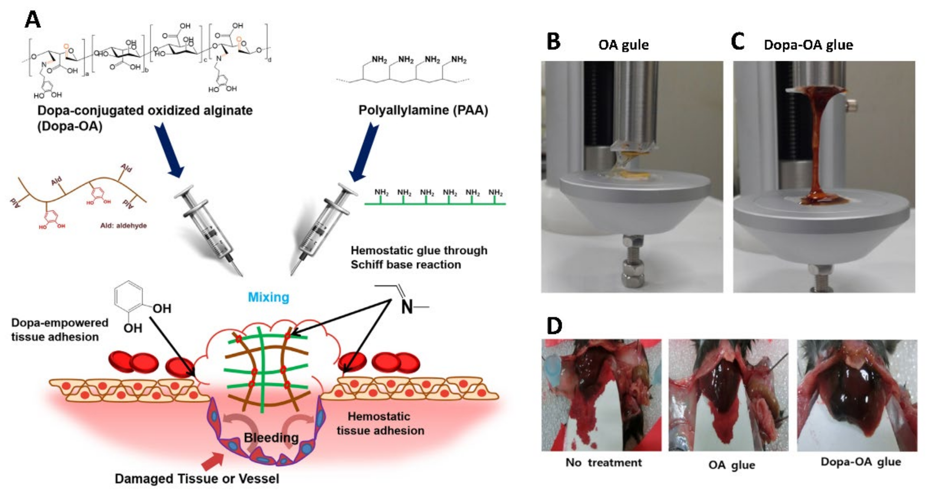
- Song, C.K.; Kim, M.-K.; Lee, J.; Davaa, E.; Baskaran, R.; Yang, S.-G. Dopa-Empowered Schiff Base Forming Alginate Hydrogel Glue for Rapid Hemostatic Control. Macromol. Res. 2019, 27, 119–125. [Google Scholar] [CrossRef]
- Balakrishnan, B.; Joshi, N.; Jayakrishnan, A.; Banerjee, R. Self-Crosslinked Oxidized Alginate/Gelatin Hydrogel as Injectable, Adhesive Biomimetic Scaffolds for Cartilage Regeneration. Acta Biomater. 2014, 10, 3650–3663. [Google Scholar] [CrossRef] [PubMed]
- Hou, J.; Li, C.; Guan, Y.; Zhang, Y.; Zhu, X.X. Enzymatically Crosslinked Alginate Hydrogels with Improved Adhesion Properties. Polym. Chem. 2015, 6, 2204–2213. [Google Scholar] [CrossRef]
- Zhang, H.; Bré, L.P.; Zhao, T.; Zheng, Y.; Newland, B.; Wang, W. Mussel-Inspired Hyperbranched Poly(Amino Ester) Polymer as Strong Wet Tissue Adhesive. Biomaterials 2014, 35, 711–719. [Google Scholar] [CrossRef]
- Kong, Y.; Hou, Z.; Zhou, L.; Zhang, P.; Ouyang, Y.; Wang, P.; Chen, Y.; Luo, X. Injectable Self-Healing Hydrogels Containing CuS Nanoparticles with Abilities of Hemostasis, Antibacterial Activity, and Promoting Wound Healing. ACS Biomater. Sci. Eng. 2021, 7, 335–349. [Google Scholar] [CrossRef] [PubMed]
- Shi, X.; Fang, Q.; Ding, M.; Wu, J.; Ye, F.; Lv, Z.; Jin, J. Microspheres of Carboxymethyl Chitosan, Sodium Alginate and Collagen for a Novel Hemostatic in Vitro Study. J. Biomater. Appl. 2016, 30, 1092–1102. [Google Scholar] [CrossRef] [PubMed]
- Huang, X.; Fu, Q.; Deng, Y.; Wang, F.; Xia, B.; Chen, Z.; Chen, G. Surface Roughness of Silk Fibroin/Alginate Microspheres for Rapid Hemostasis in Vitro and in Vivo. Carbohydr. Polym. 2021, 253, 117256. [Google Scholar] [CrossRef] [PubMed]
- Jin, J.; Xu, M.; Liu, Y.; Ji, Z.; Dai, K.; Zhang, L.; Wang, L.; Ye, F.; Chen, G.; Lv, Z. Alginate-Based Composite Microspheres Coated by Berberine Simultaneously Improve Hemostatic and Antibacterial Efficacy. Colloids Surf. B Biointerfaces 2020, 194, 111168. [Google Scholar] [CrossRef] [PubMed]
- Zhang, X.; Dai, K.; Liu, C.; Hu, H.; Luo, F.; Qi, Q.; Wang, L.; Ye, F.; Jin, J.; Tang, J.; et al. Berberine-Coated Biomimetic Composite Microspheres for Simultaneously Hemostatic and Antibacterial Performance. Polymers 2021, 13, 360. [Google Scholar] [CrossRef] [PubMed]
- Huang, W.; Cheng, S.; Wang, X.; Zhang, Y.; Chen, L.; Zhang, L. Noncompressible Hemostasis and Bone Regeneration Induced by an Absorbable Bioadhesive Self-Healing Hydrogel. Adv. Funct. Mater. 2021, 31, 2009189. [Google Scholar] [CrossRef]
- Huang, H.; Chen, H.; Wang, X.; Qiu, F.; Liu, H.; Lu, J.; Tong, L.; Yang, Y.; Wang, X.; Wu, H. Degradable and Bioadhesive Alginate-Based Composites: An Effective Hemostatic Agent. ACS Biomater. Sci. Eng. 2019, 5, 5498–5505. [Google Scholar] [CrossRef]
- Lee, B.-B.; Ravindra, P.; Chan, E.-S. Size and Shape of Calcium Alginate Beads Produced by Extrusion Dripping. Chem. Eng. Technol. 2013, 36, 1627–1642. [Google Scholar] [CrossRef]
- Chan, E.-S.; Lim, T.-K.; Voo, W.-P.; Pogaku, R.; Tey, B.T.; Zhang, Z. Effect of Formulation of Alginate Beads on Their Mechanical Behavior and Stiffness. Particuology 2011, 9, 228–234. [Google Scholar] [CrossRef]
- Fathi, P.; Sikorski, M.; Christodoulides, K.; Langan, K.; Choi, Y.S.; Titcomb, M.; Ghodasara, A.; Wonodi, O.; Thaker, H.; Vural, M.; et al. Zeolite-Loaded Alginate-Chitosan Hydrogel Beads as a Topical Hemostat. J. Biomed. Mater. Res. B Appl. Biomater. 2018, 106, 1662–1671. [Google Scholar] [CrossRef] [PubMed]
- Dabholkar, N.; Gorantla, S.; Waghule, T.; Rapalli, V.K.; Kothuru, A.; Goel, S.; Singhvi, G. Biodegradable Microneedles Fabricated with Carbohydrates and Proteins: Revolutionary Approach for Transdermal Drug Delivery. Int. J. Biol. Macromol. 2021, 170, 602–621. [Google Scholar] [CrossRef] [PubMed]
- Al Sulaiman, D.; Chang, J.Y.H.; Bennett, N.R.; Topouzi, H.; Higgins, C.A.; Irvine, D.J.; Ladame, S. Hydrogel-Coated Microneedle Arrays for Minimally Invasive Sampling and Sensing of Specific Circulating Nucleic Acids from Skin Interstitial Fluid. ACS Nano 2019, 13, 9620–9628. [Google Scholar] [CrossRef] [PubMed]
- Chen, W.; Tian, R.; Xu, C.; Yung, B.C.; Wang, G.; Liu, Y.; Ni, Q.; Zhang, F.; Zhou, Z.; Wang, J.; et al. Microneedle-Array Patches Loaded with Dual Mineralized Protein/Peptide Particles for Type 2 Diabetes Therapy. Nat. Commun. 2017, 8, 1777. [Google Scholar] [CrossRef] [PubMed]
- Yu, W.; Jiang, G.; Zhang, Y.; Liu, D.; Xu, B.; Zhou, J. Polymer Microneedles Fabricated from Alginate and Hyaluronate for Transdermal Delivery of Insulin. Mater. Sci. Eng. C 2017, 80, 187–196. [Google Scholar] [CrossRef] [PubMed]
- Moniz, T.; Costa Lima, S.A.; Reis, S. Marine Polymeric Microneedles for Transdermal Drug Delivery. Carbohydr. Polym. 2021, 266, 118098. [Google Scholar] [CrossRef]
- Moreira, A.F.; Rodrigues, C.F.; Jacinto, T.A.; Miguel, S.P.; Costa, E.C.; Correia, I.J. Poly (Vinyl Alcohol)/Chitosan Layer-by-Layer Microneedles for Cancer Chemo-Photothermal Therapy. Int. J. Pharm. 2020, 576, 118907. [Google Scholar] [CrossRef] [PubMed]
- Arshad, M.S.; Fatima, S.; Nazari, K.; Ali, R.; Farhan, M.; Muhammad, S.A.; Abbas, N.; Hussain, A.; Kucuk, I.; Chang, M.-W.; et al. Engineering and Characterisation of BCG-Loaded Polymeric Microneedles. J. Drug Target. 2020, 28, 525–532. [Google Scholar] [CrossRef]
- Li, Y.; Liu, F.; Su, C.; Yu, B.; Liu, D.; Chen, H.-J.; Lin, D.; Yang, C.; Zhou, L.; Wu, Q.; et al. Biodegradable Therapeutic Microneedle Patch for Rapid Antihypertensive Treatment. ACS Appl. Mater. Interfaces 2019, 11, 30575–30584. [Google Scholar] [CrossRef] [PubMed]
- Wang, Z.; Yang, Z.; Jiang, J.; Shi, Z.; Mao, Y.; Qin, N.; Tao, T.H. Silk Microneedle Patch Capable of On-Demand Multidrug Delivery to the Brain for Glioblastoma Treatment. Adv. Mater. 2022, 34, 2106606. [Google Scholar] [CrossRef] [PubMed]
- Zhang, X.; Chen, G.; Bian, F.; Cai, L.; Zhao, Y. Encoded Microneedle Arrays for Detection of Skin Interstitial Fluid Biomarkers. Adv. Mater. Deerfield Beach Fla 2019, 31, e1902825. [Google Scholar] [CrossRef] [PubMed]
- Sharma, S.; Huang, Z.; Rogers, M.; Boutelle, M.; Cass, A.E.G. Evaluation of a Minimally Invasive Glucose Biosensor for Continuous Tissue Monitoring. Anal. Bioanal. Chem. 2016, 408, 8427–8435. [Google Scholar] [CrossRef] [PubMed] [Green Version]
- Gao, J.; Huang, W.; Chen, Z.; Yi, C.; Jiang, L. Simultaneous Detection of Glucose, Uric Acid and Cholesterol Using Flexible Microneedle Electrode Array-Based Biosensor and Multi-Channel Portable Electrochemical Analyzer. Sens. Actuators B Chem. 2019, 287, 102–110. [Google Scholar] [CrossRef]
- Zhang, A.; Xiao, Z.; Liu, Q.; Li, P.; Xu, F.; Liu, J.; Tao, H.; Feng, L.; Song, S.; Liu, Z.; et al. CaCO3 -Encapuslated Microspheres for Enhanced Transhepatic Arterial Embolization Treatment of Hepatocellular Carcinoma. Adv. Healthc. Mater. 2021, 10, e2100748. [Google Scholar] [CrossRef] [PubMed]
- Jin, J.; Ji, Z.; Xu, M.; Liu, C.; Ye, X.; Zhang, W.; Li, S.; Wang, D.; Zhang, W.; Chen, J.; et al. Microspheres of Carboxymethyl Chitosan, Sodium Alginate, and Collagen as a Hemostatic Agent in Vivo. ACS Biomater. Sci. Eng. 2018, 4, 2541–2551. [Google Scholar] [CrossRef] [PubMed]
- Zhang, Y.; Jiang, G.; Yu, W.; Liu, D.; Xu, B. Microneedles Fabricated from Alginate and Maltose for Transdermal Delivery of Insulin on Diabetic Rats. Mater. Sci. Eng. C 2018, 85, 18–26. [Google Scholar] [CrossRef] [PubMed]


| Type of Reaction | Materials | Methods Used | Form of Gels | Properties | Ref. |
|---|---|---|---|---|---|
| Ionic cross-linking | Alginate + ZnO | Casting and solvent evaporation | Bi-layered hydrogel films | Antibacterial properties, promotes healing capacity | [58] |
| Ionic cross-linking | Alginate + shellac | Microfluidic technology | Amphiphilic hydrogel–solid dimer particles | Amphiphilic structure, biocompatibility | [45] |
| Ionic cross-linking | AuNP-CaCO3 + sodium alginate | / | Hydrogel precipitation | Fast response, pH-response, and ultrahigh sensitivity | [59] |
| Electrostatic interaction and divalent chelation | Alginate + N-carboxymethyl chitosan | / | Dual-crosslinked hydrogels | Tunable mechanical properties, efficient wound closure | [60] |
| Hydrogen bonds and Schiff cross-linking | Oxidized sodium alginate + dopamine | / | Hydrogels | Self-healing, high tensile strength and stretchability | [61] |
| Molecular entanglements | Alginate + carboxymethylcellulose sodium + chitosan | Thermoforming, high-speed blending | Composite hydrogels | Water vapor permeability | [62] |
| Molecular entanglements | Alginate + PVA + PVP | Freezing-thawing method | Hydrogels | Bioadhesive strength and mechanical properties | [63] |
| Self-assemble | Alginate + copolymer F127 | / | Hydrogels | Thermo-responsive behavior | [64] |
| Guest and host reaction | PEG-Adamantane + β-CD + alginate | / | Hydrogels | Self-healing | [65] |
| Photo-cross-linking | Alginate + PVP + chitosan | Gamma-radiation | Hydrogel pads | Photoreactivity, hygroscopicity | [66] |
| Ionically cross-linking, covalent cross-linking | laponite + PVA + alginate | Nanohybrid hydrogels | Blood coagulation | [67] | |
| Covalent cross-linking, ionically cross-linking | OA + ECM + Amine-rGO | Double-network hydrogel | Improved mechanical properties and electrical conductivity | [68] |
| Type | Materials | Active Ingredients | Properties | Indications | Ref. |
|---|---|---|---|---|---|
| Fibrous dressings | Alginate + nZnO | nZnO | High porosity, antibacterial properties | Severe bleeding and wounds at risk of infection | [85] |
| Alginate + chitosan + hyaluronic acid | / | High swelling absorption properties | Moist wound care | [86] | |
| Films and membranes | Sodium alginate (SA) + carboxymethyl chitosan (CMCH) | Sr2+, Zn2+ | Cell adhesion enhancement and antibacterial properties | Various stages of wound healing | [92] |
| Hyaluronic acid (HA) + sodium alginate (SA) | Sulfadiazine, AgNPs | Mechanical property enhancement, antibacterial properties | Local wound hemostasis and care | [94] | |
| Gel-ADH+ SA-mCHO | / | Rapid spray-filming performance | Rapid and massive hemostasis | [96] | |
| Sponges | Sodium alginate (SA) + graphene oxide (GO) + polyvinyl alcohol (SPG) | Norfloxacin (NFX) | High water uptake and gas permeability | Hemostasis in superficial wounds, Wound dressings | [99] |
| Oxidized cellulose nanocrystals (TOCN) + Sodium alginate (SA) | / | High chemical stability and water absorption | Rapid local hemostasis | [100] | |
| Hm-alginate | / | High adhesiveness | Dressing for hemostasis | [101] | |
| Hemostatic needles | Alginate + CaCl2 | / | Coating, hemostasis in situ | Prevention of bleeding after vascular and tissue puncture | [103] |
| SA + HA + calcium carbonate | CD34 | Coating, hemostasis in situ | Stab wound hemostasis and healing | [104] | |
| SA + HA + calcium carbonate | / | Coating, hemostasis in situ | Hemorrhage after AVF or AVG cannulation | [105] | |
| Embolic materials | Alginate + CaCl2·2H2O | / | Minimal trauma and good hemostatic effect | Hemostasis in solid visceral organ rupture and hemorrhage | [121] |
| Alginate + CaCl2 | Thrombin | Embolic hemostasis | Parenchymal visceral hemorrhage | [123] | |
| CaCO3-Alginate | / | High safety and availability | Hepatocellular carcinoma therapy | [152] | |
| Injectable Hydrogels | Pept-1 + ALG | Pept-1 | High mechanical strength and hemostatic efficiency | Hemostasis in noncompressible wounds and irregular wounds | [124] |
| Oxidized sodium alginate (OA) + dopamine | Dopamine | High stiffness and elasticity | Hemostasis in deep tissue | [126] | |
| N-carboxyethyl chitosan + oxidized sodium alginate | CuS-NPs | Injectability and self-healing | Hemostasis of in situ wounds | [130] | |
| Hemostatic powders | Carboxymethyl chitosan + sodium alginate + collagen | / | High surface roughness and good biodegradability | Hemostasis in emergency conditions | [153] |
| Alginate + silk fibroin | / | High surface roughness | Rapid hemostasis in vitro and in vivo | [132] | |
| Alginate + collagen + TF liposomes | Tissue factor (TF) | Efficient and inexpensive, adaptation to various shapes of wounds | Rapid hemostasis in traumatic injury and deep wounds | [79] | |
| Microneedles | Alginate + maltose + CaCl2 | Insulin | Excellent mechanical strength and toughness, great swelling and dissolution properties | Sustained release transdermal delivery of insulin | [154] |
| Sodium alginate + Bacillus Calmette–Guérin (BCG) | Bacillus Calmette–Guérin (BCG) | Small risk of inducing inflammatory response and skin damage | Transcutaneous immunization with vaccine | [146] |
Publisher’s Note: MDPI stays neutral with regard to jurisdictional claims in published maps and institutional affiliations. |
© 2022 by the authors. Licensee MDPI, Basel, Switzerland. This article is an open access article distributed under the terms and conditions of the Creative Commons Attribution (CC BY) license (https://creativecommons.org/licenses/by/4.0/).
Share and Cite
Xie, Y.; Gao, P.; He, F.; Zhang, C. Application of Alginate-Based Hydrogels in Hemostasis. Gels 2022, 8, 109. https://doi.org/10.3390/gels8020109
Xie Y, Gao P, He F, Zhang C. Application of Alginate-Based Hydrogels in Hemostasis. Gels. 2022; 8(2):109. https://doi.org/10.3390/gels8020109
Chicago/Turabian StyleXie, Yue, Pan Gao, Fangfang He, and Chun Zhang. 2022. "Application of Alginate-Based Hydrogels in Hemostasis" Gels 8, no. 2: 109. https://doi.org/10.3390/gels8020109
APA StyleXie, Y., Gao, P., He, F., & Zhang, C. (2022). Application of Alginate-Based Hydrogels in Hemostasis. Gels, 8(2), 109. https://doi.org/10.3390/gels8020109