
A Laboratory-Developed Assay for the Simultaneous Detection of Aspergillus fumigatus and Pneumocystis jirovecii Pulmonary Pathogens
 , ,
, ,  , ,
, ,  ,
,  and
and 
Abstract
1. Introduction
2. Materials and Methods
2.1. Study Samples
2.2. Panther Fusion-Based LDA
2.2.1. Primers and Probes
2.2.2. Target Plasmid DNA
2.2.3. Optimization and Performance Experiments
2.2.4. BALF Sample Testing
2.3. Statistical Analysis
3. Results
4. Discussion
Supplementary Materials
Author Contributions
Funding
Institutional Review Board Statement
Informed Consent Statement
Data Availability Statement
Acknowledgments
Conflicts of Interest
Abbreviations
| AF | Aspergillus fumigatus |
| AIDS | Acquired immunodeficiency syndrome |
| BALF | Bronchoalveolar lavage fluid |
| BDG | Beta-d-glucan |
| BLAST | Basic local alignment search tool |
| CI | Confidence interval |
| Ct | Cycle threshold |
| CV | Coefficient of variation |
| DHFR | Dihydrofolate reductase |
| EORTC/MSGERC | European Organization for Research and Treatment of Cancer/Mycoses Study Group Education and Research Consortium |
| GM | Galactomannan |
| HIV | Human immunodeficiency virus |
| IFD | Invasive fungal disease |
| ITS1 | Internal transcribed spacer 1 |
| IVD | In vitro diagnostic assay |
| LDA | Laboratory-developed assay |
| LDT | Laboratory-developed test |
| LoD | Limit of detection |
| NPA | Negative percent agreement |
| PcP | Pneumocystis pneumonia |
| PCR | Polymerase chain reaction |
| PJ | Pneumocystis jirovecii |
| PPA | Positive percent agreement |
| PPR | Primer-probe reconstitution |
| SARS-CoV-2 | Severe acute respiratory syndrome-coronavirus 2 |
| SD | Standard deviation |
| WHO | World Health Organization |
References
- El-Baba, F.; Gao, Y.; Soubani, A.O. Pulmonary Aspergillosis: What the Generalist Needs to Know. Am. J. Med. 2020, 133, 668–674. [Google Scholar] [CrossRef]
- Apostolopoulou, A.; Fishman, J.A. The Pathogenesis and Diagnosis of Pneumocystis jiroveci Pneumonia. J. Fungi 2022, 8, 1167. [Google Scholar] [CrossRef] [PubMed]
- Pham, D.; Sivalingam, V.; Tang, H.M.; Montgomery, J.M.; Chen, S.C.; Halliday, C.L. Molecular Diagnostics for Invasive Fungal Diseases: Current and Future Approaches. J. Fungi 2024, 10, 447. [Google Scholar] [CrossRef] [PubMed]
- Wickes, B.L.; Wiederhold, N.P. Molecular Diagnostics in Medical Mycology. Nat. Commun. 2018, 9, 5135. [Google Scholar] [CrossRef] [PubMed]
- Stellrecht, K.A.; Cimino, J.L.; Maceira, V.P. The Panther Fusion System with Open Access Functionality for Laboratory-Developed Tests for Influenza A Virus Subtyping. J. Clin. Microbiol. 2020, 58, e00188-20. [Google Scholar] [CrossRef]
- Stellrecht, K.A.; Wilson, L.I.; Castro, A.J.; Maceira, V.P. Automated Real-Time PCR Detection of Tickborne Diseases Using the Panther Fusion Open Access System. Microbiol. Spectr. 2022, 10, e0280822. [Google Scholar] [CrossRef]
- Grant, J.; Atapattu, N.; Dilcher, M.; Tan, C.E.; Elvy, J.; Ussher, J.E. Development of Real-Time RT-PCR Assays to Detect Measles Virus on the Hologic Panther Fusion® System. J. Clin. Virol. 2023, 159, 105355. [Google Scholar] [CrossRef]
- Villa, R.D.; Pentella, M.A.; Benfer, J.L. A Laboratory-Developed Assay for Clade II Human Mpox Virus on the Panther Fusion Open Access System. J. Infect. Dis. 2024, 229, S132–S136. [Google Scholar] [CrossRef]
- Lindroth, Y.; Hansson, L.; Forslund, O. An Automated Commercial Open Access Assay for Detection of Mycoplasma genitalium macrolide resistance. APMIS 2025, 133, e13477. [Google Scholar] [CrossRef]
- Hologic®. Panther Fusion® System. Available online: https://www.hologic.com/hologic-products/diagnostic-solutions/panther-fusion-system (accessed on 13 January 2025).
- Zhen, W.; Smith, E.; Manji, R.; Schron, D.; Berry, G.J. Clinical Evaluation of Three Sample-to-Answer Platforms for Detection of SARS-CoV-2. J. Clin. Microbiol. 2020, 58, e00783-20. [Google Scholar] [CrossRef]
- World Health Organization. WHO Fungal Priority Pathogens List to Guide Research, Development and Public Health Action; WHO: Geneva, Switzerland, 2022. [Google Scholar]
- Albert, E.; Alcaraz, M.J.; Giménez, E.; Clari, M.Á.; Torres, I.; Colomina, J.; Olea, B.; Tormo, M.; Piñana, J.L.; Oltra, R.; et al. Comparative Performance of the Platelia Aspergillus Antigen and Aspergillus Galactomannan Antigen Virclia Monotest Immunoassays in Serum and Lower Respiratory Tract Specimens: A “Real-Life” Experience. Microbiol. Spectr. 2024, 12, e0391023. [Google Scholar] [CrossRef]
- Price, J.S.; Fallon, M.; Posso, R.; Backx, M.; White, P.L. An Evaluation of the OLM PneumID Real-Time Polymerase Chain Reaction to Aid in the Diagnosis of Pneumocystis Pneumonia. J. Fungi 2023, 9, 1106. [Google Scholar] [CrossRef] [PubMed]
- Del Corpo, O.; Butler-Laporte, G.; Sheppard, D.C.; Cheng, M.P.; McDonald, E.G.; Lee, T.C. Diagnostic Accuracy of Serum (1-3)-β-D-Glucan for Pneumocystis jirovecii Pneumonia: A Systematic Review and Meta-Analysis. Clin. Microbiol. Infect. 2020, 26, 1137–1143. [Google Scholar] [CrossRef] [PubMed]
- Donnelly, J.P.; Chen, S.C.; Kauffman, C.A.; Steinbach, W.J.; Baddley, J.W.; Verweij, P.E.; Clancy, C.J.; Wingard, J.R.; Lockhart, S.R.; Groll, A.H.; et al. Revision and Update of the Consensus Definitions of Invasive Fungal Disease from the European Organization for Research and Treatment of Cancer and the Mycoses Study Group Education and Research Consortium. Clin. Infect. Dis. 2020, 71, 1367–1376. [Google Scholar] [CrossRef]
- de Heer, K.; Gerritsen, M.G.; Visser, C.E.; Leeflang, M.M. Galactomannan Detection in Bronchoalveolar Lavage Fluid for Invasive Aspergillosis in Immunocompromised Patients. Cochrane Database Syst. Rev. 2019, 5, CD012399. [Google Scholar] [CrossRef]
- Gits-Muselli, M.; White, P.L.; Mengoli, C.; Chen, S.; Crowley, B.; Dingemans, G.; Fréalle, E.; Gorton, R.L.; Guiver, M.; Hagen, F.; et al. The Fungal PCR Initiative’s Evaluation of In-House and Commercial Pneumocystis jirovecii qPCR Assays: Toward a Standard for a Diagnostics Assay. Med. Mycol. 2020, 58, 779–788. [Google Scholar] [CrossRef]
- Walsh, T.J.; Wissel, M.C.; Grantham, K.J.; Petraitiene, R.; Petraitis, V.; Kasai, M.; Francesconi, A.; Cotton, M.P.; Hughes, J.E.; Greene, L.; et al. Molecular Detection And Species-Specific Identification of Medically Important Aspergillus species by Real-Time PCR in Experimental Invasive Pulmonary Aspergillosis. J. Clin. Microbiol. 2011, 49, 4150–4157. [Google Scholar] [CrossRef]
- Liotti, F.M.; Posteraro, B.; De Angelis, G.; Torelli, R.; De Carolis, E.; Speziale, D.; Menchinelli, G.; Spanu, T.; Sanguinetti, M.A. New PCR-Based Assay for Testing Bronchoalveolar Lavage Fluid Samples from Patients with Suspected Pneumocystis jirovecii Pneumonia. J. Fungi 2021, 7, 681. [Google Scholar] [CrossRef]
- Bustin, S.A.; Benes, V.; Garson, J.A.; Hellemans, J.; Huggett, J.; Kubista, M.; Mueller, R.; Nolan, T.; Pfaffl, M.W.; Shipley, G.L.; et al. The MIQE Guidelines: Minimum Information for Publication of Quantitative Real-Time PCR Experiments. Clin. Chem. 2009, 55, 611–622. [Google Scholar] [CrossRef]
- Motooka, D.; Fujimoto, K.; Tanaka, R.; Yaguchi, T.; Gotoh, K.; Maeda, Y.; Furuta, Y.; Kurakawa, T.; Goto, N.; Yasunaga, T.; et al. Fungal ITS1 Deep-Sequencing Strategies to Reconstruct the Composition of a 26-Species Community and Evaluation of the Gut Mycobiota of Healthy Japanese Individuals. Front. Microbiol. 2017, 8, 238. [Google Scholar] [CrossRef]
- Yusuf, E.; Schijffelen, M.J.; Leeflang, M. How to Verify and Validate a Clinical Microbiology Test before It Can Be Used in Routine Diagnostics: A Practical Guide. Clin. Microbiol. Infect. 2024, 30, 1261–1269. [Google Scholar] [CrossRef] [PubMed]
- Kidd, S.E.; Chen, S.C.; Meyer, W.; Halliday, C.L. A New Age in Molecular Diagnostics for Invasive Fungal Disease: Are We Ready? Front. Microbiol. 2020, 10, 2903. [Google Scholar] [CrossRef] [PubMed]
- Wickes, B.L.; Romanelli, A.M. Diagnostic Mycology: Xtreme Challenges. J. Clin. Microbiol. 2020, 58, e01345-19. [Google Scholar] [CrossRef] [PubMed]
- Montesinos, I.; Albichr, I.S.; Collinge, E.; Delaere, B.; Huang, T.D.; Bogaerts, P.; Deckers, C.; Hamouda, M.; Honoré, P.M.; Bulpa, P.; et al. Diagnostic Value of Serum Biomarkers for Invasive Aspergillosis in Haematologic Patients. J. Fungi 2024, 10, 661. [Google Scholar] [CrossRef]
- Brown, L.; Rautemaa-Richardson, R.; Mengoli, C.; Alanio, A.; Barnes, R.A.; Bretagne, S.; Chen, S.C.A.; Cordonnier, C.; Donnelly, J.P.; Heinz, W.J.; et al. Polymerase Chain Reaction on Respiratory Tract Specimens of Immunocompromised Patients to Diagnose Pneumocystis Pneumonia: A Systematic Review and Meta-analysis. Clin. Infect. Dis. 2024, 79, 161–168. [Google Scholar] [CrossRef]

| Reagent | Concentrations | |
|---|---|---|
| AF-LDA | PJ-LDA | |
| MgCl2, mM | 4 | 4 |
| KCl, mM | 50 | 50 |
| Tris, mM | 12.5 | 12.5 |
| Forward primer, µM | 0.2 | 0.2 |
| Reverse primer, µM | 0.2 | 0.2 |
| Probe, µM | 0.1 | 0.1 |
| Internal control | 0.5× | 0.5× |
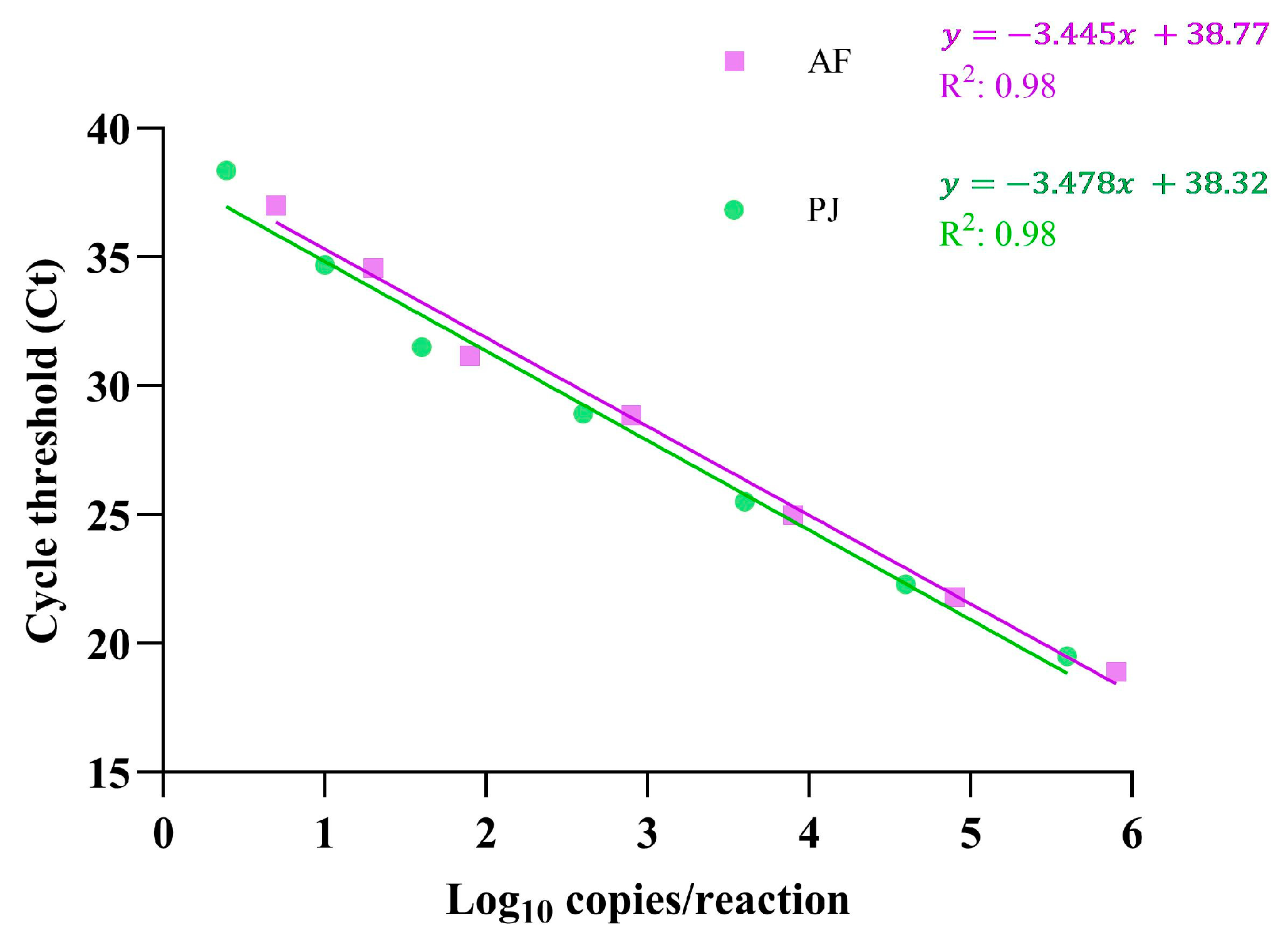
| Assay Target | Concentration (log10 Copies/Reaction) | No. of Replicates Detected/No. of Replicates Tested | Mean Ct | Standard Deviation | Coefficient of Variation (%) a |
|---|---|---|---|---|---|
| AF | |||||
| 5.90 | 5/5 | 18.9 | 0.30 | 1.60 | |
| 4.90 | 5/5 | 2.8 | 0.35 | 1.62 | |
| 3.90 | 5/5 | 24.9 | 0.21 | 0.86 | |
| 2.90 | 5/5 | 28.8 | 0.32 | 1.13 | |
| 1.90 | 5/5 | 31.1 | 0.23 | 0.75 | |
| 1.30 | 5/5 | 34.5 | 0.55 | 1.60 | |
| 0.70 | 2/5 | 37.1 | 0.52 | 1.42 | |
| PJ | |||||
| 5.60 | 5/5 | 19.5 | 0.47 | 2.42 | |
| 4.60 | 5/5 | 22.3 | 0.33 | 1.50 | |
| 3.60 | 5/5 | 25.4 | 0.35 | 1.37 | |
| 2.60 | 5/5 | 28.9 | 0.43 | 1.49 | |
| 1.60 | 5/5 | 31.5 | 0.23 | 0.72 | |
| 1.00 | 5/5 | 34.7 | 0.90 | 2.59 | |
| 0.39 | 2/5 | 38.3 | 0.52 | 1.38 |
| Panther Fusion LDA | No. of Positive (with Values) or Negative Results for Samples Tested by: | |||||||
|---|---|---|---|---|---|---|---|---|
| AF Culture (BALF Samples, n = 239) | GM Assay (BALF Samples, n = 57) | PneumID Assay (BALF Samples, n = 239) | BDG Assay (Serum Samples, n = 64) | |||||
| Positive | Negative | ≥1 Cutoff | <1 Cutoff | ≤35 Ct | >35 Ct | ≥80 pg/mL | <80 pg/mL | |
| AF positives (n = 22) | 22 | 0 | 11 (2–>10) | 2 | – | – | – | – |
| AF negatives (n = 216) | 1 a | 215 | 9 (2–>10) | 35 | – | – | – | – |
| PJ positives (n = 11) | – | – | – | – | 11 (17–29) | 0 | 11 (>500) | 0 |
| PJ negatives (n = 227) | – | – | – | – | 1 (31) b | 226 | 0 | 52 |
| AF/PJ positives (n = 1) | 1 | 0 | – | – | 1 (23) | 0 | 1 (>500) | 0 |
| Results | AF | PJ |
|---|---|---|
| No. matched positive | 23 | 12 |
| No. matched negative | 215 | 226 |
| No. missed | 1 | 1 |
| Range of Ct values | 25.1–35.7 | 22.6–34.3 |
| PPA (95% CI) | 95.8% (78.9–99.9%) | 92.3% (64.0–100%) |
| NPA (95% CI) | 100% (98.3–100%) | 100% (98.4–100%) |
Disclaimer/Publisher’s Note: The statements, opinions and data contained in all publications are solely those of the individual author(s) and contributor(s) and not of MDPI and/or the editor(s). MDPI and/or the editor(s) disclaim responsibility for any injury to people or property resulting from any ideas, methods, instructions or products referred to in the content. |
© 2025 by the authors. Licensee MDPI, Basel, Switzerland. This article is an open access article distributed under the terms and conditions of the Creative Commons Attribution (CC BY) license (https://creativecommons.org/licenses/by/4.0/).
Share and Cite
Cacaci, M.; Talamonti, D.; Menchinelli, G.; Squitieri, D.; Torelli, R.; De Carolis, E.; De Angelis, G.; Sanguinetti, M.; Posteraro, B. A Laboratory-Developed Assay for the Simultaneous Detection of Aspergillus fumigatus and Pneumocystis jirovecii Pulmonary Pathogens. J. Fungi 2025, 11, 280. https://doi.org/10.3390/jof11040280
Cacaci M, Talamonti D, Menchinelli G, Squitieri D, Torelli R, De Carolis E, De Angelis G, Sanguinetti M, Posteraro B. A Laboratory-Developed Assay for the Simultaneous Detection of Aspergillus fumigatus and Pneumocystis jirovecii Pulmonary Pathogens. Journal of Fungi. 2025; 11(4):280. https://doi.org/10.3390/jof11040280
Chicago/Turabian StyleCacaci, Margherita, Debora Talamonti, Giulia Menchinelli, Damiano Squitieri, Riccardo Torelli, Elena De Carolis, Giulia De Angelis, Maurizio Sanguinetti, and Brunella Posteraro. 2025. "A Laboratory-Developed Assay for the Simultaneous Detection of Aspergillus fumigatus and Pneumocystis jirovecii Pulmonary Pathogens" Journal of Fungi 11, no. 4: 280. https://doi.org/10.3390/jof11040280
APA StyleCacaci, M., Talamonti, D., Menchinelli, G., Squitieri, D., Torelli, R., De Carolis, E., De Angelis, G., Sanguinetti, M., & Posteraro, B. (2025). A Laboratory-Developed Assay for the Simultaneous Detection of Aspergillus fumigatus and Pneumocystis jirovecii Pulmonary Pathogens. Journal of Fungi, 11(4), 280. https://doi.org/10.3390/jof11040280