
Technical Data of Heterologous Expression and Purification of SARS-CoV-2 Proteases Using Escherichia coli System


Abstract
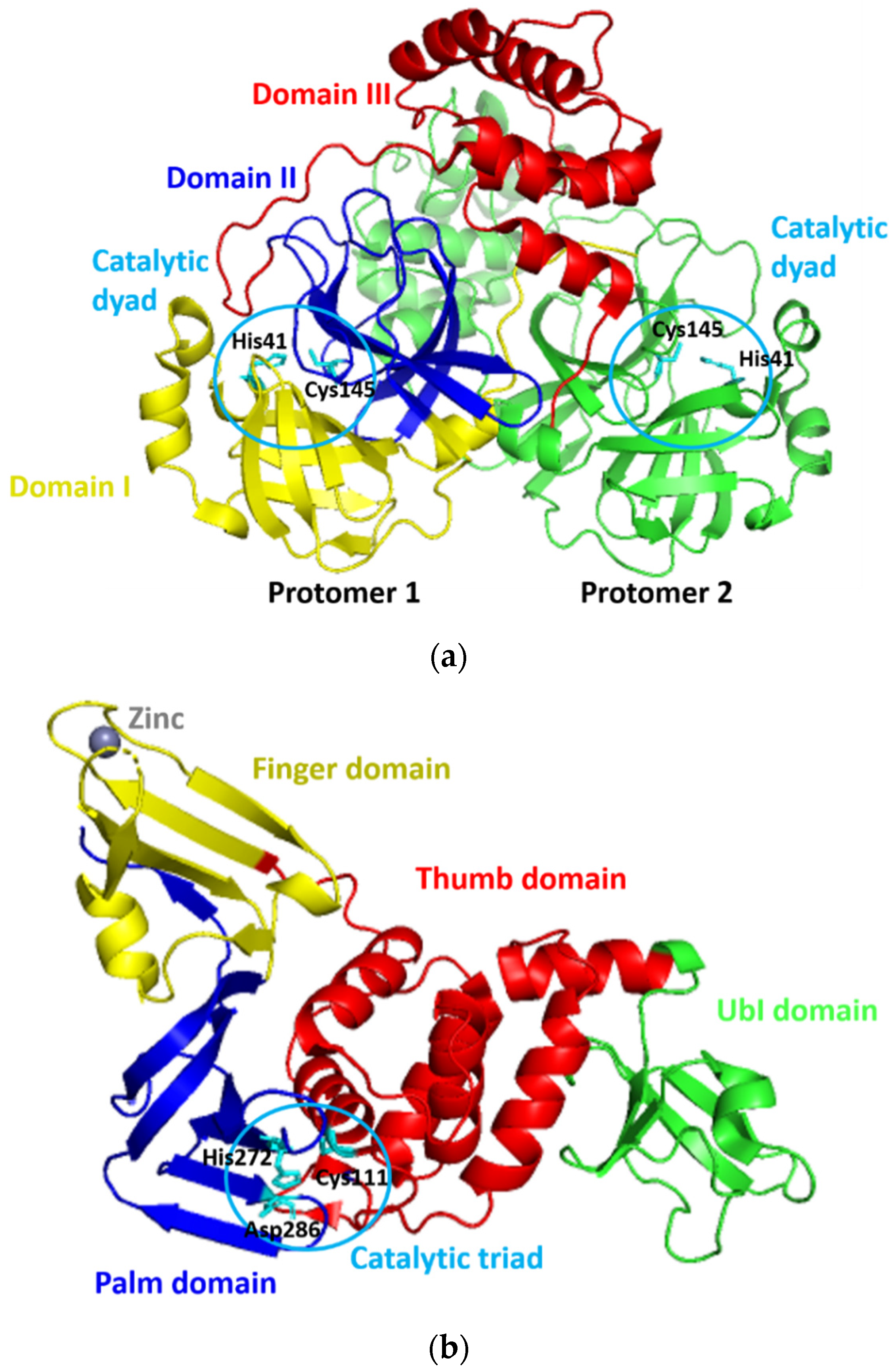
:1. Summary
2. Data Description
3. Methods
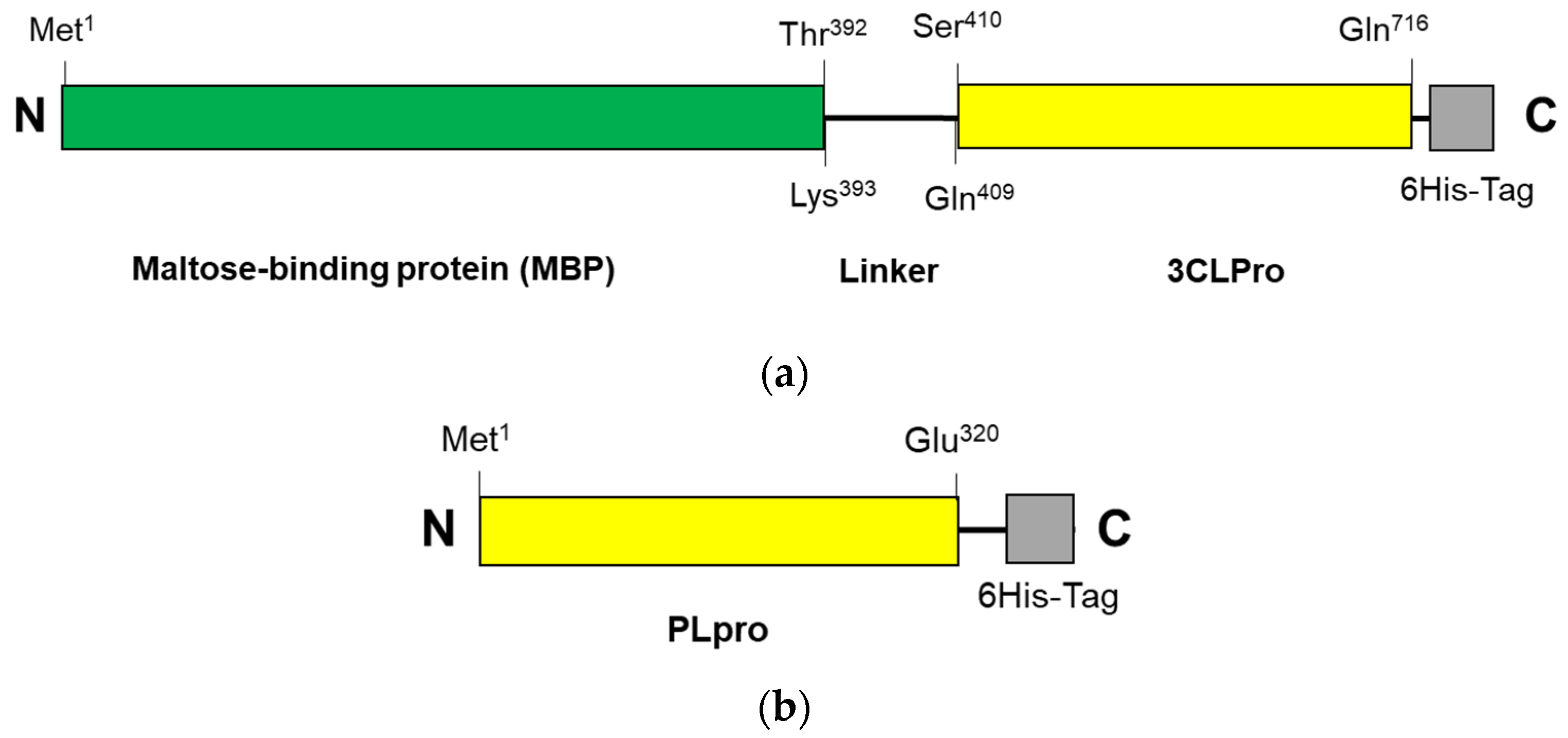
3.1. Expression and Purification of 3CLpro-CoV2
3.2. Expression and Purification of PLpro-CoV2
3.3. Cell Harvesting
3.4. Purification of Recombinant Proteins
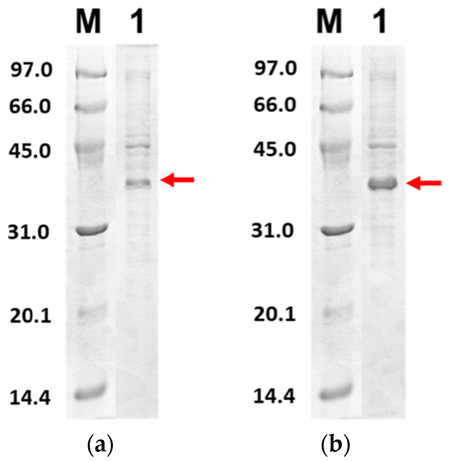
3.5. SDS-PAGE
3.6. Purification Profiles
Author Contributions
Funding
Institutional Review Board Statement
Informed Consent Statement
Data Availability Statement
Acknowledgments
Conflicts of Interest
References
- Zhou, P.; Yang, X.L.; Wang, X.G.; Hu, B.; Zhang, L.; Zhang, W.; Si, H.R.; Zhu, Y.; Li, B.; Huang, C.L.; et al. A pneumonia outbreak associated with a new coronavirus of probable bat origin. Nature 2020, 579, 270–273. [Google Scholar] [CrossRef] [PubMed] [Green Version]
- Wu, F.; Zhao, S.; Yu, B.; Chen, Y.M.; Wang, W.; Song, Z.G.; Hu, Y.; Tao, Z.W.; Tian, J.H.; Pei, Y.Y.; et al. A New Coronavirus associated with human respiratory disease in China. Nature 2020, 579, 265–269. [Google Scholar] [CrossRef] [PubMed] [Green Version]
- Gorbalenya, A.E.; Haagmans, B.L.; Sola, I. The species severe acute respiratory syndrome-related coronavirus: Classifying 2019-Ncov and naming it SARS-CoV-2. Nat. Microbiol. 2020, 5, 536–544. [Google Scholar] [CrossRef] [Green Version]
- Altay, O.; Mohammadi, E.; Lam, S.; Turkez, H.; Boren, J.; Nielsen, J.; Uhlen, M.; Mardinoglu, A. Current status of COVID-19 therapies and drug repositioning applications. iScience 2020, 23, 101303. [Google Scholar] [CrossRef] [PubMed]
- Ziebuhr, J. Molecular biology of severe acute respiratory syndrome coronavirus. Curr. Opin. Microbiol. 2004, 7, 412–419. [Google Scholar] [CrossRef] [PubMed]
- Thiel, V.; Ivanov, K.A.; Putics, Á.; Hertzig, T.; Schelle, S.; Bayer, S.; WeiBbrich, B.; Snijder, E.J.; Rabenau, H.; Doerr, H.W.; et al. Mechanisms and enzymes involved in SARS coronavirus genome expression. J. Gen. Virol. 2003, 84, 2305–2315. [Google Scholar] [CrossRef]
- Ul Qamar, M.H.; Alqahtani, S.M.; Alamri, M.A.; Chen, L.L. Structural basis of SARS-CoV-2 3CLpro and anti-COVID-19 drug discovery from medicinal plants. J. Pharm. Anal. 2020, 10, 313–319. [Google Scholar] [CrossRef]
- Alamri, M.A.; ul Qamar, M.T.; Mirza, M.U.; Alqahtani, S.M.; Froeyen, M.; Chen, L.L. Discovery of human coronaviruses pan-papain-like protease inhibitors using computational approaches. J. Pharm. Anal. 2020, 10, 546–559. [Google Scholar] [CrossRef]
- Rut, W.; Lv, Z.; Zmudzinski, M.; Patchett, S.; Nayak, D.; Snipas, S.J.; El Oualid, F.; Huang, T.T.; Bekes, M.; Drag, M.; et al. Activity profiling and crystal structures of inhibitor-bound SARS-CoV-2 papain-like protease: A framework for anti–COVID-19 drug design. Sci. Adv. 2020, 6, eabd4596. [Google Scholar] [CrossRef]
- Devaraj, S.G.; Wang, N.; Chen, Z.; Zhen, Z.; Tseng, M.; Barretto, N.; Lin, R.; Peters, C.J.; Tseng, C.K.; Baker, S.C.; et al. Regulation of IRF-3-dependent innate immunity by the papain-like protease domain of the severe acute respiratory syndrome coronavirus. J. Biol. Chem. 2007, 282, 32208–32221. [Google Scholar] [CrossRef] [Green Version]
- Clementz, M.A.; Chen, Z.; Banach, B.S.; Wang, Y.; Sun, L.; Ratia, K.; Baez-Santos, Y.M.; Wang, J.; Tukuyama, J.; Ghosh, A.K.; et al. Deubiquitinating and interferon antagonism activities of coronavirus papain-like proteases. J. Virol. 2019, 84, 4619–4629. [Google Scholar] [CrossRef] [PubMed] [Green Version]
- Frieman, M.; Ratia, K.; Johnston, R.E.; Mesecar, A.D.; Baric, R.S. Severe acute respiratory syndrome coronavirus papain-like protease ubiquitin-like domain and catalytic domain regulate antagonism of IRF3 and NF-kappa B signaling. J. Virol. 2009, 83, 6689–6705. [Google Scholar] [CrossRef] [PubMed] [Green Version]
- Osipiuk, J.; Azizi, S.A.; Dvorkin, S.; Endres, M.; Jedrzejczak, R.; Jones, K.A.; Kang, S.; Kathayat, R.S.; Kim, Y.; Lisnyak, V.G.; et al. Structure of papain-like protease from SARS-CoV-2 and its complexes with non-covalent inhibitors. Nat. Commun. 2021, 12, 743. [Google Scholar] [CrossRef]
- Kneller, D.W.; Phillips, G.; O’neil, H.M.; Jedrzejczak, R.; Langan, P.; Joachimiak, A.; Coates, L.; Kovalevsky, A. Structural plasticity of SARS-CoV-2 3CL Mpro active site cavity revealed by room temperature X-ray crystallography. Nat. Commun. 2020, 11, 3202. [Google Scholar] [CrossRef]
- Forstner, M.; Leder, L.; Mayr, L.M. Optimization of protein expression systems for modern drug discovery. Expert Rev. Proteomic 2007, 4, 67–78. [Google Scholar] [CrossRef]
- Smith, C. Striving for purity: Advances in protein purification. Nat. Methods 2005, 2, 71–77. [Google Scholar] [CrossRef]
- De Marco, A.; Deuerling, E.; Mogk, A.; Tomoyasu, T.; Bukau, B. Chaperone-based procedure to increase yields of soluble recombinant proteins produced in E. coli. BMC Biotechnol. 2007, 7, 32. [Google Scholar] [CrossRef] [Green Version]
- LaVallie, E.R.; DiBlasio, E.A.; Kovacic, S.; Grant, K.L.; Schendel, P.F.; McCoy, J.M. A thioredoxin gene fusion expression system that circumvents inclusion body formation in the E. coli cytoplasm. Biotechnology 1993, 11, 187–193. [Google Scholar] [CrossRef] [PubMed]
- Froger, A.; Hall, J.E. Transformation of plasmid DNA into E. coli using the heat shock method. J. Vis. Exp. 2007, 6, 253. [Google Scholar] [CrossRef] [PubMed]
- Razali, R.; Kumar, V.; Budiman, C. Structural insights into the enzymatic activity of protease bromelain of MD2 pineapple. Pak. J. Biol. Sci. 2020, 23, 829–838. [Google Scholar] [CrossRef]
- Laemmli, U.K. Cleavage of structural proteins during the assembly of the head of bacteriophage T4. Nature 1970, 227, 680–685. [Google Scholar] [CrossRef]
- Goodwin, T.; Morton, R. The spectrophotometric determination of tyrosine and tryptophan in proteins. Biochem. J. 1946, 40, 628. [Google Scholar] [CrossRef] [PubMed] [Green Version]
- Cheng, S.; Chang, G.; Chou, C. Mutation of Glu-166 blocks the substrate-induced dimerization of SARS coronavirus main protease. Biophys. J. 2010, 98, 1327–1336. [Google Scholar] [CrossRef] [PubMed] [Green Version]
- Bala, M.; Mel, M.; Jami, M.S.; Amid, A.; Salleh, H.M. Kinetic studies on recombinant stem bromelain. Adv. Enzym. Res. 2013, 1, 52–60. [Google Scholar] [CrossRef]


| Subject | Biological Sciences |
| Specific Subject Area | Biotechnology and biochemistry |
| Type of Data | Table Figure |
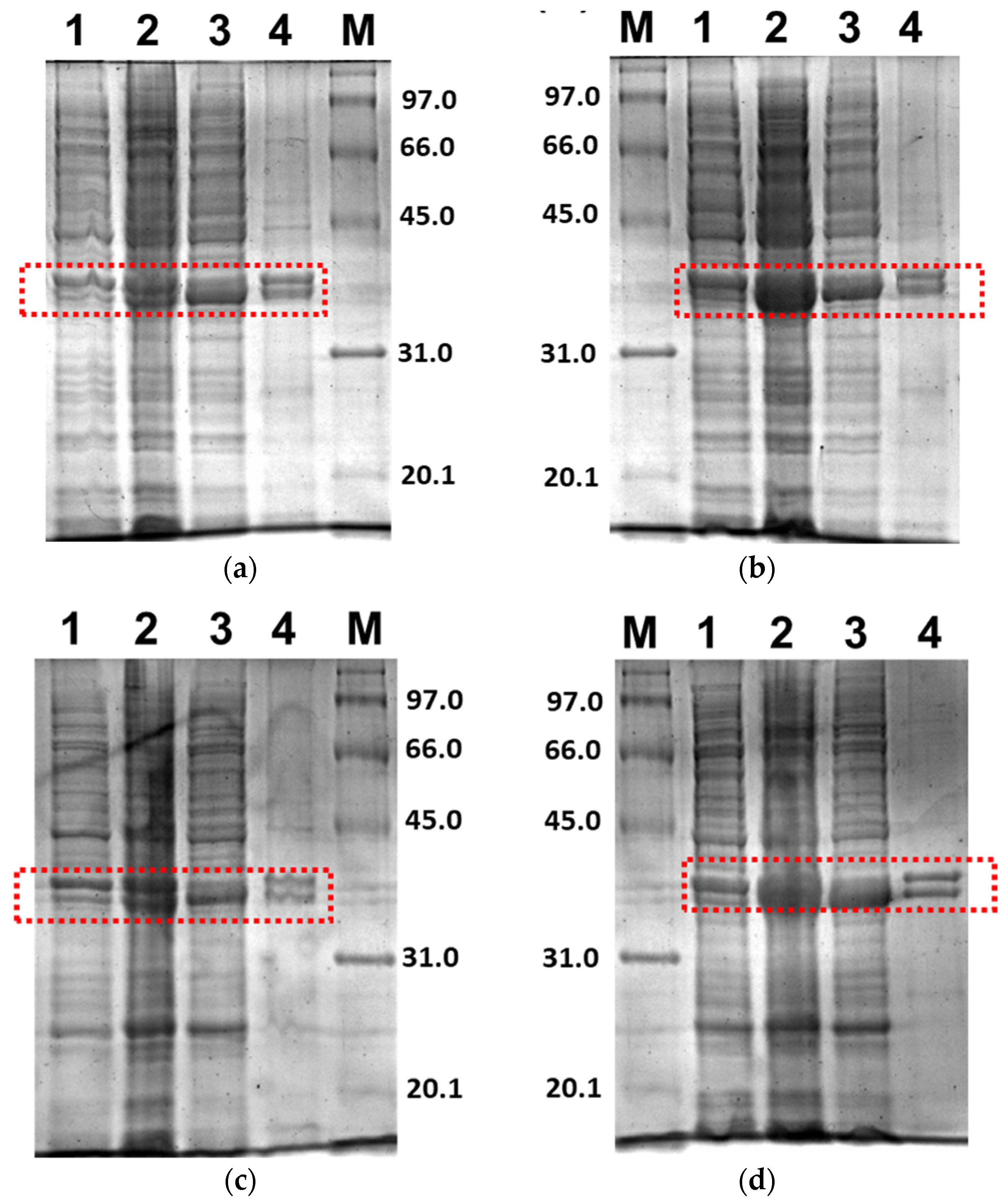
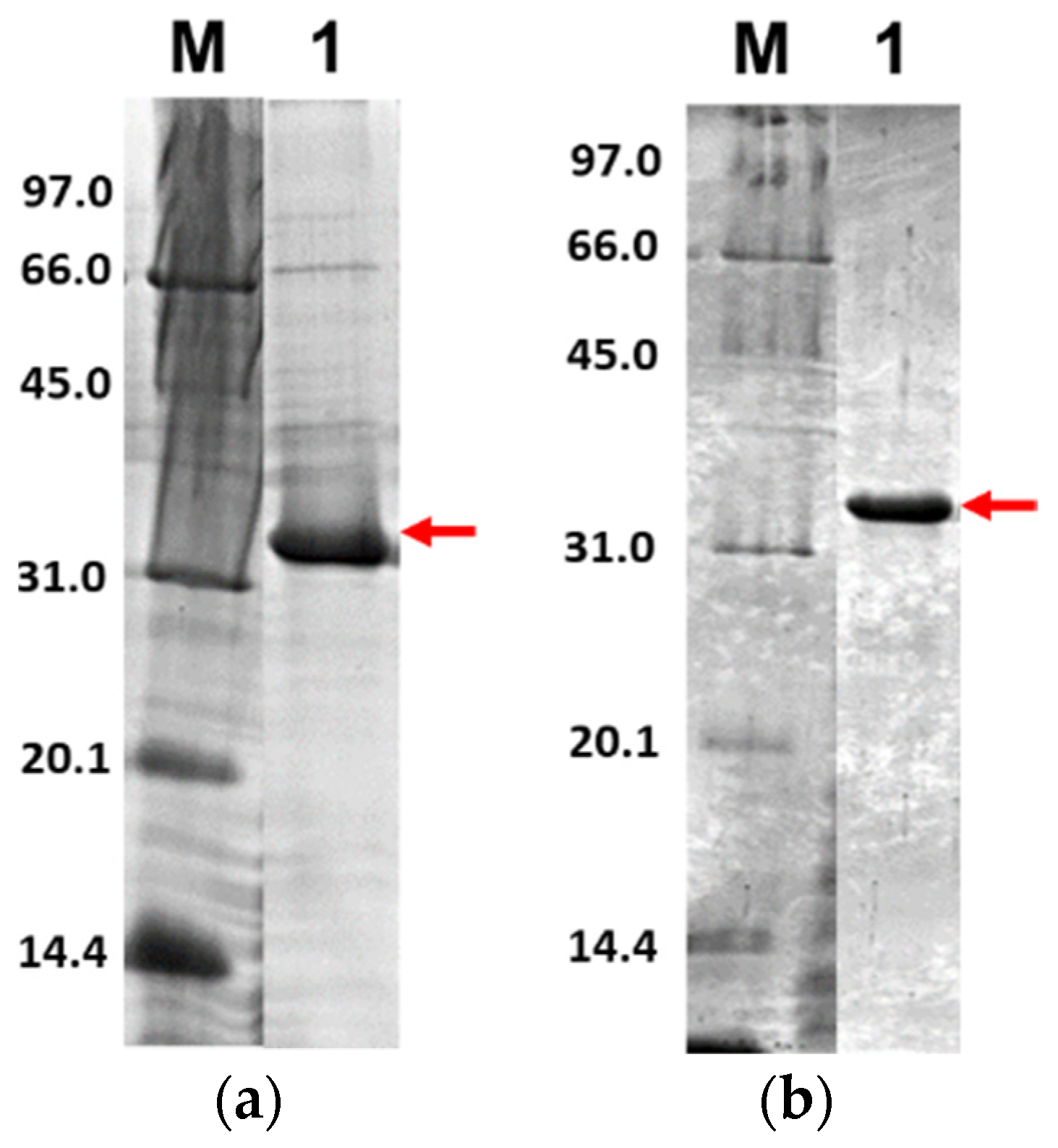
| How Data Were Acquired | The expression system for 3CLpro-CoV2 or PLpro-CoV2 was transformed into E. coli BL21(DE3) or E. coli BL21-CodonPlus(DE3) strains. The expression of both proteases was obtained by isopropyl β-D-1-thiogalactopyranoside (IPTG) induction. The expression of target proteins was analysed using SDS-PAGE and observed using Gel DocTM XR+ imager (Biorad, CA, USA). Purification profiles of both proteases from the selected conditions were obtained through purification under a single Ni2+-NTA affinity chromatography, followed by quantification of protein amount and enzymatic activity. |
| Data Format | Raw (Purification Table) Analyzed |
| Parameters for Data Collection | Concentration of IPTG for protein expression induction (mM); optical density at 600 nm (OD600); incubation temperature of protein expression (°C); incubation time of protein expression (h); volume of sample (mL); amount of protein (mg); total activity (U); specific activity (U/mg); yield (%) and purification fold. |
| Description of Data Collection | The data was collected along the production and purification flows of 3CLpro-CoV2 and PLpro-CoV2 through two steps. The first step involved the over-expression of 3CLpro-CoV2 and PLpro-CoV2 in the E. coli host cells under several conditions. The data collected included the expression and solubility observed under sodium dodecyl sulphate-polyacrylamide gel electrophoresis (SDS-PAGE). The second step involved the purification of the proteins using Ni2+-NTA affinity chromatography. The data of purification performances were collected based on the amount of protein, activity, yield and purification fold. |
| Data Source Location | Whole experiments and data collection were performed at Biotechnology Research Institute, Universiti Malaysia Sabah, Kota Kinabalu, Sabah, Malaysia. |
| Data Accessibility | With the article. |
| Condition | Host Cell | OD600 | Induction | Expression Condition | Expression Result |
|---|---|---|---|---|---|
| 3CLpro-CoV2 | |||||
| 1 | E. coli BL21(DE3) | 0.8 | 0.5 mM IPTG | 37 °C, 5 h, 180 rpm | Expressed in soluble forms |
| 2 | 18 °C, 18 h, 180 rpm | Expressed in soluble forms | |||
| PLpro-CoV2 | |||||
| 3 | E. coli BL21(DE3) | 0.8 | 1 mM IPTG | 37 °C, 5 h, 180 rpm | Expressed in insoluble forms |
| 4 | 1.5 | 0.1 mM IPTG, 0.1 mM ZnSO4 | Expressed in insoluble forms | ||
| 5 | 0.8 | 1 mM IPTG | 18 °C, 18 h, 180 rpm | Expressed in soluble forms | |
| 6 | 1.5 | 0.1 mM IPTG, 0.1 mM ZnSO4 | Expressed in soluble forms | ||
| 7 | E. coli BL21-CodonPlus(DE3) | 0.8 | 1 mM IPTG | 37 °C, 5 h, 180 rpm | Expressed in insoluble forms |
| 8 | 1.5 | 0.1 mM IPTG, 0.1 mM ZnSO4 | Expressed in insoluble forms | ||
| 9 | 0.8 | 1 mM IPTG | 18 °C, 18 h, 180 rpm | Expressed in soluble forms | |
| 10 | 1.5 | 0.1 mM IPTG, 0.1 mM ZnSO4 | Expressed in soluble forms | ||
| Condition | Step. | Volume (mL) | Total Protein (mg) | Total Activity (U) | Specific Activity (U/mg) | Yield (%) | Purification (Fold) |
|---|---|---|---|---|---|---|---|
| 3CLpro-CoV2 | |||||||
| (2) Expressed in E. coli BL21(DE3) at 18 °C, 180 rpm for 18 h. Induced with 1 mM IPTG | Soluble fraction (Crude) | 100.5 | 1095.33 | 23.22 | 0.02 | 100 | 1 |
| Ni2+-NTA affinity chromatography | 15.6 | 20.43 | 21.30 | 1.04 | 92 | 49 | |
| PLpro-CoV2 | |||||||
| (9) Expressed in E. coli BL21 CodonPlus(DE3) at 18 °C, 180 rpm for 18 h. Induced with 1 mM IPTG | Soluble fraction (Crude) | 100.33 | 1330 | 11.88 | 0.01 | 100 | 1 |
| Ni2+-NTA affinity chromatography | 36.83 | 15.88 | 9.88 | 0.63 | 83 | 71 | |
| (10) Expressed in E. coli BL21 CodonPlus(DE3) at 18 °C, 180 rpm for 18 h. Induced with 0.1 mM IPTG and 0.1 mM ZnSO4 | Soluble fraction (Crude) | 97.67 | 1239.33 | 5.39 | 0.004 | 100 | 1 |
| Ni2+-NTA affinity chromatography | 38.33 | 9.59 | 4.65 | 0.49 | 86 | 112 | |
| Host Cell | OD600 | Induction | Expression Condition | |
|---|---|---|---|---|
| 3CLpro-CoV2 | ||||
| 1 | E. coli BL21(DE3) | 0.8 | 0.5 mM IPTG | 37 °C, 5 h, 180 rpm |
| 2 | 18 °C, 18 h, 180 rpm | |||
| PLpro-CoV2 | ||||
| 3 | E. coli BL21(DE3) | 0.8 | 1 mM IPTG | 37 °C, 5 h, 180 rpm |
| 4 | 1.5 | 0.1 mM IPTG, 0.1 mM ZnSO4 | ||
| 5 | 0.8 | 1 mM IPTG | 18 °C, 18 h, 180 rpm | |
| 6 | 1.5 | 0.1 mM IPTG, 0.1 mM ZnSO4 | ||
| 7 | E. coli BL21-CodonPlus(DE3) | 0.8 | 1 mM IPTG | 37 °C, 5 h, 180 rpm |
| 8 | 1.5 | 0.1 mM IPTG, 0.1 mM ZnSO4 | ||
| 9 | 0.8 | 1 mM IPTG | 18 °C, 18 h, 180 rpm | |
| 10 | 1.5 | 0.1 mM IPTG, 0.1 mM ZnSO4 | ||
Publisher’s Note: MDPI stays neutral with regard to jurisdictional claims in published maps and institutional affiliations. |
© 2021 by the authors. Licensee MDPI, Basel, Switzerland. This article is an open access article distributed under the terms and conditions of the Creative Commons Attribution (CC BY) license (https://creativecommons.org/licenses/by/4.0/).
Share and Cite
Razali, R.; Subbiah, V.K.; Budiman, C. Technical Data of Heterologous Expression and Purification of SARS-CoV-2 Proteases Using Escherichia coli System. Data 2021, 6, 99. https://doi.org/10.3390/data6090099
Razali R, Subbiah VK, Budiman C. Technical Data of Heterologous Expression and Purification of SARS-CoV-2 Proteases Using Escherichia coli System. Data. 2021; 6(9):99. https://doi.org/10.3390/data6090099
Chicago/Turabian StyleRazali, Rafida, Vijay Kumar Subbiah, and Cahyo Budiman. 2021. "Technical Data of Heterologous Expression and Purification of SARS-CoV-2 Proteases Using Escherichia coli System" Data 6, no. 9: 99. https://doi.org/10.3390/data6090099
APA StyleRazali, R., Subbiah, V. K., & Budiman, C. (2021). Technical Data of Heterologous Expression and Purification of SARS-CoV-2 Proteases Using Escherichia coli System. Data, 6(9), 99. https://doi.org/10.3390/data6090099