Abstract
The separation and discrete detection of isomeric sequence peptides with similar properties are important tasks for analytical science. Three different peptide isomers of 12 amino-acid residues long, containing direct and reverse regions of the alanine-valine-proline-isoleucine (AVPI) motif, were partially separated and discretely detected from their mixture using two approaches. Capillary electrophoresis enabled the separation and optical detection of the peptide sequence isomers close to the baseline. The ability to separate these sequence isomers from the mixture and discretely identify them from mass spectra has also been demonstrated by ion-mobility tandem mass spectrometry. Moreover, for the first time, capillary electrophoresis and ion-mobility mass spectrometry connected online have shown their ability for a discrete detection of the multidirectional sequence isomers.
1. Introduction
Peptide sequence inversions are a significant class of isomers of proteolytic peptides that are important for protein identification [1]. A short section of the sequence can be partially inverted at some peptide region (e.g., due to alternative splicing [2,3]), making this change tracking inapplicable by sequencing of the coding DNA. These inversions may change the peptides’ properties and functions due to the differences in the resulting three-dimensional structure. The ability to separate and discretely identify peptide isomers from their mixtures is still a significant technological challenge. Along with the classical method of peptide sequencing using Edman degradation [4], mass spectrometry (MS) has become the gold standard for peptide identification in modern bioanalysis [5]. The use of MS conventionally coupled to liquid chromatography to analyze mixtures containing peptide isomers is limited due to the inability to separate them by charge or mass and basic chemical properties [6]. The same total masses and similar charges make their separation by commonly used techniques a challenging task. Due to the noticeable differences in isomer arrangements in three-dimensional structures, only the methods based on detecting diversities in their size or shape (expressed in collisional cross sections [7]) are effective for isomer separation from their mixtures. Capillary electrophoresis (CE) has proven to be a high-power technique for separating very closely shaped molecules, such as phosphorylated peptide isomers [8,9], as well as a method capable of determining isomeric phosphorylation sites [10]. Studies of the isomeric peptides with CE have typically been carried out with laser-induced fluorescence detection (LIF) [11].
While LIF-based CE with optical detection has been readily used for the analysis of amino acids, peptides are more complex molecules and for untargeted analysis it is required to couple the CE with more legible MS detectors to achieve sequence information about the peptides [12]. Comprehensive reviews containing common state-of-the-art advances in the use of CE-MS for the study of peptides have been compiled every few years by the Kašička group [13,14]. However, the outlook on this problem regarding the peptide isomers is relatively smaller. Thus, the separation of peptide isomers deamidated in various positions by CE-MS was reported in [15]. Some interesting studies are devoted to the methodology of peptide stereoisomers separation by CE [16,17]. Regarding the separation and detection of sequence isomeric peptides, a promising analytical platform for dipeptide structural isomer separation and analysis by CE-MS was developed by Ozawa et al. [18].
The detection and separation of peptide sequence isomers in the gas phase have become possible by ion mobility separation (IMS) devices coupled with mass spectrometers [1,9]. The ability to separate four β-amyloid peptide isomers using IMS-MS was demonstrated in [19]. The Li group provided a method to distinguish between two isomers of melanocyte-stimulating peptide hormone in a single run by combining LC–MS with IMS [20]. The possibility of inverted isomer separation with IMS has been shown for such small biomolecules as dipeptides [21]. However, there is still a technical challenge in separating partially reversed isomers that contain only a short reversed section (up to four amino-acid residues) of the overall sequence.
In this work, we demonstrated the CE and the IMS instruments’ efficiency for the separation and qualitative detection of short peptide sequence isomers in their mixture. Then we used the combination of these two methods coupled online to illustrate the possibility of their application for effective peptide separation. To identify the mixture components, we used two basic approaches: LIF optical detection in the ultraviolet (UV) range and tandem mass spectrometry (MS/MS).
For technical experiments on the separation of isomers, synthetic peptides containing two multidirectional alanine-valine-proline-isoleucine (AVPI) motifs were used. The AVPI binding motif is an N-terminal part of Smac protein [22], released from mitochondria into the cytosol in response to apoptotic stimuli. The AVPI-containing synthetic small-molecule Smac mimetics are widely used to design potential anticancer agents [23]. The different directions of the AVPI motif are intended to increase the specificity and effectiveness of a potential anticancer drug.
2. Materials and Methods
2.1. Chemicals
The following reagents and consumables were used: ammonium acetate (Fisher Scientific, Fair Lawn, NJ, USA), acetic acid (Bio Basic Inc., Markham, ON, Canada), HPLC grade methanol (Sigma-Aldrich, Oakville, ON, Canada), bare silica capillary (Polymicro Technologies, Phoenix, AZ, USA). All solutions were made using Milli-Q-quality deionized water and filtered through a 0.22 μm filter.
2.2. Synthetic Peptides
Isomeric peptides were provided by Robert N. Ben (Department of Chemistry and Biomolecular Sciences, University of Ottawa, Ottawa, ON, Canada) and were synthesized as described previously [24]. A stock solution of 10 mM peptide in methanol was prepared. Before each experiment, the stock solution was diluted to 50 μM with 10 mM ammonium acetate buffer adjusted to various pH with acetic acid.
2.3. CE Separation
The CE experiments were made using the ProteomeLab PA 800 capillary electrophoresis system (Beckman-Coulter, Brea, CA, USA). UV absorbance was measured by a photodiode array detector. The best peaks were obtained at 200 nm. Separations were carried out using a bare fused silica capillary of 85 cm in total length (75 cm from injection to the detection point) with an outer diameter of 365 μm and an inner diameter of 75 μm. Hydrodynamic injections of 15.7 nL were made by pressure. The separations were conducted by applying an electric field of 300 V/cm (positive charge at the inlet and ground at the outlet). The voltage of 25 kV was set, and a current of 12.8 μA was observed. The temperature of the capillary was 20 °C. The running buffer was 10 mM ammonium acetate adjusted to pH 3.45 with acetic acid.
2.4. IMS-MS/MS Experiments
The IMS-MS/MS experiments were made using the Synapt G2 HDMS QuanTOF mass spectrometer (Waters, Milford, MA, USA). The capillary electrophoresis machine injected sample solutions directly to the nanoelectrospray source. The pressure push of 1–4 psi was implemented through a bare fused silica capillary with an inner diameter of 50 μm. Electrospray injection (ESI) was carried out in the positive polarity mode. The ESI voltage was set to 4.0 kV, and the capillary tip was heated up to 80 °C. The ion-mobility cell parameters were set to default: IMS gas flow was 90 mL/min, IMS wave velocity was 650 m/s. The transfer cell collision energy was set to 60 V in the MS/MS mode. MassLynx 4.1 with BioLynx package (Waters, Milford, MA, USA.) was used to identify the MS/MS fragments.
2.5. CE Coupled with IMS-MS/MS
The ProteomeLab PA 800 Plus capillary electrophoresis system was coupled with the above-described mass spectrometer. CE-ESI connection with sheath fluid interface was used. A bare fused silica capillary of 90 cm in length with an inner diameter of 50 μm was used in the CE-MS/MS experiments. Injections of 11.7 nL of sample solution were made by pressure to fill the capillary tip. Then, an electric field of 290 V/cm with normal polarity was applied. The voltage of 26 kV was set, and currents of 3.5–4.0 μA were observed. The temperature of the capillary was 20 °C. The run buffer and sample concentrations were the same as in the CE-only experiments. The sheath fluid of 50% methanol and 1% formic acid was injected into the electrospray at a 1.2 μL/min rate. The trap collision energy was 40 V due to the MS/MS regime without the IMS. The transfer collision energy was set to 50 V with the IMS involvement. The other settings of the MS instrument were set the same as they were during the IMS-MS/MS experiments.
3. Results and Discussion
Three synthetic isomeric peptides with partially inverted sequence sections were used in the experiments. They consisted of 12 amino-acid residues and contained two areas of the AVPI sequence group, which were inverted in various ways. Both alanines were methylated. The first peptide had a sequence of AVPI-GGG-AVPI-G (direct-direct or DD sequence). The sequence of the second peptide was AVPI-GGG-IPVA-G (direct-reverse or DR). The third peptide sequence was IPVA-GGG-IPVA-G (reverse-reverse or RR). All residues in the peptides were aliphatic, which determined their neutral pI value. The absence of easily chargeable basic residues makes dealing with these peptides in an ionized state difficult.
3.1. CE Separation with UV Detection
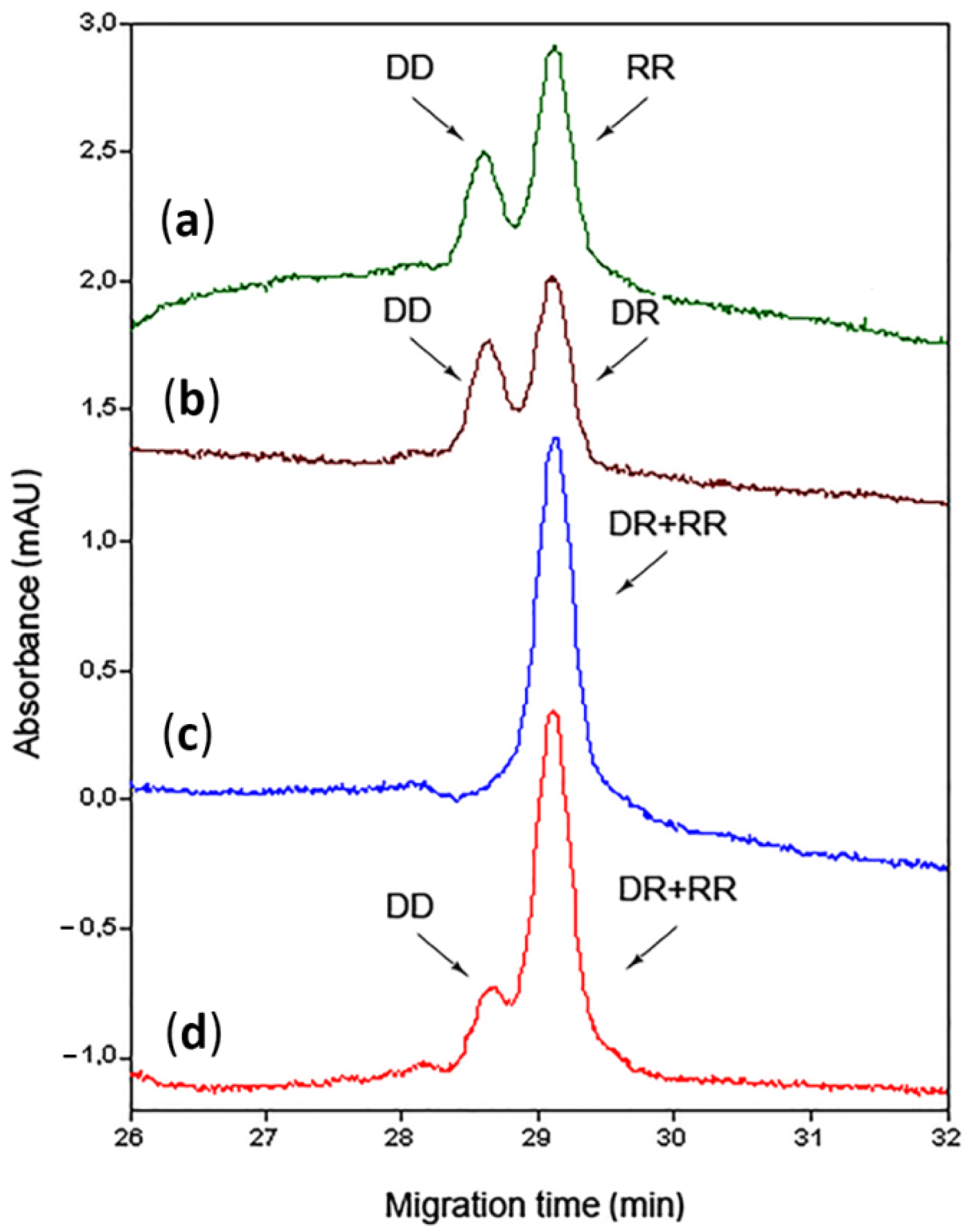
Choosing the separation buffer for the CE experiments is an important point. It should be suitable for peptide separation as well as compatible with mass spectrometry. The optimum buffer pH depends strongly on the peptides’ isoelectric point (pI) values [25]. The 10 mM ammonium acetate water solution was used for both running buffer and solvent to dilute stock samples. The CE experiments with the buffer of normal pH of 6.2 showed no separation of the peptide mixture. It migrated in capillary as neutral substance with the same velocity and was not detached from the electroosmotic flow (EOF), which carried all non-ionized components. The buffers with lowered pH showed the effect of the peptide mixture separation from the EOF. The peptides were quite far from the EOF peak and separated after lowering the pH below 4. Adjusting the pH of the buffer to 3.45 provided the separation close to the baseline (Figure 1).
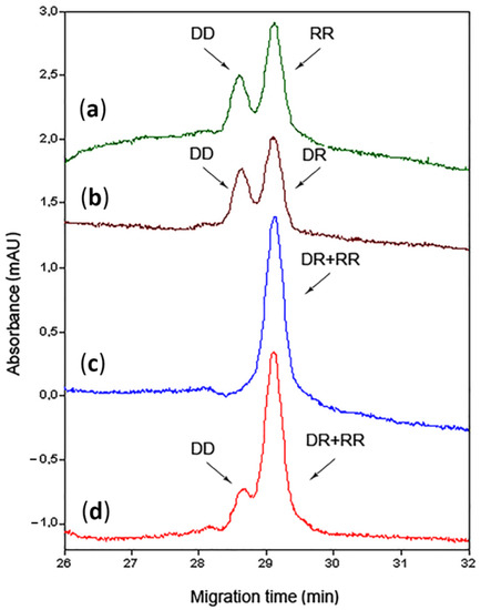
Figure 1.
Capillary electrophoresis (CE) electropherograms for 50 μM peptide mixtures. Ultraviolet (UV) absorbance measured at 200 nm. Buffer: 10 mM ammonium acetate adjusted to pH of 3.45 with acetic acid. (a) Mixture of direct-direct (DD) and reverse-reverse (RR) peptides; (b) Mixture of DD and direct-reverse (DR) peptides; (c) Mixture of DR and RR peptides; (d) Mixture of DD, DR, and RR peptides. The peak of electroosmotic flow outcomes at 42 min (not shown).
The mixture of DD and RR (Figure 1a) showed the difference in the migration time between the components of 0.48 min (1.61%). The same result was established by the mixture of DD and DR (Figure 1b). The peaks of DR and RR in their mixture (Figure 1c,d) overlapped under all tested conditions. A series of experiments with other buffers and various conditions provided no separation results. These isomers showed the same behavior in the capillary.
Obviously, all the peptide isomers remained in their zwitterion form in neutral aqueous solution. The initial amine group was protonated and the carboxylic group at the final G was dissociated. More acidic conditions caused an additional protonation of this group, which imparted a positive charge to the entire structure. This explains the increase in peptide peak distance from the EOF with a decrease in pH, since the charged ions move faster than the neutral ones. However, the increase in mobility occurred differently for DD and RR (and DR) peptides. The most possible primary protonation site in the absence of basic residues is the ammonium group at the N-terminus of the peptide chain [26,27]. The same charge location of the isomers provided no apparent difference in the migration time because the folding of the molecules remained the same. The additional charge on diverse positions may affect the different mobility of the isomeric peptides. The second charge should be closer to the C-terminus and most likely at the AVPI (or IPVA) section because it is the only difference between the DD and the DR and the same feature of the DR and the RR peptides. The different locations of the second charge can affect the diverse folding of double-charged molecules [28]. The closer the second charge to the first charge, the longer the polymer chain’s uncharged free end. This extended free uncharged end could be folded more compactly and, thus, the whole molecule became more compact. The charges located far away provided the entire molecule with a less compact elongated shape due to the repulsive Coulombic forces [14]. The probable location of the additional proton at the AVPI and IPVA sections can be verified by the MS/MS fragmentation of the molecular ions.
3.2. IMS-MS/MS Experiments
The ion mobility experiments were conducted in the MS/MS mode. The mobility was measured in the positive mode for the single-charged molecular ions. The precursor ions were fragmented after the IMS cell by collision energy inside the transfer part of the instrument to identify them by the fragment’s spectra.
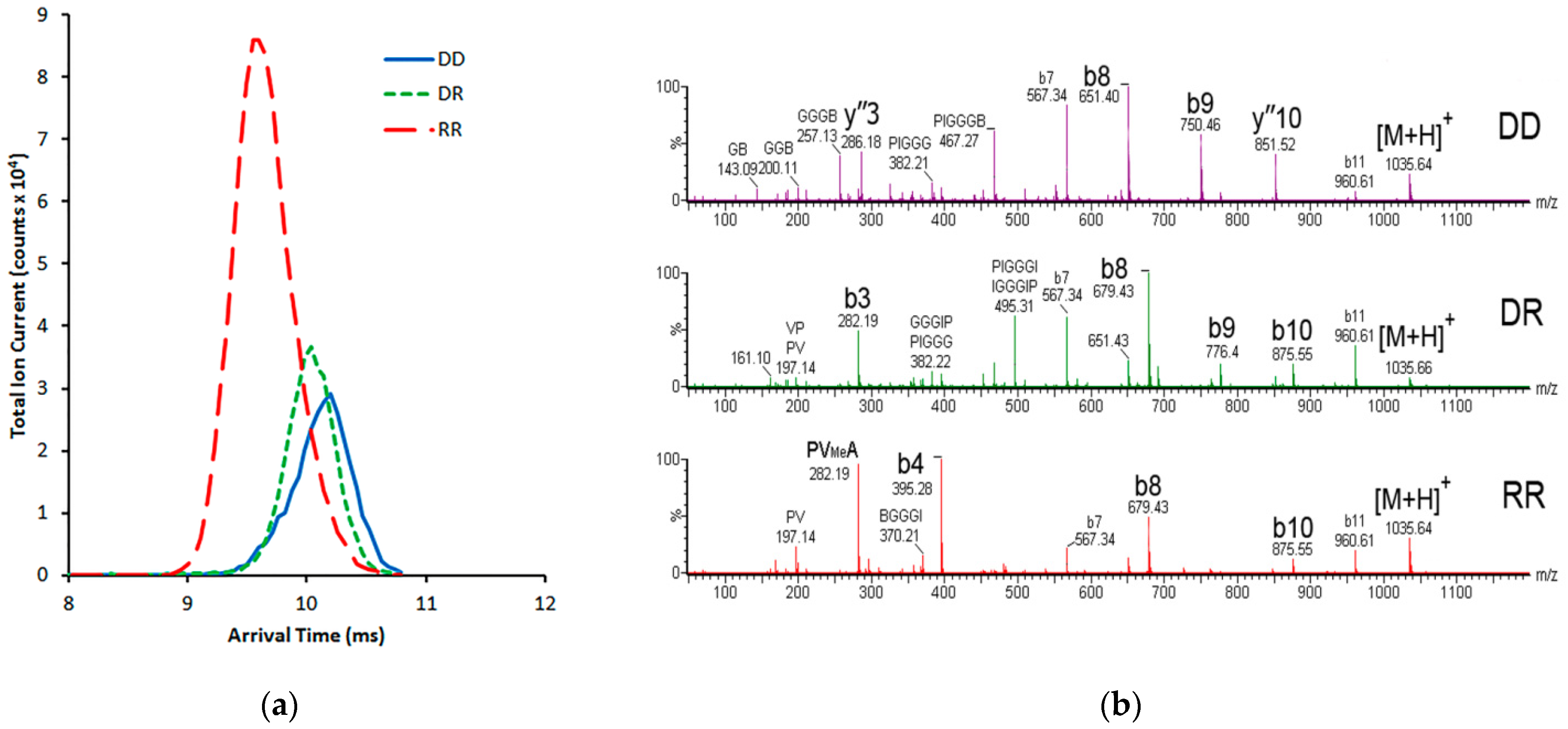
At first, the arrival times for single charged peptide ions were measured separately (Figure 2a). The difference between the DD and the RR was 0.6 ms or 5.9%; between the DD and the DR it was 0.16 ms (1.6%); between the DR and the RR it was 0.44 ms (4.2%). The measurements were made with the IMS parameters set to default. The IMS gas flow of 90 mL/min and the IMS wave velocity of 650 m/s proved to be the beginning parameters of the apparent separation of isomer peaks. Higher gas flow or wave rates could have provided a better peak resolution. However, the unstable single charged molecular ions of DD and DR were easily destroyed with the increase of these parameters.
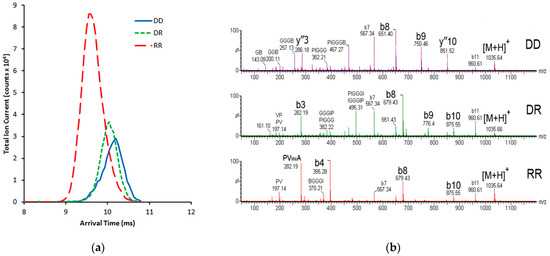
Figure 2.
(a) The ion-mobility separation (IMS) arrival times for 50 μM peptides were measured separately. The IMS gas flow was 90 mL/min, the IMS wave velocity was 650 m/s. (b) The tandem mass spectra (MS/MS) of the sequence-isomeric peptides.
The MS/MS spectra of the separately run peptides showed the sequence-specific fragment ions, identifying the isomers from their mixtures (Figure 2b).
The MS/MS spectrum of the DD peptide showed a set of characteristic peaks. There were b8 (AVPI-GGG-A) and b9 (AVPI-GGG-AV), which were strictly sequence-specific, as well as the y10 fragment (PI-GGG-AVPI-G). The last one was the only characteristic y-fragment in the high-m/z side of the spectra and was formed by breaking the V-P bond in the left AVPI part. There were also peaks of the internal fragment GGG-A, and a peak of the characteristic y3 ion. These fragments do not represent a specific sequence for the DD peptide. However, they had not been seen in other spectra.
The MS/MS spectrum of the DR peptide had b8 (AVPI-GGG-I), b9 (AVPI-GGG-IP), and b10 (AVPI-GGG-IPV) peaks. These fragments are characteristic for the right-side IPVA sequence section and are specific for both the DR and the RR. The low-m/z part contained a b3 (AVP) fragment, which was isomeric with the PVA fragment of the RR peptide.
The MS/MS spectrum of the RR peptide showed intense peaks of b8 (IPVA-GGG-I) and b10 (IPVA-GGG-IPV) fragments which were isomeric with the corresponding fragments of the DR peptide. The b4 (IPVA) fragment was the only specific fragment of the RR peptide from the low m/z part. Despite its isomerism with the AVPI fragments, this peak showed significant intensity in the RR spectrum, notable as a specific peak.
The MS/MS fragmentation of double-charged precursors and consideration of the ions of y-series could solve the ambiguities about the differences in the N-terminal part of AVPI and IPVA, which distinguish the DD and the DR from the RR. However, the RR peptide provided an insufficient amount of double-charged ions for the MS/MS analysis.
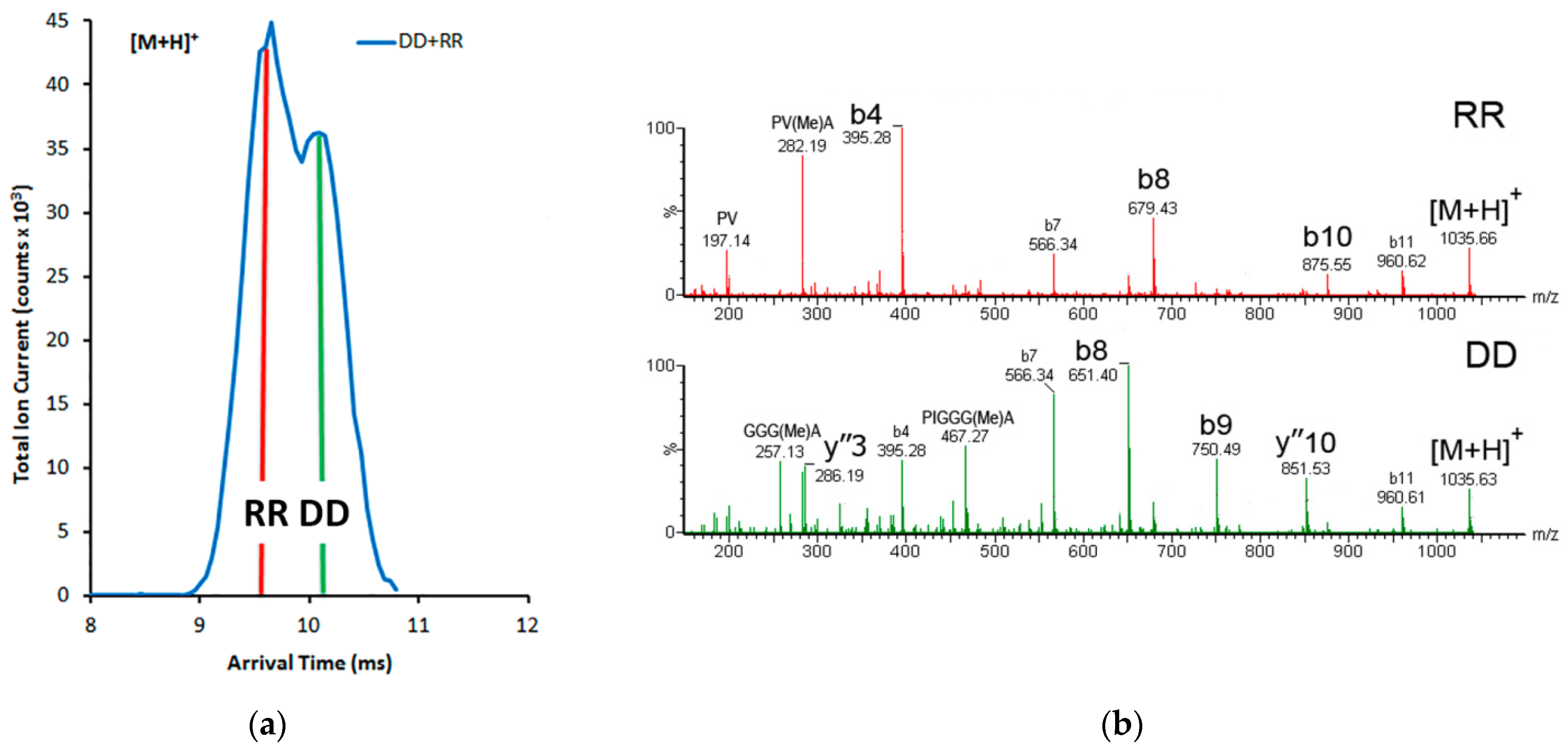
An attempt to separate the two most different peptides (DD and RR) from their mixture by the IMS was made. The probes of equal concentrations did not show equal currents of the single charged ions for each component. So, the comparison of the IMS peaks in a mixture of DD and RR was made with the adjusted concentrations. The mix of a 3:1 ratio of the DD and the RR peptides showed the combined IMS peak (Figure 3a).
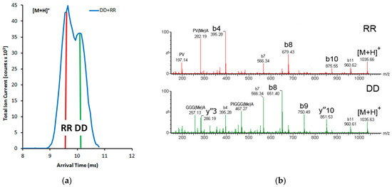
Figure 3.
(a) The IMS arrival time of the mixture of 150 μM DD and 50 μM RR peptides; (b) the MS/MS scans of the peak’s sides show the characteristic ions of the different peptides. The left fragment mass spectrum from 9.55 ms has RR peptide fragments; the right fragment mass spectrum from 10.15 ms shows the ions of the DD peptide.
The MS/MS scans (Figure 3b) of the different sides of the double peak showed the characteristic fragments of both peptides. However, their intensity varied considerably. In the top scan, the spectrum with the most intensive characteristic peaks b4, b8, and b10 of the RR peptide is shown. In the bottom scan, the DD peptide’s specific peaks y3, b8, b9, and y10 are presented. The less intensive peaks of fragments of the adjacent peptides emerge from the overlapping base of the double peak.
3.3. CE-MS/MS Experiments
The CE-UV experiments have shown that the DR and RR peptides overlap on the corresponding electropherograms (Figure 1c,d), showing the inefficiency of their separation by CE. This result was also expected for the CE-MS experiments. To avoid overlapping of the DR and RR peaks, the CE-MS/MS separation experiments were made with peptide mixtures in pairs of the DD with the DR and the DD with the RR peptides. The DR and RR peptides provided the same behavior in the capillary due to the identical sequence of their right part, as was shown above. The components separated in the capillary were verified by MS/MS in the positive ionization mode. Single charged molecular ions were fragmented by collision energy inside the transfer part of the instrument to identify them by the fragments’ spectra.
All conditions of electrophoresis were the same as for CE-UV experiments except for the capillary. It was 90 cm in length and had an inner diameter of 50 μm. The same acidic buffer gave a similar near-baseline separation of the components (Figure 4a).
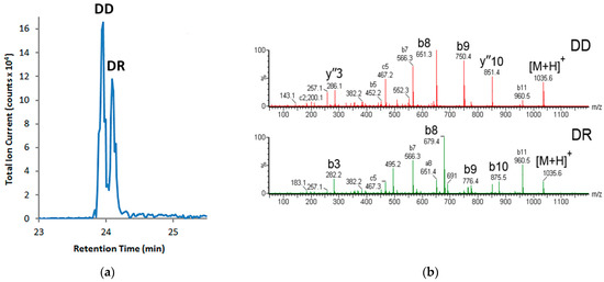
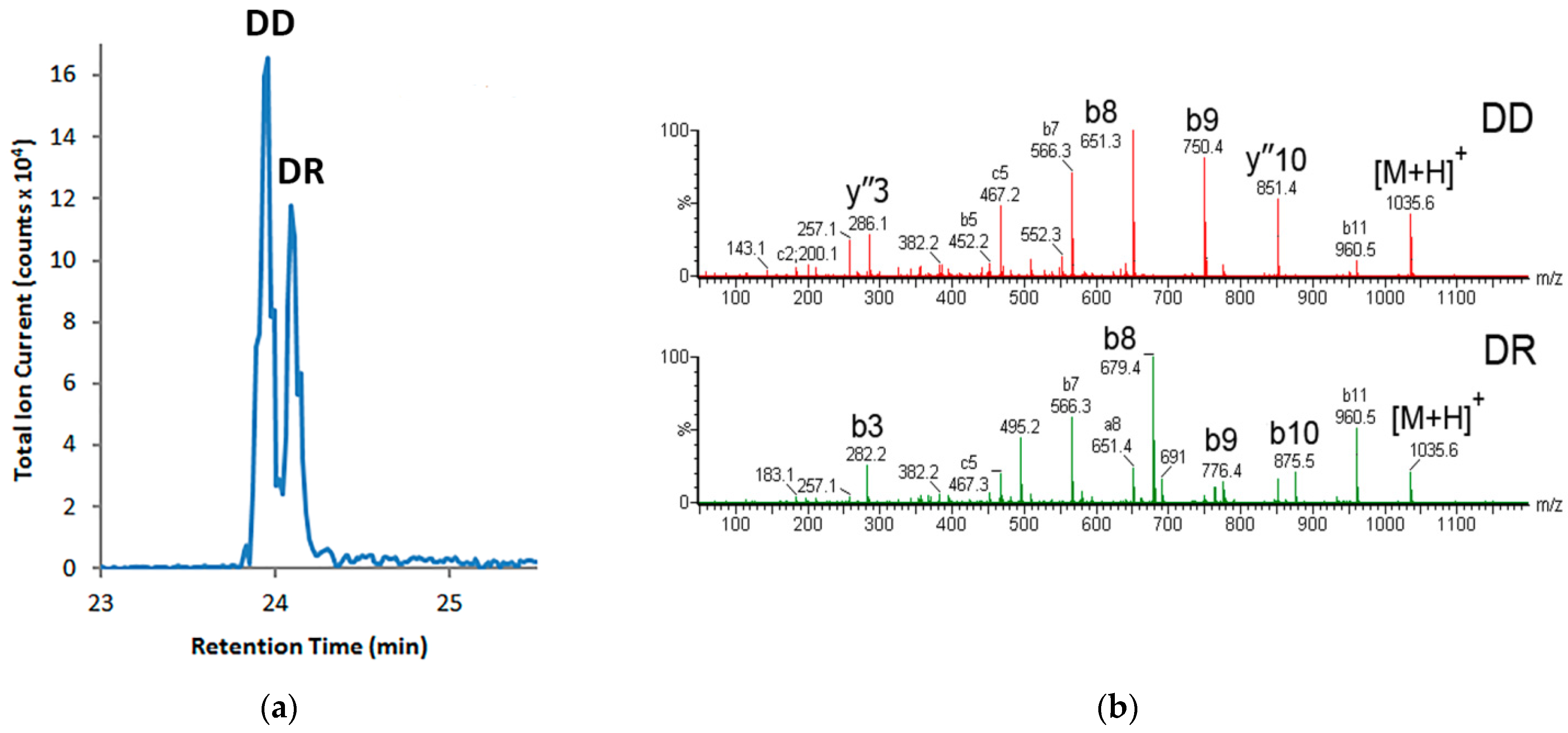
Figure 4.
(a) The CE-MS/MS chromatogram of the mixture of 50 μM DD and DR peptides. (b) The MS/MS scans of 23.94 min peak shows the characteristic ions of the DD peptide and 24.09 min peak shows the fragments of the DR peptide.
The mixture of the DD and the DR peptides showed a significant difference between the components in capillary migration time, which was 0.15 min (0.62%). This difference is considerably less than the migration time of the same mixture components during the CE-UV experiments without the MS/MS detection. This feature can be explained by almost three times less current inside the capillary observed during the CE-MS experiments. The lower current was due to the specific parameters of the CE-ESI electric circuit and a thinner capillary. Nevertheless, this difference was sufficient to detect the peaks by MS/MS.
The sheath liquid in the CE–MS interface acts as an electrically conductive medium for creating an electrical potential during the CE [29]. The sheath fluid rate in the CE–ESI interface in our experiments was set to 1.2 μL/min, which significantly exceeds the estimated electroosmotic flow in the capillary (rated as about 73.6 nl/min). This difference could reduce the overall sensitivity of the method to a certain extent due to the dilution of the analytes [30]. However, in our case, the sensitivity was sufficient for the separate detection of the isomeric peptides, which was the experimental goal. Furthermore, the time interval between the releases of components was sufficient for their effective separation without interference from the sheath liquid. The scan of the first peak shows the distinctive features of the DD peptide, and the scan of the second peak contains all the characteristic ions of the DR peptide (Figure 4b).
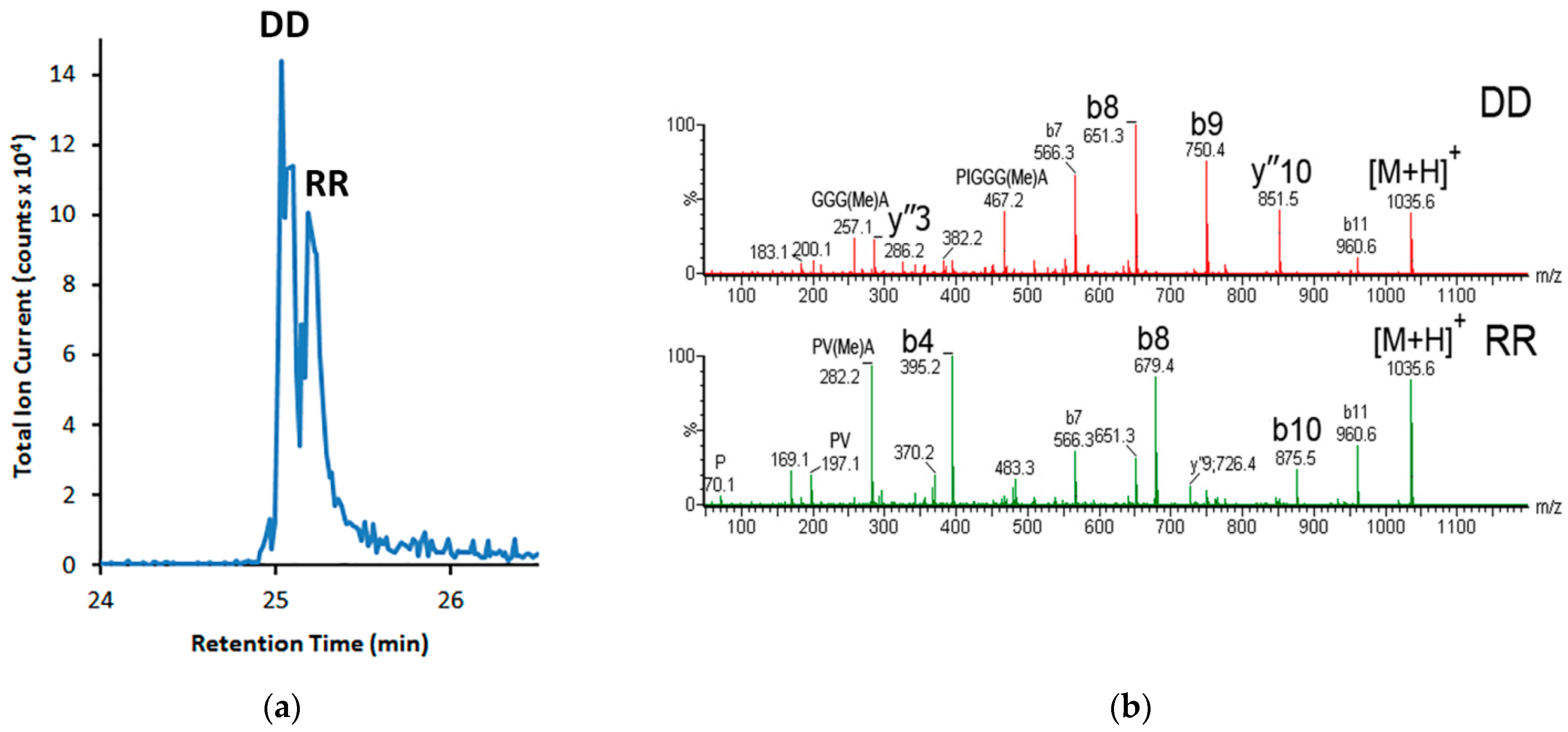
The CE-MS/MS experiment for separating the DD and RR peptide mixture (Figure 5a) showed the same difference in migration time between the components as was observed with the DD-DR mixture. The partly separated peptides were identified by the MS/MS as well. The scan of the first peak shows the distinctive features of the DD peptide, and the scan of the second peak contains all the characteristic ions of the RR peptide (Figure 5b).
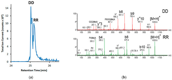
Figure 5.
(a) The CE-MS/MS chromatogram of the mixture of 50 μM DD and RR peptides; (b) the MS/MS scans of 25.03 min peak shows the characteristic ions of the DD peptide and 25.20 min peak shows the characteristic ions of the RR peptide.
The electropherograms of paired mixtures (Figure 4 and Figure 5) show the same relative release time of the DR and RR peptides, which indicate their inevitable overlap in a possible experiment with all three peptides. For this reason, CE-MS/MS experiments with all three peptides in the mixture were not carried out.
3.4. CE-IMS-MS/MS Experiments
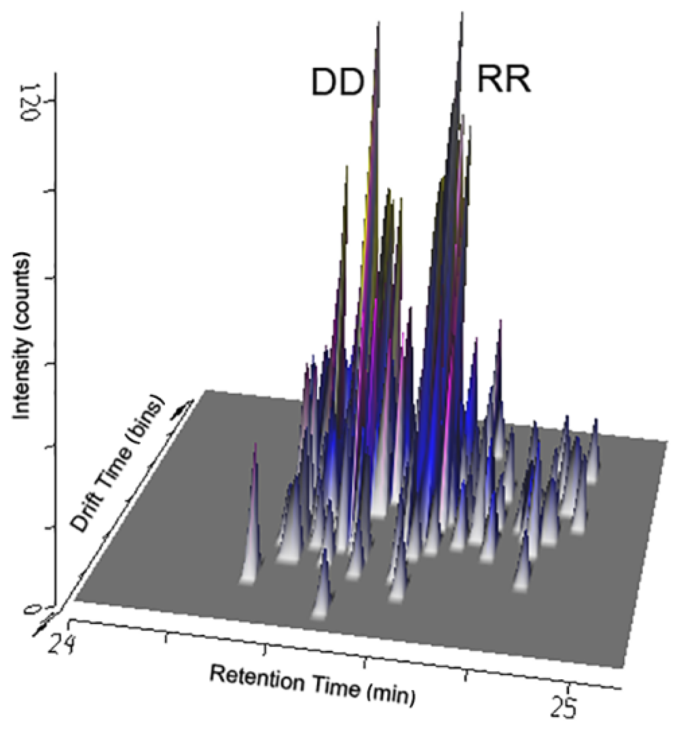
The capillary electrophoresis coupled with the ion mobility mass spectrometry allowed us to compare the two separation methods in the online combination. Coupled after the CE separation and before the MS/MS detection, the ion mobility differentiation stage had some difficulties. The investigated peptides are neutral because of their composition and showed relatively poor ionization ability. The additional separation gas added to the IMS cell acted as an interfering factor, reducing the sensitivity output. So the simultaneous application of these two techniques allowed us to lower the measured ion current by ten times and preserve the effect of isomer separation from a mixture.
The concurrent involvement of the CE and the IMS separation showed the two groups of peaks that have an apparent shift in the CE migration time (recorded by the detector as retention time) and significant overlay in the IMS drift time dimension (Figure 6).

Figure 6.
The combinative diagram of the CE retention time and the IMS drift time was drawn during the DD and RR peptide mixture separation.
4. Conclusions
Methods based on the separation of molecules according to their cross section and regardless of their masses and chemical properties possess significant potential. Capillary electrophoresis and ion-mobility mass spectrometry showed their separation abilities and provided discrete qualitative detection and identification of peptide sequence isomers that contain inverted regions of the tetrapeptide AVPI. This motif was inserted twice in various inverted combinations into a 12-amino-acid-long synthetic peptide, designed as a new anticancer drug candidate. In addition, both of these methods have shown high performance when connected online. This experimental result illustrates significant prospects for the development of combined instrumental techniques for separating complex mixtures containing biopolymers with similar properties but different biological functions.
Supplementary Materials
The following supporting information can be downloaded at: https://www.mdpi.com/article/10.3390/separations9050106/s1.
Author Contributions
Conceptualization, Y.E.G. and M.V.B.; methodology, Y.E.G. and G.G.M.; writing—original draft preparation, Y.E.G.; writing—review and editing, A.S.K. and M.V.B.; supervision, M.V.B. All authors have read and agreed to the published version of the manuscript.
Funding
This research was funded by the Ministry of Science and Higher Education of the Russian Federation; project FWES-2022-0005 for Y.E.G. and A.S.K.
Institutional Review Board Statement
Not applicable.
Informed Consent Statement
Not applicable.
Data Availability Statement
The data presented in this study are available in Supplementary material.
Acknowledgments
The authors thank Robert N. Ben for providing the synthetic peptides.
Conflicts of Interest
The authors declare no conflict of interest.
References
- Shvartsburg, A.A.; Creese, A.J.; Smith, R.D.; Cooper, H.J. Separation of a Set of Peptide Sequence Isomers Using Differential Ion Mobility Spectrometry. Anal. Chem. 2011, 83, 6918–6923. [Google Scholar] [CrossRef] [PubMed] [Green Version]
- Wang, Y.; Liu, J.; Huang, B.; Xu, Y.-M.; Li, J.; Huang, L.-F.; Lin, J.; Zhang, J.; Min, Q.-H.; Yang, W.-M.; et al. Mechanism of alternative splicing and its regulation. Biomed. Rep. 2015, 3, 152–158. [Google Scholar] [CrossRef] [PubMed] [Green Version]
- Chaudhary, S.; Khokhar, W.; Jabre, I.; Reddy, A.S.N.; Byrne, L.J.; Wilson, C.M.; Syed, N.H. Alternative Splicing and Protein Diversity: Plants Versus Animals. Front. Plant Sci. 2019, 10, 708. [Google Scholar] [CrossRef] [PubMed] [Green Version]
- Edman, P.; Begg, G. A Protein Sequenator. Eur. J. Biochem. 1967, 1, 80–91. [Google Scholar] [CrossRef] [Green Version]
- Mann, M. The Rise of Mass Spectrometry and the Fall of Edman Degradation. Clin. Chem. 2016, 62, 293–294. [Google Scholar] [CrossRef] [PubMed] [Green Version]
- Srebalus Barnes, C.A.; Hilderbrand, A.E.; Valentine, S.J.; Clemmer, D.E. Resolving Isomeric Peptide Mixtures: A Combined HPLC/Ion Mobility-TOFMS Analysis of a 4000-Component Combinatorial Library. Anal. Chem. 2002, 74, 26–36. [Google Scholar] [CrossRef] [PubMed]
- Harvey, D.J.; Watanabe, Y.; Allen, J.D.; Rudd, P.; Pagel, K.; Crispin, M.; Struwe, W.B. Collision Cross Sections and Ion Mobility Separation of Fragment Ions from Complex N-Glycans. J. Am. Soc. Mass Spectrom. 2018, 29, 1250–1261. [Google Scholar] [CrossRef] [PubMed]
- Tadey, T.; Purdy, W.C. Capillary electrophoretic resolution of phosphorylated peptide isomers using micellar solutions and coated capillaries. Electrophoresis 1995, 16, 574–579. [Google Scholar] [CrossRef] [PubMed]
- Muetzelburg, M.V.; Hoffmann, R. Separation of multiphosphorylated peptide isomers by CZE. Electrophoresis 2008, 29, 4381–4385. [Google Scholar] [CrossRef]
- Gamble, T.N.; Ramachandran, C.; Bateman, K.P. Phosphopeptide Isomer Separation Using Capillary Zone Electrophoresis for the Study of Protein Kinases and Phosphatases. Anal. Chem. 1999, 71, 3469–3476. [Google Scholar] [CrossRef] [PubMed]
- Gassmann, E.; Kuo, J.E.; Zare, R.N. Electrokinetic Separation of Chiral Compounds. Science 1985, 230, 813–814. [Google Scholar] [CrossRef] [PubMed] [Green Version]
- Jansson, E.T. Strategies for analysis of isomeric peptides. J. Sep. Sci. 2018, 41, 385–397. [Google Scholar] [CrossRef]
- Kašička, V. Recent developments in capillary and microchip electroseparations of peptides (2017–mid 2019). Electrophoresis 2020, 41, 10–35. [Google Scholar] [CrossRef] [PubMed]
- Kašička, V. Recent developments in capillary and microchip electroseparations of peptides (2019–mid 2021). Electrophoresis 2022, 43, 82–108. [Google Scholar] [CrossRef] [PubMed]
- Dominguez-Vega, E.; De Vijlder, T.; Romijn, E.P.; Somsen, G.W. Capillary electrophoresis-tandem mass spectrometry as a highly selective tool for the compositional and site-specific assessment of multiple peptide-deamidation. Anal. Chim. Acta 2017, 982, 122–130. [Google Scholar] [CrossRef] [PubMed]
- Wan, H.; Blomberg, L.G. Chiral separation of amino acids and peptides by capillary electrophoresis. J. Chromatogr. A 2000, 875, 43–88. [Google Scholar] [CrossRef]
- Hussain, A.; Hussain, A.; Hussain, I.; Al-Ajmi, M.F.; Ali, I. Stereo-separations of Peptides by Capillary Electrophoresis and Chromatography. Protoc. Exch. 2014. [Google Scholar] [CrossRef]
- Ozawa, H.; Hirayama, A.; Ishikawa, T.; Kudo, R.; Maruyama, M.; Shoji, F.; Doke, T.; Ishimoto, T.; Maruyama, S.; Soga, T.; et al. Comprehensive Dipeptide Profiling and Quantitation by Capillary Electrophoresis and Liquid Chromatography Coupled with Tandem Mass Spectrometry. Anal. Chem. 2020, 92, 9799–9806. [Google Scholar] [CrossRef] [PubMed]
- Zheng, X.; Deng, L.; Baker, E.S.; Ibrahim, Y.M.; Petyuk, V.A.; Smith, R.D. Distinguishing d- and l-aspartic and isoaspartic acids in amyloid β peptides with ultrahigh resolution ion mobility spectrometry. Chem. Commun. 2017, 53, 7913–7916. [Google Scholar] [CrossRef]
- Jia, C.; Lietz, C.B.; Yu, Q.; Li, L. Site-Specific Characterization of d-Amino Acid Containing Peptide Epimers by Ion Mobility Spectrometry. Anal. Chem. 2014, 86, 2972–2981. [Google Scholar] [CrossRef]
- Blagojevic, V.; Chramow, A.; Schneider, B.B.; Covey, T.R.; Bohme, D.K. Differential Mobility Spectrometry of Isomeric Protonated Dipeptides: Modifier and Field Effects on Ion Mobility and Stability. Anal. Chem. 2011, 83, 3470–3476. [Google Scholar] [CrossRef] [PubMed]
- Du, C.; Fang, M.; Li, Y.; Li, L.; Wang, X. Smac, a Mitochondrial Protein that Promotes Cytochrome c–Dependent Caspase Activation by Eliminating IAP Inhibition. Cell 2000, 102, 33–42. [Google Scholar] [CrossRef] [Green Version]
- Sun, H.; Nikolovska-Coleska, Z.; Yang, C.-Y.; Qian, D.; Lu, J.; Qiu, S.; Bai, L.; Peng, Y.; Cai, Q.; Wang, S. Design of Small-Molecule Peptidic and Nonpeptidic Smac Mimetics. Acc. Chem. Res. 2008, 41, 1264–1277. [Google Scholar] [CrossRef] [PubMed] [Green Version]
- Eniade, A.; Ben, R.N. Fully Convergent Solid Phase Synthesis of Antifreeze Glycoprotein Analogues. Biomacromolecules 2001, 2, 557–561. [Google Scholar] [CrossRef] [PubMed]
- Schwartz, H.; Pritchett, T. Separation of Proteins and Peptides by Capillary Electrophoresis: Application to Analytical Biotechnology; Beckman Coulter, Inc.: Fullerton, CA, USA, 1994. [Google Scholar]
- Nair, H.; Wysocki, V.H. Are peptides without basic residues protonated primarily at the amino terminus? Int. J. Mass Spectrom. Ion Processes 1998, 174, 95–100. [Google Scholar] [CrossRef]
- Unnithan, A.G.; Myer, M.J.; Veale, C.J.; Danell, A.S. MS/MS of protonated polyproline peptides: The influence of N-terminal protonation on dissociation. J. Am. Soc. Mass Spectrom. 2007, 18, 2198–2203. [Google Scholar] [CrossRef] [Green Version]
- Hill, H.H.; Hill, C.H.; Asbury, G.R.; Wu, C.; Matz, L.M.; Ichiye, T. Charge location on gas phase peptides. Int. J. Mass Spectrom. 2002, 219, 23–37. [Google Scholar] [CrossRef]
- Sun, L.; Zhu, G.; Zhang, Z.; Mou, S.; Dovichi, N.J. Third-Generation Electrokinetically Pumped Sheath-Flow Nanospray Interface with Improved Stability and Sensitivity for Automated Capillary Zone Electrophoresis–Mass Spectrometry Analysis of Complex Proteome Digests. J. Proteome Res. 2015, 14, 2312–2321. [Google Scholar] [CrossRef] [PubMed] [Green Version]
- Mokaddem, M.; Gareil, P.; Belgaied, J.-E.; Varenne, A. New insight into suction and dilution effects in CE coupled to MS via an ESI interface. II-Dilution effect. Electrophoresis 2009, 30, 1692–1697. [Google Scholar] [CrossRef] [PubMed]
Publisher’s Note: MDPI stays neutral with regard to jurisdictional claims in published maps and institutional affiliations. |
© 2022 by the authors. Licensee MDPI, Basel, Switzerland. This article is an open access article distributed under the terms and conditions of the Creative Commons Attribution (CC BY) license (https://creativecommons.org/licenses/by/4.0/).