Abstract
Hyperkeratosis lenticularis perstans, also known as Flegel’s disease (FD), is a rare cutaneous disorder affecting mainly the lower extremities of middle-aged people. Due to its rarity, this disease is usually not recognized by physicians resulting in a delay in diagnosis, especially in those cases with atypical cutaneous involvement. Herein, we present a 72-year-old woman who developed FD characterized by a generalized distribution, involving, in addition to the lower limbs, the trunk and the upper limbs as well. We performed a description of the dermoscopic and pathologic features of this rare entity, also carrying out a brief reappraisal of the cases of FD with a diffuse, atypical and generalized distribution that have been described in the literature. Histopathology with clinical correlation is the cornerstone of the diagnosis, even and especially in atypical cases. This patient with a disease duration of 58 years also represents the longest-lasting case of FD reported in the literature.
1. Introduction
Hyperkeratosis lenticularis perstans, also known as Flegel’s disease (FD), is a rare benign cutaneous disorder (first described in 1958) presenting with multiple reddish-brown keratotic papules on the lower extremities of mid-to-older age people. Rarely, FD can affect other body sites or present with a diffuse and generalized involvement, making diagnosis more difficult. Herein, we report the case of a woman who developed a generalized form of FD with a disease duration of 58 years, describing its histopathologic and dermoscopic findings. A brief review of the atypical and generalized cases of this rare entity reported in the literature so far are also described.
2. Case Report
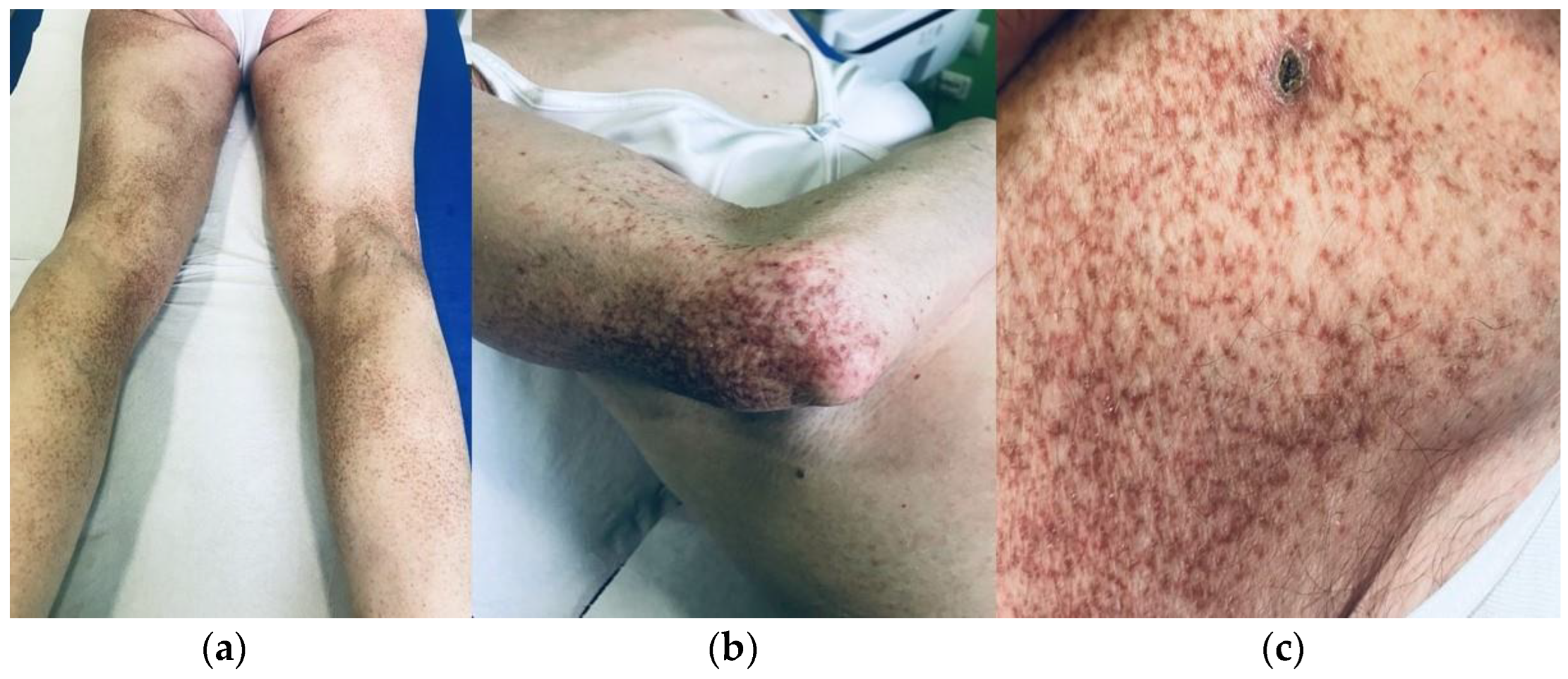
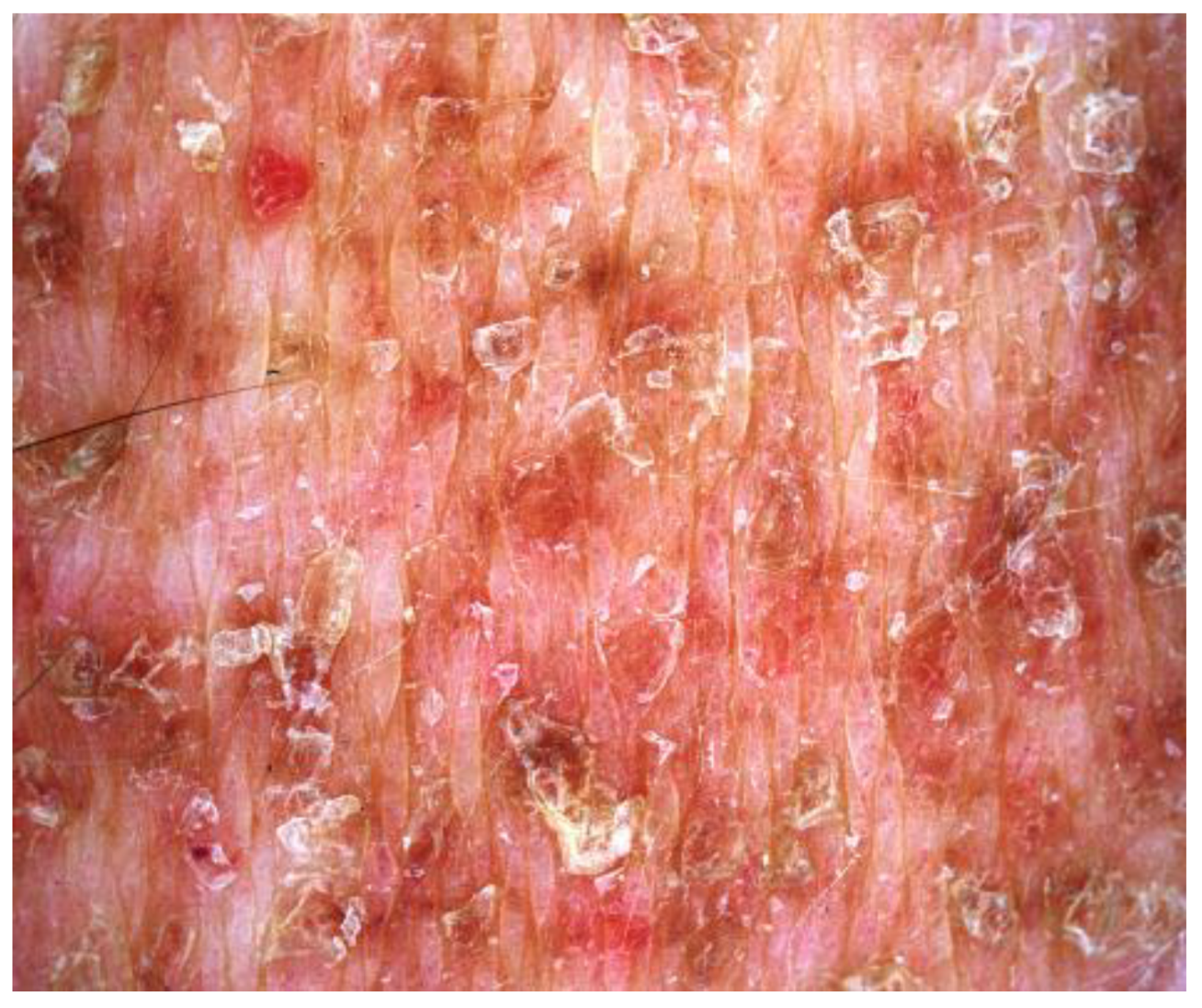
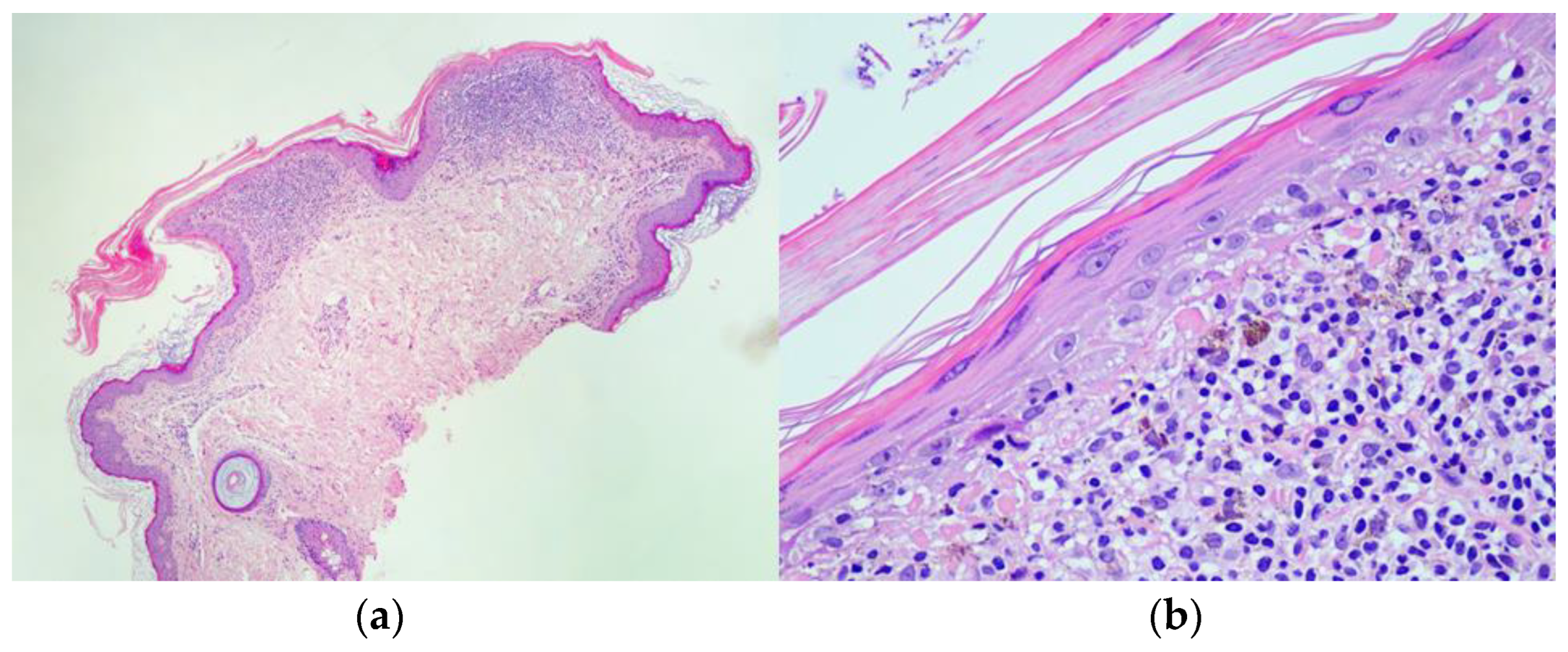
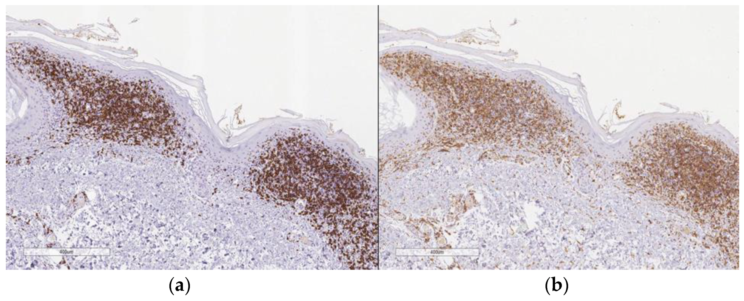
A 72-year-old Caucasian woman was seen for a 58 year old history of multiple small, reddish and brownish, keratotic papules, initially located on her lower limbs, which subsequently spread, involving the upper limbs and the trunk (Figure 1a,b). Her personal medical history was negative for cutaneous and systemic diseases, except for an in situ well differentiated cutaneous squamous cell carcinoma on the leg, which was removed two years before. Dermoscopy of one cutaneous lesion showed brownish structureless areas, with an also brownish pseudo-reticular appearance on an erythematous base, some scattered lacunae-like ectatic vessels and hyperkeratosis with fine superficial desquamation (Figure 2). Histopathology of a lesion on the trunk showed a compact, thick lamellar hyperkeratosis with focal parakeratosis overlying an atrophic epidermis associated with a dense, lichenoid infiltrate of small lymphocytes in the superficial dermis with focal accentuation in the papillomatous areas. Some melanophages, focal basal vacuolization with scattered areas of exocytosis, were also present (Figure 3a,b). Immunohistochemical studies showed a similar proportion of CD4 and CD8T-lymphocytes (Figure 4a,b) with sparse to absent CD20 cells. According to the clinico-pathologic correlation, a final diagnosis of generalized hyperkeratosis lenticularis perstans (Flegel’s disease) was made. A therapeutic attempt with acitretin 20 mg/die was made and stopped after 3 months due to scarce improvement and poor satisfaction of the patient, in the absence of side effects.
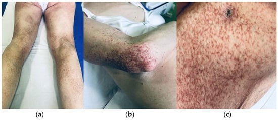
Figure 1.
Multiple small, reddish and brownish, hyperkeratotic papules, initially present only in the lower limbs (a) and subsequently involving also on upper limbs (b) and the trunk (b,c).
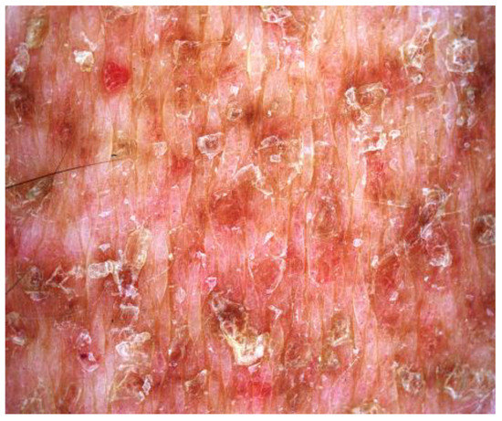
Figure 2.
Dermoscopy showed brownish structureless areas, with a brownish pseudo-reticular appearance on an erythematous base, rare ectatic lacunae-like vessels and hyperkeratosis with fine superficial desquamation.
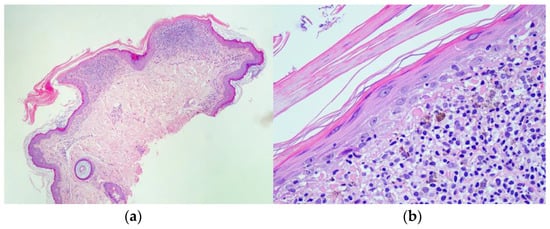
Figure 3.
Histopathology showed a dense, lichenoid lymphocytic infiltrate in the superficial dermis, with focal accentuation in the papillomatous areas, hyper and parakeratosis, and thin, atrophic cutaneous epidermis under hyper-parakeratotic scales (a; Hematoxylin and Eosin 10×); Close-up of the lesion showing atrophic epidermis under hyper-parakeratotic scales, lichenoid infiltrate in the superficial dermis characterized by the presence of multiple small lymphocytes, with scattered melanophages, exocytosis and basal vacuolization of the epidermis (b; Hematoxylin and Eosin 30×).
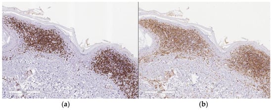
Figure 4.
CD8 immunostain (a; 10×); CD4 immunostain (b; 10×).
3. Discussion
Flegel’s disease (FD), first described in 1958, is a rare cutaneous disorder that usually arises in the fourth/fifth decade of life, often confused with other cutaneous keratotic pathologies. To the best of our knowledge, about 80 articles dealing with FD cases have been reported in the literature [1,2,3,4,5,6,7,8,9,10,11,12,13,14,15,16,17,18,19,20,21,22,23]. According to the literature, there is a slight prevalence of the female gender with a ratio 1.6:1 and a median age of 50 years, ranging between 18 and 82 years [1,2,3,4,5,6,7,8,9,10,11,12,13,14,15,16,17,18,19,20]. The median duration of the disease before reaching the diagnosis is reasonably long, with a median of eight years. FD is mainly characterized by small and multiple erythematous brownish and hyperkeratotic papules, from 1 mm to 5 mm in diameter, whose typical location is on the dorsa of the feet and lower part of the legs in 87% of cases. An atypical presentation with involvement of unusual sites such as the axillae, eyes, palms/soles, a single leg, trunk, upper extremities, buttocks, forehead, pinnae, breast, hands and oral mucosa has been also reported [1,2,3,4,5,6,7,8,9,10,11]. A diffused or generalized involvement of the body is a very rare presentation with only three cases described [2,5,6]. In Table 1 we summarize all the cases of FD in the literature that showed a generalized distribution, as well as cases with involvement of atypical anatomic sites, highlighting how the duration of the disease in all these cases is long, with a mean time of 16 years before reaching a diagnosis, ranging between 5 years and 30 years. In this regard, the main point of interest in the present case is the longstanding history of the disease, lasting 58 years, with an atypical, widespread and generalized distribution of the lesions involving the lower limbs, the upper limbs and the chest.

Table 1.
Review of the clinical features of generalized hyperkeratosis lenticularis perstans including involvement of atypical areas, reported so far in the literature [2,3,4,5,6,7,8,9,10,11,20].
Regarding the pathogenesis, since there are familial cases, FD has been hypothesized to be an autosomal dominant disease (although no specific candidate genes have yet been discovered); however, since most cases seem to occur sporadically and usually start late in adult life, the hypothesis of a genetic pathogenesis is not strengthened. It has been suggested that FD is a primary keratinization disorder. In this regard, while some authors have found some changes in the membrane-coating granules (MCGs), inducing structural alterations in the epidermis, other authors have not found these alterations [1,13,14]. A possible correlation of FD with other conditions, such as Borrelia burgdorferi [15], basal cell carcinoma, squamous cell carcinoma, urinary bladder tumor, lung cancer and digestive cancers has also been reported [16]. Anecdotal association with hyperaldosteronism and hyperthyroidism, as well as a coexistence with Kyrle disease and Mibelli porokeratosis have been also described [17]. The personal medical history of our patient was negative, except for a cutaneous squamous cell carcinoma on her leg that was removed two years before. Therefore, the correlation between FD and cutaneous keratinocyte malignancies should be better evaluated in future reports.
The main clinico-pathologic differential diagnoses of FD are stucco keratoses, disseminated superficial actinic porokeratosis (DSAP), Kyrle’s disease, acrokeratosis verruciformis of Hopf and porokeratosis of Mibelli [21,22]. In all these cases, the histological examination and the clinico-pathologic correlation help perform a correct diagnosis.
To date, dermoscopy of FD has been performed in only two cases [18,19] presenting with superficial whitish scaling areas (induced by hyper and parakeratosis), brownish structureless areas (induced by melanophages in the dermis, produced by the lichenoid infiltrate and the pigment incontinence) and grayish areas induced by lichenoid infiltrate. In our case, we found similar dermoscopic findings, with some superficial scaling areas, brownish reticulated areas and multiple lacunae-like areas corresponding to superficial dermis ectatic vessels.
The treatment of FD is challenging; the main treatments include emollients, topical steroids (betamethasone diproprionate 0.05%), 5-fluorouracil (5-FU) cream, topical or systemic retinoids, Vitamin D3 and PUVA. Ablative treatments (local excision, CO2 laser, curettage, dermabrasion, cryotherapy and electrocoagulation) can also be taken into consideration [1]. However, all these treatments offer only partial therapeutic responses and are aimed only at ameliorating the aesthetic impact of the lesions, rather than at their definitive treatment.
4. Conclusions
FD, or hyperkeratosis lenticularis perstans, is a rare disease whose diagnosis is based upon a good clinico-pathological correlation, as there are no genetic or laboratory exams specific to this disease. When an atypical presentation occurs, such as in our case, with a diffuse and generalized involvement, the diagnosis is more difficult and can be delayed. Histopathology with the typical findings of focal, thick, compact hyperkeratosis with parakeratosis, thinned epidermis and a dense band-like lymphocytic inflammatory infiltrate in the papillary dermis is mandatory to confirm the diagnosis. Our patient is also the longest-enduring case of FD, lasting 58 years, with progressive spreading from the legs to a generalized involvement.
Author Contributions
Conceptualization, G.P. and F.R.; methodology, G.P.; software; validation, G.P., G.S., N.R. and F.R.; formal analysis, F.R.; investigation, G.P.; resources, G.S.; data curation, G.S.; writing—original draft preparation, G.P.; writing—review and editing, G.P, F.R.; visualization, G.P.; supervision, F.R. All authors have read and agreed to the published version of the manuscript.
Funding
This research received no external funding.
Institutional Review Board Statement
Not applicable.
Informed Consent Statement
Written informed consent has been obtained from the patient to publish this paper.
Data Availability Statement
Not applicable.
Conflicts of Interest
The authors declare no conflict of interest.
References
- Bortoluzzi, P.; Cusini, M.; Veraldi, S.; Nazzaro, G. Hyperkeratosis lenticularis perstans (Flegel’s disease): Our experience and review of the literature. Int. J. Dermatol. 2021, 60, 33–38. [Google Scholar] [CrossRef] [PubMed]
- Krishnan, A.; Kar, S. Photoletter to the editor: Hyperkeratosis lenticularis perstans (Flegel’s disease) with unusual clinical presentation. Response isotretinoin therapy. J. Dermatol. Case Rep. 2012, 6, 93–95. [Google Scholar] [CrossRef] [PubMed]
- Fernandez-Crehuet, P.; Rodr ıguez-Rey, E.; Rıos-Martın, J.J.; Camacho, F.M. Hyperkeratosis lenticularis perstans, or Flegel disease, with palmoplantar involvement. Actas Dermosifiliogr. 2009, 100, 157–159. [Google Scholar] [CrossRef]
- Miranda-Romero, A.; Sanchez-Sambucety, P.; del Pozo, C.B. Unilateral hyperkeratosis lenticularis perstans (Flegel’s disease). J. Am. Acad. Dermatol. 1998, 39, 655–657. [Google Scholar] [CrossRef]
- Jang, K.A.; Choi, J.H.; Sung, K.J.; Moon, K.C.; Koh, J.K. Hyperkeratosis lenticularis perstans (Flegel’s disease): Histologic, immunohistochemical, and ultrastructural features in a case. Am. J. Dermatopathol. 1999, 21, 395–398. [Google Scholar] [CrossRef] [PubMed]
- Kocsard, E.; Bear, C.L.; Constance, T.J. Hyperkeratosis lenticularis perstans (Flegel). Dermatology 1968, 136, 35–42. [Google Scholar] [CrossRef]
- Price, M.L.; Jones, E.W.; Macdonald, D.M. A clinicopathological study of Flegel’s disease (hyperkeratosis lenticularis perstans). Br. J. Dermatol. 1987, 116, 681–691. [Google Scholar] [CrossRef]
- Urbina, F.; Sudy, E.; Misad, C. A case of localized, unilateral hyperkeratosis lenticularis perstans on a woman’s breast. J. Dtsch. Dermatol. Ges. 2016, 14, 416–418. [Google Scholar] [CrossRef]
- Massone, L.; Pestarino, A.; Gambini, C.; Isola, V. Hyperkeratosis lenticularis perstans. A clinical case and review of the literature. G. Ital. Dermatol. Venereol. 1990, 125, 281–284. [Google Scholar]
- Van de Staak, W.J.; Bergers, A.M.; Bongaarts, P. Hyperkeratosis lenticularis perstans (Flegel). Dermatology 1980, 161, 340–346. [Google Scholar] [CrossRef]
- Matarredona, J.; García-Abellán, J.; Ramírez, I.V. Unilateral Asymptomatic Progressive Eruption of Hyperkeratotic Papules. JAMA Dermatol. 2019, 155, 739–740. [Google Scholar] [CrossRef]
- Flegel, H. Hyperkeratosis lenticularis perstans. Der Hautarzt Z. Dermatol. Venerol. Verwandte Geb. 1958, 9, 363–364. [Google Scholar]
- Ishibashi, A.; Tsuboi, R.; Fujita, K. Familial hyperkeratosis lenticularis perstans: Associated with cancers of the digestive organs. J. Dermatol. 1984, 11, 407–409. [Google Scholar] [CrossRef] [PubMed]
- Ando, K.; Hattori, H.; Yamauchi, Y. Histopathological differences between early and old lesions of hyperkeratosis lenticularis perstans (Flegel’s disease). Am. J. Dermatopathol. 2006, 28, 122–126. [Google Scholar] [CrossRef]
- Schwarzova, K.; Kozub, P.; Szep, Z.; Golovchenko, M.; Rudenko, N. Detection of Borrelia burgdorferi sensu stricto and Borrelia garinii DNAs in patient with Hyperkeratosis lenticularis perstans (Flegel disease). Folia Microbiol. 2016, 61, 359–363. [Google Scholar] [CrossRef]
- Beveridge, G.W.; Langlands, A.O. Familial hyperkeratosis lenticularis perstans associated with tumours of the skin. Br. J. Dermatol. 1973, 88, 453–458. [Google Scholar] [CrossRef] [PubMed]
- Zhang, H.; Miao, C.; Zhang, X. Hyperkeratosis lenticularis perstans in a patient with primary hyperaldosteronism. J. Dtsch. Dermatol. Ges. 2018, 16, 72–73. [Google Scholar] [CrossRef] [PubMed]
- Errichetti, E.; Turina, M.; Pizzolitto, S.; Stinco, G. Dermoscopy of hyperkeratosis lenticularis perstans (Flegel disease). J. Dermatol. 2019, 46, e298–e299. [Google Scholar] [CrossRef]
- Valdebran, M.; Terrero, D.; Xue, R. Dermoscopic findings in hyperkeratosis lenticularis perstans. J. Am. Acad. Dermatol. 2016, 75, e211–e213. [Google Scholar] [CrossRef]
- Miljković, J. An unusual generalized form of hyperkeratosis lenticularis perstans (Flegel’s disease). Wien. Klin. Wochenschr. 2004, 116 (Suppl. 2), 78–80. [Google Scholar]
- Kocsard, E.; Palmer, G.; Constance, T.J. Coexistence of hyperkeratosis lenticularis perstans (Flegel) and hyperkeratosis follicularis et parafollicularis in cutem penetrans (Kyrle) in a patient. Acta Derm. Venereol. 1970, 50, 385–390. [Google Scholar] [PubMed]
- Bianchi, C.; Casala, A.; Bianchi, O.; Stringa, S.; Balsa, R. Hyperkeratosis lenticularis perstans. Ses rapports avec la porokératose de Mibelli [Hyperkeratosis lenticularis perstans. Its relations to Mibelli’s porokeratosis]. Ann. Dermatol. Syphiligr. 1974, 101, 145–154. [Google Scholar]
- Diaz De La Pinta, F.J.; Eraña, I.; Jo Velasco, M.; Diaz Recuero, J.L.; Requena, L. Hyperkeratosis lenticularis perstans (Flegel disease) with back and breast involvement. J. Cutan. Pathol. 2021, 48, 1525–1527. [Google Scholar] [CrossRef] [PubMed]
Disclaimer/Publisher’s Note: The statements, opinions and data contained in all publications are solely those of the individual author(s) and contributor(s) and not of MDPI and/or the editor(s). MDPI and/or the editor(s) disclaim responsibility for any injury to people or property resulting from any ideas, methods, instructions or products referred to in the content. |
© 2023 by the authors. Licensee MDPI, Basel, Switzerland. This article is an open access article distributed under the terms and conditions of the Creative Commons Attribution (CC BY) license (https://creativecommons.org/licenses/by/4.0/).

