Article

Long-Term Skin Safety Effect of Chlorine-Rich Water Treatment on C57BL/6 Mice

1
Department of Convergence Medicine, Wonju College of Medicine, Yonsei University, Wonju 26426, Gangwon-do, Republic of Korea
2
Department of Laboratory Medicine, Wonju College of Medicine, Yonsei University, Wonju 26426, Gangwon-do, Republic of Korea
3
NUGA BEST Bldg, 9, Bongeunsa-ro 68-gil, Gangnam-gu, Seoul 06153, Republic of Korea
*
Author to whom correspondence should be addressed.
These authors contributed equally to this work.
Processes 2023, 11(7), 1914; https://doi.org/10.3390/pr11071914
Submission received: 29 April 2023 / Revised: 22 June 2023 / Accepted: 23 June 2023 / Published: 26 June 2023
(This article belongs to the Section Biological Processes and Systems)

Abstract

:
Background: The weakened keratinocyte cohesion, wrinkle formation, loss of elasticity, decreased sebum production, increased water loss, and other skin problems caused by a compromised skin barrier can eventually result in various skin diseases. Given its current usage as a disinfectant and its potential use in medicine, it is crucial to assess the safety of chlorinated water. Objective: The study’s objective was to evaluate the long-term skin safety effects of chlorine-rich sterilized water (CIRW) treatment on C57BL/6 mice in vivo. Methods: Reactive oxygen species (ROS), nitric oxide (NO), glutathione peroxides (GPx), and catalase (CAT), as well as cytokines involved in inflammation (such as interleukin (IL)-1α, IL-1 β, IL-13, GM-CS, and TNF-α) were tested as oxidative stress markers in both serum and skin. The skin parameters such as moisture level, elasticity, sebum, pore size, and wrinkles were measured. Results: The CIRW group showed higher elasticity and humidity than the NC group. Similarly, the ROS and NO levels were decreased significantly in the CIRW group compared with those in the NC group by the oxidative stress markers in the skin. We also observed the ClRW group serum IL-5, IL-6, and IL-12 to be significantly lower than those in the NC group. Conclusions: Our results indicate that ClRW does not generally exhibit skin toxicity in vivo with long-term care or immune redox reactions such as oxidative stress, inflammation, and allergic reactions.

1. Introduction

The body’s largest organ, the skin, functions as a barrier between the interior and exterior domains. The rate of absorption in the skin determines the local and systemic toxicities of many chemicals. Different factors determine the absorption, such as the concentration and volume of the chemical, duration of exposure, skin temperature, chemical binding to the skin, vehicle, or solvent for the chemical, and chemical characteristics of the material applied. Drug distribution into and through the skin is still difficult, even though effective skin disorders treatment requires medications in the various skin compartments, such as the epidermis, dermis, or hair follicles [1]. The weakened keratinocyte cohesion and wrinkle formation caused by a compromised skin barrier can eventually result in many skin illnesses. These skin problems include a loss of elasticity, a decrease in sebum production, an increase in water loss, and the strengthening of keratinocyte cohesion.
In recent years, the development of different machines and compounds to help maintain good health as well as prevent and cure diseases has become the focus of medical research. One of the developments is the use of functional water. It has recently been studied extensively for its health advantages and prevention of diseases [2]. A type of functional water is acidic water. It has an acidic pH and a high chlorine concentration. It is used to disinfect food equipment, vegetables, fruits, poultry, and meat. Acidic water has been demonstrated to inactivate germs, fungi, viruses, and toxins [3]. It has also been studied for its efficacy in handwashing [4], hospital bactericidal effects [5], and cleaning medical apparatuses such as endoscopes [6]. This antimicrobial effect is considered to be owing to the water properties. One of these properties is its high chlorine concentration, particularly hypochlorous acid (HOCl). However, HOCl has long been known for its antiseptic and antibacterial actions. However, HOCl is being utilized in the fields of hygiene, food safety, water treatment, and health. Many papers reporting the use of HOCl have surfaced in the previous two decades (e.g., for hospital disinfection, wound antisepsis, long-term care facilities, and animal husbandry) [7,8]. It was recently reported that HOCl inhibits the expression of nuclear factor kappa B (NF-ĸB)-dependent genes, decreases the disease severity, prevents skin ulceration, and reduces the skin aging process [9,10]. At a normal pH, HOCl is in equilibrium with its anionic form, hypochlorite (OCl), and can cause tissue damage by oxidizing and chlorinating biomolecules that target peptides (such as glutathione), proteins, lipids, and nucleic acids [11,12]. Human HOCl exposure can occur through a variety of external environmental routes in addition to the endogenous HOCl produced during physiological antibacterial and inflammatory responses [13,14].
However, developing substances that can provide better protection without damage remains a significant challenge. Therefore, it is necessary to test this innovation to prevent future health conflicts. This study demonstrated that long-term supplementation with chlorine-rich sterilized water (ClRW), which has a high chlorine concentration, affects immunological responses, including inflammation, allergic reactions, oxidative stress, and immune responses.

2. Materials and Methods

2.1. Animal Groupings and Water Supplementation

Six-week-old female hairless mice with a mean weight of 25 ± 1.2 g were purchased from Orient Bio Inc. (Swingman, Republic of Korea). These were acclimatized for seven days in a controlled environment at a temperature of 22 ± 2 °C and humidity of 40–60% under a 12:12 h light–dark cycle prior to treatment. Thirty mice were randomly divided into three groups (n = 10 per group) as follows: a group without water supplementation (normal control (NC), group with tap water (TW) supplementation, and group with ClRW supplementation with chlorine concentration (5 ppm) for up to 90 days for the study period. The Institutional Animal Care and Use Committee (IACUC) at the Wonju Campus of Yonsei University, Gangwon, Wonju, Republic of Korea, approved the study protocol (ethical approval number: YWC-180615-1). For three months, 4 mL of TW and ClRW was administered by spraying the dorsal region of hairless mice.

2.2. Water Preparation and Their Properties

LG Electronics Inc. (Seoul, Republic of Korea) developed and prepared a machine (Model: ABQ76861001) that produces ClRW. A pH and ORP meter from the DKK-TOA Corporation in Japan was used to measure the pH, a chlorine meter from Lutron Electronic Enterprise, Co., LTD in Taiwan was used to measure the available chlorine concentration (ACC), and a dissolved hydrogen meter from VZOR LLC in Russia was used to measure the concentration of hydrogen. The properties of the water produced by the machine are listed in Table 1. To ensure a consistent chlorine concentration, weekly measurements of ACC were performed. Adjustments were made when the concentration was altered.

2.3. Serum and Skin Sample Preparation

The serum samples were obtained by centrifuging the blood in a BD Microtainer tube (Lot 9305148, Becton, Dickinson and Company, Franklin Lakes, NJ, USA) at 14,000 rpm for 5 min at 4 °C. The serum samples were then kept at −80 °C until use. To prepare the skin tissue sample, skin tissue (2 × 2 mm in size) was cut and placed in ice-cold radioimmunoprecipitation assay (RIPA) buffer (Pierce Biotechnology Inc., Waltham, MA, USA) with protease inhibitor compounds (Sigma Chemical Co., St. Louis, MO, USA) (Sigma Chemical Co., St. Louis, MO, USA). The skin tissue was homogenized for 10 min at 14,000 rpm and centrifuged for 5 min, and the supernatant of the skin lysate was used as a sample.

2.4. Histological Examination by Hematoxylin and Eosin (H&E) Staining

Each group’s representative dorsal region of the skin was sectioned, fixed in 10% neutral buffered formalin (0.1 M phosphate buffer, pH 7.4), dehydrated using a graduated ethanol series, cleaned with xylene, and then embedded in paraffin wax (Polyscience, Washington, DC, USA). Using a microtome (Reichert, Inc., New York, NY, USA), a 4 m thick paraffin section was created. The sections were stained with hematoxylin and eosin and examined under a light microscope (BA300; Motic Ltd., Hong Kong, China).

2.5. Skin Parameters Measurement

The skin parameters were measured using two devices. First, a skin sensitivity test using A-ONE TAB (Bomtech Electronics Co., LTD, Seoul, Republic of Korea) measured the moisture level, elasticity, sebum, pore size, and wrinkles. Following the manufacturer’s instructions, the device was placed on the skin of the mice. The photographs and measurement readings were recorded. Another device that measures the skin sensitivity using the MSIP-CRM-G10 barrier sensor (Gpower, Republic of Korea) evaluated the barrier score, barrier strength, and transepidermal water loss (TEWL). A mobile application was downloaded through a Bluetooth connection. The measurements were recorded on a mobile phone upon the placement of the device on the skin of the mice. For the qualitative data, numerical values for each measurement were assigned to measure the significant differences.

2.6. Total Reactive Oxygen Species (ROS) Detection

The total ROS formation in serum and skin tissue lysates was determined by oxidating 2-4-dichlorodihydrofluorescein diacetate (DCFH-DA) (Abcam, Cambridge, MA, USA), as described earlier [15].

2.7. Nitric Oxide (NO) Assay

To assess the NO production in the blood and skin tissue lysates, the Griess reagent (Promega Corp., Madison, WI, USA) was used as described earlier [15].

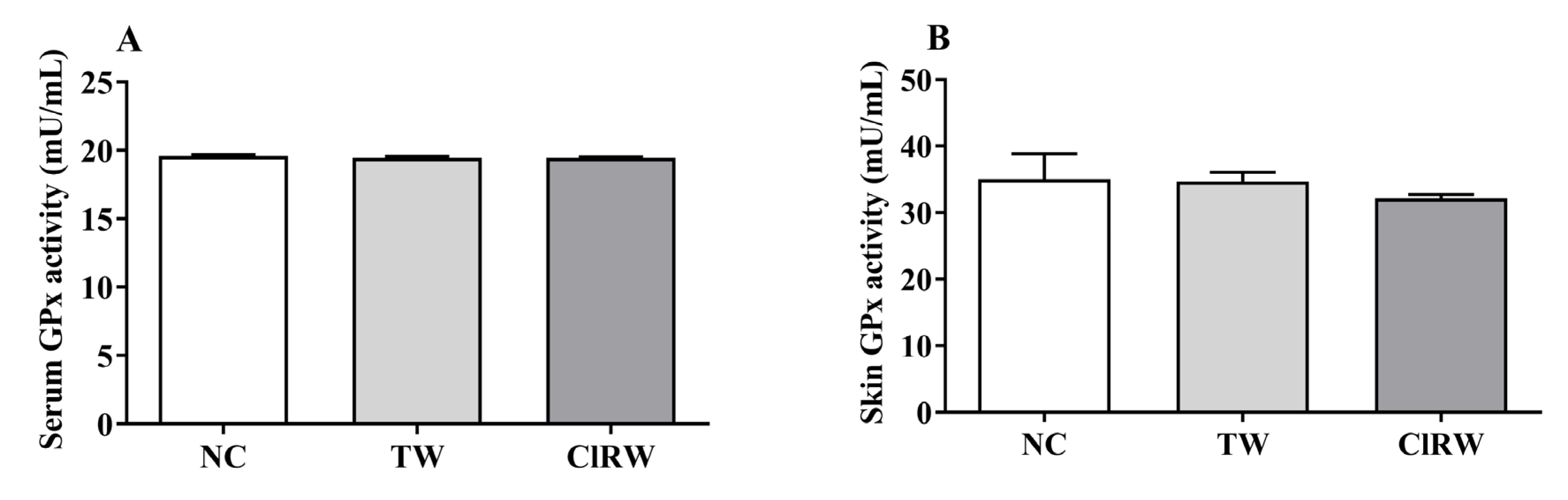
2.8. Measurement of Antioxidant Enzyme Activities

The glutathione peroxidase (GPx) and catalase (CAT) activities were measured in both serum and skin lysates using a BioVision kit (Milpitas, CA, USA). Briefly, normalized protein concentrations were used to measure the activities of the two antioxidant enzymes (GPx and CAT) according to the manufacturer’s instructions. Each antioxidant assay followed the standards and reaction processes. The absorbance to obtain the concentration was read at the following absorbance: GPx (340 nm) and CAT (510 nm).

2.9. Cytokine Analysis

Following the manufacturer’s instructions, a bead array Suspension Multiplex Kit (Bio-Rad, San Diego, CA, USA) was used to analyze the Th1/Th2 cytokines such as interleukin (IL)-2, IL-4, IL-5, IL-6, IL-10, and IL-12, in both serum and skin. Inflammatory cytokines such as IL-1α, IL-1β, IL-13, GM-CSF, and TNF-α were also examined. The plate was run on a Luminex 200 Bioplex instrument (Bio-Rad Laboratories). The raw data were analyzed using a five-parameter logistic method.

2.10. White Blood Cell (WBC) and its Differential Count Analysis

Retro-orbital plexus blood was drawn and placed in tubes coated with ethylenediaminetetraacetic acid (EDTA). An automatic blood analyzer (HEMAVET HV950 FS; Drew Scientific Inc., Dallas, TX, USA) was used to quantify the total WBC count and differential counts of lymphocytes, monocytes, neutrophils, eosinophils, and basophils.

2.11. Statistical Analysis

Statistical analysis was performed using analysis of variance (ANOVA) followed by a multiple comparison test (Tukey’s post hoc test) using the GraphPad Prism version 8.0 software packages (GraphPad Software, La Jolla, CA, USA). The differences were considered statistically significant at * p < 0.05.

3. Results

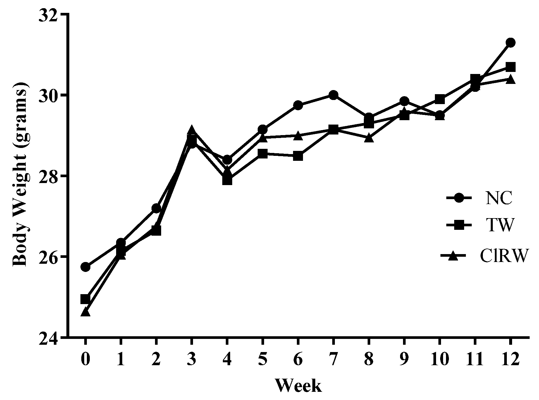
3.1. Effects of Chlorine-Rich Sterilized Water (ClRW) on Body Weight Measurements

To observe the effect of long-term treatment with CIRW in vivo on mice, the body measurements were performed weekly (Figure 1). This revealed the body weight among the three groups for 12 weeks. The body weights of mice in both groups did not vary significantly.

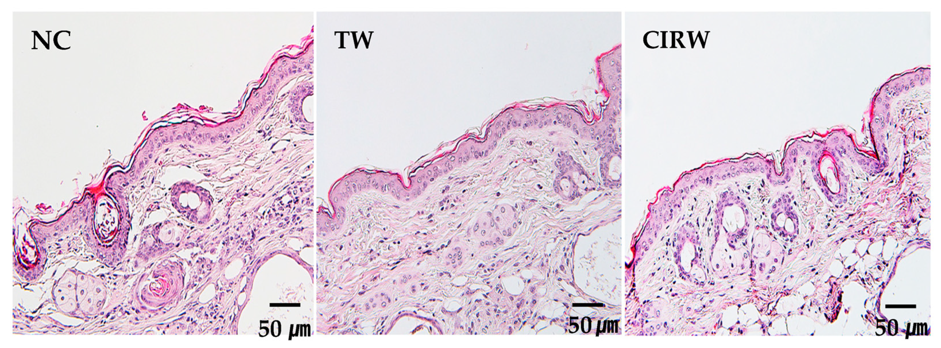
3.2. Effects of ClRW on Skin Histological Analysis

Figure 2 shows the histological observation of the skin. It verifies that there were no structural differences among the groups after long-term treatment with ClRW or tap water or without treatment. Epidermal hyperproliferation, inflammation, or other abnormal skin conditions were not observed in any group.

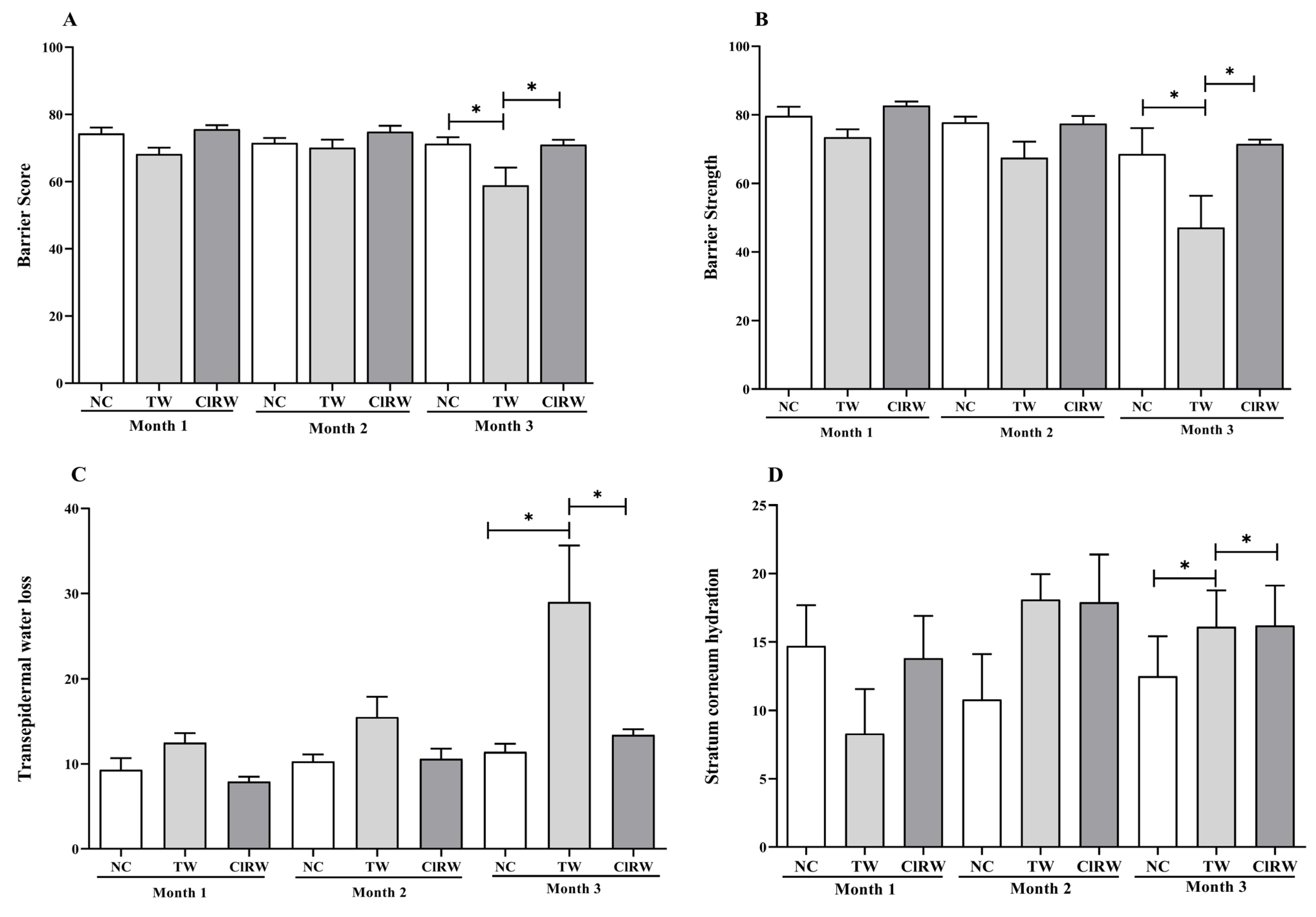
3.3. Effect of ClRW on Skin Parameters

In this study, the A-ONE TAB (Bomtech Electronics Co., LTD, Seoul, Republic of Korea) was used to measure the skin characteristics such as moisture content, elasticity, sebum, pore size, and wrinkles. (Figure 3). There were no significant differences in any of these skin parameters between the NC and ClRW groups except for the skin elasticity at month 3 (* p < 0.05). The photographs, in conjunction with the measurements, verified these results. Other skin parameters, such as barrier score, in Figure 3A, barrier strength in Figure 3B, and transepidermal water loss (TEWL) (Figure 3C), were also measured using MSIP-CRM-G10 barrier sensor (Gpower, Republic of Korea). The results showed no significant differences in skin parameters during the first two months of supplementation. However, in the third month, the TW group had a significantly lower barrier score than the NC and ClRW groups (* p < 0.05). Similarly, the barrier strength was significantly lower in the TW group than in the NC (* p < 0.05) and ClRW (* p < 0.05) groups. Moreover, the TEWL was significantly higher in the TW group than that in the NC (* p < 0.05) and ClRW (* p < 0.05) groups. In addition, stratum corneum hydration levels were observed in TW (* p < 0.05) and CIRW (* p < 0.05) as compared to the NC group.

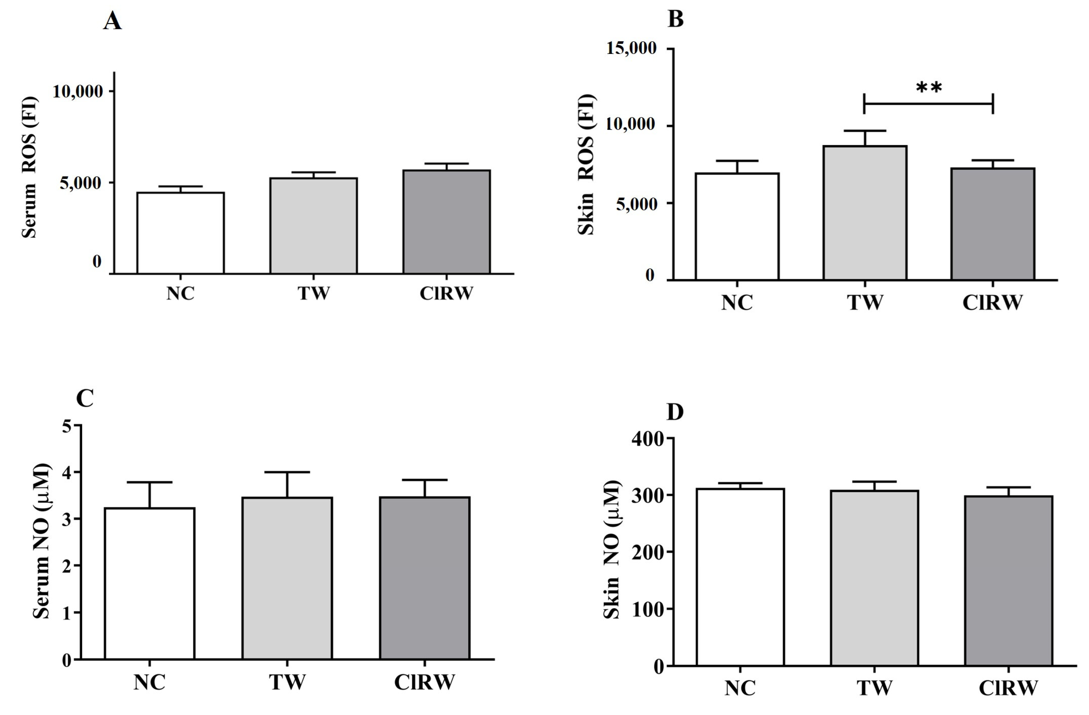
3.4. Effects of Chlorine-Rich Sterilized Water on Oxidative Stress Response

To test the effect of long-term supplementation with ClRW on the skin, we measured the oxidative stress markers in both serum and skin (Figure 4). The results showed that the ClRW group had a significantly higher ROS than that of the NC group (* p < 0.05) in the serum. However, no statistically significant variation was observed in the skin (Figure 4A,B). Moreover, nitric oxide did not show any significant differences in either the serum or skin (Figure 4C,D). Similarly, the antioxidant GPx levels in the serum and skin were not significantly different (Figure 5A,B). However, the serum catalase activity was significantly lower in the ClRW group than in the NC (*** p < 0.001) and TW (* p < 0.05) groups. In addition, the catalase activity in the skin of the ClRW group was significantly lower than that in both NC and TW groups (* p < 0.05) (Figure 5C,D).

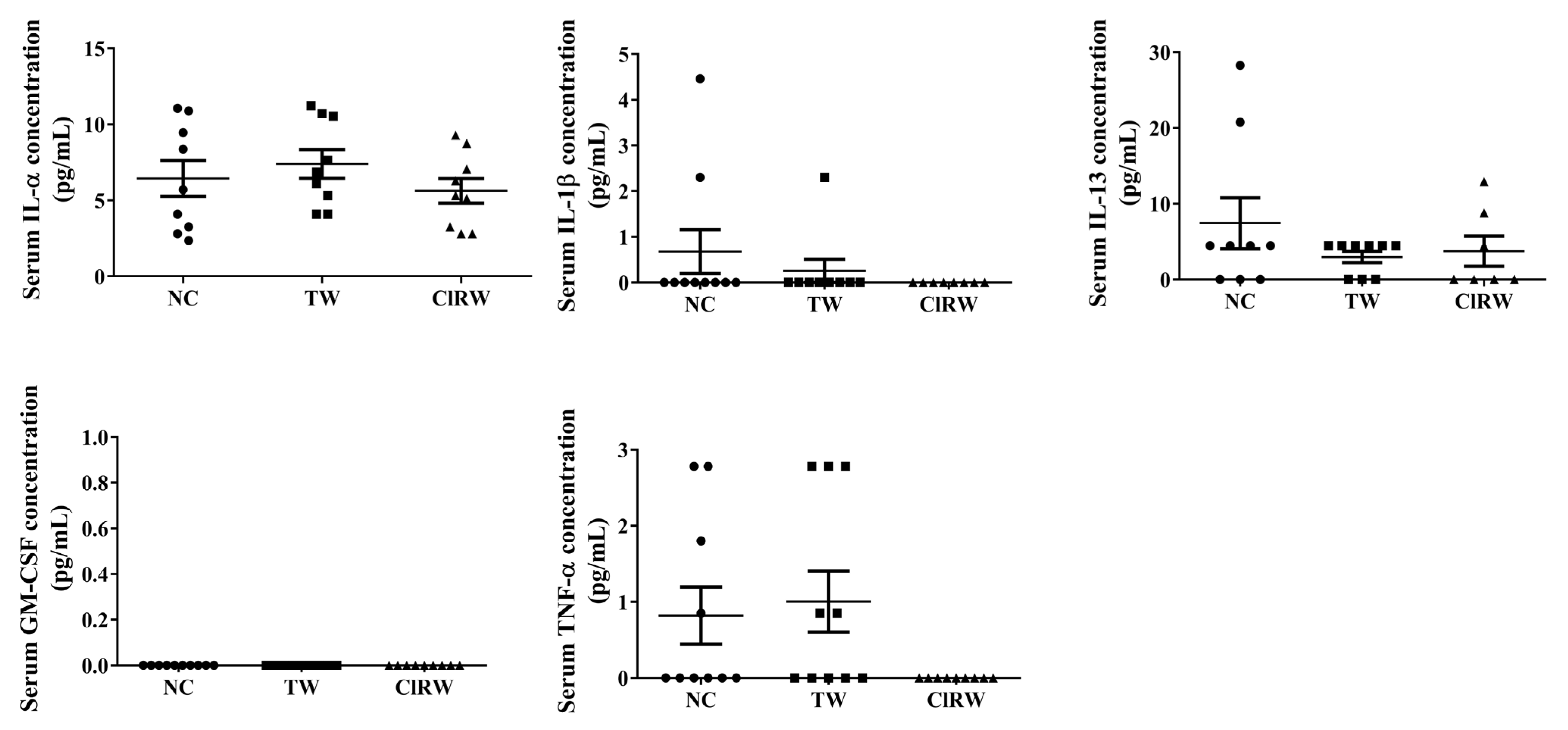
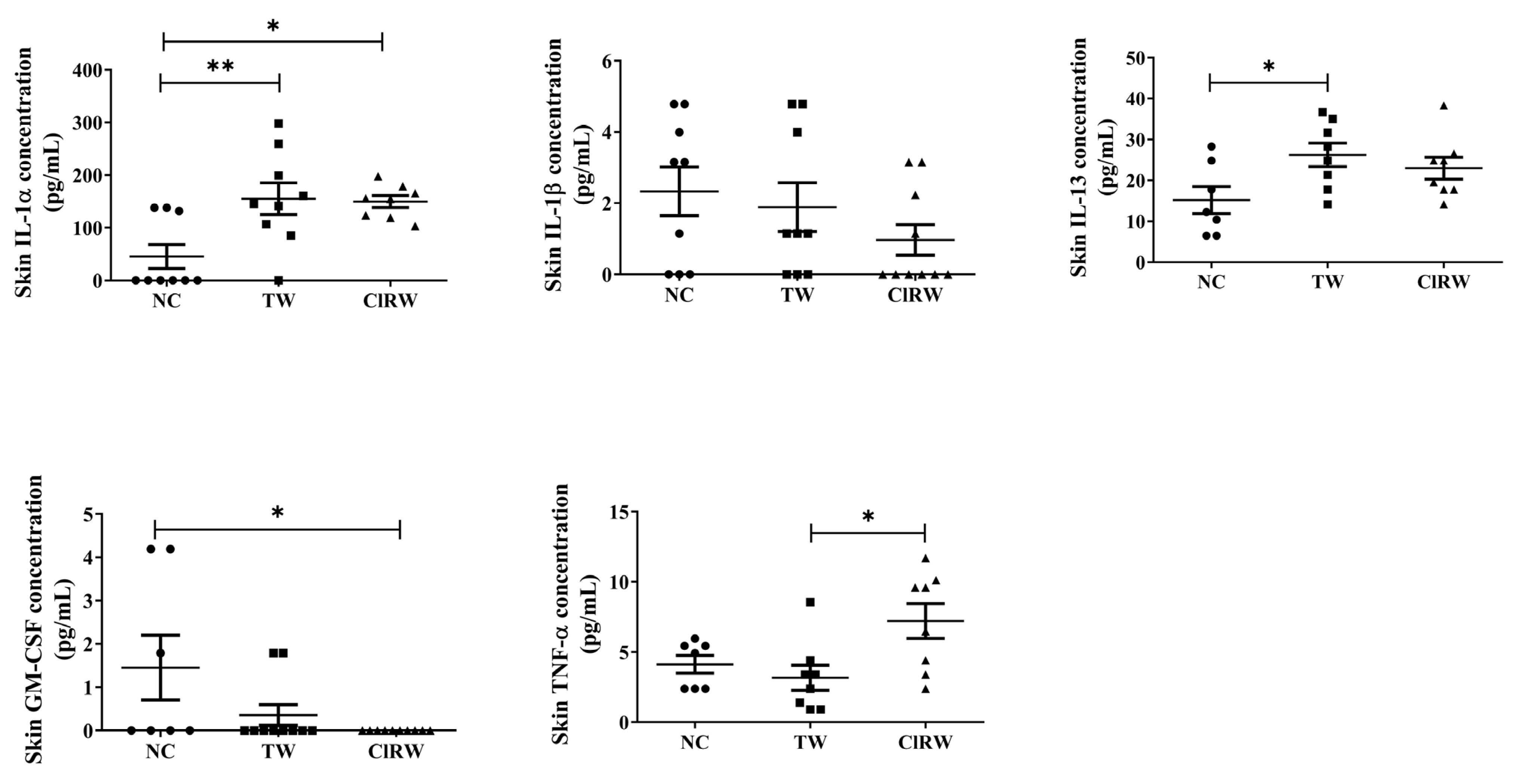
3.5. Effects of Chlorine-Rich Sterilized Water (ClRW) on Inflammatory Response

Furthermore, to test the effect of the long-term supplementation of ClRW on the inflammatory response, we tested the cytokines involved in inflammation (such as IL-1α, IL-1β, IL-13), granulocyte/macrophage colony-stimulating factor (GM-CSF), and tumor necrosis factor (TNF)-α in both serum and skin (Figure 6). Our results showed no significant variations in the production of these cytokines in the serum. However, in the skin, IL-1α showed a significant increase in the TW (** p < 0.01) and ClRW (* p < 0.05) groups compared with the NC group. Similarly, an increase in IL-13 production was observed in TW (* p < 0.05) but not in ClRW. Moreover, the GM-CSF levels were significantly lower in the ClRW group than in the NC group (* p < 0.05). The results also demonstrated that TNF-α increased significantly compared with that in the TW group (* p < 0.05).

3.6. Effects of Chlorine-Rich Sterilized Water (ClRW) on Allergic Response

The Th1/Th2 cytokine levels were analyzed. Moreover, the concentrations of IL-2, IL-4, IL-5, IL-6, IL-10, and IL-12 in both serum and skin were measured to test the effect of long-term treatment on the allergic response (Table 2). The results showed that the IL-5, IL-6, IL-10, and IL-12 levels were significantly lower in the ClRW group than in the NC and TW groups (Table 2). However, no significant differences were observed in the levels of these cytokines in the skin.

3.7. Effects of Chlorine Rich Sterilized Water (ClRW) on WBC Count

Our results showed that ClRW supplementation did not significantly alter the hematological parameters. The total WBC, neutrophil, lymphocyte, monocyte, eosinophil, and basophil counts were similar among the three groups (see Table 3).

4. Discussion

This study showed that ClRW (with characteristics such as a normal pH and high ACC) did not display systemic and local skin toxicity. To demonstrate this assertion, several tests were performed on both serum and skin to examine different markers related to the immune and oxidative stress responses. First, no significant differences in body weight were observed between the NC and ClRW groups. In addition, the morphology of the internal organs, such as the liver, kidney, and spleen, were normal. These results implied that the three-month supplementation with ClRW did not cause any hazardous physiological effect on the entire body. To further analyze the results, the skin parameters were measured to assess their effects on the skin. No significant differences in skin parameters, such as sebum, pores, wrinkles, and pigments, were observed among the three groups. However, the higher moisture level and elasticity in the ClRW group than in the NC group may have been an indication that ClRW has the potential to maintain moisture and elasticity, which are essential for normal skin barrier function. Similarly, the skin barrier score, barrier strength, and TEWL were similar to those of the NC group. This indicated that even under different water properties, particularly a high chlorine concentration, the ClRW group did not alter these skin parameters. This assertion is supported by the fact that the histological analysis yielded normal microscopy results for the epidermis and dermis in all the groups.
However, it has been proposed that during routine metabolic processes, ROS plays a significant role in the regulation of physiological intracellular signal transductions [16]. The fundamental formulation that has been approved is HOCl, which can remain stable without cytotoxicity for up to twelve months [17]. Additionally, its pH neutralization can improve skin tolerability, stability, and therapeutic action. Interestingly, ROS, such as HOCl, penetrate bacterial cell membranes to kill pathogens through chlorination or oxidation, which destroys the essential metabolic building blocks [18]. Our study revealed that the effect of the three-month supplementation of ClRW on the oxidative stress response showed that the ROS increased in the serum but not in the skin. Our study found that free radicals such as ROS are known to play a significant role in the pathogenesis of diseases. Moreover, NO production is important for health and disease control, including its role in oxidative stress- and inflammation-related pathways [19]. Our results showed that NO had similar levels among all the groups, indicating that the ClRW supplementation had a normal effect on NO production. Thus, it did not alter the normal physiological function of NO in the body. Many studies demonstrated that antioxidants eliminated excess ROS to prevent cellular damage [20,21]. In addition, in our study, we found that GPx levels were similar to those of NC in both serum and skin, indicating normal antioxidant activity. Furthermore, the decreased catalase activity in both serum and skin of the TW and ClRW groups may have been an indication that catalase balances ROS production during supplementation.
The immune responses in the skin are essential to protect it from hazardous effects. It regulates the crosstalk between the components of the skin at the cellular and microbial levels for its function in host defense, skin homeostasis, and prevention of chronic diseases [22]. In addition to bathroom fixtures like ice makers and sink drains, bacteria may also be found there. Additionally, infections at the site of surgery sometimes involve skin-related superficial infections [23,24]. Meanwhile, infections at other surgical sites, which may affect organs, tissues beneath the skin, or even implanted materials, may be more deadly [25,26]. However, the concentration of chlorine (Cl2, OCl, and HOCl), ORP, and pH all directly affect how effective electrolyzed water is against microorganisms. HOCl is the chlorine group’s most potent inactivation chemical [27,28]. Inflammation is defined as the response of the body to regeneration. It protects against infections and hazards [29]. The WBC test showed no significant differences in these immune cells, indicating normal function even after a three-month ClRW supplementation. Next, the results showed that serum inflammatory cytokine levels did not increase compared with those in the NC group, indicating its relative safety against inflammation. A study reported that dendritic cell-derived IL-1β may be an important molecule for the onset of the primary immune responses in the skin [30]. The results wherein pro-inflammatory cytokines such as IL-1β and GM-CSF decreased compared with those in NC may imply its action on the skin’s immune balance. Conversely, TNF-α, IL-13, and IL-1α increased significantly in the skin. However, these cytokines activated the immune cells, such as macrophages and T-cells [31]. An allergic response is an approach to measuring skin toxicity. The chemicals entering the body can be hazardous and cause allergic responses. B-cells or macrophages attack allergens that enter the body through the skin. Allergen-specific T-cells, primarily Th2 cells, release IL-4, IL-5, and IL-10 to combat allergic reactions [32]. Our results showed that the serum IL-5, IL-6, and IL-12 levels in both ClRW- and TW-supplemented groups were significantly lower than those in the NC group. This may indicate their role in fighting allergic responses. There was no difference between TW and ClRW, notwithstanding the differences in their chemical properties. In addition, no allergic response was detected in the skin of the ClRW groups. This demonstrated that ClRW could cause an allergic response. We observed that the ClRW therapy substantially reduced the skin-induced oxidative stress and stabilized the ROS levels. The activities of the antioxidant enzymes (catalase [CAT], superoxide dismutase [SOD], glutathione peroxidase [GPX], and glutathione reductase [GR]) responsible for ROS regulation were assessed to understand the mechanism by which ClRW influences redox regulation. In ClRW-treated cells, the GPX activity was stimulated dramatically [33]. These results indicated that the glutathione system was significantly activated in the ClRW-treated sample. This, in turn, implied that the glutathione system may play an important role in the ClRW-mediated regulation of ROS. The final result was observed after long-term treatment with ClRW. The ROS label increased in the skin because of the presence of a large amount of hydrogen and chlorine molecules.

5. Conclusions

The clinical application of this approach should be highly emphasized in conditions where the adjustment of the skin water balance by strategies other than pharmacological treatments may significantly improve skin quality. The present methodology provides an objective clinical approach to studying the effects of dietary ClRW on normal skin physiology. Collectively, our results indicated that ClRW did not show skin toxicity after long-term treatment (90 days) in vivo in skin sensitivity and immune redox reactions such as oxidative stress, inflammation, and allergic response. In addition, a few of the effects shown by ClRW maintained moisture better than the normal control and had a tendency to decrease naturally induced inflammation. Further clinical studies are required to identify the mechanisms by which ClRW specifically mediates these biological processes.

Author Contributions

Conceptualization, K.-J.L.; methodology, A.F. and M.H.R.; software, A.F., M.H.R. and J.B.; validation, M.H.R., A.F. and J.B.; formal analysis, M.H.R.; investigation, C.-S.K.; resources, C.-S.K. and G.S.H.; data curation, A.F. and M.H.R.; writing—original draft preparation, M.H.R. and A.F.; writing—review and editing, J.B., C.S.H. and C.-S.K.; visualization, C.-S.K.; supervision, C.-S.K.; project administration, K.-J.L.; funding acquisition, K.-J.L. All authors have read and agreed to the published version of the manuscript.

Funding

This study received no external funding.

Data Availability Statement

Not Applicable.

Conflicts of Interest

The authors declare no conflict of interest.

References

  1. Gorzelanny, C.; Mess, C.; Schneider, S.W.; Huck, V.; Brandner, J.M. Skin barriers in dermal drug delivery: Which barriers have to be overcome and how can we measure them? Pharmaceutics 2020, 12, 684. [Google Scholar] [CrossRef]
  2. Johansson, B. Functional water-in promotion of health beneficial effects and prevention of disease. Intern. Med. Rev. 2016, 3. [Google Scholar] [CrossRef]
  3. Iram, A.; Wang, X.; Demirci, A. Electrolyzed oxidizing water and its applications as sanitation and cleaning agent. Food Eng. Rev. 2021, 13, 411–427. [Google Scholar] [CrossRef]
  4. Kawada, J.; Yamada, H.; Matsuba, Y.; Ogawa, H. Effectiveness of Hand-washing Using Acidic Electrolyzed Water-A Comparative Study of Weak Acidic Electrolyzed Water, Strong Acidic Electrolyzed Water and Tap Water. Ski. Res. 2000, 42, 137–142. [Google Scholar]
  5. Vorobjeva, N.V.; Vorobjeva, L.I.; Khodjaev, E.Y. The bactericidal effects of electrolyzed oxidizing water on bacterial strains involved in hospital infections. Artif. Organs 2004, 28, 590–592. [Google Scholar] [CrossRef]
  6. Lee, J.H.; Rhee, P.L.; Kim, J.H.; Kim, J.J.; Paik, S.W.; Rhee, J.C.; SONG, J.H.; Yeom, J.S.; Lee, N.Y. Efficacy of electrolyzed acid water in reprocessing patient-used flexible upper endoscopes: Comparison with 2% alkaline glutaraldehyde. J. Gastroenterol. Hepatol. 2004, 19, 897–903. [Google Scholar] [CrossRef]
  7. Williams, J.; Rasmussen, E.; Robins, L.; Nguyen, U. Hypochlorous Acid: Harnessing an Innate Response. Infect. Prev. Strategy (TIPS) 2017, 1–9. [Google Scholar]
  8. Rahman, M.H.; Bajgai, J.; Cho, Y.; Fadriquela, A.; Sharma, S.; Thuy, T.T.; Cho, S.H.; Jeong, Y.J.; Goh, S.H.; Kim, Y. Immune Redox Modulation Effects of Non-Electrolyzed Hypochlorous Acid Water on Helicobacter pylori-Infected C57BL/6 Mouse Model. Processes 2023, 11, 1474. [Google Scholar] [CrossRef]
  9. Pan, G.J.; Rayner, B.S.; Zhang, Y.; van Reyk, D.M.; Hawkins, C.L. A pivotal role for NF-κB in the macrophage inflammatory response to the myeloperoxidase oxidant hypothiocyanous acid. Arch. Biochem. Biophys. 2018, 642, 23–30. [Google Scholar] [CrossRef] [Green Version]
  10. Leung, T.H.; Zhang, L.F.; Wang, J.; Ning, S.; Knox, S.J.; Kim, S.K. Topical hypochlorite ameliorates NF-κB–mediated skin diseases in mice. J. Clin. Investig. 2013, 123, 5361–5370. [Google Scholar] [CrossRef] [Green Version]
  11. Andrés, C.M.C.; Pérez de la Lastra, J.M.; Juan, C.A.; Plou, F.J.; Pérez-Lebeña, E. Hypochlorous Acid Chemistry in Mammalian Cells—Influence on Infection and Role in Various Pathologies. Int. J. Mol. Sci. 2022, 23, 10735. [Google Scholar] [CrossRef]
  12. Bauer, G. HOCl and the control of oncogenesis. J. Inorg. Biochem. 2018, 179, 10–23. [Google Scholar] [CrossRef] [PubMed]
  13. Water, S.; Organization, W.H. Guidelines for Safe Recreational Water Environments. In Swimming Pools and Similar Environments; World Health Organization: Geneva, Switzerland, 2006; Volume 2. [Google Scholar]
  14. Prest, E.I.; Hammes, F.; Van Loosdrecht, M.C.; Vrouwenvelder, J.S. Biological stability of drinking water: Controlling factors, methods, and challenges. Front. Microbiol. 2016, 7, 45. [Google Scholar] [CrossRef] [PubMed] [Green Version]
  15. Jeong, E.-S.; Bajgai, J.; You, I.-S.; Rahman, M.; Fadriquela, A.; Sharma, S.; Kwon, H.-U.; Lee, S.-Y.; Kim, C.-S.; Lee, K.-J. Therapeutic effects of hydrogen gas inhalation on trimethyltin-induced neurotoxicity and cognitive impairment in the C57BL/6 mice model. Int. J. Mol. Sci. 2021, 22, 13313. [Google Scholar] [CrossRef] [PubMed]
  16. Oyinloye, B.; Adenowo, A.; Kappo, A. Reactive oxygen species, apoptosis, antimicrobial peptides and human inflammatory diseases. Pharmaceuticals 2015, 8, 151–175. [Google Scholar] [CrossRef] [Green Version]
  17. Fukuyama, T.; Martel, B.; Linder, K.; Ehling, S.; Ganchingco, J.; Bäumer, W. Hypochlorous acid is antipruritic and anti-inflammatory in a mouse model of atopic dermatitis. Clin. Exp. Allergy 2018, 48, 78–88. [Google Scholar] [CrossRef]
  18. Sam, C.-H.; Lu, H.-K. The role of hypochlorous acid as one of the reactive oxygen species in periodontal disease. J. Dent. Sci. 2009, 4, 45–54. [Google Scholar] [CrossRef] [Green Version]
  19. Luiking, Y.C.; Engelen, M.P.; Deutz, N.E. Regulation of nitric oxide production in health and disease. Curr. Opin. Clin. Nutr. Metab. Care 2010, 13, 97. [Google Scholar] [CrossRef] [PubMed] [Green Version]
  20. Masaki, H. Role of antioxidants in the skin: Anti-aging effects. J. Dermatol. Sci. 2010, 58, 85–90. [Google Scholar] [CrossRef]
  21. Pham-Huy, L.A.; He, H.; Pham-Huy, C. Free radicals, antioxidants in disease and health. Int. J. Biomed. Sci. 2008, 4, 89. [Google Scholar]
  22. Pasparakis, M.; Haase, I.; Nestle, F.O. Mechanisms regulating skin immunity and inflammation. Nat. Rev. Immunol. 2014, 14, 289. [Google Scholar] [CrossRef]
  23. Edmiston, C.E.; McBain, A.; Kiernan, M.; Leaper, D. A narrative review of microbial biofilm in postoperative surgical site infections: Clinical presentation and treatment. J. Wound Care 2016, 25, 693–702. [Google Scholar] [CrossRef]
  24. Iskandar, K.; Sartelli, M.; Tabbal, M.; Ansaloni, L.; Baiocchi, G.L.; Catena, F.; Coccolini, F.; Haque, M.; Labricciosa, F.M.; Moghabghab, A. Highlighting the gaps in quantifying the economic burden of surgical site infections associated with antimicrobial-resistant bacteria. World J. Emerg. Surg. 2019, 14, 1–14. [Google Scholar] [CrossRef] [Green Version]
  25. Stevens, D.L. Treatments for skin and soft-tissue and surgical site infections due to MDR Gram-positive bacteria. J. Infect. 2009, 59, S32–S39. [Google Scholar] [CrossRef]
  26. Gomila, A.; Carratalà, J.; Badia, J.M.; Camprubí, D.; Piriz, M.; Shaw, E.; Diaz-Brito, V.; Espejo, E.; Nicolas, C.; Brugués, M. Preoperative oral antibiotic prophylaxis reduces Pseudomonas aeruginosa surgical site infections after elective colorectal surgery: A multicenter prospective cohort study. BMC Infect. Dis. 2018, 18, 1–9. [Google Scholar] [CrossRef]
  27. da Cruz Nizer, W.S.; Inkovskiy, V.; Overhage, J. Surviving reactive chlorine stress: Responses of gram-negative bacteria to hypochlorous acid. Microorganisms 2020, 8, 1220. [Google Scholar] [CrossRef]
  28. Memar, M.Y.; Ghotaslou, R.; Samiei, M.; Adibkia, K. Antimicrobial use of reactive oxygen therapy: Current insights. Infect. Drug Resist. 2018, 11, 567. [Google Scholar] [CrossRef] [Green Version]
  29. Dainichi, T.; Hanakawa, S.; Kabashima, K. Classification of inflammatory skin diseases: A proposal based on the disorders of the three-layered defense systems, barrier, innate immunity and acquired immunity. J. Dermatol. Sci. 2014, 76, 81–89. [Google Scholar] [CrossRef] [Green Version]
  30. Enk, A.H.; Angeloni, V.; Udey, M.; Katz, S. An essential role for Langerhans cell-derived IL-1 beta in the initiation of primary immune responses in skin. J. Immunol. 1993, 150, 3698–3704. [Google Scholar] [CrossRef] [PubMed]
  31. Arango Duque, G.; Descoteaux, A. Macrophage cytokines: Involvement in immunity and infectious diseases. Front. Immunol. 2014, 5, 491. [Google Scholar] [CrossRef] [Green Version]
  32. Kidd, P. Th1/Th2 balance: The hypothesis, its limitations, and implications for health and disease. Altern. Med. Rev. 2003, 8, 223–246. [Google Scholar]
  33. Ren, A.; Liu, R.; Miao, Z.G.; Zhang, X.; Cao, P.F.; Chen, T.X.; Li, C.Y.; Shi, L.; Jiang, A.L.; Zhao, M.W. Hydrogen-rich water regulates effects of ROS balance on morphology, growth and secondary metabolism via glutathione peroxidase in Ganoderma lucidum. Environ. Microbiol. 2017, 19, 566–583. [Google Scholar] [CrossRef]
Figure 1. Body weight measurement. The data are presented as the mean ± SEM (n = 10). ANOVA Tukey’s test (p < 0.05) was used to determine the significant difference.
Processes 11 01914 g001
Figure 2. Histological observation of the skin in normal control (NC); the group with tap water (TW) supplementation; and the group with ClRW supplementation. The histological sections were stained with hematoxylin and eosin. Scale bar = 50 μm.
Processes 11 01914 g002
Figure 3. Measurement of skin parameters such as moisture level, elasticity, sebum, pore size, and wrinkle among three groups. The data are presented as the mean ± SEM (n = 10). ANOVA Tukey’s test (p < 0.05) was used to determine the significant difference. (A) Barrier score, (B) Barrier strength, (C) Transepidermal water loss, (D) Stratum corneum hydration. * p < 0.05.
Processes 11 01914 g003
Figure 4. Oxidative stress markers such as the ROS and NO levels in both serum and skin lysates. The data are presented as the mean ± SEM (n = 10). ANOVA Tukey’s test (p < 0.05) was used to determine the significant difference. (A) Serum ROS (FI), (B) Skin ROS (FI), (C) Serum NO (µM), (D) Skin NO (µM). ** p < 0.01.
Processes 11 01914 g004
Figure 5. Oxidative stress markers such as the GPx and CAT levels in both serum and skin lysates. The data are presented as the mean ± SEM (n = 10). ANOVA Tukey’s test (p < 0.05) was used to determine the significant difference. (A) Serum GPx (mU/mL), (B) Skin GPx (uM/mL), (C) Serum CAT (mU/mL), (D) Skin CAT (mU/mL). * p < 0.05, *** p < 0.001.
Processes 11 01914 g005aProcesses 11 01914 g005b
Figure 6. Inflammatory markers in both levels in both serum and skin lysates among three groups. The data are presented as the mean ± SEM (n = 10). ANOVA Tukey’s test (p < 0.05) was used to determine the significant difference. * p < 0.05, ** p < 0.01.
Processes 11 01914 g006aProcesses 11 01914 g006b
Table 1. Properties of tap water (TW) and chlorine-rich sterilized water (ClRW) before and after using the machine.
BeforeAfter
Temp.21.421.3
pH7.757.11
ORP500 mV450 mV
ACC0.7 ppm5.0 ppm
H236 ppm235 ppm
ORP: Oxidation-reduction potential; ACC: Available chlorine concentration; PPM: Parts per million.
Table 2. The analysis of Th1/Th2 cytokines in both serum and skin.
Serum and Skin Count (pg/mL)NCTWClRW
Serum IL-22.97 ± 0.982.72 ± 0.652.82 ± 0.42
Serum IL-42.12 ± 1.561.02 ± 0.590.99 ± 1.56 *
Serum IL-52.60 ± 0.712.09 ± 0.621.65 ± 0.53 *
Serum IL-62.83 ± 0.472.14 ± 0.601.83 ± 0.50
Serum IL-1012.27 ± 9.240.70 ± 1.730.11 ± 0.23 *
Serum IL-1278.22 ± 21.6457.06 ± 13.8843.52 ± 18.69 *
Skin IL-21.24 ± 0.952.04 ± 1.071.95 ± 0.97
Skin IL-40.00 ± 0.000.00 ± 0.000.00 ± 0.00
Skin IL-50.00 ± 0.000.00 ± 0.000.10 ± 0.10
Skin IL-60.70 ± 0.120.64 ± 0.160.76 ± 0.12
Skin IL-100.00 ± 0.000.00 ± 0.000.00 ± 0.00
Skin IL-126.20 ± 2.046.08 ± 2.037.05 ± 2.99
The data are presented as the mean ± SEM (n = 10). Tukey’s test (p < 0.05) was used to determine the significant difference. NC: Normal Control, TW: Tap water, CIRW: Chlorine Rich Sterilized Water. * p < 0.05.
Table 3. Total white blood cells (WBC) and WBC differential counts.
WBC Count (K/μL)NCTWClRW
Total WBC5.94 ± 1.246.29 ± 1.177.15 ± 1.31 *
Neutrophils1.67 ± 0.301.83 ± 0.341.93 ± 036
Lymphocytes4.15 ±1.004.23 ± 1.025.04 ± 1.00 *
Monocytes0.11 ± 0.050.20 ± 0.150.16 ± 0.11
Eosinophils0.02 ± 0.010.01 ± 0.010.01 ± 0.008
Basophils0.002 ± 0.000.005 ± 0.0050.006 ± 0.006
The data are presented as the mean ± SEM (n = 10). Tukey’s test (p < 0.05) was used to determine the significant difference. NC: Normal Control, TW: Tap water, CIRW: Chlorine Rich Sterilized Water. * p < 0.05.
Disclaimer/Publisher’s Note: The statements, opinions and data contained in all publications are solely those of the individual author(s) and contributor(s) and not of MDPI and/or the editor(s). MDPI and/or the editor(s) disclaim responsibility for any injury to people or property resulting from any ideas, methods, instructions or products referred to in the content.

