Article

Pharmacological Profile of Nigella sativa Seeds in Combating COVID-19 through In-Vitro and Molecular Docking Studies

1
Department of Biology, College of Sciences, University of Ha’il, Ha’il 2440, Saudi Arabia
2
Department of Chemistry, College of Sciences, University of Ha’il, Ha’il 2440, Saudi Arabia
3
Molecular Diagnostics and Personalized Therapeutics Unit, University of Ha’il, Ha’il 2440, Saudi Arabia
4
Department of Pathology, Sub-Division of Medical Microbiology, College of Medicine, University of Ha’il, Ha’il 2440, Saudi Arabia
5
Department of Basic Dental and Medical Sciences, University of Ha’il, Ha’il 2440, Saudi Arabia
6
Department of Physiotherapy, College of Applied Medical Sciences, University of Ha’il, Ha’il 2440, Saudi Arabia
7
Department of Chemistry, College of Science, King Faisal University, Al-Ahsa 31982, Saudi Arabia
*
Author to whom correspondence should be addressed.
Processes 2022, 10(7), 1346; https://doi.org/10.3390/pr10071346
Submission received: 18 May 2022 / Revised: 22 June 2022 / Accepted: 5 July 2022 / Published: 11 July 2022

Abstract

:
COVID-19 infection is associated with elevated oxidative stress, systemic hyper-inflammatory responses, endothelial dysfunction, and red blood cell membrane deformability. Nigella sativa extract is widely used in alternative and complementary medicine systems in a large population, due to its highly therapeutic, economic, natural, and safe nature. The aim of this study was to evaluate the effect of N. sativa extract on oxidative stress, hemolysis, proteolysis, and glycation through in vitro studies, as well as to find out its anti-viral potential against severe acute respiratory syndrome coronavirus-2 (SARS-CoV-2) using in silico studies. N. sativa seed extract (at 600 µg/mL) displayed 67.33% scavenging activity in the 2,2-diphenyl-1-picrylhydrazyl (DPPH) test, and 70.28% hydrogen peroxide reducing activity. N. sativa exhibited anti-proteolytic activity by decreasing heat-induced denaturation of bovine serum albumin (BSA) and egg albumin by 63.14% and 57.95%, respectively, and exhibited anti-proteinase potential of 66.28% at 600 μg/mL. In addition, heat-induced hemolysis and hypersalinity-induced hemolysis were inhibited by 57.86% and 61.7%, respectively, by the N. sativa seeds. N. sativa also inhibited browning intensity by 56.38%, and percent aggregation index by 51.38%, amyloid structure by 48.28%, and AGE-specific fluorescence by 52.18%, thereby protecting the native structure of BSA from glycation. The binding interactions between bioactive molecules of N. sativa seed with SARS-CoV-2 spike glycoprotein were proven by using in silico molecular docking tools. The functional amino acids involved in the interactions are Asp467, Thr108, Thr114, Ile468, Asn234, Gln155, Glu465, Arg466, Gly232, and Ile233, indicating the inhibiting property of N. sativa on SARS-CoV-2. Finally, we may infer that phytoconstituents of N. sativa seeds have the potential to protect against the spike protein of SARS-CoV-2. Studies on N. sativa seeds might act as a path to develop a potent alternative therapy against viral infections, especially COVID-19 infection, in the future. However, the limitations linked with the use of natural products are also needed to be considered in this regard.

1. Introduction

The coronavirus disease-2019 (COVID-19) is caused by SARS-CoV-2) infection. The condition in infected individuals varies from asymptomatic, mild to severe symptoms [1]. Severe outcomes may include acute respiratory distress syndrome, cardiac problems, multiple organ failure syndrome, septic shock, and death. Cytokine storm has been implicated in most hyper-inflammatory complications, often associated with co-morbidities [2,3]. Changes in the blood cell phenotype, lead to coagulation abnormalities and vascular constrictions, contributing to vascular disease [4].
The coronavirus entry into the host cell is mediated by a glycoprotein transmembrane spike protein on the viral envelope. The functional subunits (S1 and S2) in the spike protein attach to the host cell surface receptor to facilitate the virus-host cell fusion [5]. Angiotensin-converting enzyme 2 (ACE2) has a pivotal role in viral entry via membrane fusion [6]. Recently, the crystallographic structure of the ACE2 receptor-S protein (spike protein from SARS-CoV-2) complex -was published, suggesting that the disruption of this complex formation might be a promising target for drug development.
Angiotensin and ACE2 interacting proteins have been identified on the surface of red blood cell (RBC) membranes [7]. Thus, RBCs are susceptible to invasion by the virus. COVID-19 can lead to an intense acute-phase response, linked to complement dysregulation [8]. COVID-19 infection causes disruption in the homeostasis of the RBC membrane. A study indicates that RBCs from COVID-19 patients show altered lipid metabolism, in addition to an increase in glycolytic intermediates, as well as oxidation and fragmentation of erythrocyte membrane domains [9]. COVID-19 can also lead to impaired function of inherent antioxidant capacity of mature RBCs. Proteasome and degradation machinery components were found in higher concentrations in RBCs, indicating increased RBC protein breakdown in patients. Thus, RBCs in COVID-19 show higher susceptibility to oxidative stress-induced lysis [9].
In diabetic patients, the expression of ACE2 increases substantially [10]. Non-enzymatic glycosylation i.e., glycation of ACE2 has been suggested to have a direct effect on the mechanism of the SARS-CoV-2 virus [11]. Hyperglycemic condition is well known to increase the glycation of various biomolecules and increase the formation of advanced glycation end products (AGEs) [12,13]. AGEs are linked to increased COVID-19 risk factors. AGEs bind with its membrane-bound receptor (mRAGE) and stimulate intracellular signals to express various inflammatory transcription factors. The role of AGE/RAGE signaling has been associated with an increase in oxidative stress leading to hyperglycemia-mediated diabetes mellitus [14]. Antioxidant enzyme inactivation by glycation, can affect the cellular antioxidant defense mechanism, contributing to a variety of diseases associated with long-term diabetes consequences leading to exacerbation of oxidant-antioxidant imbalance [15].
In COVID-19 patients with sepsis, the inflammatory response can cause cellular oxidative stress and activation of redox pathways, resulting in an increase in circulatory inflammatory mediators, including cytokines [16]. Oxidative stress and mitochondrial activity suppression lead to tissue injuries in COVID-19, which cause damage to endothelial, alveolar, and cardiac cells [17]. Moreover, inflammation has been linked to protein denaturation, membrane changes, vascular permeability, endothelial dysfunction, and pain, thereby exacerbating the disease [18]. Hypoalbuminemia may be considered a severity marker of epithelial-endothelial damage in COVID-19 patients [19]. Administration of agents with a potential to stabilize endothelial membrane and suppress albumin denaturation or supply a high dose of albumin, might be beneficial in the management of acute inflammation associated with COVID-19 [20].
Treatment and management of diseases in various traditional medicinal systems using medicinal plants have been found to be a safe and economically sound alternative to allopathic medicines. Nigella sativa (family: Ranunculaceae) is a medicinal spice and is recognized as an effective remedy to treat various health problems. Previous studies have reported its effects, such as antihypertensive, antibacterial, antidiarrheal, digestive, liver tonic, diuretic, analgesic, and skin protectant. It is also reported to have therapeutic effects on neurological diseases, heart disease, tumor progression, hyperglycemia, and inflammation [21,22]. Despite the identification of the beneficial effects of N. sativa in various chronic illnesses and risk factors, the actual mechanism has yet to be elucidated.
The research and development of new medications is a time-consuming and costly procedure. In the United States, it takes roughly up to 12 years to disclose a novel medicine, including trials and regulatory clearance [23]. Currently, there are no particular antivirals available to battle COVID-19. So many approaches are used to tackle this problem such as RNA-dependent RNA polymerase (RdRp) as a novel drug target due to its essential role in virus replication. turmeric-derived compounds were chosen and subjected to in-silico analysis to evaluate their binding affinity against the RdRp-RNA complex of SARS-CoV-2 [24]. Non-structural protein 1 (Nsp1), a virulence agent of SARS-CoV-2, has emerged as an important target for drug discovery. The semi-synthetically derived aminoarylbenzosu-berenes (AAB) from Himachalenes (an isomeric mixture of sesquiterpenes found in Cedrus deodara oil) were used for elucidating their therapeutic potential against COVID-19 [25]. The SARS-CoV-2 main protease (Mpro) is an attractive target in the COVID-19 drug development process. It catalyzes the polyprotein’s translation from viral RNA and specifies a particular cleavage site. Due to the absence of identical cleavage specificity in human cell proteases, targeting Mpro with a chemical compound (synthesized 3 h) can obstruct the replication of the virus [26]. The in silico work is developed with this in mind to evaluate the medicinal potentials of major phytochemicals in N. sativa. Our aim was to examine the effect of N. sativa on oxidative stress, inflammation, glycation, and other chronic disease risk factors linked with COVID-19 to understand its therapeutic potential, using in vitro and in silico studies. Exploring the medicinal plants and using a drug repurposing strategy to determine potential anti-COVID-19 drugs, are important in order to find a potent natural therapy for COVID-19 in the current circumstances.

2. Materials and Methods

Ascorbic acid, Folin-Ciocalteau reagent, gallic acid, trichloroacetic acid (TCA), DPPH, ferric chloride, potassium ferricyanide, quercetin, trypsin, and Congo red were purchased from Sigma-Aldrich, St. Louis, MI, USA. Dimethyl sulfoxide (DMSO), ethanol, methanol, hydrochloric acid, aluminum chloride, sodium carbonate, sodium hydroxide, hydrogen peroxide, monosodium dihydrogen phosphate, and disodium hydrogen phosphate were purchased from Merck, Darmstadt, Germany. The solvents used were of HPLC grade.

2.1. Extract Preparation from Nigella sativa Seeds

The methanol extract of N. sativa seeds was prepared by following the standardized extraction procedure published earlier [27]. In a nutshell, distilled water was used to clean the seeds. After drying, the seeds were crushed using a mortar and pestle. The dry crushed seeds were coarsely powdered in an automated mixer grinder. The seed powder was soaked in 97% methanol (50 g in 500 mL) for three days at 37 °C in a magnetic shaker. To get a crude active component, the extract was filtered and condensed using rotary evaporators at 40 °C under reduced pressure. The condensed extract was kept at 4 °C for further study. Using the following equation, the percent yield of extract was computed. ‘
Yield (%) of N. sativa seed extract = [W/IW] × 100.
where, W refers to the weight of seed extract, and IW denotes the initial weight of dry seeds.

2.2. Determination of Antioxidant Capacity Using Hydrogen Peroxide (H2O2)

A previously published procedure was followed to investigate the antioxidant capacity of the extract [27,28,29] using hydrogen peroxide (1000 µL of 40 mM H2O2, prepared in phosphate buffer of pH 7.4) and extract (50, 75, 100, 200, 300, 400, 500, and 600 μg/mL) or ascorbic acid (100 and 200 µg/mL). The absorbance of the control and experimental samples was noted at 230 nm after 10 min of mixing. Phosphate buffer was used as a blank. The experiments were performed in triplicate.
H2O2 reducing potential (%) = [(Uc − Us)/Uc] × 100
where Us is the absorbance of the sample that contains both extract (or ascorbic acid) and H2O2 solution, while Uc is just the absorbance of control H2O2 solution [27].

2.3. DPPH Assay for the Evaluation of Antioxidant Activity

DPPH assay used 2.5 mL of ascorbic acid (100 and 200 µg/mL) or extract (50, 75, 100, 200, 300, 400, 500, and 600 µg/mL) along with 1 mL of DPPH (0.3 mM in methanol) to confirm antioxidant potential of the extract [27,28,29]. After half an hour incubation under dark conditions, the absorbance of all solutions was measured against methanol at 517 nm. In contrast, a DPPH solution in methanol was employed as a control.
DPPH scavenging activity (%) = [(Uc − Us)/Uc] × 100
Here, Us is indeed the absorbance of the sample that contains both extract (or ascorbic acid) and DPPH solution, while Uc is just the absorbance of control.

2.4. Evaluation of RBC Membrane Stability

2.4.1. Preparation of RBC Suspension

The blood samples from three healthy and fit volunteers were transferred to tubes with an equal amount of Alsever’s solution (sterilized), and then the tubes were centrifuged at 3000 rpm for 10 min. The erythrocytes were collected in separate tubes and washed thrice with an equal volume of saline. An RBC suspension (10% v/v) was prepared using isotonic phosphate buffer (pH 7.4) [27,30].

2.4.2. Inhibition of Heat-Induced Hemolysis

Inhibition of hemolysis is very significant in the context of the treatment of various diseases. With a few modifications, heat-induced erythrocyte hemolysis was performed as described in previous works [27,31]. A total of 500 µL of either extract (50, 75, 100, 200, 300, 400, 500, and 600 µg/mL) or aspirin (100, and 200 µg/mL) was mixed with 500 µL of RBC solution (10% v/v) and incubated at 56 °C. After 20 min, the tubes were cooled under running tap water, and were centrifuged at 2500 rpm for 5 min. The absorbance of the different supernatants was recorded at 540 nm against phosphate buffer. The control consisted of buffer and RBC suspension only.
Percentage protection = [(Uc − Us)/Uc] × 100
Here, Us is the absorbance of the sample that contains both extract/aspirin) and RBC solution, while Uc is just the absorbance of the control solution.

2.4.3. Inhibition of Hyposaline Induced Hemolysis

The protecting ability of N. sativa extract against hemolysis was tested as described in the previous studies with a few changes [27,31]. A total of 250 µL of phosphate buffer (pH 7.4, 0.1 M), 500 µL of hyposaline, and 125 µL RBC suspension (10% v/v) were combined with 1 mL of extract (0–600 µg/mL) or diclofenac (100, and 200 µg/mL). In contrast, the control solution solely utilized distilled water. All tubes were kept at 37 °C for 30 min. The tubes were centrifuged for 10 min at 3000 rpm after the incubation period. The absorbance of the supernatant was measured at 560 nm.
Protection (%) = 100 − [(Us/Uc) × 100]
Here, Us is the absorbance of the sample that contains both extract/diclofenac and RBC solution, while Uc is just the absorbance of the control solution [27].

2.5. Screening of Anti-Glycating and AGEs Formation Inhibition Potentials

2.5.1. In Vitro System of Protein Glycation System

With minimal modifications, Brownlee’s technique was used [32]. In phosphate buffer (0.1 M, pH 7.4), glucose (500 mM) was mixed with BSA (10 mg/mL) with or without seed extract (50, 75, 100, 200, 300, 400, 500, and 600 µg/mL). The combinations were incubated in a shaker for 15 days at room temperature, without any direct sunlight. To remove unbound glucose, the glycated samples were dialyzed at 37 °C against phosphate buffer (50 mM, pH 7.4) for 12 h.

2.5.2. Assessment of Browning Intensity

The degree of glycation can be correlated with the browning intensity of glycated samples. The degree of browning was investigated by recording the absorbance of different samples at 420 nm [15] after dilution with distilled water.
Percentage protection = [(Uc − Us)/Uc] × 100
Here, Us is the absorbance of the sample having BSA and glucose with extract, while Uc is just the absorbance of the control solution having BSA and glucose.

2.5.3. Protein Aggregation Index

The protective ability of extract against protein aggregate formation was determined by measuring the absorbance of glycated samples incubated in the presence of extract (50, 75, 100, 200, 300, 400, 500, and 600 µg/mL) or absence [27,33]. The aggregation index was derived using the following formula given below:
Percentage of protein aggregation index = [U340/(U280 − U340)] × 100
where U340 = Absorbance at 340 nm and U280 = Absorbance at 280 nm.

2.6. Biophysical Studies to Investigate AGEs Formation Inhibiting Properties of Extract

To induce glycation, BSA (0.2 mg/mL) was incubated with glucose (500 mM) in 20 mM sodium phosphate buffer (pH 7.4) for 15 days in a shaking water bath at 37 °C. The control had BSA incubated with phosphate buffer (20 mM, pH 7.4) [27,28]. The addition of sodium azide (3 mM) prevented bacterial contamination. The dialysis of each sample was done at the end of incubation using sodium phosphate buffer (0.1 M, pH 7.4) at 4 °C.
BSA (0.2 mg/mL) that was incubated with glucose (0.5 M) in sodium phosphate buffer (20 mM, pH 7.4) with N. sativa seed extract (50, 75, 100, 200, 300, 400, 500, and 600 µg/mL) to investigate, whether N. sativa could prevent glycation and inhibit AGE formation. The protection against glycation and AGEs formation was evaluated using the biophysical method, AGEs specific fluorescence [28,29,34,35]. All fluorescence measurements were performed with a Shimadzu spectrofluorometer (model RF-5301PC). However, with excitation at 350 nm and emission in the range of 400–480 nm, protection against the generation of fluorescent AGE products was investigated. For both excitation and emission, the slit widths were 3 nm.

2.7. Molecular Docking Studies

Molecular docking studies of N. sativa compounds were conducted to identify potential natural anti-virals for COVID-19 treatment with compounds other than the selected ligands; as well, it provided the precise idea for this preliminary study [36].

2.7.1. The Receptors

To forecast the interaction of key protein SARS-CoV-2 spike glycoprotein with metabolites of N. sativa seed extract, molecular docking analysis was used. The SARS-CoV-2 spike glycoprotein (closed state, PDB id: 6VXX) 3-D structure was downloaded from Protein Databank (https://www.rcsb.org/, accessed on 23 January 2022). The selection of the structure was based on resolution, source organism, and availability of bound ligand for reference of the active site. The crystal structure was obtained by X-ray diffraction at the wavelength of 2.8 Å. Spike glycoprotein has three chains, A, B, and C, which do not participate in binding with ligands. NAG or 2-acetamido-2-deoxy-beta-D-glucopyranose is a ligand that is already bound with spike glycoprotein chains. During docking analysis, the previously bound ligands, including NAG, should be removed and are needed to be replaced with test ligands, to check the interaction of test ligand binding with the receptor. Therefore, we removed NAG bound with spike glycoprotein chains.

2.7.2. The Ligands

Thymoquinone, dithymoquinone, nigellicine, pentadecanoic acid, octadecadienoic acid, (Z)6-pentadecen-1-ol, 9,12-octadecadien-1-ol, and dioctyl phthalate were identified an active constituent of N. sativa extract [37]. Three-dimensional structures of all the ligands were downloaded from the NCBI PubChem compounds database (https://www.pubchem.ncbi.nm.nih.gov/, accessed on 23 January 2022) in SDF format. Then these files were converted into PDB format using Open Babel software (https://sourceforge.net/projects/openbabel/, accessed on 23 January 2022). Details of the ligands with their properties are given in Table 1.

2.7.3. In Silico Molecular Docking

The present in silico work is developed with the consideration of claimed medicinal potentials of N. sativa major components. Molecular docking is a powerful computational modeling tool in evaluating the binding of a ligand (phytochemicals or others) to the active site of a protein or receptor. Molecular docking was performed to identify the interaction between substrate and protein. AutoDock vina (https://vina.scripps.edu/, accessed on 23 January 2022) was used for docking, and the protein was prepared accordingly in Biovia Discovery Studio Visualizer (https://discover.3ds.com/discovery-studio-visualizer-download/, accessed on 23 January 2022).
Before docking, all crystallographic water molecules were removed, the hydrogen atoms, charges were added, and default settings were selected for other parameters. The grid size of the docking was, center_x = 181.11, center_y = 233.11, center_z = 243.26 along the size 20 for X, Y, and Z-axis for 6VXX.
To minimize the energy, before performing the docking experiment, polar hydrogens were added to the protein receptor, and Kollman’s partial atomic charges were applied. The processed structure of receptors was saved in PDBQT file format, which contains hydrogens in all polar residues. MGL tools (https://ccsb.scripps.edu/mgltools/downloads/, accessed on 23 January 2022) were used to process the receptor and ligands by adding hydrogen atoms [38]. The maximum number of runs was set to 8. The best docking in terms of free energy of binding (expressed as negative values) was considered for further analysis.

2.8. Statistical Analysis

Experiments were performed in triplicate. Values were expressed as means ± Standard Error. ANOVA was used for statistical analysis. The probability, p < 0.05 was considered statistically significant for a test.

3. Results

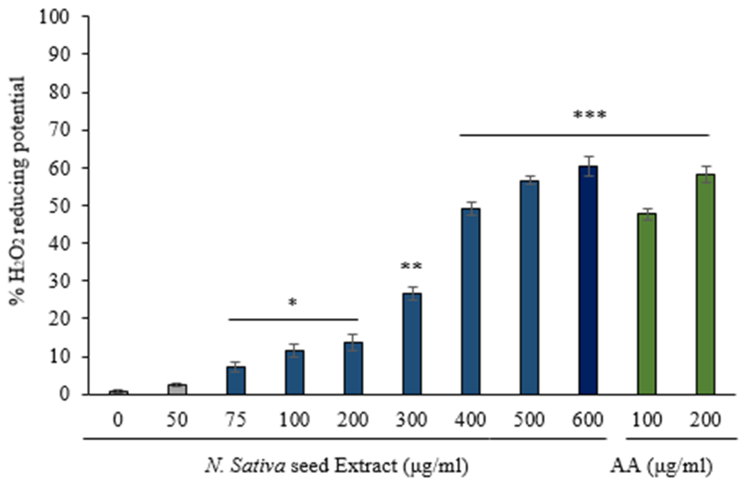
3.1. H2O2 Reducing Activity of N. sativa Seed Extract

In vitro antioxidant activity of N. sativa seed extract was evaluated by measuring the H2O2 reducing potential of the extract. Our data in Figure 1 shows that the extract possesses a significant H2O2 reducing activity as compared to the negative control (0 mg/mL of extract) in a concentration-dependent manner. The radical scavenging activity of Nigella extract at 400, 500, and 600 µg/mL has been found to be significantly comparable to the radical scavenging activity of ascorbic acid (p ≤ 0.001). However, the antioxidant activity of ascorbic acid was higher than the equivalent concentration of the extract.

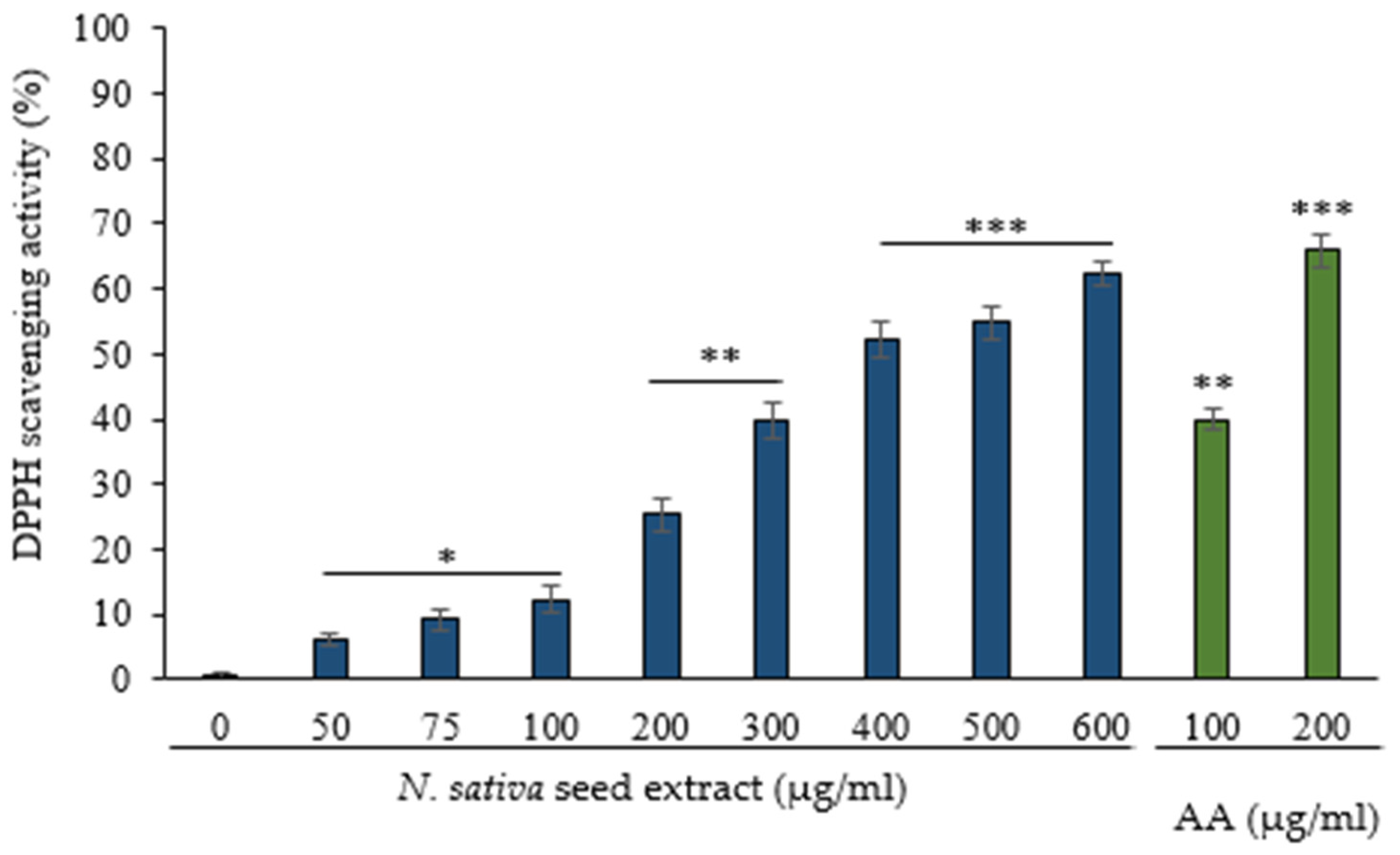
3.2. Scavenging of DPPH Radical by N. sativa Seed Extract

The DPPH assay was employed in the current investigation for the screening of the free radical-scavenging activity of the extract because it is sensitive even at low concentrations. Antioxidants react with DPPH free radical to form 1,1-diphenyl-2-picryl hydrazine, and the amount of discoloration reveals the ability of N. sativa to scavenge free radicals. From 50 mg/mL concentration, N. sativa seed extract showed significant DPPH radical scavenging activity in a concentration-dependent manner (Figure 2), and displayed 67.33% scavenging activity at 600 µg/mL. The radical scavenging potential of the extract at 200 and 300 mg/mL is significantly equivalent to (p ≤ 0.01) 100 mg/mL of ascorbic acid, whereas 400 to 600 mg/mL of extract is significantly equivalent to (p ≤ 0.001) 200 mg/mL of ascorbic acid.

3.3. Effect of N. sativa on Membrane Stabilization

The role of RBC membrane stabilization and inhibition in its lysis by N. sativa seed extract was investigated to measure the anti-inflammatory activity of N. sativa.

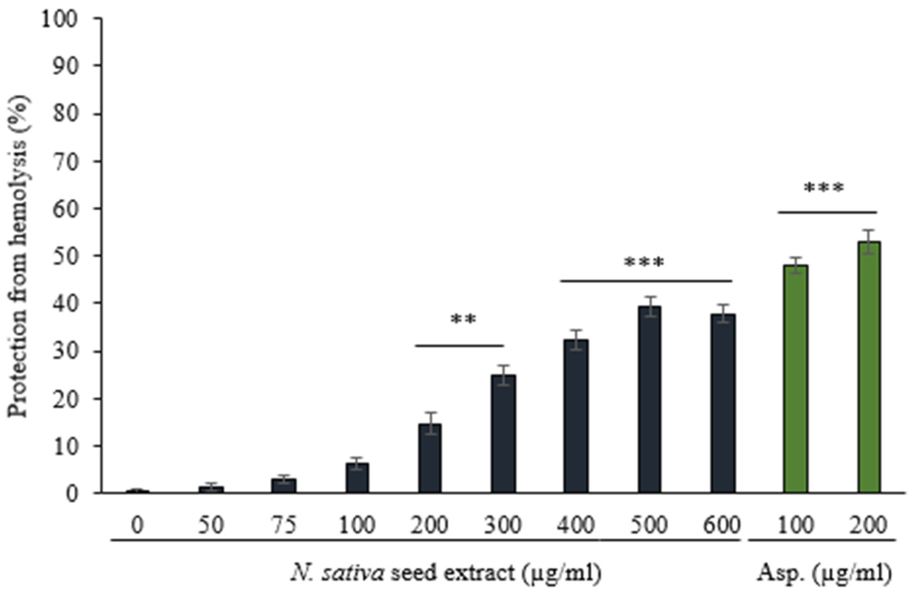
3.3.1. Protection against Hemolysis Caused by Heat

Methanol extract of N. sativa seed had substantial membrane stabilizing activity from 200 mg/mL concentrations (Figure 3). The maximal membrane stabilization was found to be 57.86 ± 0.81% at 600 µg/mL of N. Sativa extract against heat-induced hemolysis. This effect of extract from 400 to 600 µg/mL is significantly similar (p ≤ 0.001)) to the effect of a traditional allopathic anti-inflammatory medicine, aspirin (100 and 200 µg/mL). However, aspirin provided the best protection against hemolysis at 200 µg/mL concentration by 52.97 ± 0.77%.

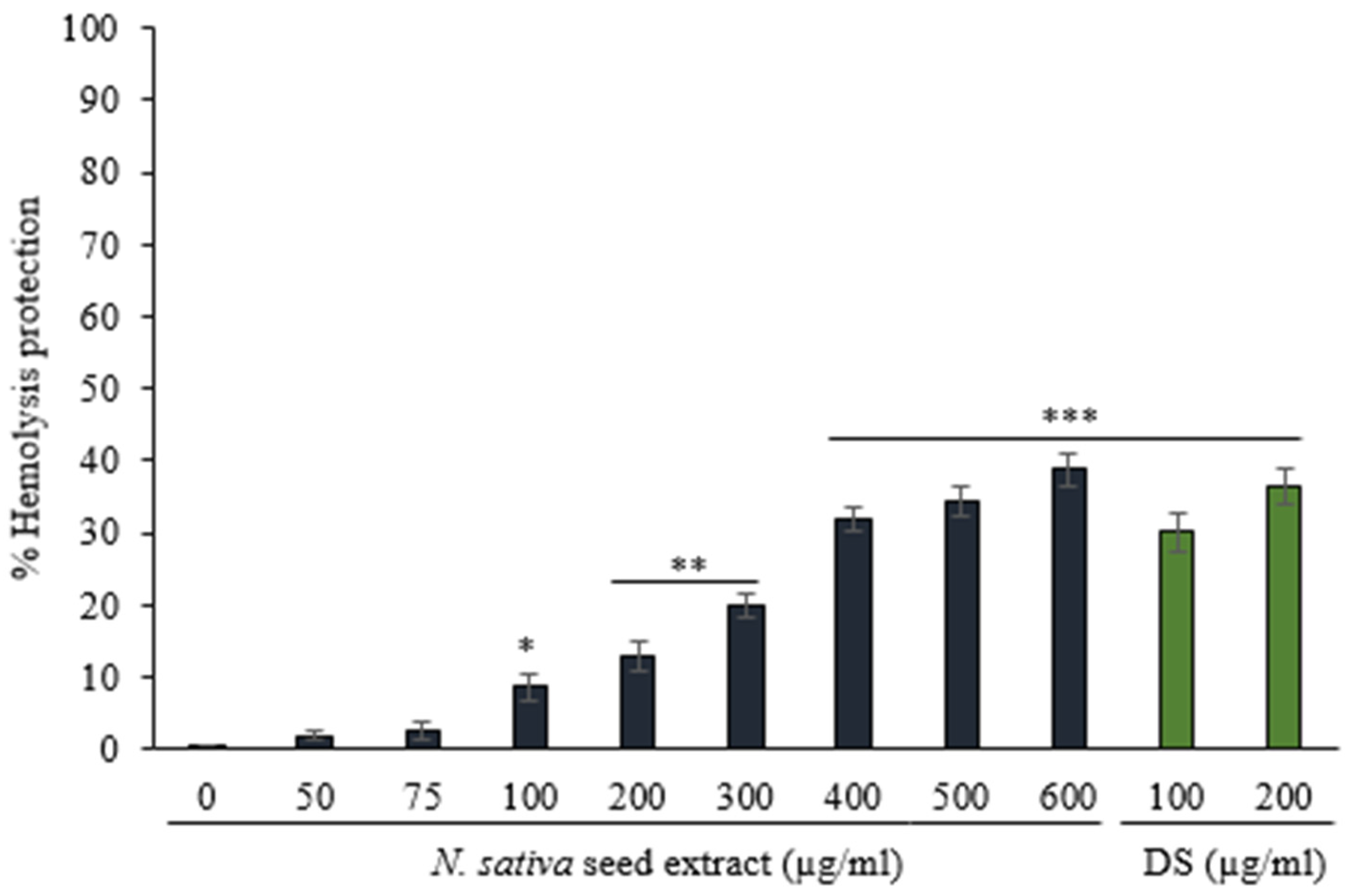
3.3.2. Protection against Hemolysis Caused by Hyposaline

N. sativa seed extract protect against hyposaline-induced hemolysis of RBCs in a dose-dependent manner from 100 mg/mL. (Figure 4). The protective effect of the extract (400–600 mg/mL) is equivalent to the standard reference drug diclofenac sodium (p ≤ 0.001). In the studied concentrations, the extract exhibited a maximal inhibition of 38.72%.

3.4. Screening of Anti-Glycating and AGEs Formation Inhibition Potentials

Hyperglycemia leads to glycation of various biomolecules, and AGEs formation [12,13] and AGEs are also linked to increased COVID-19 risk factors. Thus, it is important to investigate the role of N. sativa seeds in inhibition of glycation, formation of AGEs as well as glycation associated effects on biomolecules.

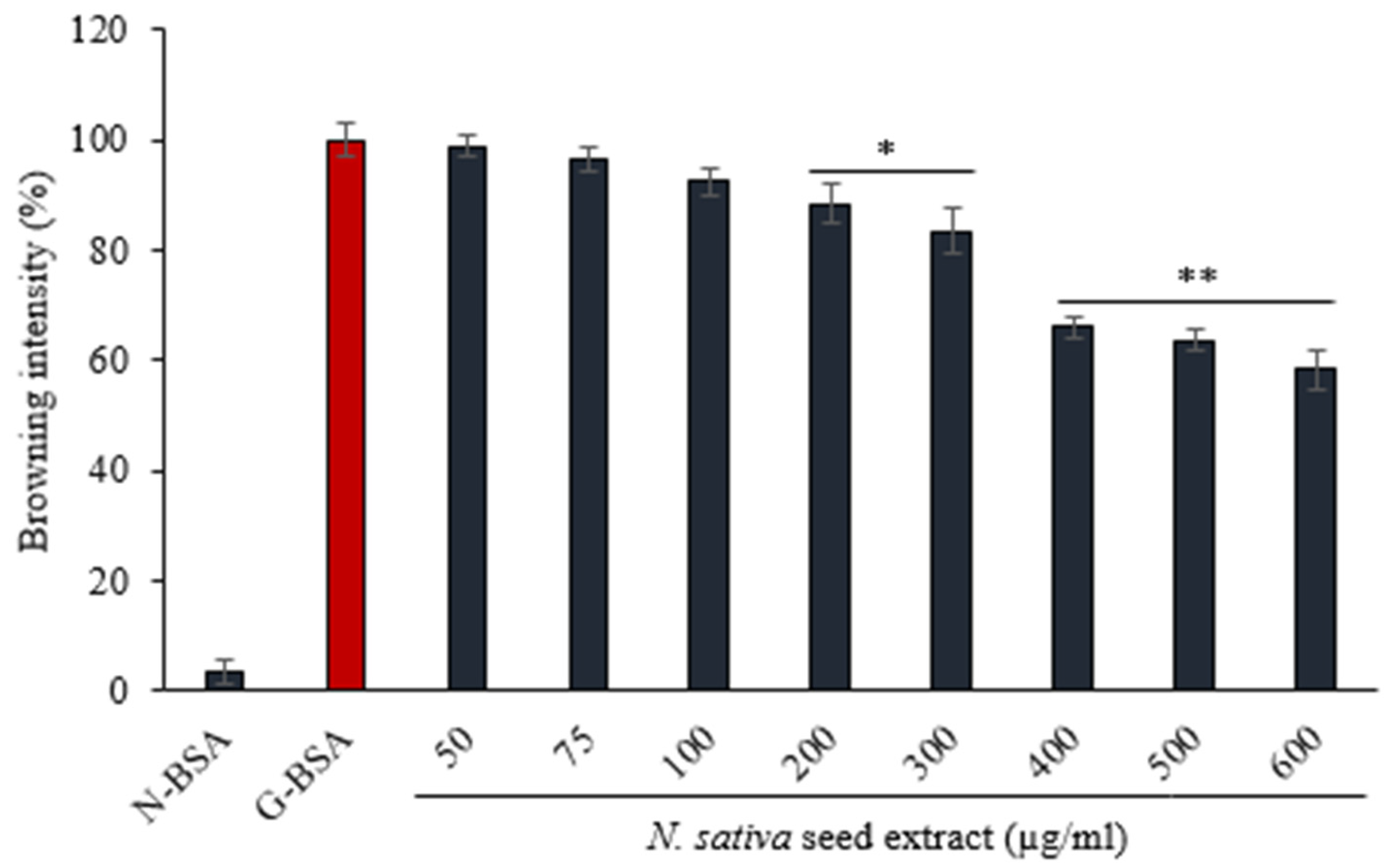
3.4.1. Effect of N. sativa on Browning Intensity

Glycation of various biomolecules, including proteins, can be detected by its browning intensity [27,29]. To determine the effect of N. sativa extract against glycation, the browning intensity of glycated BSA (G-BSA) was measured (Figure 5). The browning of BSA, glycated by glucose, in the absence of N. sativa extract was considered to be 100% (G-BSA: negative control). In our experiment, methanol extract of N. sativa significantly reduced the browning of BSA from 200 mg/mL concentration as compared to the negative control. The extract significantly prevented 43.91% browning at 600 µg/mL (p ≤ 0.01).

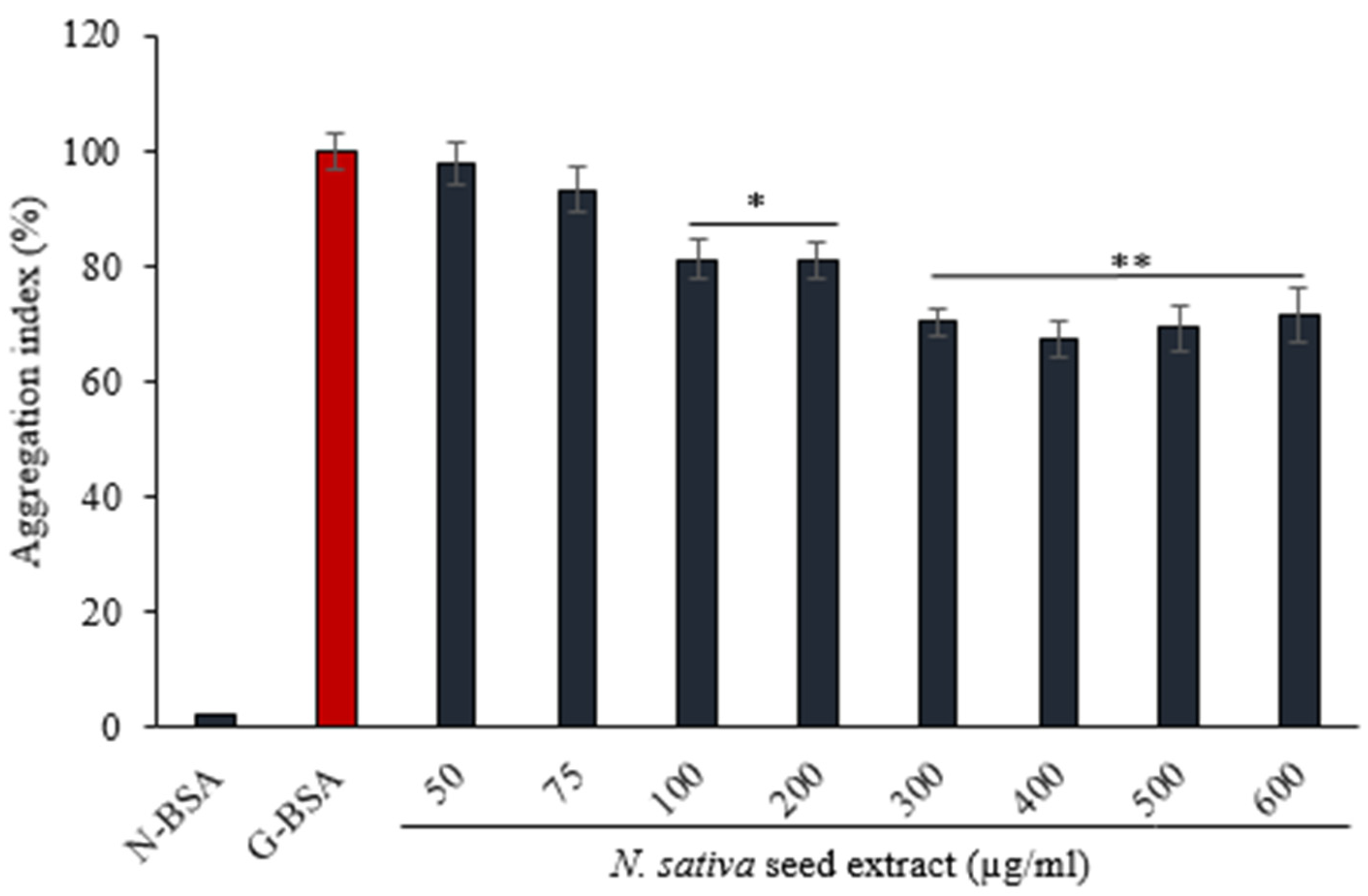
3.4.2. Effect of on N. sativa Seed Extract on Protein Aggregation Index

Non-enzymatic protein glycation leads to the formation of aggregates [27]. Incubation of BSA with glucose for 15 days results in the formation of misfolded aggregates, which is considered as 100% aggregation index (negative control). BSA samples incubated with glucose in the presence of N. sativa seed extract showed a reduced aggregation index as compared to the negative control (Figure 6). The effect of the extract is significant from 100 µg/mL (p ≤ 0.05) and the maximum effect is seen from 300–600 µg/mL.

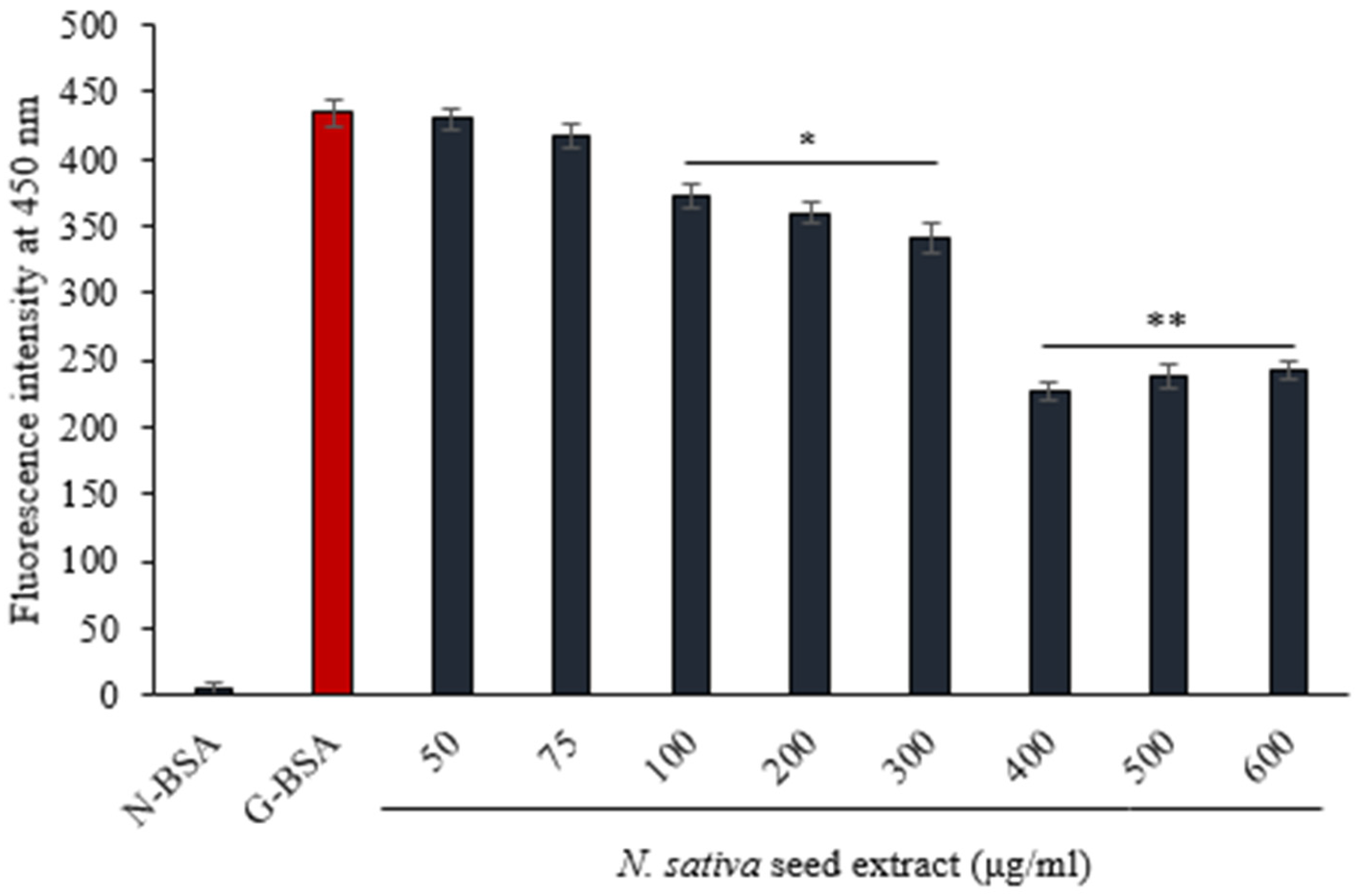
3.4.3. Fluorescence of AGE

The fluorescence intensity of BSA treated with glucose was at its peak at 450 nm. BSA incubated with glucose is considered as negative control. With the increase in the concentration of N. sativa seed extract, the co-incubated BSA showed a continuous decrease in AGEs specific fluorescence at 450 nm (Figure 7). The highest inhibition (52.18%) of AGE fluorescence was achieved at 400 µg/mL. Our findings suggest that N. sativa seed extract protects against the formation of AGEs due to glycation.

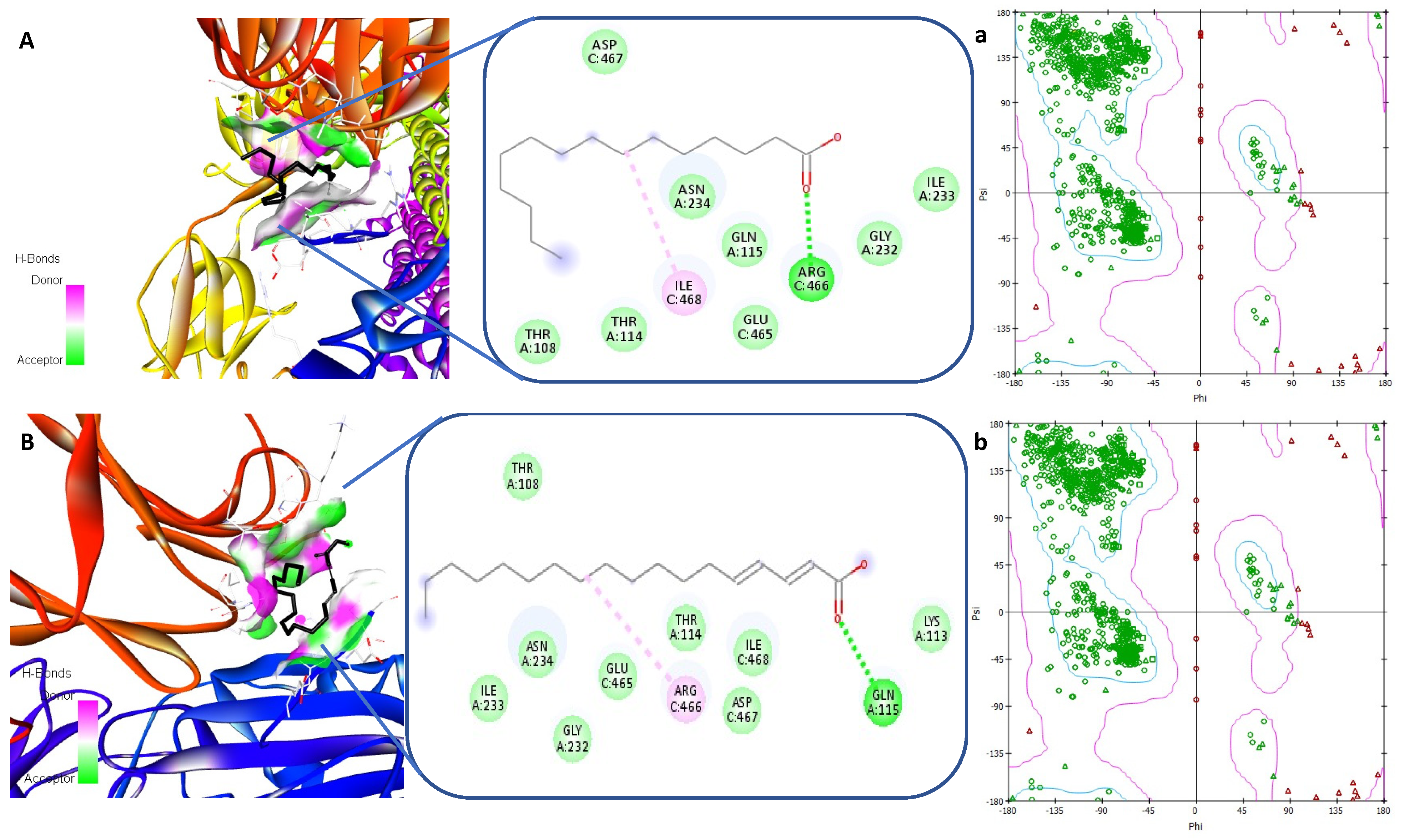
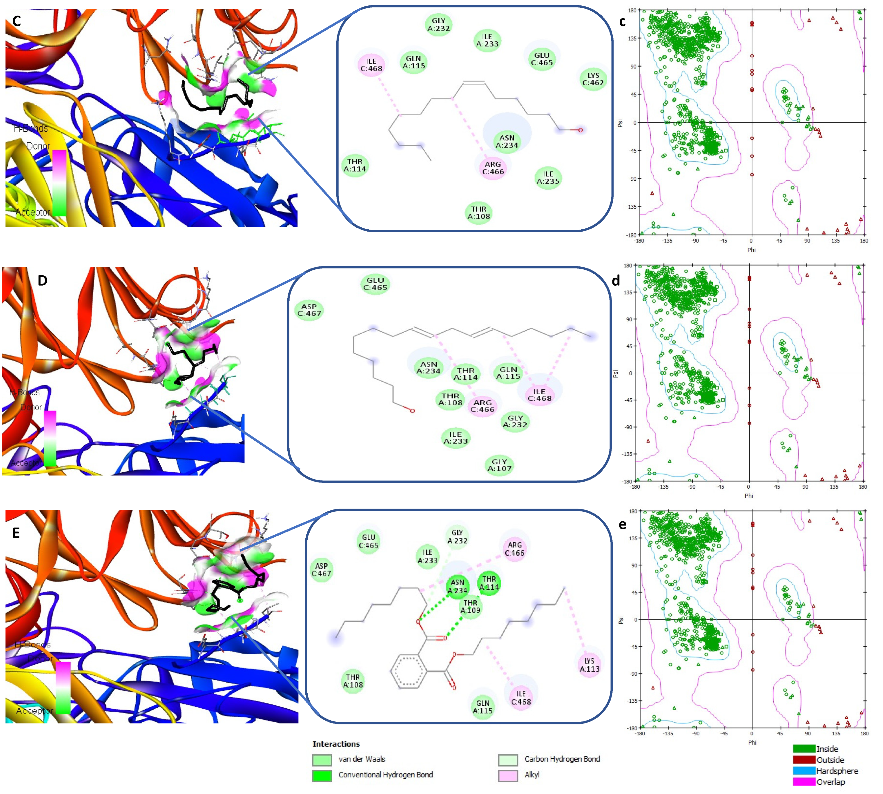
3.5. Receptor-Ligand Interaction Study by Molecular Docking

N. sativa was selected in this study because of its considerable documented therapeutic effects including antihypertensive, diuretics, antidiarrheal, appetite stimulant, analgesics, digestive, antibacterial, as well as its protective properties against neurological and mental illness, cardiovascular disorders, cancer, diabetes, and inflammatory conditions. Due to the diverse range of protective functions and their clinical implications in COVID-19, the involvement of certain compounds in N. sativa were investigated in this study for their therapeutic potential. The compounds used in this study were chosen because peaks of these compounds have been reported to be very significant in the GCMS profile of N. sativa seed extract as investigated by a previous researcher [36]. The selected phytoconstituents in this study including thymoquinone, dithymoquinone, nigellicine, pentadecanoic acid, octadecadienoic acid, (Z)6-pentadecen-1-ol, 9,12-octadecadien-1-ol, and dioctyl phthalate have been identified to be biologically active constituents of N. sativa extract [36].
However, we found that molecular interactions of thymoquinone, dithymoquinone, thymohydroquinone with necessary Coronavirus spike protein using autodoc vena are already published [36]. Therefore, the data linked is not being presented here due to similar findings. The binding energy and functional residues of the protein are shown in Table 1. Based on the results, all the ligands bind with protein. The active residues mainly found in these interactions are Asp467, Thr108, Thr114, Ile468, Asn234, Gln155, Glu465, Arg466, Gly232, and Ile233 as shown in Figure 8. Ramachandran plot analysis suggests that most residues are in favored regions [39]. The hydrogen bond, pi-sigma, pi-alkyl, and alkyl type interactions were the main forces that held the compounds in the protein pockets.

4. Discussion

SARS-CoV-2 virus infection rate and transmissibility are very high. Currently, there is no clinically authorized medication to treat COVID-19 and SARS-CoV-2. COVID-19 immunization aids in the prevention of COVID-19 infection. Several researchers are investigating the possibility of repurposing current antiviral medications to evaluate their efficiency against the SARS-CoV-2 virus. Meanwhile, understanding the pathogenic mechanism of SARS-CoV2 is critical for developing effective antiviral medicines or vaccines. Several studies have identified possible SARS-CoV-2 therapeutic medication targets based on the virus’s biology and clinical symptoms [40].
The imbalance of numerous defense systems in response to oxidative stress is one of the causative factors in the development of illnesses [27,35]. Khan and his group have suggested that it might be feasible to regulate or restrict the disease severity and improve prognosis by reducing oxidative stress [41]. The increased production of reactive oxygen species (ROS) and the depletion of antioxidant systems can impact COVID-19 incidence, progression, and severity. Interconnected pathways arising from the interplay of transcription factors with opposing effects explain the links between oxidative damage and inflammation. A well-balanced diet high in antioxidants is thought to have antioxidant properties, as well as the ability to avoid oxidative stress, inflammation, and immune system damage, and may be useful in the prevention, treatment, and management of COVID-19. The study revealed that N. sativa seed extract contains prominent radical scavenging and antioxidant activities that may be due to the presence of phenolic compounds. Therefore, N. sativa seed extract might have the ability to protect from oxidative stress-induced lung injury in COVID-19.
Glycation is a significant cause of oxidative stress [34,35]. Glycation has been shown to cause structural alterations in a variety of biomolecules, including proteins, and the formation of aggregates may occur as a result of the structural defect. The formation of protein aggregates has also been found to be further related to a variety of diseases and associated repercussions [42]. Human blood contains Albumin in large amounts, which is glycation-prone [34,35]. Our data reveal that a methanol extract of Nigella seed has a significant ability to prevent browning of BSA and hence, it has the ability to inhibit glycation of BSA.
As previously noted, AGEs through the interaction with RAGE and other molecules are the underlying cause of COVID-19 and related comorbidities, as well as non-enzymatic glycation, might stimulate ACE2 activity [18]. RAGE binds damage-associated molecular patterns and transduces the pathogenic signals in the vascular system, including blood vessels (in the diabetic population), and adipose tissue (in obesity-suffering people). Damage-associated molecular pattern-RAGE enrichment in these priming tissues, as well as in the lungs and heart during an active infection, is thought to play a role in SARS-CoV-2-induced broad tissue damage [43]. Our data shows that N. sativa seed extract decreases glycation and AGEs formation as indicated by a reduction in the browning intensity and aggregation index. AGEs specific fluorescence investigation confirmed the anti-glycating and AGEs formation inhibition potential of N. sativa seed extract.
The most numerous blood cells (80%) involved in oxygen transport from the lungs to the bodily tissues are red blood cells (RBCs), also known as erythrocytes. Furthermore, poor red blood cell (RBC) deformability may have a role in inflammation and hypoxia in COVID-19 patients [44]. Besides, COVID-19 vaccines have been reported to induce severe hemolysis in paroxysmal nocturnal hemoglobinuria [45]. These findings showed that N. sativa seed extract is potent to decrease hemolysis, which suggests that the membrane stability is a secondary mechanism of their anti-inflammatory activity. This impact might prevent neutrophils from releasing lysosomal material at the site of inflammation. In this regard, N. sativa seed can be an important therapeutic drug candidate as indicated by its RBC membrane-stabilizing potential in our study.
Inflammation in endothelial cells is thought to be triggered by oxidative stress, which is mediated via the production of adhesion molecules on the cell surface. Changes in the immune system and RAAS, as well as inflammatory condition, oxidative damage, and endothelial dysfunction in diabetes, have the potential to exaggerate the response triggered by SARS-CoV-2, which subsequently leads to pulmonary thrombosis, increased vascular permeability, and/or cytokine storm, resulting in respiratory failure [10]. N. sativa seed extract has shown antioxidant activity, and protection from denaturation of albumin, proteinase inhibition, as well as from hemolysis. Molecular docking research was conducted to find inhibitors of the SARS-CoV-2 virus; to confirm the chemical constituents of N. sativa seed extract which can possibly bind to viral protein and therefore can inhibit its entry to the human cell. SARS-CoV-2 spike glycoproteins promote entry into cells, and could be the main target of antibodies. It was shown that SARS-CoV-2 S uses ACE2 to enter cells. The receptor-binding domains of SARS-CoV-2 spike glycoprotein and SARS-CoV have been reported to bind to human ACE2 with similar affinities, correlating with the efficient spread of SARS-CoV-2 among humans [46]. The BNT162b2 vaccine candidate from Pfizer/BioNTech and the mRNA-1273 vaccine candidate from Moderna are both based on stabilized mRNA encoding prefusion SARS-2-S that can be created after the mRNA is introduced into the human cell and translated. SARS-2-S is split into two subunits, S1 and S2, with S1 functioning as a receptor binder and S2 serving as a membrane fusion protein [47]. In our molecular docking analysis, we took five bioactive compounds of N. sativa seed extract. We investigated the possible interaction of these ligands with Coronavirus spike glycoprotein (6VXX) and proved their interactions. Our docking study shows that N. sativa seed extract has a broad range of affinity for angiotensin binding spike glycoproteins of SARS-CoV-2. As a result, N. sativa seed extract might be able to subsequently block the angiotensin binding spike proteins and can stop the inflammatory/vascular impairment. Similar to our docking analysis, various research groups have verified an optimal interaction between N. sativa compounds and SARS-CoV-2 receptors and have predicted N. sativa as a plausible inhibitor to disrupt viral-host interactions. Shaikh et al. [48] have conducted a similar docking study using N. sativa phytoconstituents as ligands and receptor-binding site of SARS-CoV-2 (PDB ID: 2AJF). The study reported that dithymoquinone, thy-mohydroquinone, thymol, and thymoquinone from this medicinal plant might inhibit COVID-19 infection giving the same or better energy score compared to chloroquine [48]. Ahmad and group found that dithymoquinone could interact with the SARS-CoV-2:ACE2 interface with a high binding affinity [36]. Koshak et al., have confirmed that N. sativa supplementation was linked to a quicker recovery of symptoms than standard therapy alone for individuals with moderate COVID-19 infection [49].
Other medicinal plants such as Turmeric possess beneficial features such as analgesic, anti-inflammatory, antiviral, and antimicrobial activities, and hence are extensively used in Ayurveda and Siddha medical systems. The turmeric-derived compounds were computationally screened to evaluate their binding affinity with the RdRp of SARS-CoV-2. A set of six turmeric-derived compounds was docked with the SARS-CoV-2 RdRp-RNA; Diacetylcurcumin (69.54 kcal/mol), Curcumin (64.10 kcal/mol), Tetrahydrocurcumin (62.1 kcal/mol), Dimethylcurcumin (58.99 kcal/mol), Demethoxycurcumin (52.95 kcal/mol), Bis-demethoxycurcumin (51.15 kcal/mol) [24].
In 2022, Anwar and group confirmed the beneficial health effects of ajwa against the pathophysiology of various diseases that might be involved in the severity and mortality linked with SARS-CoV-2 infection in diabetic conditions through in vitro studies [50]. This research group has further suggested that agents with membrane-stabilizing properties and inhibiting potential of protein denaturation [27,30] might be very beneficial against inflammatory diseases like COVID-19 [50]. The health beneficial effects of traditional Chinese medicine (TCM) have been published in response to the epidemics of COVID-19 and SARS-CoV. It is suggested that TCM reduces the severity of COVID-19 through numerous mechanisms [51], such as reducing NF-B signaling and 3CLpro activity [52]. In this respect, our in vitro investigations on the antioxidant, anti-inflammatory, antiglycating, and anti-AGEs production properties of N. sativa seed extract suggest that Nigella sativa seed might be recommended for the protection, treatment, and management of COVID-19-associated comorbidities in diabetic conditions.
In silico study is an excellent tool to screen a drug because it can predict the protein-ligand binding site, can reduce laboratory experiments, and can accelerate the drug discovery process. Virtual screening to uncover potential drug leads using molecular docking simulations has been extremely successful over the last decade. However, in terms of using such compounds as medicines for specific indications and/or diseases, conventional, model-dependent screening of compounds with limited targets has significant limitations. Furthermore, when selecting a prospective therapeutic lead in a model-dependent strategy, the interactions of the compounds with all biomolecules, cells, and tissues (i.e., systems biology) are not taken into account, and such non-systems biology approaches may be leading to the existing dried-up drug development pipelines [53].

5. Conclusions

In a summary, we may infer that the Nigella sativa seeds are involved in a variety of health-promoting mechanisms that may reduce the severity of many diseases, as well as its phytoconstituents, may interact with spike proteins of COVID-19. Besides, the main cause of death among COVID-19 patients is acute respiratory distress syndrome, linked with a storm of pro-inflammatory cytokines. The in silico molecular docking analysis proves the binding interactions between bioactive compounds of N. sativa seed extract with SARS-CoV-2 spike glycoprotein. Given the in vitro anti-inflammatory and antioxidant effects of N. sativa seed extract as well as the protective effects on glycation and AGEs formation, this herb may be useful in the treatment and management of COVID-19 and its complications in diabetics. Clinical trials, on the other hand, are necessary to back up its efficacy under in vivo conditions. If verified on experimental models by in vivo investigations, various phytoconstituents used in this study might be utilized to treat COVID-19 and might act as a path for the development of more potent natural antivirals against COVID-19 in the future. Further, in vitro experiments; molecular simulations are required to validate these in silico results. The limitations like interactions of the compounds with all biomolecules, cells, and tissues (i.e., systems biology) are further needed to be adjusted before selecting Nigella as a prospective therapeutic lead.

Author Contributions

Conceptualization, S.S., M.W.A.K.; methodology, S.S., S.R., M.W.A.K., M.S., S.K.; formal analysis, S.S., M.K., S.K., M.W.A.K., F.O.A.; investigation, S.S., S.R., M.W.A.K.; resources, S.S., M.W.A.K., M.S.; data curation, S.S., M.W.A.K., M.R., S.K.; writing—original draft preparation, S.S., M.W.A.K.; writing—review and editing, S.R., M.R., M.K., S.K., F.O.A.; supervision, S.S.; funding acquisition, S.S. All authors have read and agreed to the published version of the manuscript.

Funding

This research has been funded by the Scientific Research Deanship at the University of Ha’il—Saudi Arabia through project number RG-20 077.

Institutional Review Board Statement

This study protocol was reviewed and approved by Research Ethics Committee (REC) at the University of Hail dated: 12/04/2021 and approved by university president letter number 42/5/44372 dated 12/04/2021.

Informed Consent Statement

Written informed consent was obtained from participants to participate in the study.

Data Availability Statement

Not applicable.

Conflicts of Interest

The authors have no conflicts of interest to declare.

References

  1. Guan, W.J.; Ni, Z.Y.; Hu, Y.; Liang, W.H.; Qu, C.Q.; He, J.X.; Liu, L.; Shan, H.; Lei, C.L.; Hui, D.S.C.; et al. Clinical characteristics of coronavirus disease 2019 in China. N. Engl. J. Med. 2020, 382, 1708–1720. [Google Scholar] [CrossRef] [PubMed]
  2. Ragab, D.; Salah Eldin, H.; Taeimah, M.; Khattab, R.; Salem, R. The COVID-19 Cytokine Storm; What We Know So Far. Front. Immunol. 2020, 11, 1446. [Google Scholar] [CrossRef] [PubMed]
  3. Sherwani, S.; Khan, M.W.A. Cytokine Response in SARS-CoV-2 Infection in the Elderly. J. Inflamm. Res. 2020, 13, 737–747. [Google Scholar] [CrossRef] [PubMed]
  4. Kubánková, M.; Hohberger, B.; Hoffmanns, J.; Fürst, J.; Herrmann, M.; Guck, J.; Kräter, M. Physical phenotype of blood cells is altered in COVID-19. Biophys. J. 2021, 120, 2838–2847. [Google Scholar] [CrossRef]
  5. Shang, J.; Wan, Y.; Luo, C.; Ye, G.; Geng, Q.; Auerbach, A.; Li, F. Cell entry mechanisms of SARS-CoV-2. Proc. Natl. Acad. Sci. USA 2020, 117, 11727–11734. [Google Scholar] [CrossRef] [PubMed]
  6. Zhang, H.; Penninger, J.M.; Li, Y.; Zhong, N.; Slutsky, A.S. Angiotensin-converting enzyme 2 (ACE2) as a SARS-CoV-2 receptor: Molecular mechanisms and potential therapeutic target. Intensive Care Med. 2020, 46, 586–590. [Google Scholar] [CrossRef] [PubMed] [Green Version]
  7. Salamanna, F.; Maglio, M.; Landini, M.P.; Fini, M. Body Localization of ACE-2: On the Trail of the Keyhole of SARS-CoV-2. Front. Med. (Lausanne) 2020, 7, 594495. [Google Scholar] [CrossRef] [PubMed]
  8. Jarlhelt, I.; Nielsen, S.K.; Jahn, C.X.H.; Hansen, C.B.; Pérez-Alós, L.; Rosbjerg, A.; Bayarri-Olmos, R.; Skjoedt, M.O.; Garred, P. SARS-CoV-2 Antibodies Mediate Complement and Cellular Driven Inflammation. Front. Immunol. 2021, 12, 767981. [Google Scholar] [CrossRef]
  9. Thomas, T.; Stefanoni, D.; Dzieciatkowska, M.; Issaian, A.; Nemkov, T.; Hill, R.C.; Francis, R.O.; Hudson, K.E.; Buehler, P.W.; Zimring, J.C.; et al. Evidence of Structural Protein Damage and Membrane Lipid Remodeling in Red Blood Cells from COVID-19 Patients. J. Proteome Res. 2020, 19, 4455–4469. [Google Scholar] [CrossRef]
  10. Roberts, J.; Pritchard, A.L.; Treweeke, A.T.; Rossi, A.G.; Brace, N.; Cahill, P.; MacRury, S.M.; Wei, J.; Megson, I.L. Why Is COVID-19 More Severe in Patients With Diabetes? The Role of Angiotensin-Converting Enzyme 2, Endothelial Dysfunction and the Immunoinflammatory System. Front. Cardiovasc. Med. 2021, 7, 629933. [Google Scholar] [CrossRef]
  11. Maiuolo, J.; Mollace, R.; Gliozzi, M.; Musolino, V.; Carresi, C.; Paone, S.; Scicchitano, M.; Macrì, R.; Nucera, S.; Bosco, F.; et al. The Contribution of Endothelial Dysfunction in Systemic Injury Subsequent to SARS-CoV-2 Infection. Int. J. Mol. Sci. 2020, 21, 9309. [Google Scholar] [CrossRef] [PubMed]
  12. Nabi, R.; Alvi, S.S.; Shah, A.; Chaturvedi, C.P.; Faisal, M.; Alatar, A.A.; Ahmad, S.; Khan, M.S. Ezetimibe attenuates experimental diabetes and renal pathologies via targeting the advanced glycation, oxidative stress and AGE-RAGE signalling in rats. Arch. Physiol. Biochem. 2021, 29, 1–16. [Google Scholar] [CrossRef] [PubMed]
  13. Ahmad, S.; Akhter, F.; Shahab, U.; Rafi, Z.; Khan, M.S.; Nabi, R.; Khan, M.S.; Ahmad, K.; Ashraf, J.M.; Moinuddin. Do all roads lead to the Rome? The glycation perspective! Semin. Cancer Biol. 2018, 49, 9–19. [Google Scholar] [CrossRef] [PubMed]
  14. Ahmad, S.; Uddin, M.; Habib, S.; Shahab, U.; Alam, K.; Ali, A. Autoimmune response to AGE modified human DNA: Implications in type 1 diabetes mellitus. J. Clin. Transl. Endocrinol. 2014, 1, 66–72. [Google Scholar] [CrossRef] [PubMed] [Green Version]
  15. Khan, M.A.; Anwar, S.; Aljarbou, A.N.; Al-Orainy, M.; Aldebasi, Y.H.; Islam, S.; Younus, H. Protective effect of thymoquinone on glucose or methylglyoxal-induced glycation of superoxide dismutase. Int. J. Biol. Macromol. 2014, 65, 16–20. [Google Scholar] [CrossRef] [PubMed]
  16. Beltrán-García, J.; Osca-Verdegal, R.; Pallardó, F.V.; Ferreres, J.; Rodríguez, M.; Mulet, S.; Sanchis-Gomar, F.; Carbonell, N.; García-Giménez, J.L. Oxidative Stress and Inflammation in COVID-19-Associated Sepsis: The Potential Role of Anti-Oxidant Therapy in Avoiding Disease Progression. Antioxidants 2020, 9, 936. [Google Scholar] [CrossRef]
  17. De la Cruz-Enríquez, J.; Rojas-Morales, E.; Ruíz-García, M.G.; Tobón-Velasco, J.C.; Jiménez-Ortega, J.C. SARS-CoV-2 induces mitochondrial dysfunction and cell death by oxidative stress/inflammation in leukocytes of COVID-19 patients. Free Radic. Res. 2021, 55, 982–995. [Google Scholar] [CrossRef]
  18. Sartore, G.; Ragazzi, E.; Faccin, L.; Lapolla, A. A role of glycation and methylation for SARS-CoV-2 infection in diabetes? Med. Hypotheses 2020, 144, 110247. [Google Scholar] [CrossRef]
  19. Anwar, S.; Khan, S.; Almatroudi, A.; Khan, A.A.; Alsahli, M.A.; Almatroodi, S.A.; Rahmani, A.H. A review on mechanism of inhibition of advanced glycation end products formation by plant derived polyphenolic compounds. Mol. Biol. Rep. 2021, 48, 787–805. [Google Scholar] [CrossRef]
  20. Younus, H.; Anwar, S. Prevention of non-enzymatic glycosylation (glycation): Implication in the treatment of diabetic complication. Int. J. Health Sci. (Qassim) 2016, 10, 261–277. [Google Scholar] [CrossRef]
  21. Ahmad, A.; Husain, A.; Mujeeb, M.; Alam Khan, S.; Najmi, A.K.; Siddique, N.A.; Damanhouri, Z.A.; Anwar, F. A review on therapeutic potential of Nigella sativa: A miracle herb. Asian Pac. J. Trop. Biomed. 2013, 3, 337–352. [Google Scholar] [CrossRef] [Green Version]
  22. Hadi, V.; Pahlavani, N.; Malekahmadi, M.; Alam Khan, S.; Najmi, A.K.; Siddique, N.A.; Damanhouri, Z.A.; Anwar, F. Nigella sativa in controlling Type 2 diabetes, cardiovascular, and rheumatoid arthritis diseases: Molecular aspects. J. Res. Med. Sci. 2021, 26, 20. [Google Scholar] [PubMed]
  23. Norman, G.A.V. Drugs, Devices, and the FDA: Part 1: An Overview of Approval Processes for Drugs. JACC Basic Transl. Sci. 2016, 1, 170–179. [Google Scholar]
  24. Singh, R.; Bhardwaj, V.K.; Purohit, R. Potential of turmeric-derived compounds against RNA-dependent RNA polymerase of SARS-CoV-2: An in-silico approach. Comp. Biol. Med. 2021, 139, 104965. [Google Scholar] [CrossRef] [PubMed]
  25. Singh, R.; Bhardwaj, V.K.; Das, P.; Purohit, R. A computational approach for rational discovery of inhibitors for non-structural protein 1 of SARS-CoV-2. Comp. Biol. Med. 2021, 135, 104555. [Google Scholar] [CrossRef]
  26. Singh, R.; Bhardwaj, V.K.; Das, P.; Bhattacherjee, D.; Zyryanov, G.V.; Purohit, R. Benchmarking the ability of novel compounds to inhibit SARS-CoV-2 main protease using steered molecular dynamics simulations. Comp. Biol. Med. 2022, 146, 105572. [Google Scholar] [CrossRef]
  27. Anwar, S.; Almatroudi, A.; Allemailem, K.S.; Jacob-Joseph, R.; Khan, A.A.; Rahmani, A.H. Protective Effects of Ginger Extract against Glycation and Oxidative Stress-Induced Health Complications: An In Vitro Study. Processes 2020, 8, 468. [Google Scholar] [CrossRef]
  28. Khan, M.W.A.; Otaibi, A.A.; Alhumaid, A.F.M.; Alsukaibi, A.K.D.; Alshamari, A.K.; Alshammari, E.M.; Al-Zahrani, S.A.; Almudyani, A.Y.M.; Sherwani, S. Garlic Extract: Inhibition of Biochemical and Biophysical Changes in Glycated HSA. Appl. Sci. 2021, 11, 11028. [Google Scholar] [CrossRef]
  29. Khan, M.W.A.; Otaibi, A.A.; Alsukaibi, A.K.D.; Alshammari, E.M.; Al-Zahrani, S.A.; Sherwani, S.; Khan, W.A.; Saha, R.; Verma, S.R.; Ahmed, N. Biophysical, Biochemical, and Molecular Docking Investigations of Anti-Glycating, Antioxidant, and Protein Structural Stability Potential of Garlic. Molecules 2022, 27, 1868. [Google Scholar] [CrossRef]
  30. Anwar, S.; Almatroudi, A.; Alsahli, M.A.; Khan, M.A.; Khan, A.A.; Rahmani, A.H. Natural Products: Implication in Cancer Prevention and Treatment through Modulating Various Biological Activities. Anticancer Agents Med. Chem. 2020, 20, 2025–2040. [Google Scholar] [CrossRef]
  31. Chanda, S.; Juvekar, A. In vitro anti-inflammatory activity of syringic acid. Int. J. Pharm. Pharm. Sci. 2019, 11, 71–73. [Google Scholar] [CrossRef]
  32. Brownlee, M.; Vlassara, H.; Kooney, A.; Ulrich, P.; Cerami, A. Aminoguanidine prevents diabetes-induced arterial wall protein cross-linking. Science 1986, 232, 1629–1632. [Google Scholar] [CrossRef] [PubMed]
  33. Kumar, D.; Ali, A. Antiglycation and antiaggregation potential of thymoquinone. Nat. Volatiles Essent. Oils 2019, 6, 25–33. [Google Scholar]
  34. Khan, M.W.A.; Rasheed, Z.; Khan, W.A.; Ali, R. Biochemical, biophysical, and thermodynamic analysis of in vitro glycated human serum albumin. Biochemistry 2007, 72, 146–152. [Google Scholar] [PubMed]
  35. Khan, M.W.A.; Otaibi, A.A.; Al-Zahrani, S.A.; Alshammari, E.M.; Haque, A.; Alouffi, S.; Khan, W.A.; Khan, S.N. Experimental and theoretical insight into resistance to glycation of bovine serum albumin. J. Mol. Struc. 2021, 1230, 129645. [Google Scholar] [CrossRef]
  36. Ahmad, S.; Abbasi, H.W.; Shahid, S.; Gul, S.; Abbasi, S.W. Molecular docking, simulation and MM-PBSA studies of nigella sativa compounds: A computational quest to identify potential natural antiviral for COVID-19 treatment. J. Biomol. Struct. Dyn. 2021, 39, 4225–4233. [Google Scholar] [CrossRef] [PubMed]
  37. Tiruppur Venkatachallam, S.K.; Pattekhan, H.; Divakar, S.; Kadimi, U.S. Chemical composition of Nigella sativa L. seed extracts obtained by supercritical carbon dioxide. J. Food Sci. Technol. 2010, 47, 598–605. [Google Scholar] [CrossRef] [Green Version]
  38. Morris, G.M.; Huey, R.; Lindstrom, W.; Sanner, M.F.; Belew, R.L.; Goodsell, D.S.; Olson, A.J. AutoDock4 and AutoDockTools4: Automated docking with selective receptor flexibility. J. Comput. Chem. 2009, 30, 2785–2791. [Google Scholar] [CrossRef] [Green Version]
  39. Ramachandran, G.N.; Ramakrishnan, C.; Sasisekharan, V. Stereochemistry of polypeptide chain configurations. J. Mol. Biol. 1963, 7, 95–99. [Google Scholar] [CrossRef]
  40. Swargiary, A.; Mahmud, S.; Saleh, M.A. Screening of phytochemicals as potent inhibitor of 3-chymotrypsin and papain-like proteases of SARS-CoV2: An in silico approach to combat COVID-19. J. Biomol. Struct. Dyn. 2022, 40, 2067–2081. [Google Scholar] [CrossRef] [PubMed]
  41. Alouffi, S.; Sherwani, S.; Al-Mogbel, M.S.; Sherwani, M.K.A.; Khan, M.W.A. Depression and Smoking Augment the Production of Circulating Autoantibodies against Glycated HSA in Rheumatoid Arthritis Patients. Int. Arch. Allergy Immunol. 2018, 177, 170–180. [Google Scholar] [CrossRef] [PubMed]
  42. Bouma, B.; Kroon-Batenburg, L.M.; Wu, Y.P.; Brünjes, B.; Posthuma, G.; Kranenburg, O.; de Groot, P.G.; Voest, E.E.; Gebbink, M.F. Glycation induces formation of amyloid cross-beta structure in albumin. J. Biol. Chem. 2003, 278, 41810–41819. [Google Scholar] [CrossRef] [PubMed] [Green Version]
  43. Roy, D.; Ramasamy, R.; Schmidt, A.M. Journey to a Receptor for Advanced Glycation End Products Connection in Severe Acute Respiratory Syndrome Coronavirus 2 Infection. Arterioscler. Thromb. Vasc. Biol. 2021, 41, 614–627. [Google Scholar] [CrossRef] [PubMed]
  44. Akhter, N.; Ahmad, S.; Alzahrani, F.A.; Dar, S.A.; Wahid, M.; Haque, S.; Bhatia, K.; Sr Almalki, S.; Alharbi, R.A.; Sindi, A.A.A. Impact of COVID-19 on the cerebrovascular system and the prevention of RBC lysis. Eur. Rev. Med. Pharmacol. Sci. 2020, 24, 10267–10278. [Google Scholar] [PubMed]
  45. Gerber, G.F.; Yuan, X.; Yu, J.; Cher, B.A.Y.; Braunstein, E.M.; Chaturvedi, S.; Brodsky, R.A. COVID-19 vaccines induce severe hemolysis in paroxysmal nocturnal hemoglobinuria. Blood 2021, 137, 3670–3673. [Google Scholar] [CrossRef] [PubMed]
  46. Walls, A.C.; Park, Y.J.; Tortorici, M.A.; Wall, A.; McGuire, A.T.; Veesler, D. Structure, Function, and Antigenicity of the SARS-CoV-2 Spike Glycoprotein. Cell 2020, 181, 281–292.e6, Erratum in: 2020, 183, 1735. [Google Scholar] [CrossRef]
  47. Xia, X. Domains and Functions of Spike Protein in SARS-CoV-2 in the Context of Vaccine Design. Viruses 2021, 13, 109. [Google Scholar] [CrossRef]
  48. Shaikh, Y.I.; Shaikh, V.S.; Ahmed, K.; Nazeruddin, G.M.; Pathan, H.M. The revelation of various compounds found in Nigella sativa L.(Black Cumin) and their possibility to inhibit COVID-19 infection based on the molecular docking and physical properties. Eng. Sci. 2020, 11, 31–35. [Google Scholar] [CrossRef]
  49. Koshak, A.E.; Koshak, E.A.; Mobeireek, A.F.; Badawi, M.A.; Wali, S.O.; Malibary, H.M.; Atwah, A.F.; Alhamdan, M.M.; Almalki, R.A.; Madani, T.A. Nigella sativa for the treatment of COVID-19: An open-label randomized controlled clinical trial. Complement. Ther. Med. 2021, 61, 102769. [Google Scholar] [CrossRef]
  50. Anwar, S.; Raut, R.; Alsahli, M.A.; Almatroudi, A.; Alfheeaid, H.; Alzahrani, F.M.; Khan, A.A.; Allemailem, K.S.; Almatroodi, S.A.; Rahmani, A.H. Role of Ajwa Date Fruit Pulp and Seed in the Management of Diseases through In Vitro and In Silico Analysis. Biology 2022, 11, 78. [Google Scholar] [CrossRef]
  51. Yang, Y.; Islam, M.S.; Wang, J.; Li, Y.; Chen, X. Traditional Chinese Medicine in the Treatment of Patients Infected with 2019-New Coronavirus (SARS-CoV-2): A Review and Perspective. Int. J. Biol. Sci. 2020, 16, 1708–1717. [Google Scholar] [CrossRef] [PubMed]
  52. Du, A.; Zheng, R.; Disoma, C.; Li, S.; Chen, Z.; Li, S.; Liu, P.; Zhou, Y.; Shen, Y.; Liu, S.; et al. Epigallocatechin-3-gallate, an active ingredient of Traditional Chinese Medicines, inhibits the 3CLpro activity of SARS-CoV-2. Int. J. Biol. Macromol. 2021, 176, 1–12. [Google Scholar] [CrossRef] [PubMed]
  53. Minie, M.; Chopra, G.; Sethi, G.; Horst, J.; White, G.; Roy, A.; Hatti, K.; Samudrala, R. CANDO and the infinite drug discovery frontier. Drug Discov. Today 2014, 9, 1353–1363. [Google Scholar]
Figure 1. H2O2 reducing potential of N. sativa seed extract. Ascorbic acid (AA) was used as the positive control. The results are presented as means ± SEM (n = 3). The statistical analysis was performed by using ANOVA (* p ≤ 0.05, ** p ≤ 0.01, *** p ≤ 0.001).
Processes 10 01346 g001
Figure 2. DPPH scavenging activity (%) of N. sativa seed methanolic extract. Ascorbic acid (AA) was used as the positive control. The results are presented as means ± SEM (n = 3. The statistical analysis was performed by using ANOVA (* p ≤ 0.05, ** p ≤ 0.01, *** p ≤ 0.001).
Processes 10 01346 g002
Figure 3. Effect of N. sativa seed extract on heat induced hemolysis. Aspirin was used as the positive control. The results are presented as means ± SEM (n = 3, p < 0.05). The statistical analysis was performed by using ANOVA (** p ≤ 0.01, *** p ≤ 0.001).
Processes 10 01346 g003
Figure 4. Effect of N. sativa seed extract on hyposaline-induced hemolysis diclofenac sodium was used as the positive control. The results are presented as means ± SEM (n = 3. The statistical analysis was performed by using ANOVA (* p ≤ 0.05, ** p ≤ 0.01, *** p ≤ 0.001).
Processes 10 01346 g004
Figure 5. Inhibition of browning of glycated BSA by N. sativa seed extract: Native BSA incubated alone for 15 days. G-BSA: BSA incubated with glucose for 15 days and considered as negative control. The results are presented as means ± SEM (n = 3). The statistical analysis was performed by using ANOVA (* p ≤ 0.05, ** p ≤ 0.01).
Processes 10 01346 g005
Figure 6. Effect of N. sativa seed extract on aggregation index. N-BSA: Native BSA incubated alone for 15 days. G-BSA: BSA incubated with glucose for 15 days and considered as negative control. The results are presented as means ± SEM (n = 3). The statistical analysis was performed by using ANOVA (* p ≤ 0.05, ** p ≤ 0.01).
Processes 10 01346 g006
Figure 7. Effect of N. sativa seed extract on AGEs formation. N-BSA: Native BSA incubated alone for 15 days. G-BSA: BSA incubated with glucose for 15 days and considered as negative control. The results are presented as means ± SEM (n = 3, (* p ≤ 0.01, ** p <0.05).
Processes 10 01346 g007
Figure 8. The molecular docking studies of Spike protein S (6VXX) with active constituents of N. sativa extract with ligands (1) Pentadecanoic acid (A); (2) Octadecadienoic acid (B); (3) (Z)6-Pentadecen-1-ol (C); (4) 9,12-Octadecadien-1-ol (D), and (5) Dioctyl phthalate (E). The Ramachandran plots of respective interactions are provided with each ligand-protein interaction (ae).
Processes 10 01346 g008aProcesses 10 01346 g008b
Table 1. Details of molecular docking study; the ligands, binding energy, and interacting amino acids interacting with protein.
LigandsPubChem
CID
Spike Protein S (6VXX)
Binding Energy (kcal/mol)Interacting Amino Acids
Pentadecanoic acid13849−3.4Asp467, Thr108, Thr114, Ile468, Asn234, Gln155, Glu465, Arg466, Gly232, Ile233
Octadecadienoic acid5312457−3.9The108, Ile233, Asn234, Gly232, Glu465, Arg466, Thr114, Asp467, Ile468, Gln115, Lys113
(Z)6-Pentadecen-1-ol5365626−3.7Ile468, Arg466, Gln115, Gly232, Ile233, Gle465, Lys462, Thr114, Thr108, Asn234, Ile235
9,12-Octadecadien-1-ol5462912−3.6Ile468, Arg466, Gln115, Gly232, Ile233, Thr114, Thr108, Asn234, Ile233, Asp467, Glu465, Gly107
Dioctyl phthalate8346−4.2Asp467, Glu465, Ile233, Gly232, Arg466, Asn234, Thr114, Thr109, Thr108, Gln115, Ile468, Lys113
Publisher’s Note: MDPI stays neutral with regard to jurisdictional claims in published maps and institutional affiliations.

