Abstract
The current study aimed to develop carbomer based hydrogel dressings, incorporating ethanolic extracts of Rosmarinus officinalis aerial parts, Achillea millefolium and Calendula officinalis flowers. The pharmaceutical properties of the obtained hydrogels, as well as their texture and antimicrobial activity, were further evaluated. Five wound dressing formulations based on carbopol were prepared. The addition of the ethanolic extracts to the formulation slightly lowered the pH of the hydrogels, as expected. The Rosmarinus officinalis aerial parts extract loaded hydrogel proved to be the firmest one. In terms of consistency and viscosity, the behavior of the five hydrogels was relatively similar. Based on the texture analysis, the texture of the hydrogels has been affected to some extent by the addition of the ethanolic extracts, decreasing their consistency, firmness, and adhesiveness. The hydrogel loaded with Rosmarinus officinalis aerial parts extract and the one incorporating the blend of extracts (mixture of the three above-mentioned extracts) proved to have a good antimicrobial activity. The studied hydrogel formulations could serve as a basis for the development of novel wound dressing materials, although more extended in vivo studies would be needed in order to support current results.
1. Introduction
Hydrogels are among the most used dressing materials that have confirmed their effectiveness in wet wound therapy. The three-dimensional polymer networks formed in hydrogels have the capacity to incorporate large quantities of water, ensuring not only the humid environment necessary for wound healing, but also an excellent biocompatibility [1]. The water retaining properties of the hydrogel dressings are induced by the presence of hydrophilic groups in the polymer chains, with the higher water content assuring a porous, soft, and elastic structure, thus enhancing the compatibility with biological tissues [2].
Hydrogels are obtained from natural polymers, such as cellulose, alginate, chitosan, gelatin, dextran, or from synthetic ones, like polyvinyl alcohol (PVA), polyvinylpyrrolidone (PVP), or polyethylene glycol (PEG). Their physicochemical properties—which influence the clinical behavior of the dressing—depend on the chemical nature of the monomer (natural or synthetic), on the structure of the polymer chain (the resistance of the covalent bonds and intermolecular forces), and molecular weight, but also on the synthesis method of the polymer [3,4].
Hydrogel wound dressings are recommended for their healing promoting properties, accelerating the process of granulation and epithelialization, being recognized as a first aid measure for burn wound management [5,6,7]. However, given their occlusive nature, their use is not recommended in the case of infected lesions.
Research studies on obtaining hydrogels with antimicrobial properties mention two possible strategies: the use of hydrogels as native antimicrobial supports, or the loading of hydrogels with antimicrobial substances, with various antimicrobial agents being physically incorporated or chemically conjugated with hydrogels. Among the hydrogels obtained from materials with intrinsic antimicrobial activity, the most relevant are those based on peptides, chitosan, collagen, alginate, or synthetic polymers with antimicrobial functional groups [2,8,9].
In order to increase the antimicrobial efficacy even more, the loading of the wound dressings with various bioactive compounds was proposed. Many researches proved that silver or gold nanoparticles [10,11,12] or various antibiotics [13,14] offer consistent antimicrobial properties. Moreover, taking into consideration the fact that various medicinal plants enhance the wound healing process in all its phases, some authors have described the development of hydrogel wound dressings loaded with antimicrobial compounds [15,16,17], some of which are proving to have great potential in wound healing management [18,19,20].
Several herbal products are known for their healing enhancing potential. Among these, rosemary (Lamiaceae family) is a plant known for its antiseptic, antimicrobial, and healing assets, due to its leaves containing volatile oils (camphor), polyphenols (rosmarinic acid, hesperidin), diterpene, and triterpene acids [21,22]. The possible mechanisms of action of rosemary—which would point to further studies—are related to the enhanced formation of granular tissue, deposition of collagen, neovascularization, and a decrease in bacterial contamination [23].
A second herbal product that arouses interest would be the flowers of Achillea millefolium L. (Asteraceae family), containing volatile oils, tannins, and alkaloids. This composition offers antimicrobial, antiseptic, but also anti-inflammatory and hemostatic properties, thus indicating that this vegetal product could be used topically in the treatment of slow-healing wounds [24].
The Calendula flowers L. (Asteraceae family) are another herbal product recognized for its biological properties with relevance in wound healing. Their antiseptic, antimicrobial, anti-inflammatory, antioedematous, emollient, trophic, and antioxidant properties (due to their content in flavonoids, carotenoids, saponins, tannins, essential oils, etc.) recommend their use in various topical preparations for the treatment of minor wounds and various skin disorders [25,26,27].
Thus far, few research studies have incorporated extracts of Rosmarini herba, Millefolii flos, and Calendulae flos in a hydrogel formulation.
For instance, Shafeie, N. et al. compared gels with different concentrations of Calendula officinalis extracts, using ethanolic flowers extracts added to the hydrogel (based on carbopol). The histopathological results showed that the gel incorporating a 7% concentration of Calendula officinalis extract proved to be effective on cutaneous wound healing [28]. Recently, Chanaj-Kaczmarek et al. proposed a formulation of 3% Calendulae flos lyophilized extracts (obtained from water-ethanolic extract) with chitosan (known as a natural biopolymer with antimicrobial activity used for wound healing). Their combination (in weight ratio 1:1) implies a synergy of action towards hyaluronidase inhibition and microbiological activity [29]. Furthermore, a formulation with Achillea millefolium extract was reported by Andleeb et al. The new formulation proposed the encapsulation of an ethanolic plant extract in nanoethosomal vesicles, followed by its incorporation in a carbopol based gel. The researchers concluded that the formulation is stable and could be a promising product in order to increase skin penetration [30].
In order to improve the wound healing process, other researchers proposed the use of Rosemary essential oil as a biologically active compound. Such a hydrogel formulation was prepared in various weight ratios of polyvinyl alcohol and gelatin. The best results were obtained with the 1:4 weight ratio, with the incorporation of rosemary essential oil not affecting the physicochemical characteristics of the gel (swelling degree, fluid uptake, and porosity) [31].
Zanfirescu et al. developed an interesting gel formulation by combining Achillea millefolium and Taxodium distichum essential oils with extracts of Aesculus hippocastanum seeds and Plantago lanceolata leaves. The active constituents of the incorporated essential oils and extracts proved to present in vivo anti-inflammatory properties [32].
Our researcher group has also previously developed and evaluated a chitosan film formulation loaded with bioactive compounds (a mixture of six herbal extracts: Calendula officinalis, Plantago lanceolata, Arnica montana, Tagetes patula, Symphytum officinale, and Geum urbanum), which demonstrated antioxidant activity, proliferative effect, and biocompatibility, with potential use in the acceleration of wound healing.
The present study aimed to develop stable wound dressings, using carbomer as a gel forming agent, as well as to investigate the possibility of incorporating high potent ethanolic herbal extracts into the polymeric matrix, while at the same time maintaining the pharmaceutical qualities of the product. Furthermore, the physicochemical properties of the formulated hydrogels, as well as their antimicrobial activity, were evaluated.
2. Materials and Methods
2.1. Herbal Extracts Preparation
The selected vegetal materials, represented by blooming aerial parts of Rosmarinus officinalis L. and Achillea millefolium L., as well as flowers of Calendula officinalis L., were harvested from Cluj County, Romania. Voucher specimens of each vegetal material are deposited in the Herbarium of the Pharmacognosy Department of the Faculty of Pharmacy, Cluj-Napoca (Voucher no. 187, 188 and 189).
A solvent extraction method using ethanol as a reagent has been chosen in order to obtain the bioactive compounds loaded extracts. For this purpose, the vegetal material of each species (grinded after air drying) was macerated with 70% v/v ethanol for 10 days, at room temperature. The ratio between the vegetal material and solvent was 1/10 (g/mL). The samples were then concentrated using a rotary evaporator (Buchi R 210-215, Flawil, Switzerland), cooled down, centrifuged at 4500 rpm for 15 min, and then recovering the supernatant.
In order to investigate their combined effect, a blend has also been prepared by mixing equal parts of the Rosmarini herba, Millefolii flos, and Calendulae flos extracts.
2.2. Total Polyphenolic Content Measurement
The TPC for each herbal extract and their blend was determined spectrophotometrically, using the Folin-Ciocalteu method, as described in the European Pharmacopoeia. Briefly, 2.0 mL of ethanolic extract were mixed with 1.0 mL of Folin–Ciocalteu reagent and 10.0 mL of distilled water. The mixture was further diluted to 25.0 mL with a 290 g/L solution of sodium carbonate. The absorbance was measured after 30 min at 760 nm with a Jasco V-530 spectrophotometer (Cremella, Italy), using gallic acid as reference standard (calibration curve with R2 = 0.9928), and the results were expressed as mg GAE/g dry weighted plant material.
2.3. Antioxidant Activity Assay
For the antioxidant activity assay, DPPH was purchased from Alfa-Aesar (Thermo Fisher Scientific, Dreieich, Germany). The in vitro antioxidant activity of the tested extracts was determined based on the reaction of the DPPH radical and the antioxidant compounds present in the extracts. A measure of 2 mL of each extract of different concentrations (12.5–100 μg/mL) was added to 2 mL 0.1 g/L DPPH methanolic solution and maintained for 30 min in a thermostatized bath at 40 °C. The absorbance was then measured at 517 nm, with the decrease in absorbance indicating an increased scavenging activity. Further, the inhibition of the DPPH radical was calculated, and the antioxidant capacity was expressed as IC50 (µg/mL), which was, respectively, the concentration of vegetal material required to cause a 50% DPPH inhibition [33]. In comparison, Trolox (Hoffmann-La Roche AG, Basel, Switzerland), with known antioxidative activity, has been used as standard.
2.4. Development of the Hydrogel Dressing
Carbopol 980NF polymer (Lubrizol Pharmaceuticals, Bethlehem, PA, USA) was used as a gelling agent, Polyethylene glycol 400 (Sigma-Aldrich, St. Louis, MO, USA) as plasticizer, and a 10% sodium hydroxide solution as neutralizing agent. All reagents were of analytical grade.
At first, some preliminary experiments (data not shown) were performed in order to find an optimal hydrogel formulation that was able to incorporate the ethanolic plant extracts. Further, the formulations were prepared by dissolving the carbomer and PEG into water, adding the herbal extracts, and, under continuous mechanical stirring, neutralizing the system with the appropriate amount of sodium hydroxide solution. The proportion of each excipient is presented in Table 1. Five wound dressing formulations were prepared: a blank one, serving as control, three containing the mentioned herbal ethanolic extracts, and a last one containing a blend of the extracts (each extract in equal proportion).

Table 1.
Hydrogel wound dressing formulations.
2.5. Pharmaceutical Characterization of the Hydrogels
The previously formulated products were analyzed regarding their pH, tensile capacity, viscosity of structure, consistency, and texture.
The pH values were determined using a laboratory Mettler Toledo pH meter. In order to reduce the viscosity of the gel, 2 g of hydrogel were diluted with 18 g of distilled water and were magnetically stirred for 30 min until a homogeneous sample was obtained.
The stretching capacity was assessed by applying an extensiometric method. This technique consists of placing 2 g of gel between two horizontal glass plates and gradually increasing the weight over the upper plate, at certain time intervals, until the surface of the gel stretched between the plates remains constant. For these particular formulations, the weight was increased with 50 g at 2-min intervals, up to 350 g.
The consistency of the developed hydrogels was evaluated by the cone penetration method, using a Koehler Instrument Company (New York, NY, USA) apparatus. This technique is actually a static test, depending on the shear strength of the gel’s structure. The metallic cone was set to sink into 40 g of hydrogel poured into a 50 mL Berzelius glass and the depth of the cone was assessed at 10-s intervals. The obtained values were graphically plotted.
The structural viscosity was measured using a Brookfield RVDV-E digital rotary rheometer (Brookfield Engineering Laboratories, Middleboro, MA, USA). The viscometrical properties were assessed by registering the deformation and actual viscosity as functions of tangential stress.
2.6. Texture Analysis of the Hydrogels
The evaluation of the hydrogels’ structure and behavior has been performed by using a CT3 Texture Analyzer (Brookfield Engineering Laboratories, Middleboro, MA, USA), controlled with a TexturePro CT V 1.9 software, by following a previously described method [34], with a few modifications. The method relied on compressing the sample by using the TA10 probe of the texture analyzer. Once the trigger value of 5 g was reached, the probe descended to the target deformation of 30 mm, and then returned to the starting position. Both the test and post-test speeds were 1 mm/s. The values of specific parameters such as consistency, firmness, and adhesiveness were calculated and the deformation curves were generated.
2.7. Antimicrobial Activity
The antimicrobial effects of the previously formulated hydrogels against common wound pathogens such as Escherichia coli (ATCC25922), Staphylococcus aureus (ATCC6538P), Pseudomonas aeruginosa (ATCC9027), and Candida albicans (ATCC10231) were determined by the disk diffusion method, as previously described [35]. Briefly, after cultivation, each microbial culture was diluted with saline water, so that an optical density of 0.5 McFarland (108 CFU/mL) was obtained. Petri plates with Mueller-Hinton agar medium were then inoculated with 150 µL of microbial suspension. Further, round cut-outs (6 mm diameter) of the five formulated hydrogels were placed on the agar plate with the aid of sterile forceps. The plates were then placed in a fridge for 2 h, in order to allow the diffusion of the active compounds into the media, and afterwards were incubated at 37 °C for 24 h. Lastly, the diameter of the microbial inhibition zone (mm) was measured and evaluated. All experiments were conducted in triplicate and the data are expressed as mean with standard deviations (±SD).
3. Results and Discussion
3.1. Total Polyphenolic Content and Antioxidant Properties of the Extracts
A solvent extraction method using ethanol has been chosen in order to obtain the bioactive compounds loaded products. Ethanol is one of the most popular and safe reagents, mostly because it allows the extraction of both lipophilic and hydrophilic compounds and leaves behind a safe to use, non-toxic product.
The selected extracts of Rosmarini herba, Millefolii flos, and Calendulae flos, known for their healing and antimicrobial properties, were characterized regarding their total polyphenolic content (TPC) and their antioxidant properties. Moreover, a mixture obtained by blending these three extracts in equal proportion was also tested.
The standardization of the extracts in terms of their properties would allow us to obtain them again whenever needed, using additional herbal materials harvested at different times or even from different areas.
Generally, the antioxidant capacity of plant extracts is related to their total polyphenolic content. The TPC values for the selected extracts are presented in Table 2, with the values ranging from 30.12 to 75.2 mg GAE/g.
Table 2.
Total polyphenolic content and half maximal inhibitory concentration of the in vitro assessed antioxidant activities for the tested extracts.
The results regarding the antioxidant capacity, assessed by applying a method using 2,2-diphenyl-1-picrylhydrazyl (DPPH) as a reagent, are also presented in Table 2. Considering the half maximal inhibitory concentration (IC50) values, the Rosmarini herba extract showed the greatest radical scavenging activity (IC50 = 70.5 µg/mL), while the Calendulae flos extract registered the lowest capacity (IC50 = 111 µg/mL). The antioxidant capacity of the extract blend was, as expected, somewhere in the middle of the antioxidant domain obtained for the individual extracts and well above the positive control value.
It is a well-known fact that the presence of high concentrations of oxygen and nitrogen centered reactive species in wound sites induces harmful effects on cells and tissues, promoting oxidative stress, which further generates lipid peroxidation, damage of deoxyribonucleic acid (DNA), and enzyme inactivation [36]. Many medicinal plants have been proven to possess antioxidant properties, including Rosmarinus officinalis, Achillea millefolium, or Calendula officinalis. The involvement and use of plant extracts with antioxidant effects in the development of new dressing materials may represent a potential therapeutic tool to enhance and accelerate the wound healing process.
3.2. Pharmaceutical Properties of the Hydrogels
For the hydrogel formulation, shown in Table 1, carbopol has been chosen as a gel forming agent. A particularity of this polymer is that, in order to achieve maximum viscosity, its solution needs to be neutralized.
In a presolvated state, the carbopol macromolecules are tightly coiled. After preparing the 1% aqueous solution, the molecules hydrated and uncoiled to some extent, resulting in a gel with significant viscosity, but which still allowed the homogenous incorporation of the herbal extracts.
In order to maximize the polymer’s performance, i.e., to increase the system’s viscosity after the incorporation of the bioactive compounds, the complete uncoiling and extension of the carbopol macromolecules needed to be attained. In this aqueous system, the best way to achieve this was by adding a 10% solution of a strong inorganic monovalent base. The neutralization ionized the carbopol polymer and generated negative charges along its backbone. Repulsions of like charges then caused uncoiling of the molecule into an extended structure, with the reaction being a rapid one, instantaneously thickening the product.
The 10% herbal extract concentration in the hydrogel formulation was chosen based on preliminary experiments, as well as on the experience of the developers, in order to strike a balance between ensuring the desired effect and preserving the properties of the hydrogel.
As a plasticizer, polyethylene glycol has been used, with the aim of improving the flexibility and stretchability properties of the product.
Figure 1 depicts the five gels developed and tested in the study.
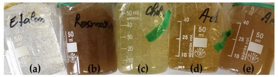
Figure 1.
Aspect of the hydrogel: (a) blank formulation; (b) Rosmarini herba extract formulation; (c) Calendulae flos extract formulation; (d) Millefolii flos extract formulation; (e) Extract blend formulation.
Taking into consideration the fact that the addition of the 70% ethanolic extracts to the formulation was expected to slightly lower the pH, due not only to the alcohol per se (functional-hydroxyl group, which reduces the pH value), but also due to the content in active principles, the blank hydrogel formulation was developed to have a slightly higher pH than the physiological values of the epiderma, of 4.8 to 6.0. Therefore, the pH value of the blank formulation was measured at 6.12, while the registered pH values of the formulated dressings were 6.05 for the Rosmarini herba formulation, 5.78 for the Achillea formulation, 5.95 for the Calendula formulation, and 5.81 for the herbal extract blend, respectively. As the results showed, the extract blend loaded into the hydrogel formulation influenced the pH in a relatively similar manner as the extracts used alone.
The results obtained by performing the extensiometric method in order to assess and compare the stretching capacity of the hydrogels are presented in Figure 2, as a plot of the stretched surface and weight load applied over the top glass plate. The highest stretching capacity has been registered for the formulation containing the herbal extract blend, 24% higher than the blank hydrogel. This result suggests that the inclusion of such a blend should be performed with caution, in order not to alter the structure of the gel. The Rosmarini herba formulation was the firmest one, registering a stretching capacity 10% lower than the blank. Overall, the firmness differences registered for all the five tested formulations were relatively low, so that the gels would maintain their structure after their application as wound dressings.
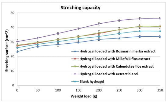
Figure 2.
Graphical representation of the stretching capacity of the formulated hydrogels.
The consistency of the developed gels was evaluated by the cone penetration method, which is a static test, actually assessing the shear strength of the gel’s structure. The behavior of each formulation during the test is graphically depicted by representing the mean penetration depth of the cone, as a function of time, in Figure 3. As presented in the figure, the consistency values of the gels are close, within the ±3% range compared to the blank, and the behavior of the gels is relatively similar, suggesting that the structure of the polymer was not affected by the addition of the concentrated ethanolic herbal extracts.
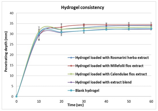
Figure 3.
Gels consistency analyzed by applying the cone penetration method.
Structural viscosity defines the stability of the hydrogel matrix, with the results being presented in Figure 4 as deformation and actual viscosity as functions of tangential stress registered by the eccentric rotation of the rheometer’s spindle.
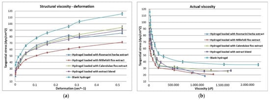
Figure 4.
Viscosity plots of the tested samples. (a) Deformation; (b) Actual viscosity evolution.
The first observation that can be made based on the obtained results would be that, even if the bioactive compounds loaded hydrogels had a similar consistency with the blank formulation, their viscosity is significantly lower. Seventeen to 38% less energy was necessary to achieve gel deformation, with the lowest value corresponding to the Millefolii extract formulation. Figure 4b represents the evolution of the actual measured viscosity of each formulation, with the first point of the graph representing the pressure needed to be applied in order to break the gel’s structure and start the stirring of the viscosimeter spindle. As depicted, the actual measured viscosity of the extract loaded formulations was also considerably lower than the one registered for blank, with their structure being also easier to shear.
Products with pharmaceutical properties as the registered ones may be spread evenly over potential wound surfaces, also reaching cavitary areas. However, such hydrogels cannot maintain their own structure so they cannot be stored in conventional unitary dressing packaging. Conditioning them in a multidose packaging, such as large diameter syringes, would be more appropriate and would even facilitate the application of the product.
3.3. Texture Analysis of the Hydrogels
The texture analysis has been performed by compressing each sample, using a specific probe, at a constant speed in order to evaluate the texture attributes of the hydrogels. The resistance of each hydrogel sample was recorded as load values plotted over time and a graphical representation was generated for every compression cycle. The parameters were automatically calculated for the recorded results. In order to provide a better overview, a graph representing the overlapped analysis of the blank and extract blend formulations is presented in Figure 5. The positive part of the plot is generated as the probe travels through the hydrogel matrix, whereas the negative part is registered as the probe returns to its starting point. The texture parameters are calculated as follows: the maximum force registered on the graph represents the firmness, the area under the positive half of the plot represents the consistency, and the peak negative force of the graph shows the result for the adhesive force [34].
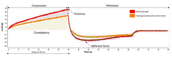
Figure 5.
Texture analysis plot representing the consistency, firmness, and adhesiveness tests for the blank hydrogel and hydrogel loaded with extract blend.
The texture analysis has been performed in triplicate for all five formulations, with the obtained values being presented in Table 3. By comparing the blank formulation with the bioactive compounds loaded hydrogels, it is noticeable that the texture of the hydrogels has been affected to some extent by the ethanolic extract, decreasing the consistency, firmness, and adhesive force values. Similar to the viscometric properties, the largest decrease regarding all three assessed texture properties was recorded for the formulation loaded with the Millefolii flos extract, although all formulated hydrogels maintained suitable consistency and adhesive properties, in order to be used as topical products. Regarding the previously assessed pharmaceutical properties, the texture analysis results related to the extract blend formulation fall in the middle of the generally obtained value range. Our results are in agreement with the study published by Tasić-Kostov et al. where the addition of the herbal extracts led to the decrease of both the hardness and adhesiveness of the hydrogel vehicle while keeping the product’s desirable qualities [37].
Table 3.
Texture analysis results.
3.4. Antimicrobial Activity of the Formulated Hydrogels
A distinct zone of inhibition was observed around the cut-outs of the hydrogel incorporating the Rosmarini herba extract in the plates inoculated by Staphylococcus aureus (inhibition zone of 10 mm) and Pseudomonas aeruginosa (inhibition zone of 10 mm), as well as by Candida albicans (inhibition zone of 15 mm) (Figure 6). Rosmarini herba are known to contain flavonoids (such as luteolin, genkwanin, hesperidin, diosmin), phenolic acids (mainly rosmarinic acid, as well as caffeic, chlorogenic, and ferulic acid), diterpenoids (mainly rosmanol, carnesol, carnosic acid), and triterpenoids (betulinic, ursolic, or oleanolic acid), as well as alkaloids, tannins, saponins, and essential oils, having strong antioxidant, anti-inflammatory, and antimicrobial properties [38].
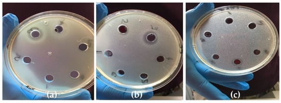
Figure 6.
Antimicrobial activity of the hydrogel incorporating the Rosmarini herba extract against (a) Staphylococcus aureus, (b) Pseudomonas aeruginosa, and (c) Candida albicans.
No antimicrobial activity was identified in the case of the hydrogels incorporating the extracts of Milefolii flos and Calendulae flos.
None of the tested hydrogel formulations proved to be efficient against Escherichia coli, excepting the one incorporating the blend of extracts (inhibition zone of 8 mm), probably suggesting a synergic antimicrobial effect of the concentrated alcoholic extracts (70%) of Rosmarini herba, Millefolii flos, and Calendulae flos.
Thus far, few research studies have incorporated extracts of Rosmarini herba, Millefolii flos, and Calendulae flos in a hydrogel formulation.
For instance, hydrogels containing chitosan with 3% Calendulae flos lyophilized extract were found to have a bactericidal effect against Staphylococcus aureus, Propionibacterium acnes, and Escherichia coli [29]. A chitosan film formulation loaded with bioactive compounds (a mixture of six herbal extracts: Calendula officinalis, Plantago lanceolata, Arnica montana, Tagetes patula, Symphytum officinale, and Geum urbanum) developed and evaluated by our research group also proved to be effective against Staphylococcus aureus, Pseudomonas aeruginosa, and Escherichia coli [17]. However, it must be noted that all the above-mentioned hydrogels were chitosan-based ones, a compound known to possess important antimicrobial properties.
Almost all wounds may become infected due to exposure to non-sterile conditions. Inflammation and bacteria in the wound negatively influence the process of wound healing, causing the formation of exudate, which destroys growth factors and body proteins, as well as the extracellular matrix. Therefore, treating wounds with extract-loaded hydrogels with antimicrobial properties could be beneficial, considering their ease of application and rare systemic toxicity [39,40].
4. Conclusions
Hydrogels containing plant extracts with antimicrobial properties were developed, intended for further use as a possible aid in the acceleration of wound healing. The proposed formulation implies the use of a different gel forming agent than the ones described in previous studies (in which the most common compounds used were PVA, PVP, and PEG) and evaluates how the properties of the carbomer gel matrix are affected by the ethanolic herbal extracts incorporated in the hydrogel.
The studied hydrogel formulations could serve as a basis for the development of novel wound dressing materials. However, an optimized formulation should be further tested in terms of advanced pharmaceutical properties and in vivo effect, in order to support current results.
Author Contributions
Conceptualization, S.M., D.H. and N.K.O.; methodology, M.A., A.G., L.C. and C.B.; software, A.G. and C.B.; validation, A.G. and L.C.; formal analysis, A.G.; investigation, A.G. and C.B.; resources, D.H., N.K.O. and M.A.; data curation, S.M. and A.G.; writing—original draft preparation, A.G. and L.C.; writing—review and editing, L.C. and S.M.; visualization, S.M.; supervision, S.M.; project administration, S.M. All authors have read and agreed to the published version of the manuscript.
Funding
This research received no external funding.
Informed Consent Statement
Not applicable.
Data Availability Statement
Not applicable.
Conflicts of Interest
The authors declare no conflict of interest.
References
- Madaghiele, M.; Demitri, C.; Sannino, A.; Ambrosio, L. Polymeric hydrogels for burn wound care: Advanced skin wound dressings and regenerative templates. Burn. Trauma 2014, 2, 153–161. [Google Scholar] [CrossRef] [PubMed]
- Veiga, A.; Joel, P. Antimicrobial hydrogels for the treatment of infection. Biopolymers 2013, 100, 637–644. [Google Scholar] [CrossRef] [PubMed]
- Peppas, N.A.; Bures, P.; Leobandung, W.; Ichikawa, H. Hydrogels in pharmaceutical formulations. Eur. J. Pharm. Biopharm. 2000, 50, 27–46. [Google Scholar] [CrossRef]
- Li, Q.; Wang, J.; Shahani, S.; Sun, D.D.N.; Sharma, B.; Elisseeff, J.H.; Leong, K.W. Biodegradable and photocrosslinkable polyphosphoester hydrogel. Biomaterials 2006, 27, 1027–1034. [Google Scholar] [CrossRef]
- Boateng, J.; Catanzano, O. Advanced Therapeutic Dressings for Effective Wound Healing—A Review. J. Pharm. Sci. 2015, 104, 3653–3680. [Google Scholar] [CrossRef]
- Koehler, J.; Brandl, F.P.; Goepferich, A. Hydrogel wound dressings for bioactive treatment of acute and chronic wounds. Eur. Polym. J. 2018, 100, 1–11. [Google Scholar] [CrossRef]
- Goodwin, N.S.; Spinks, A.; Wasiak, J. The efficacy of hydrogel dressings as a first aid measure for burn wound management in the pre-hospital setting: A systematic review of the literature. Int. Wound J. 2016, 13, 519–525. [Google Scholar] [CrossRef]
- Vichayarat, R.; Nuttaporn, P.; Pitt, S. Development of gelatin hydrogel pads as antibacterial wound dressings. Macromol. Biosci. 2009, 9, 1004–1015. [Google Scholar] [CrossRef]
- Zahid, M.; Lodhi, M.; Afzal, A.; Rehan, Z.A.; Mehmood, M.; Javed, T.; Shabbir, R.; Siuta, D.; Althobaiti, F.; Dessok, E.S. Development of Hydrogels with the Incorporation of Raphanus sativus L. Seed Extract in Sodium Alginate for Wound-Healing Application. Gels 2021, 7, 107. [Google Scholar] [CrossRef]
- Verma, J.; Kanoujia, J.; Parashar, P.; Tripathi, C.B.; Saraf, S.A. Wound healing applications of sericin/chitosan-capped silver nanoparticles incorporated hydrogel. Drug Deliv. Transl. Res. 2017, 7, 77–88. [Google Scholar] [CrossRef]
- Rajendran, N.K.; Kumar, S.S.D.; Houreld, N.N.; Abrahamse, H. A review on nanoparticle based treatment for wound healing. J. Drug Deliv. Sci. Technol. 2018, 44, 421–430. [Google Scholar] [CrossRef]
- Vijayakumar, V.; Samal, S.K.; Mohanty, S.; Nayak, S.K. Recent advancements in biopolymer and metal nanoparticle-based materials in diabetic wound healing management. Int. J. Biol. Macromol. 2019, 122, 137–148. [Google Scholar] [CrossRef] [PubMed]
- Alavarse, A.C.; de Oliveira Silva, F.W.; Colque, J.T.; da Silva, V.M.; Prieto, T.; Venancio, E.C.; Bonvent, J.J. Tetracycline hydrochloride-loaded electrospun nanofibers mats based on PVA and chitosan for wound dressing. Mater. Sci. Eng. C 2017, 77, 271–281. [Google Scholar] [CrossRef] [PubMed]
- Grolman, J.; Singh, M.; Mooney, D.; Eriksson, E.; Nuutila, K. Antibiotic-Containing Agarose Hydrogel for Wound and Burn Care. J. Burn Care Res. 2019, 40, 900–906. [Google Scholar] [CrossRef]
- Chin, C.; Jalil, J.; Ng, P.Y.; Ng, S. Development and formulation of Moringa oleifera standardised leaf extract film dressing for wound healing application. J. Ethnopharmacol. 2018, 212, 188–199. [Google Scholar] [CrossRef]
- Tan, S.P.; McLoughlin, P.; O’Sullivan, L.; Prieto, M.L.; Gardiner, G.E.; Lawlor, P.G.; Hughes, H. Development of a novel antimicrobial seaweed extract-based hydrogel wound dressing. Int. J. Pharm. 2013, 456, 10–20. [Google Scholar] [CrossRef]
- Colobatiu, L.; Gavan, A.; Mocan, A.; Bogdan, C.; Mirel, S.; Tomuta, I. Development of bioactive compounds-loaded chitosan films by using a QbD approach—A novel and potential wound dressing material. React. Func. Polym. 2019, 138, 46–54. [Google Scholar] [CrossRef]
- Colobatiu, L.; Gavan, A.; Potarniche, A.-V.; Rus, V.; Diaconeasa, Z.; Mocan, A.; Tomuta, I.; Mirel, S.; Mihaiu, M. Evaluation of bioactive compounds-loaded chitosan films as a novel and potential diabetic wound dressing material. React. Funct. Polym. 2019, 145. [Google Scholar] [CrossRef]
- El-Kased, R.; Amer, R.; Attia, D.; Elmazar, M. Honey-based hydrogel: In vitro and comparative in vivo evaluation for burn wound healing. Sci. Rep. 2017, 7, 1–11. [Google Scholar] [CrossRef]
- Kamoun, E.A.; Kenawy, S.; Chen, X. A review on polymeric hydrogel membranes for wound dressing applications: PVA-based hydrogel dressings. J. Adv. Res. 2017, 8, 217–233. [Google Scholar] [CrossRef]
- Benedec, D.; Hanganu, D.; Oniga, I.; Tiperciuc, B.; Olah, N.-K.; Raita, O.; Bischin, C.; Silaghi-Dumitrescu, R.; Vlase, L. Assessment of rosmarinic acid content in six Lamiaceae species extracts and their antioxidant and antimicrobial potential. Pak. J. Pharm. Sci. 2015, 28, 2297–2303. [Google Scholar]
- Olah, N.K.; Osser, G.; Câmpean, R.F.; Furtuna, F.R.; Benedec, D.; Filip, L.; Raita, O.; Hanganu, D. The study of polyphenolic compounds profile of some Rosmarinus officinalis L. extracts. Pak. J. Pharm. Sci. 2016, 29, 2355–2361. [Google Scholar] [CrossRef] [PubMed]
- Alizargar, J.; Kuchaki, E.; Shaabani, A.; Namazi, M. Properties of Wound Healing Activities of Rosemary Extract. J. Biol. Act. Prod. from Nat. 2013, 2, 218–224. [Google Scholar] [CrossRef]
- Benedec, D.; Vlase, L.; Oniga, I.; Mot, A.C.; Damian, G.; Hanganu, D.; Duma, M.; Silaghi-Dumitrescu, R. Polyphenolic composition, antioxidant and antibacterial activities for two Romanian subspecies of Achillea distans Waldst. et Kit. ex Willd. Molecules 2013, 18, 8725–8739. [Google Scholar] [CrossRef] [PubMed]
- Preethi, K.C.; Kuttan, G.; Kuttan, R. Antioxidant potential of an extract of Calendula officinalis flowers in vitro and in vivo. Pharm. Biol. 2006, 44, 691–697. [Google Scholar] [CrossRef]
- Quave, C.L. Wound Healing with Botanicals: A Review and Future Perspectives. Curr. Dermatol. Rep. 2018, 7, 287–295. [Google Scholar] [CrossRef] [PubMed]
- Leach, M.J. Calendula officinalis and Wound Healing: A Systematic Review. Wounds Compend. Clin. Res. Pract. 2008, 20, 236–243. [Google Scholar]
- Shafeie, N.; Naini, A.T.; Jahromi, H.K. Comparison of different concentrations of Calendula officinalis gel on cutaneous wound healing. Biomed. Pharmacol. J. 2015, 8, 979–992. [Google Scholar] [CrossRef]
- Chanaj-Kaczmarek, J.; Paczkowska, M.; Osmałek, T.; Kaproń, B.; Plech, T.; Szymanowska, D.; Karaźniewicz-Łada, M.; Kobus-Cisowska, J.; Cielecka-Piontek, J. Hydrogel Delivery System Containing Calendulae flos Lyophilized Extract with Chitosan as a Supporting Strategy for Wound Healing Applications. Pharmaceutics 2020, 12, 634. [Google Scholar] [CrossRef]
- Andleeb, M.; Shoaib Khan, H.M.; Daniyal, M. Development, Characterization and Stability Evaluation of Topical Gel Loaded With Ethosomes Containing Achillea millefolium L. Extract. Front. Pharmacol. 2021, 12, 1–11. [Google Scholar] [CrossRef]
- Radulescu, D.; Radulescu, D.; Constantinescu, G.; Chirila, L.; Popescu, A. PVA-gelatin hydrogels containing rosemary essential oil for wound dressings. In Proceedings of the TexTeh IX ed., Bucharest, Romania, 24–25 October 2019; pp. 1–5. [Google Scholar]
- Zanfirescu, A.; Nitulescu, G.; Stancov, G.; Radulescu, D.; Trif, C.; Nitulescu, G.M.; Negres, S.; Olaru, O.T. Evaluation of Topical Anti-Inflammatory Effects of a Gel Formulation with Plantago Lanceolata, Achillea Millefolium, Aesculus Hippocastanum and Taxodium Distichum. Sci. Pharm. 2020, 88, 26. [Google Scholar] [CrossRef]
- Prior, R.L.; Wu, X.; Schaich, K. Standardized Methods for the Determination of Antioxidant Capacity and Phenolics in Foods and Dietary Supplements. J. Agric. Food Chem. 2005, 53, 4290–4302. [Google Scholar] [CrossRef] [PubMed]
- Bogdan, C.; Iurian, S.; Tomuta, I.; Moldovan, M. Improvement of skin condition in striae distensae: Development, characterization and clinical efficacy of a cosmetic product containing Punica granatum seed oil and Croton lechleri resin extract. Drug Des. Devel. Ther. 2017, 11, 521–531. [Google Scholar] [CrossRef]
- Anjum, S.; Arora, A.; Alam, M.S.; Gupta, B. Development of antimicrobial and scar preventive chitosan hydrogel wound dressings. Int. J. Pharm. 2016, 508, 92–101. [Google Scholar] [CrossRef] [PubMed]
- Barku, V.Y.A. Wound healing: Contributions from plant secondary metabolite antioxidants. Wound Heal. Perspect. 2019, 5, 50–63. [Google Scholar] [CrossRef]
- Tasić-Kostov, M.; Arsić, I.; Pavlović, D.; Stojanović, S.; Najman, S.; Naumović, S.; Tadić, V. Towards a modern approach to traditional use: In vitro and in vivo evaluation of Alchemilla vulgaris L. gel wound healing potential. J. Ethnopharmacol. 2019, 238. [Google Scholar] [CrossRef]
- Michalak, M.; Zielinska, A.; Paradowska, K. Phenolic content, antioxidant activity and pharmaceutical availability of hydrogels with extracts of Rosmarinus officinalis L. And Sambucus nigra L. Acta Pol. Pharm. -Drug Res. 2021, 78, 219–226. [Google Scholar] [CrossRef]
- McCarty, S.M.; Percival, S.L. Proteases and Delayed Wound Healing. Adv. Wound Care 2013, 2, 438–447. [Google Scholar] [CrossRef]
- Frykberg, R.G.; Banks, J. Challenges in the Treatment of Chronic Wounds. Adv. Wound Care 2015, 4, 560–582. [Google Scholar] [CrossRef]
Publisher’s Note: MDPI stays neutral with regard to jurisdictional claims in published maps and institutional affiliations. |
© 2022 by the authors. Licensee MDPI, Basel, Switzerland. This article is an open access article distributed under the terms and conditions of the Creative Commons Attribution (CC BY) license (https://creativecommons.org/licenses/by/4.0/).