
Patellar Osteomyelitis in a 9-Year-Old Patient with Chronic Granulomatous Disease: A Case Report


Abstract
:1. Introduction
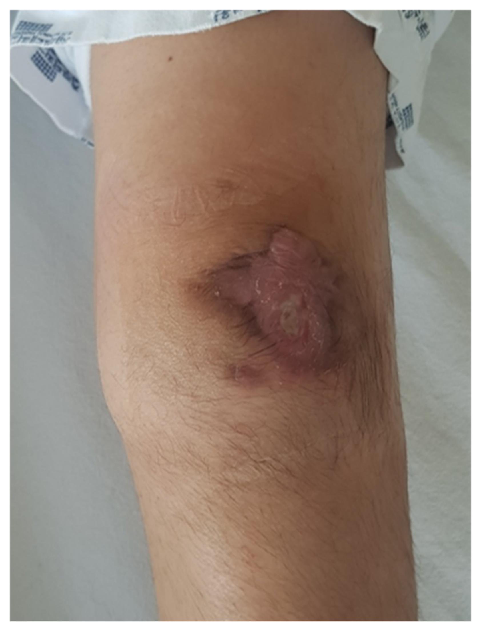
2. Case Report
3. Discussion
4. Conclusions
Author Contributions
Funding
Institutional Review Board Statement
Informed Consent Statement
Data Availability Statement
Conflicts of Interest
References
- Roy, D.R.; Greene, W.B.; Gamble, J.G. Osteomyelitis of the patella in children. J. Pediatr. Orthop. 1991, 11, 364–366. [Google Scholar] [CrossRef] [PubMed]
- Sperl, M.; Novak, M.; Sperl, D.; Svehlik, M.; Singer, G.; Kraus, T. Osteomyelitis of the Patella in a 10-Year-Old Girl: A Case Report and Review of the Literature. Case Rep. Orthop. 2017, 6573271. [Google Scholar] [CrossRef] [PubMed] [Green Version]
- Dartnell, J.; Ramachandran, M.; Katchburian, M. Haematogenous acute and subacute paediatric osteomyelitis: A systematic review of the literature. J. Bone Jt. Surg. Br. 2012, 94, 584–595. [Google Scholar] [CrossRef] [PubMed]
- Holland, S.M. Chronic granulomatous disease. Hematol. Oncol. Clin. N. Am. 2013, 27, 89–99. [Google Scholar] [CrossRef] [PubMed] [Green Version]
- Chiriaco, M.; Salfa, I.; Di Matteo, G.; Rossi, P.; Finocchi, A. Chronic granulomatous disease: Clinical, molecular, and therapeutic aspects. Pediatr. Allergy Immunol. 2016, 27, 242–253. [Google Scholar] [CrossRef] [PubMed]
- Dotis, J.; Roilides, E. Osteomyelitis due to Aspergillus spp. in patients with chronic granulomatous disease: Comparison of Aspergillus nidulans and Aspergillus fumigatus. Int. J. Infect. Dis. 2004, 8, 103–110. [Google Scholar] [CrossRef] [PubMed] [Green Version]
- Choi, H.R. Patellar osteomyelitis presenting as prepatellar bursitis. Knee 2007, 14, 333–335. [Google Scholar] [CrossRef] [PubMed]
- Cahill, B.R. Nontraumatic osteomyelitis of the patella. Clin. Orthop. Relat. Res. 1978, 132, 177–179. [Google Scholar] [CrossRef]
- Tanikawa, M.; Murai, T.; Aizawa, Y.; Horikoshi, Y.; Kinoshita, K.; Yoshihiko, M.; Norikazu, O.; Satoshi, S. Pediatric Patellar Osteomyelitis. Pediatric. Infect. Dis. 2017, 2, 55. [Google Scholar] [CrossRef]
- Angella, J.J. Osteomyelitis of the patella. Am. J. Dis. Child. 1967, 113, 590–593. [Google Scholar] [CrossRef] [PubMed]
- Evans, D.K. Osteomyelitis of the patella. J. Bone Jt. Surg. 1962, 44, 319–323. [Google Scholar] [CrossRef]
- Wadlington, W.B.; Hatcher, H.; Turner, D.J. Osteomyelitis of the patella. Gentamicin therapy associated with encephalopathy. Clin. Pediatr. 1971, 10, 577–580. [Google Scholar] [CrossRef] [PubMed]
- Vaninbroukx, J.; Martens, M.; Verhelst, M.; Mulier, J.C. Haematogenous osteomyelitis of the patella. Report of three cases. Acta Orthop. Scand. 1976, 47, 566–569. [Google Scholar] [CrossRef] [PubMed]
- Papavasiliou, V.A.; Sferopoulos, N.K. Acute hematogenous osteomyelitis of the patella. Apropos of 3 cases. Rev. Chir. Orthop. Reparatrice Appar. Mot. 1989, 75, 130–132. [Google Scholar] [PubMed]
- Moyikoua, A.; Bouity-Buang, J.C.; Ngatse, O.; Pena-Pitra, B. Acute osteomyelitis of the patella. Acta Orthop. Belg. 1993, 59, 98–99. [Google Scholar] [PubMed]
- Masuda, S.; Shinozaki, T.; Watanabe, H.; Takagishi, K.; Fukuda, T. Acute osteomyelitis of the patella in a child. Jpn. J. Rheumatol. 1999, 9, 189–194. [Google Scholar]
- Durani, Y.; Attia, M.W. An unusual case of knee pain. Pediatr. Emerg. Care 2006, 22, 426–429. [Google Scholar] [CrossRef] [PubMed]
- De Gheldere, A. Haematogenous osteomyelitis of the patella in a child. Acta Orthop. Belg. 2009, 75, 554–556. [Google Scholar] [PubMed]
- Gil-Albarova, J.; Gómez-Palacio, V.E.; Herrera, A. Hematogenous osteomyelitis of the patella. J. Pediatr. Orthop. B 2012, 21, 411–414. [Google Scholar] [CrossRef] [PubMed]
- Traverso, A.; Tschopp, B.; Mekdade, T.; Kwiatkowski, B.; Lutz, N. Patella Osteomyelitis Mimicking Sinding-Larsen and Johansson Apophysitis: A Pitfall Not to Miss. Case Rep. Orthop. 2020, 2020. [Google Scholar] [CrossRef] [PubMed]


| Date | White Blood Cell Counts (/µL) | Neutrophils (%) | C-Reactive Protein (mg/dL) | Erythrocyte Sedimentation Rate (mm/h) |
|---|---|---|---|---|
| 2018-10 | 10,000 | 85.6 | 10.14 | 120 |
| Initial presentation of knee pain and difficulty in ambulation | ||||
| 2018-11 | 10,300 | 52.4 | 5.03 | 120 |
| 4 weeks after the use of teicoplanin and cefotaxime since initial presentation | ||||
| 2020-05 | 7100 | 54.6 | 0.37 | 19 |
| Admission to the department of orthopedic surgery before surgery | ||||
| 2020-06 | 7200 | 46.9 | 0.17 | 26 |
| Postoperative 4th week after use of cefotaxime for 4 weeks | ||||
| 2020-06 | 7500 | 55.6 | 0.35 | 39 |
| Postoperative 6th week at out-patient clinic follow-up after discharge | ||||
| 2021-06 | 8000 | 58.5 | 0.34 | 29 |
| Postoperative 1st year at out-patient clinic follow-up | ||||
| Author | Cases | Operative Treatment | Culture | |
|---|---|---|---|---|
| Age | Sex | |||
| Evans 1962 [11] | 5 | F | No | No growth |
| 7 | F | No | Staphylococcus aureus | |
| 7 | M | Yes | Staphylococcus aureus | |
| Angella 1967 [10] | 7 | F | Yes | Staphylococcus aureus |
| 11 | M | No | No growth | |
| Wadlington et al. 1971 [12] | 9 | M | No | Pseudomonas aeruginosa |
| Vaninbroukx et al. 1976 [13] | 3 | F | Yes | Not available |
| 6 | M | Yes | No growth | |
| 5 | M | Yes | Not available | |
| Cahill 1978 [8] | 7 | M | Yes | Gram positive cocci |
| 8 | M | Yes | No growth | |
| Papavasiliou et al. 1989 [14] | 6 | F | Yes | Staphylococcus aureus |
| 7 | M | Yes | Staphylococcus aureus | |
| 8 | M | Yes | Staphylococcus aureus | |
| Roy et al. 1991 [1] | 9 | F | Yes | Staphylococcus aureus |
| 6 | F | Yes | Staphylococcus aureus | |
| 7 | M | Yes | Staphylococcus aureus | |
| 8 | M | Yes | Clostridium bifermentans | |
| Moyikoua et al. 1993 [15] | 8 | M | No | Not available |
| Masuda et al. 1999 [16] | 8 | M | Yes | No growth |
| Durani et al. 2006 [17] | 7 | F | Yes | Staphylococcus aureus |
| Sperl et al. 2007 [2] | 10 | F | Yes | Staphylococcus aureus |
| Choi et al. 2007 [7] | 9 | M | Yes | No growth |
| 7 | M | Yes | Staphylococcus aureus | |
| De Gheldere 2009 [18] | 10 | M | Yes | Staphylococcus aureus |
| Gil-Albarova et al. 2012 [19] | 8 | F | Yes | Staphylococcus aureus |
| Tanikawa et al. 2017 [9] | 9 | F | Yes | Staphylococcus aureus |
| Traverso et al. 2020 [20] | 12 | M | No | Staphylococcus aureus |
Publisher’s Note: MDPI stays neutral with regard to jurisdictional claims in published maps and institutional affiliations. |
© 2022 by the authors. Licensee MDPI, Basel, Switzerland. This article is an open access article distributed under the terms and conditions of the Creative Commons Attribution (CC BY) license (https://creativecommons.org/licenses/by/4.0/).
Share and Cite
Park, Y.; Yoo, S.; Chu, Y.; Lim, C. Patellar Osteomyelitis in a 9-Year-Old Patient with Chronic Granulomatous Disease: A Case Report. Children 2022, 9, 76. https://doi.org/10.3390/children9010076
Park Y, Yoo S, Chu Y, Lim C. Patellar Osteomyelitis in a 9-Year-Old Patient with Chronic Granulomatous Disease: A Case Report. Children. 2022; 9(1):76. https://doi.org/10.3390/children9010076
Chicago/Turabian StylePark, Yonggeun, Seungjin Yoo, Yongyeon Chu, and Chaemoon Lim. 2022. "Patellar Osteomyelitis in a 9-Year-Old Patient with Chronic Granulomatous Disease: A Case Report" Children 9, no. 1: 76. https://doi.org/10.3390/children9010076
APA StylePark, Y., Yoo, S., Chu, Y., & Lim, C. (2022). Patellar Osteomyelitis in a 9-Year-Old Patient with Chronic Granulomatous Disease: A Case Report. Children, 9(1), 76. https://doi.org/10.3390/children9010076