Error in Figure
In the original publication [1], there was a mistake in Figure 1B as published. We recently discovered that one of the images in Figure 1B of our published article was accidentally taken from one of our previous publications during figure assembly. The corrected version of Figure 1B appears below. The authors state that the scientific conclusions are unaffected. This correction was approved by the Academic Editor. The original publication has also been updated.
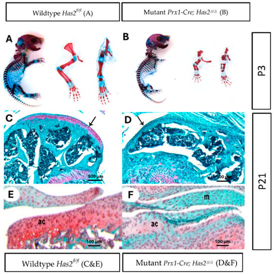
Figure 1.
Limb bud mesenchyme-specific knockout of Has2 induced severe joint defects and impaired the differentiation and formation of articular cartilage. (A,B) Whole mount staining of cartilage and mineralized bone with Alcian Blue and Alizarin Red, respectively, in neonatal day 3 (P3) (A) wildtype Has2fl/fl and (B) mutant Prx1-Cre; Has2Δ/Δ mice. Left to right: whole body, forelimb, hindlimb (0.63×). (C–F) Paraffin sections of the joint surface of 3-week-old (C) wildtype Has2fl/fl and (D) mutant Prx1-Cre; Has2Δ/Δ femurs stained with Safranin-O (red) for proteoglycan accumulation (counterstained with fast green). The wildtype Has2fl/fl femur is capped by a thick layer of articular cartilage, which stains intensely with Safranin-O, while the hyaluronan-deficient femur is capped by a thin layer of tissue that does not stain with Safranin-O, indicating normal articular cartilage is not present (4×) (pointed by black arrow). (E,F) Sections of the tibia joint surface of 3-week-old (P21) (E) wildtype Has2fl/fl and (F) mutant Prx1-Cre; Has2Δ/Δ mice stained with Safranin-O/fast green. In the wildtype Has2fl/fl tibia, a layer of articular cartilage (ac) with round chondrocytes and abundant Safranin-O matrix is present, but the joint surface in the hyaluronan-deficient tibia is covered by a highly cellular tissue with flattened cells and little or no Safranin-O staining, indicating initial articular cartilage differentiation is impaired. Menisci (M) in the hyaluronan-deficient joint also showed no or very little Safranin-O staining. This underscores the importance of Has2 in joint development for different types of chondrocytes (20×).
Reference
- Li, Y.; Tress, A.; Maye, P.; Edwards, K.; Findletar, A.; Dyment, N.A.; Yamaguchi, Y.; Rowe, D.W.; Le-Chan, G.; Chan, S.S.K.; et al. Genetic Deficiency of Hyaluronan Synthase 2 in the Developing Limb Mesenchyme Impairs Postnatal Synovial Joint Formation. Biomedicines 2025, 13, 1324. [Google Scholar] [CrossRef] [PubMed]

Disclaimer/Publisher’s Note: The statements, opinions and data contained in all publications are solely those of the individual author(s) and contributor(s) and not of MDPI and/or the editor(s). MDPI and/or the editor(s) disclaim responsibility for any injury to people or property resulting from any ideas, methods, instructions or products referred to in the content. |
© 2025 by the authors. Licensee MDPI, Basel, Switzerland. This article is an open access article distributed under the terms and conditions of the Creative Commons Attribution (CC BY) license.