Abstract
Background: Mesenchymal stem cells (MSCs) have shown positive therapeutic effects on various diseases; however, their functionality can decline during in vitro expansion. Selenium (Se) supplementation has emerged as a strategy for enhancing MSC culture. This study evaluated the effects of different forms of selenium (Na2SeO4, SeMet, ebselen, and chitosan-coated selenium nanoparticles (CS-SeNPs)) on the biological functions of MSCs. Methods: Human umbilical cord-derived MSCs (HUC-MSCs) were cultured in media supplemented with various selenium compounds at specific concentrations. To investigate their biological effects, we assessed cell proliferation, morphology, surface marker expression, and differentiation potential. Furthermore, to elucidate the underlying mechanisms, we analyzed key markers of cellular senescence, including p16, p21, IL-6, IL-8, p27, p53, and reactive oxygen species (ROS) levels. Results: All the selenium treatments promoted hUC-MSC proliferation at specific concentrations. CS-SeNPs and Na2SeO4 exhibited relatively high bioavailability, whereas ebselen and SeMet demonstrated relatively low toxicity. The optimal concentration (0.5 μM CS-SeNPs or 0.25 μM Na2SeO4) significantly enhanced proliferation without altering the hUC-MSC morphology, phenotype, or differentiation capacity. Both CS-SeNPs and Na2SeO4 effectively promoted hUC-MSC proliferation and reduced the senescence of hUC-MSCs by downregulating key senescence-related effectors: the cell cycle inhibitors p16, p21, p27, and p53; and the levels of ROS and senescence-associated secretory phenotype factors (IL-6 and IL-8). Conclusions: Selenium supplementation is an effective strategy for improving MSC expansion and alleviating senescence. The beneficial effects are dependent on the specific selenium compound used, with CS-SeNPs and Na2SeO4 showing particularly strong potential for enhancing the bioavailability and function of hUC-MSCs during in vitro cultivation.
1. Introduction
In recent decades, stem cell therapy has attracted increasing interest from researchers because of its potential for treating many diseases that are refractory to multiple advanced treatments [,]. Mesenchymal stem cells (MSCs), which are derived from the stroma of various tissues, such as the bone marrow, umbilical cord and adipose tissue, hold great promise for the development of novel therapeutics [,]. Numerous studies have shown that the transplantation of MSCs has beneficial therapeutic effects in a range of diseases [,]. To date, thousands of clinical trials involving human MSCs have evaluated their potential for clinical application and effectiveness worldwide []. A phase III randomized clinical trial demonstrated that MSC infusion after acute myocardial infarction significantly reduces heart failure risk and improves cardiac function []. Recent research by Shi et al. [] in cirrhosis highlights that determining the optimal dosage and administration protocol for MSCs remains a central challenge for clinical translation. Preserving the “youthful” phenotype and function of MSCs is thus critical for producing the high-quality cells required to realize their full clinical potential.
Cell-based therapies require a substantial number of cells, which typically need to be extensively expanded in vitro. However, prolonged culture of MSCs leads to cellular phenotypic aging, a critical issue that severely constrains the production of high-quality cell products. Senescent MSCs exhibit not only reduced proliferative capacity but also diminished multilineage differentiation potential, compromised immunomodulatory function, and adoption of a proinflammatory senescence-associated secretory phenotype, collectively undermining their therapeutic efficacy. These functional declines highlight the urgent need for strategies to delay senescence and preserve the functional potency of MSCs. Researchers have used a variety of strategies, such as the optimal source of stem cells and the improved MSC culture medium, to enhance the impaired functionality of stem cells, which has improved certain aspects of cell therapy [,]. For instance, Tee et al. [] demonstrated that L-ascorbic acid-2-phosphate supplementation significantly reduces cellular senescence and improves the chondrogenic potential of MSCs, resulting in increased critical metabolic and functional properties. These findings underscore the value of chemical preconditioning in preserving MSC quality during manufacturing.
Selenium (Se), an essential microelement for humans, plays a crucial role in various physiological functions. A moderate intake of selenium can significantly improve immune function and positively influence human health. However, poor selenium levels may lead to several diseases such as Keshan disease [,]. Selenium, primarily through its incorporation into selenoproteins such as glutathione peroxidases and thioredoxin reductases. These enzymes combat cellular senescence by neutralizing reactive oxygen species and modulating key signaling pathways to mitigate oxidative stress and its associated damage [,]. However, the specific application of selenium as a culture supplement to combat MSC senescence remains underexplored, and its effects on critical MSC properties during long-term culture are not well defined. Standard MSC culture media provide essential nutrients but lack specific components to combat replication-induced senescence and functional decline. This study aims to systematically evaluate whether selenium supplementation in standard culture medium can effectively delay MSC senescence, preserve their functional potency, and ultimately enhance their therapeutic quality.
Selenium is generally divided into inorganic selenium (e.g., Na2SeO4) and organic selenium. Organic selenium includes both naturally occurring forms, such as selenomethionine (SeMet) and selenocysteine, as well as synthetic molecules, such as ebselen—a glutathione peroxidase mimetic with demonstrated clinical efficacy in inflammatory arthritis []. These different chemical forms exhibit substantial variations in their biological properties. Inorganic selenate (Na2SeO4), while naturally abundant, has greater toxicity and lower bioavailability than organic forms. While organic selenium compounds show improved absorption and incorporation into selenoproteins, they may accumulate nonspecifically in tissues. Most notably, selenium nanoparticles (SeNPs) are highly efficient molecular compounds with greater antioxidant activity and lower toxicity than regular selenium. Numerous compounds, such as polysaccharides, amino acids, and peptides, have been used to modify the surface of SeNPs [,]. Chitosan (CS) is an alkaline polysaccharide that is characterized by its good biocompatibility, non-toxic nature, low immunogenicity, and biodegradability []. Compared with both inorganic and simple organic selenium compounds, SeNPs modified with CS (CS-SeNPs) can exhibit superior properties, including enhanced stability, reduced toxicity, and improved cellular uptake []. Furthermore, its excellent biocompatibility and biodegradability minimize its cytotoxic effects, while its matrix structure modulates selenium release, protecting cells from acute oxidative stress associated with bare SeNPs [,]. These characteristics make CS-SeNPs particularly suitable for maintaining MSC functionality during long-term culture, as they ensure consistent colloidal stability, increase selenium bioavailability, and may provide synergistic antioxidant effects through the bioactivity of chitosan. These fundamental differences in pharmacokinetics and biosafety profiles guided our selection of diverse selenium forms for comparative assessment in MSC culture.
Human umbilical cord-derived MSCs (hUC-MSCs) were selected for this study because of their compelling advantages for translational applications, including abundant availability, robust proliferative capacity in vitro, and low immunogenicity suitable for allogeneic transplantation. These properties make hUC-MSCs an ideal cell source for future therapeutic development, which is why we used hUC-MSCs in our subsequent experiments.
The biological efficacy of selenium in hUC-MSCs is associated with its chemical form, and it has not yet been determined which form of selenium has better effects. On the basis of their unique properties, we hypothesized that CS-SeNPs would more effectively enhance hUC-MSC proliferation, reduce oxidative stress, and ameliorate senescence-associated phenotypes compared to their inorganic and organic counterparts. The present study was therefore designed to systematically evaluate the impact of different selenium forms in the process of hUC-MSC cultivation, aiming to establish an optimized protocol for producing a reliable and robust cell source for clinical applications.
2. Materials and Methods
2.1. Isolation and Culture of hUC-MSCs
The use of the hUC-MSCs used in this study was approved by the Clinical Research Ethics Committee of the Third Affiliated Hospital of Sun Yat-sen University (Approval number: SYSUTHEC RG2024-148-01). All umbilical cord samples (n = 5) were collected from healthy mothers with informed consent. For hUC-MSC isolation, blood vessels and the umbilical cord epithelium were removed from the umbilical cord. The obtained Wharton’s jelly was subsequently cut into tissue blocks of 2–3 mm3 and attached to culture dishes. The cells were collected and cultured with hUC-MSC serum-free medium (YOCON, Beijing, China) in a 5% CO2, 37 °C incubator. When the cells reached 80% to 90% confluence, the hUC-MSCs were passaged at a dilution ratio of 1:3 with 1× stem cell gentle digestion enzyme (YOCON, China) for 2–5 min following the manufacturer’s instructions. All the hUC-MSC cultures were routinely tested for mycoplasma contamination and sterility. Mycoplasma testing was performed via a PCR-based method with primers (forward: 5′-TGCACCATCTGTCATTCTGTTAAC-3′; reverse: 5′-GGAGCAAACAGGATTAGATACCCT-3′) and SYBR Safe DNA gel stain (Invitrogen, Carlsbad, CA, USA). Sterility was confirmed by inoculating culture supernatants into the zLABSTAR EX (Brea, CA, USA) automated blood culture system and monitoring microbial growth for 5 days. All cultures used in the experiments were confirmed to be negative for both mycoplasma and microbial contamination.
2.2. Cell Culture and Selenium Compound Treatment
HUC-MSCs at passage 3 were suspended in MSC medium with or without combinations of the following selenium compounds: CS-SeNPs, ebselen, Na2SeO4 and SeMet. Ebselen, Na2SeO4 and SeMet were purchased from Sigma-Aldrich (St. Louis, MO, USA) and dissolved in sterile PBS buffer. CS-SeNPs were synthesized as described by Liu et al. []. The culture medium was changed every two days. Cells from at least three different hUC-MSC donors were used for all the experiments. CS-SeNPs (0.5 μM) and Na2SeO4 (0.25 μM) were selected as the optimal concentrations for further experiments. The morphology of the cells across all test groups was examined via optical microscopy (Carl Zeiss MicroImaging GmbH, Oberkochen, Germany). The evaluated parameters included cell size and granularity and the proportion of cells with a classical spindle-shaped, fibroblast-like morphology versus a flattened, enlarged, or irregular shape.
2.3. Cell Counting Kit-8 Assays
The cells were plated in 96-well plates at a density of 1000 cells per well and then treated with CCK-8 solution (Dojindo Laboratories, Kumamoto, Japan). The absorbance at a wavelength of 450 nm was subsequently measured via a multifunctional microplate reader (Tecan200, Männedorf, Switzerland) to assess cell viability. Each experiment was performed with three independent biological replicates.
2.4. Flow Cytometry
To examine the expression of hUC-MSC phenotypic markers, 2 × 10⁶ cells were stained with specific antibodies and incubated in the dark for 30 min at room temperature following the manufacturer’s instructions. Flow cytometry was performed with the following antibodies: anti-CD105 (A07768), anti-CD90 (A07768), anti-CD73 (344003), anti-CD45 (A07768), anti-CD34 (A07768), anti-CD19 (A07768), anti-CD11B (IM0530), and anti-HLA-DR (A07768) were obtained from Beckman. Flow cytometry was performed with a flow cytometer from Beckman Coulter, Inc. (Brea, CA, USA). The experiment was performed with three independent biological replicates.
2.5. EdU Assays
Cell proliferation was measured via an EdU assay kit (RiboBio, Guangzhou, China), according to the manufacturer’s protocol. Briefly, cells were plated at 4 × 10³ cells per well in 96-well plates and cultured for 48 h. The cells were subsequently treated with 50 mM EdU for 2 h at 37 °C. Then, the cells were washed with PBS and fixed with 4% formaldehyde at room temperature for 30 min. The DNA contents of the cells were stained with 100 µL of Hoechst 33342 (RiboBio, Guangzhou, China) for 30 min and observed under a fluorescence microscope. The experiment was performed with three independent biological replicates.
2.6. ROS Analysis
The intracellular ROS levels were measured using the fluorescent probe 2′,7′-dichlorodihydrofluorescein diacetate (DCFH-DA) with an ROS assay kit (Beyotime, Shanghai, China) according to the manufacturer’s instructions. HUC-MSCs were resuspended at a density of 1 × 10⁶ cells/mL and treated with 10 μM DCFH-DA in a 37 °C cell culture incubator for 20 min. The cells were washed three times with serum-free cell culture medium to thoroughly remove DCFH-DA. The fluorescence of the cells was then measured via flow cytometry (Beckman Coulter, Brea, CA, USA) at excitation and emission wavelengths of 488 nm and 525 nm, respectively. The experiment was performed with five independent biological replicates.
2.7. Quantitative Real-Time PCR Assay (qPCR)
TRIzol Reagent was used for the extraction of RNA from the cells in our study (Thermo Fisher Scientific Inc., Waltham, MA, USA). Subsequently, reverse transcription was executed by employing the Evo M-MLV RT Kit with gDNA Clean (Accurate Biology, Changsha, China). ChamQ SYBR qPCR Master Mix (Vazyme, Nanjing, China) was utilized for the qPCR analysis. β-actin was utilized as the internal control gene for normalization in each qPCR assay. The relative expression of genes was determined with the comparative cycle threshold (CT) method, specifically the 2−ΔΔCT method []. All qPCR analyses were performed with at least three independent biological replicates. The forward and reverse PCR primers used are listed in Table 1.
Table 1.
QPCR primers sequences used in this study.
2.8. MSC Adipogenic Differentiation and Osteogenic Differentiation Analysis
For adipogenic differentiation, hUC-MSCs were seeded at a density of 2 × 10⁴ cells per well in six-well plates. When the cells reached 90% to 100% confluence, the hUC-MSCs were stimulated with an MSC adipogenic differentiation kit (Cyagen Biosciences, Guangzhou, China) according to the manufacturer’s instructions. The adipogenic differentiation medium was changed every 2 to 4 days. After 14 days of adipogenic induction, the cells were washed with PBS and fixed with 4% paraformaldehyde for 1 h. The induced cells were subsequently stained with Oil Red O. Stained cells were imaged using a Leica microscope (Leica, Wetzlar, Germany) and the staining intensity was quantified with ImageJ (version 1.54g, Bethesda, MD, USA) software.
For osteogenic differentiation, hUC-MSCs were seeded into six-well plates and cultured for 12 h at a density of 2 × 10⁴ cells per well. When the cells reached 70% confluence, the medium was replaced with hUC-MSC osteogenic differentiation medium (Cyagen Biosciences, Guangzhou, China) for 21 days, and the medium was refreshed every 3 days. Subsequently, the cells were washed with PBS and fixed with 4% paraformaldehyde for 1 h. Then, Alizarin Red S staining solution was added, and the cells were observed under a microscope. The staining intensity of the cells was assessed by quantification using ImageJ software. Each experiment was performed with three independent biological replicates.
2.9. Senescence-Associated β-Galactosidase (SA-β-Gal) Staining Assay
The SA-β-gal staining assay was carried out using an SA-β-gal Staining Kit from Beyotime according to the manufacturer’s protocol. Briefly, cells were plated at 1 × 10⁵ cells per well in 6-well plates and cultured for 48 h. The cells were subsequently fixed with a 4% paraformaldehyde solution for 15 min. These cells were subsequently incubated with SA-β-gal staining solution overnight in a 37 °C cell culture incubator and observed under an inverted microscope (Leica, Wetzlar, Germany). The cells exhibiting a dark blue color were identified as positive for the staining. The experiment was performed with three independent biological replicates.
2.10. Statistical Analysis
Experimental data analysis was conducted using Prism version 5.0 (GraphPad Software, La Jolla, CA, USA), employing Student’s t-test and one- or two-way ANOVA. All data were assessed for normality (Shapiro–Wilk test) and homogeneity of variances (Levene’s test) prior to parametric analysis. For the ANOVA models, Tukey’s honestly significant difference (HSD) post hoc test was applied for multiple comparisons between groups. Continuous data are presented as the means ± standard deviations (SDs) from at least three independent biological replicates. A significance level of 0.05 was chosen to determine statistical significance.
3. Results
3.1. Choice of Selenium Compounds for Improving the Culture of hUC-MSCs
To investigate the optimal concentrations of different selenium compounds, CCK8 assays were conducted to compare the proliferation rates of hUC-MSCs cultured with different concentrations of the different selenium compounds (0 μM, 1 μM, 2 μM, 4 μM, 8 μM, 16 μM, 32 μM, 64 μM, 128 μM and 256 μM) [,,]. As shown in Figure 1A, CS-SeNPs, ebselen and SeMet promoted cell proliferation to a certain degree at concentrations above 1 μM. However, 2 μM or higher concentrations of CS-SeNPs, 1 μM or higher concentrations of Na2SeO4, 8 μM or higher concentrations of ebselen and 32 μM or higher concentrations of SeMet have been shown to be toxic to hUC-MSCs and cause cell death. We further optimized the concentrations of these selenium compounds, with CS-SeNPs (0 μM, 0.5 μM, 1.0 μM and 2 μM), Na2SeO4 (0 μM, 0.1 μM, 0.25 μM, 0.5 μM and 1.0 μM), ebselen (0 μM, 4 μM, 8 μM, 12 μM and 16 μM) and SeMet (0 μM, 2 μM, 8 μM, 12 μM and 16 μM). Compared with those in the control group, the proliferation rates of hUC-MSCs were significantly increased following treatment with 0.5 μM CS-SeNPs or 0.25 μM Na2SeO4 after incubation for 72 h (Figure 1B). Thus, 0.5 μM CS-SeNPs and 0.25 μM Na2SeO4 were selected as the optimal concentrations for further experiments.
Figure 1.
Choice of selenium compounds for improving the culture of hUC-MSCs. (A) CCK8 assays were performed to compare the proliferation rates of hUC-MSCs cultured with different concentrations of selenium. (B) CCK8 assays were used to detect the proliferation rates of the hUC-MSCs. * p < 0.05, ** p < 0.01. The data are from three independent biological replicates (n = 3) and are presented as the means ± SDs.
3.2. Basic Characterization of hUC-MSCs
To investigate the potential impact of these selenium agents on the basic characteristics of hUC-MSCs, we first examined the cellular morphology of hUC-MSCs after treatment with CS-SeNPs or Na2SeO4. The morphology of cells from the control and selenium compound groups at passages P3 to P8 is shown in Figure 2A. The size of the hUC-MSCs at passages P3 and P8 was relatively uniform. Similar to the cells from the control group, no obvious morphological differences were detected in the hUC-MSCs from the CS-SeNPs group and the Na2SeO4 group. Furthermore, the phenotypic marker profiles of the hUC-MSCs were analyzed at P3 via flow cytometry. The hUC-MSCs in the CS-SeNPs, Na2SeO4 and control groups presented expression of the phenotypic markers CD105 (98.48 ± 0.44 versus 98.86 ± 0.42 versus 99.19 ± 0.34), CD73 (99.05 ± 0.47 versus 99.02 ± 0.58 versus 99.20 ± 0.14) and CD90 (99.43 ± 0.26 versus 99.44 ± 0.49 versus 99.59 ± 0.31), and low expression of the negative markers CD45, CD34, CD19, CD11b and HLA-DR (Figure 2B). These data suggested that the expression of phenotypic markers was not affected by the selenium compounds.
Figure 2.
Analysis of the hUC-MSC phenotype and differentiation potential. (A) Morphological images of cells from the control and selenium compound groups were assessed. (B) Flow cytometry was conducted to analyze the phenotypic marker profiles of the hUC-MSCs. The data are from three independent biological replicates (n = 3) and are presented as the means ± SDs. (C) Oil red O staining and Alizarin red staining were used to detect the osteogenic and adipogenic differentiation of hUC-MSCs. The data are from three independent biological replicates (n = 3) and are presented as the means ± SDs.
We next examined the role of selenium compounds in hUC-MSC osteogenic and adipogenic differentiation via Alizarin Red staining and Oil Red O staining assays. Oil Red O staining revealed that the hUC-MSCs formed many neutral lipid droplets in the cytoplasm. Alizarin Red staining was used to assess mineral accumulation and bone nodule formation. The results revealed that hUC-MSCs cultured with or without selenium agents both possessed the potential to undergo adipogenic and osteogenic differentiation (Figure 2C).
3.3. Effects of Selenium Compounds on the Proliferation Capacity of hUC-MSCs
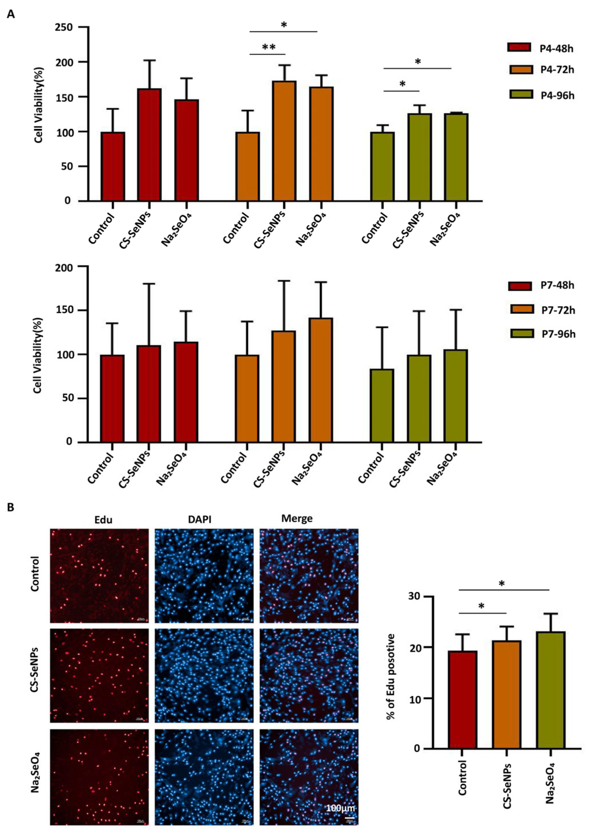
According to the previous results, we assessed the impact of culture on proliferation via the use of hUC-MSC medium or hUC-MSC medium supplemented with selenium compounds. As shown in Figure 3A, the viability of hUC-MSCs was significantly greater in the CS-SeNPs and Na2SeO4 groups than in the control group at both 72 h and 96 h in early passage P4, as determined by the CCK-8 assay. At 72 h, the viability reached 173.71 ± 22.12% and 164.70 ± 16.53% for the CS-SeNPs and Na2SeO4 groups, respectively, compared with that of the control (100.00 ± 30.60%). This promoting effect persisted at 96 h, with viability values of 126.52 ± 11.68% (CS-SeNPs) and 127.04 ± 0.66% (Na2SeO4) versus the control (100.00 ± 9.50%). In late passage P7, the viability of the hUC-MSCs in the selenium compound groups increased slightly compared with that in the control group, although the differences were not statistically significant (Figure 3A). Consistently, the results of the EdU assays revealed that the proliferation rate of P4 hUC-MSCs was significantly greater in the CS-SeNPs and Na2SeO4 groups than in the control group (19.38 ± 3.22%, 21.42 ± 2.71%, and 23.18 ± 3.47% for the control, CS-SeNPs, and Na2SeO4 groups, respectively) (Figure 3B). These results suggested that both CS-SeNPs and Na2SeO4 can effectively promote hUC-MSC proliferation at early passages.
Figure 3.
Effects of selenium compounds on the proliferative capacity of hUC-MSCs. (A) CCK8 assays were performed to assess the viability of the hUC-MSCs in the CS-SeNPs and Na2SeO4 groups. The data are from three independent biological replicates (n = 3) and are presented as the means ± SDs. (B) EdU assays were used to analyze the proliferation rate of hUC-MSCs in the CS-SeNPs and Na2SeO4 groups. The data are from three independent biological replicates (n = 3) and are presented as the means ± SDs. * p < 0.05, ** p < 0.01.
3.4. Effects of CS-SeNPs and Na2SeO4 on the Cell Senescence and Oxidative Stress of hUC-MSCs
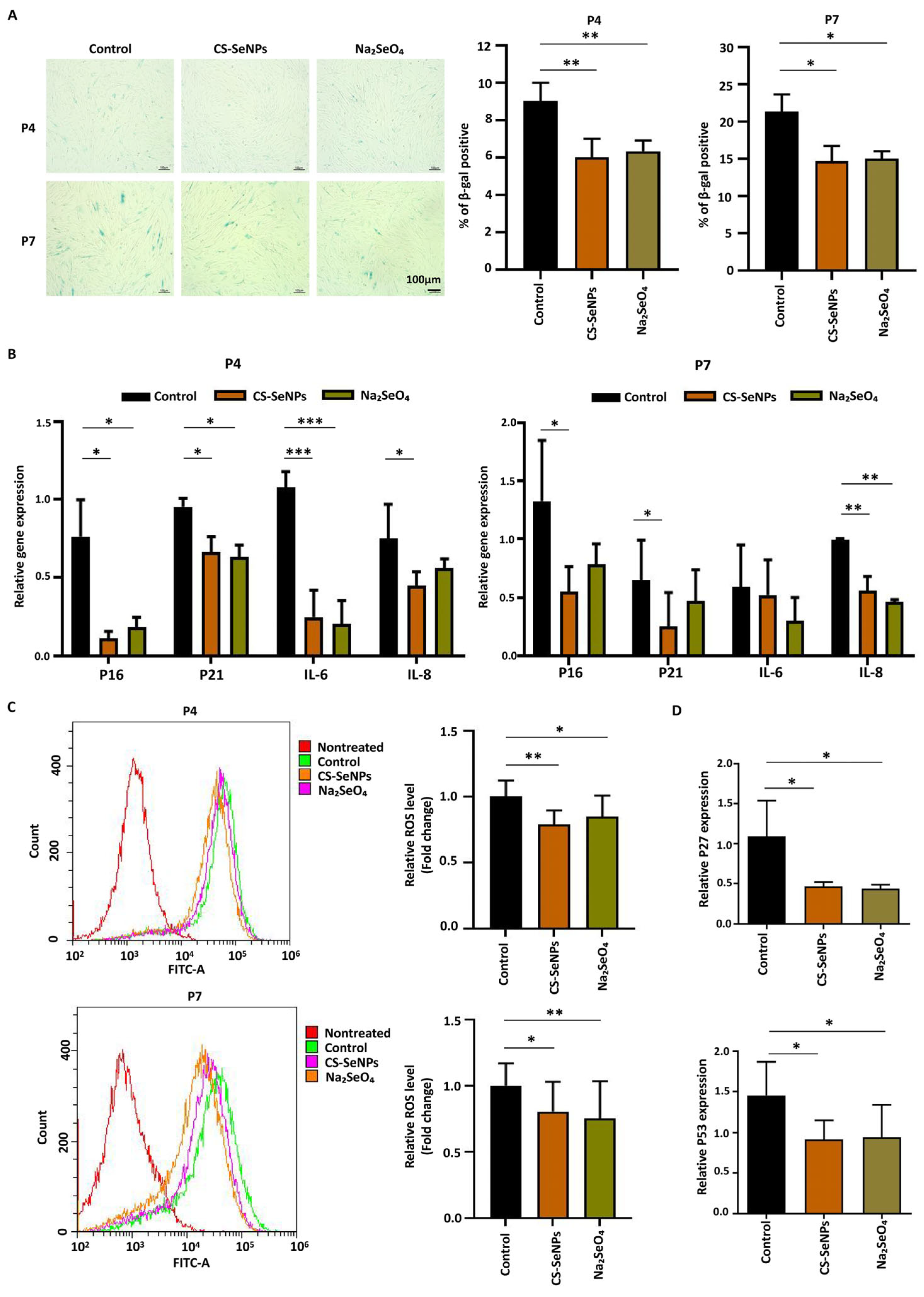
The long-term culture of hUC-MSCs leads to cellular senescence, a process characterized by a decrease in proliferative capacity, an increase in β-galactosidase activity, and the accumulation of ROS. Previous work suggested that selenium could not only improve cell survival but also reduce oxidative stress, preventing the senescence of hUC-MSCs [,]. Thus, we conducted β-galactosidase staining experiments to compare the degree of senescence between the selenium compound groups and the control group at passages P4 and P7. The presence of both CS-SeNPs and Na2SeO4 significantly reduced the percentage of β-gal-positive hUC-MSCs at both P4 (control: 9.00 ± 1.00% versus CS-SeNPs: 6.00 ± 1.00% versus Na2SeO4: 6.33 ± 0.58%) and P7 (control: 21.33 ± 2.31% versus CS-SeNPs: 14.67 ± 2.08% versus Na2SeO4: 15.00 ± 1.00%) (Figure 4A). Furthermore, we measured the expression of additional senescence markers in hUC-MSCs at passages P4 and P7 via qPCR. The results demonstrated a consistent downregulation trend across key markers of cell cycle arrest (p16 and p21) and the senescence-associated secretory phenotype (IL-6 and IL-8) upon selenium treatment (Figure 4B). Overall, these data indicated that selenium can reduce the senescence of hUC-MSCs.
Figure 4.
Effects of CS-SeNPs and Na2SeO4 on the oxidative stress and cell senescence of hUC-MSCs. (A) β-Galactosidase staining experiments were performed to compare the degree of senescence between the selenium compound groups and the control group. The data are from three independent biological replicates (n = 3) and are presented as the means ± SDs. (B) QPCR was performed to analyze the expression levels of key senescence markers, including p16, p21, IL-6 and IL-8. The data are from three independent biological replicates (n = 3) and are presented as the means ± SDs. (C) The levels of ROS in hUC-MSCs cultured with CS-SeNPs, Na2SeO4, or the control group were measured using flow cytometry. The data are from five independent biological replicates (n = 5) and are presented as the means ± SDs. (D) QPCR assays were conducted to detect the expression levels of p27 and p53. The data are from five independent biological replicates (n = 5) and are presented as the means ± SDs. * p < 0.05, ** p < 0.01, *** p < 0.001.
Next, intracellular ROS levels were measured to assess the impact of selenium on hUC-MSCs. At both P4 and P7, the relative ROS level was significantly reduced in the CS-SeNPs and Na2SeO4 groups compared to the control group. Specifically, the values were 0.79 ± 0.11 (CS-SeNPs) and 0.85 ± 0.16 (Na2SeO4) at P4, and 0.80 ± 0.23 (CS-SeNPs) and 0.75 ± 0.28 (Na2SeO4) at P7, versus the control (1.00) (Figure 4C), confirming the potent antioxidant effects of these selenium forms. These findings suggested that the proliferation-promoting effect and anti-senescence effect of selenium on hUC-MSCs are correlated with their antioxidant properties.
To gain a deeper insight into the mechanisms driving the phenotypes of cellular proliferation and senescence, we examined the expression levels of p27 and p53 to explore the molecular changes associated with cellular proliferation and senescence. P27 is an important cell cycle regulatory protein that can cause cell cycle arrest and inhibit cell proliferation. P53 is a crucial regulatory gene in mediating cellular aging and the response to DNA replication damage. As shown in Figure 4D, the relative expression levels of p27 and p53 mRNAs were decreased in the cells cultured with the CS-SeNPs and Na2SeO4. Specifically, the expression of p27 was significantly lower in the CS-SeNPs group (0.46 ± 0.05) and the Na2SeO4 group (0.44 ± 0.05) than in the control group (1.09 ± 0.45). A consistent decreasing trend was also observed for p53 expression, which was lower in both the CS-SeNPs group (0.91 ± 0.23) and the Na2SeO4 group (0.94 ± 0.40) than in the control group (1.46 ± 0.41). These findings indicate that selenium can promote the proliferation and reduce the cellular senescence of hUC-MSCs by decreasing the levels of p27, p53 and ROS.
4. Discussion
MSCs have been confirmed to have potential clinical application value in a variety of diseases because of their immune regulation, anti-inflammatory, and multipotent differentiation capabilities. To facilitate the rapid translation and implementation of this novel therapy, countries around the world have prioritized and supported the clinical application of MSCs, considering them as a key area for development. There are currently more than 1200 registered clinical trials involving MSCs and over 75,000 publications on MSCs [,]. Media formulated specifically for the cultivation of MSCs are essential for advancing the clinical application of MSCs.
Selenium, known for its antioxidant activity effect, has been extensively utilized in various studies. It was selected as a supplement to assess the effects of MSCs [,,]. An increasing number of studies have highlighted the significant impact that these selenium compounds may have on enhancing the efficacy of MSCs, indicating their potential as valuable adjuncts in the process of MSC cultivation. Different selenium compounds are metabolized via distinct pathways within cells, consequently exerting unique effects and mechanisms on cell growth [,]. It is essential to conduct a comprehensive investigation into the safety and pharmacodynamic effects of different doses and compounds of selenium to optimize its use in cell culture. The present study demonstrated that the application of selenium to hUC-MSCs resulted in increased cell proliferation, which aligns with previous studies [,]. However, different forms of selenium compounds, including organic selenium, inorganic selenium and SeNPs, have different effects on the proliferation capacity of hUC-MSCs. All these selenium compounds can promote the proliferation of hUC-MSCs at specific concentrations. Among the diverse selenium-based compounds, we found that, compared with organic selenium (Ebselen and SeMet), inorganic selenium (Na2SeO4) and SeNPs (CS-SeNPs) exhibit greater bioavailability in the process of hUC-MSC cultivation, but organic selenium (Ebselen and SeMet) has lower toxicity. The observed differences are likely attributable to variations in uptake efficiency and metabolic pathways associated with the distinct chemical forms of different selenium compounds []. CS-SeNPs enable enhanced cellular uptake and controlled selenium release, thereby promoting sustained proliferation []. Na2SeO4 is rapidly metabolized into selenoproteins, whereas SeMet is nonspecifically incorporated into proteins, limiting its availability for functional selenoprotein synthesis. Ebselen, which lacks direct selenium donation, has relatively weak proliferative effects. Studies indicate that sodium selenite supports stress erythropoiesis with minimal inflammation, whereas high-dose SeMet increases inflammatory responses []. Chen et al. [] reported that SeMet was more effective than ebselen in restoring the viability of ethanol/acetaldehyde-injured MSCs. The specific mechanisms involved require further analysis of the intracellular selenium content and speciation.
The long-term culture of MSCs leads to cellular senescence, which compromises both their biological properties and therapeutic functions. For example, Wang et al. demonstrated that long-term cultured MSCs exhibit decreased proliferation and differentiation, reduced immunosuppression abilities, and widespread alterations in gene expression patterns after long-term culture []. Cell senescence is influenced by multiple factors, including epigenetic modifications, DNA damage, and the accumulation of ROS. ROS play dual roles in MSC culture. An appropriate amount of ROS can function as intracellular signaling molecules, promoting cell proliferation, preventing cell senescence and regulating various cellular signaling pathways [,,]. Conversely, excessive ROS cause oxidative stress and damage cellular cell membranes, proteins, and DNA, potentially inhibiting cell proliferation and even leading to cell death. Studies have shown that selenium supplementation can extend the lifespan of MSCs and enhance their paracrine effects on wound healing and tissue regeneration by inhibiting ROS-mediated aging []. Therefore, understanding and controlling ROS levels is a key factor in enhancing the therapeutic efficacy and safety of MSCs in laboratory culture and clinical applications. Our findings revealed that CS-SeNPs and Na2SeO4-treated hUC-MSCs can reduce the senescence of MSCs via β-galactosidase staining experiments. Both selenium forms significantly reduced intracellular ROS levels and enhanced the antioxidant activity of the cells. Our results demonstrate that appropriate selenium supplementation enhances the cellular antioxidant capacity, helping to maintain ROS at healthy levels, and thus promoting cell function and survival. These findings are consistent with a growing body of evidence highlighting the role of selenium in oxidative stress-related pathologies. For example, one study showed that SeNPs rebalance redox homeostasis by activating GPx1, thereby reducing ROS levels and ameliorating lumbar disc degeneration []. In addition, Li et al. [] reported that transformed SeNPs activate GPx1 to reduce mitochondrial damage and ROS overproduction, reversing HAdV-14-induced oxidative damage.
To elucidate the mechanisms by which selenium treatment affects the proliferative capacity of MSCs, we analyzed the expression levels of the proliferation-associated gene p27 and the senescence-associated gene p53 via qPCR analysis. We observed that, in response to selenium treatment, p27 and p53 were dramatically downregulated. P27 acts as a negative regulator of the cell cycle by inhibiting cyclin-CDK complexes, thereby blocking the phosphorylation of key substrates required for DNA replication and cell division. Its downregulation is frequently associated with the activation of the PI3K/Akt survival pathway. This signaling cascade leads to the suppression of p27 transcription, promoting proliferation []. P53 is a crucial regulatory gene that mediates cellular aging and the response to DNA replication damage. Research has shown that, although the molecular mechanisms that activate aging signaling pathways vary, regardless of whether stem cell aging is induced by intrinsic or extrinsic factors, it ultimately exerts its effects primarily through the regulation of cell cycle-related proteins such as p53. When p53 expression is abnormal, various characteristics associated with cellular aging can be triggered. The decrease in p53, potentially achieved through a selenium-driven antioxidant response (e.g., the Nrf2 pathway) that reduces ROS and disrupts a p53-ROS feedback loop, alleviates the cellular senescence program [,]. Together with p27 downregulation, these changes collectively establish a molecular environment favorable for proliferation.
This study has several limitations. First, although we observed characteristics of cellular senescence, the senescence markers were analyzed exclusively at the mRNA level by qPCR, and we did not validate the DNA damage marker γH2AX. Second, our analysis was confined to early passages (P4 and P7). While this allowed us to capture the initial onset of senescence, it may not fully represent the more robust and mature senescent phenotype typically observed in later passages. Future work will extend the culture timeline to characterize the progression of the senescent state more comprehensively.
In conclusion, our study indicated that selenium can promote the proliferation of hUC-MSCs at specific concentrations. However, different forms of selenium compounds, including organic selenium, inorganic selenium and SeNPs, have different effects on the proliferative capacity of hUC-MSCs. Na2SeO4 and CS-SeNPs exhibit greater bioavailability in the process of hUC-MSC cultivation, but ebselen and SeMet have lower toxicity. Our results indicate that Na2SeO4 and CS-SeNPs enhance hUC-MSC proliferation and reduce senescence by inhibiting the levels of p27, p53 and ROS, suggesting potential applications in the manufacturing of clinical-grade hUC-MSCs. However, further in vivo and mechanistic studies are warranted to validate its safety and efficacy.
Author Contributions
B.N., C.L. and H.L. (Huizhu Lin) contributed equally to this work. B.N.: Investigation, Data curation, Writing—original draft; C.L.: Methodology, Investigation; H.L. (Huizhu Lin): Methodology; W.C.: Writing—review and editing; R.X.: Validation; X.C.: Methodology; H.L. (Huali Li): Validation; J.L.: Writing—review and editing; F.Y.: Funding acquisition, Writing—review and editing. All authors have read and agreed to the published version of the manuscript.
Funding
This work was supported by the National Natural Science Foundation of China (Grant No. 82460130 and 82260110), Guangdong Basic and Applied Basic Research Foundation (2024A1515013044), the Cultivation Project for National Natural Science Foundation of China from the Third Affiliated Hospital of Sun Yat-sen University (2025GZRPYMS07), the Key R&D Program of Xinjiang Uygur Autonomous Region-Department-Region Linkage Project (2024B04014, 2024B04014-3), and the Guangzhou Science and Technology Planning Project (2025A03J3207).
Institutional Review Board Statement
This study was approved by the Ethics Committee of the Third Affiliated Hospital of Sun Yat-sen University. Umbilical cord samples were collected from healthy mothers at the Third Affiliated Hospital of Sun Yat-sen University. The approved study is titled “Mechanism Study of Mesenchymal Stem Cells Regulating Gut Microbiota to Correct Treg/Th1/Th17 Cell Imbalance in the Treatment of Crohn’s Disease”, with approval granted on 21 November 2024 (Approval number: SYSUTHEC RG2024-148-01).
Informed Consent Statement
Informed consent was obtained from all participants and/or their legal guardian(s) under the ethical committee of the Third Affiliated Hospital of Sun Yat-sen University.
Data Availability Statement
All data relevant to the study are included in the article.
Acknowledgments
Special thanks to Tianfeng Chen (Jinan University) for providing the CS-SeNPs, Ebselen, Na2SeO4 and SeMet used in this study.
Conflicts of Interest
The authors declare that they have no competing interests.
References
- Chehelgerdi, M.; Behdarvand Dehkordi, F.; Chehelgerdi, M.; Kabiri, H.; Salehian-Dehkordi, H.; Abdolvand, M.; Salmanizadeh, S.; Rashidi, M.; Niazmand, A.; Ahmadi, S.; et al. Exploring the promising potential of induced pluripotent stem cells in cancer research and therapy. Mol. Cancer 2023, 22, 189. [Google Scholar] [CrossRef]
- Yadav, P.; Singh, S.K.; Rajput, S.; Allawadhi, P.; Khurana, A.; Weiskirchen, R.; Navik, U. Therapeutic potential of stem cells in regeneration of liver in chronic liver diseases: Current perspectives and future challenges. Pharmacol. Ther. 2024, 253, 108563. [Google Scholar] [CrossRef] [PubMed]
- Li, P.; Ou, Q.; Shi, S.; Shao, C. Immunomodulatory properties of mesenchymal stem cells/dental stem cells and their therapeutic applications. Cell. Mol. Immunol. 2023, 20, 558–569. [Google Scholar] [CrossRef] [PubMed]
- Yang, F.; Ni, B.; Liu, Q.; He, F.; Li, L.; Zhong, X.; Zheng, X.; Lu, J.; Chen, X.; Lin, H.; et al. Human umbilical cord-derived mesenchymal stem cells ameliorate experimental colitis by normalizing the gut microbiota. Stem Cell Res. Ther. 2022, 13, 475. [Google Scholar] [CrossRef] [PubMed]
- Xu, R.; Ni, B.; Wang, L.; Shan, J.; Pan, L.; He, Y.; Lv, G.; Lin, H.; Chen, W.; Zhang, Q. CCR2-overexpressing mesenchymal stem cells targeting damaged liver enhance recovery of acute liver failure. Stem Cell Res. Ther. 2022, 13, 55. [Google Scholar] [CrossRef]
- Li, C.; Sun, Y.; Xu, W.; Chang, F.; Wang, Y.; Ding, J. Mesenchymal Stem Cells-Involved Strategies for Rheumatoid Arthritis Therapy. Adv. Sci. 2024, 11, e2305116. [Google Scholar] [CrossRef]
- Levy, O.; Kuai, R.; Siren, E.M.J.; Bhere, D.; Milton, Y.; Nissar, N.; De Biasio, M.; Heinelt, M.; Reeve, B.; Abdi, R.; et al. Shattering barriers toward clinically meaningful MSC therapies. Sci. Adv. 2020, 6, eaba6884. [Google Scholar] [CrossRef]
- Attar, A.; Mirhosseini, S.; Mathur, A.; Dowlut, S.; Monabati, A.; Kasaei, M.; Abtahi, F.; Kiwan, Y.; Vosough, M.; Azarpira, N. Prevention of acute myocardial infarction induced heart failure by intracoronary infusion of mesenchymal stem cells: Phase 3 randomised clinical trial (PREVENT-TAHA8). BMJ 2025, 391, e083382. [Google Scholar] [CrossRef]
- Shi, L.; Zhang, Z.; Mei, S.; Wang, Z.; Xu, Z.; Yao, W.; Liu, L.; Yuan, M.; Pan, Y.; Zhu, K.; et al. Dose-escalation studies of mesenchymal stromal cell therapy for decompensated liver cirrhosis: Phase Ia/Ib results and immune modulation insights. Signal Transduct. Target. Ther. 2025, 10, 238. [Google Scholar] [CrossRef]
- Zhao, L.; Ni, B.; Li, J.; Liu, R.; Zhang, Q.; Zheng, Z.; Yang, W.; Yu, W.; Bi, L. Evaluation of the impact of customized serum-free culture medium on the production of clinical-grade human umbilical cord mesenchymal stem cells: Insights for future clinical applications. Stem Cell Res. Ther. 2024, 15, 327. [Google Scholar] [CrossRef]
- Ivanisova, D.; Bohac, M.; Culenova, M.; Smolinska, V.; Danisovic, L. Mesenchymal-Stromal-Cell-Conditioned Media and Their Implication for Osteochondral Regeneration. Int. J. Mol. Sci. 2023, 24, 9054. [Google Scholar] [CrossRef] [PubMed]
- Tee, C.; Roxby, D.; Othman, R.; Denslin, V.; Bhat, S.; Yang, Z.; Han, J.; Tucker-Kellogg, L.; Boyer, L. Metabolic modulation to improve MSC expansion and therapeutic potential for articular cartilage repair. Stem Cell Res. Ther. 2024, 15, 308. [Google Scholar] [CrossRef] [PubMed]
- Wang, P.; Chen, B.; Huang, Y.; Li, J.; Cao, D.; Chen, Z.; Li, J.; Ran, B.; Yang, J.; Wang, R.; et al. Selenium intake and multiple health-related outcomes: An umbrella review of meta-analyses. Front. Nutr. 2023, 10, 1263853. [Google Scholar] [CrossRef] [PubMed]
- Wang, Q.; Zhan, S.; Han, F.; Liu, Y.; Wu, H.; Huang, Z. The Possible Mechanism of Physiological Adaptation to the Low-Se Diet and Its Health Risk in the Traditional Endemic Areas of Keshan Diseases. Biol. Trace Elem. Res. 2022, 200, 2069–2083. [Google Scholar] [CrossRef]
- Steinbrenner, H.; Duntas, L.H.; Rayman, M.P. The role of selenium in type-2 diabetes mellitus and its metabolic comorbidities. Redox Biol. 2022, 50, 102236. [Google Scholar] [CrossRef]
- Li, L.; Zhang, W.; Cao, H.; Fang, L.; Wang, W.; Li, C.; He, Q.; Jiao, J.; Zheng, R. Nanozymes in Alzheimer’s disease diagnostics and therapy. Biomater. Sci. 2024, 12, 4519–4545. [Google Scholar] [CrossRef]
- Noguchi, N. Ebselen, a useful tool for understanding cellular redox biology and a promising drug candidate for use in human diseases. Arch. Biochem. Biophys. 2016, 595, 109–112. [Google Scholar] [CrossRef]
- Fatima, S.; Alfrayh, R.; Alrashed, M.; Alsobaie, S.; Ahmad, R.; Mahmood, A. Selenium Nanoparticles by Moderating Oxidative Stress Promote Differentiation of Mesenchymal Stem Cells to Osteoblasts. Int. J. Nanomed. 2021, 16, 331–343. [Google Scholar] [CrossRef]
- Abozaid, O.A.R.; Rashed, L.A.; El-Sonbaty, S.M.; Abu-Elftouh, A.I.; Ahmed, E.S.A. Mesenchymal Stem Cells and Selenium Nanoparticles Synergize with Low Dose of Gamma Radiation to Suppress Mammary Gland Carcinogenesis via Regulation of Tumor Microenvironment. Biol. Trace Elem. Res. 2023, 201, 338–352. [Google Scholar] [CrossRef]
- Shao, C.; Yu, Z.; Luo, T.; Zhou, B.; Song, Q.; Li, Z.; Yu, X.; Jiang, S.; Zhou, Y.; Dong, W.; et al. Chitosan-Coated Selenium Nanoparticles Attenuate PRRSV Replication and ROS/JNK-Mediated Apoptosis in vitro. Int. J. Nanomed. 2022, 17, 3043–3054. [Google Scholar] [CrossRef]
- Soleimani Asl, S.; Amiri, I.; Samzadeh-Kermani, A.; Abbasalipourkabir, R.; Gholamigeravand, B.; Shahidi, S. Chitosan-coated Selenium nanoparticles enhance the efficiency of stem cells in the neuroprotection of streptozotocin-induced neurotoxicity in male rats. Int. J. Biochem. Cell Biol. 2021, 141, 106089. [Google Scholar] [CrossRef]
- Ming, P.; Wei, Y.; Zhu, Y.; Li, K.; Zhu, W.; Qiu, J. Dual-stabilized selenium nanoparticles with chitosan and SS31 peptide: Resolving instability for enhancing ROS elimination, suppressing inflammation, and combating bacterial infections. Colloids Surf. B Biointerfaces 2025, 253, 114749. [Google Scholar] [CrossRef]
- Yang, X.; Fu, Y.; Zhang, J.; Liu, J.; Liu, X.; Peng, Y.; Kyin, S.; Zhang, M.; Zhou, D. Preparation, characterization, and antioxidant and antiapoptotic activities of biosynthesized nano-selenium by yak-derived Bacillus cereus and chitosan-encapsulated chemically synthesized nano-selenium. Int. J. Biol. Macromol. 2023, 242, 124708. [Google Scholar] [CrossRef] [PubMed]
- Liu, T.; Xu, L.; He, L.; Zhao, J.; Zhang, Z.; Chen, Q.; Chen, T. Selenium nanoparticles regulates selenoprotein to boost cytokine-induced killer cells-based cancer immunotherapy. Nanotoday 2020, 35, 100975. [Google Scholar] [CrossRef]
- Livak, K.; Schmittgen, T. Analysis of relative gene expression data using real-time quantitative PCR and the 2(-Delta Delta C(T)) Method. Methods 2001, 25, 402–408. [Google Scholar] [CrossRef] [PubMed]
- Ghasemi, F.; Khoshmirsafa, M.; Safari, E.; Asgari, M.; Alemrajabi, M.; Nojehdehi, S.; Khorrami, S. Vitamin E and selenium improve mesenchymal stem cell conditioned media immunomodulatory effects. Stem Cell Investig. 2021, 8, 9. [Google Scholar] [CrossRef]
- Gu, W.; Zhao, F.; Huang, W.; Zhu, M.; Huang, H.; Yin, H.; Chen, T. Selenium nanoparticles activate selenoproteins to mitigate septic lung injury through miR-20b-mediated RORγt/STAT3/Th17 axis inhibition and enhanced mitochondrial transfer in BMSCs. J. Nanobiotechnol. 2025, 23, 226. [Google Scholar] [CrossRef]
- Ma, S.; Xue, R.; Zhu, H.; Han, Y.; Ji, X.; Zhang, C.; Wei, N.; Xu, J.; Li, F. Selenomethionine preconditioned mesenchymal stem cells derived extracellular vesicles exert enhanced therapeutic efficacy in intervertebral disc degeneration. Int. Immunopharmacol. 2024, 132, 112028. [Google Scholar] [CrossRef]
- Li, Y.; Liu, T.; Zheng, R.; Lai, J.; Su, J.; Li, J.; Zhu, B.; Chen, T. Translational selenium nanoparticles boost GPx1 activation to reverse HAdV-14 virus-induced oxidative damage. Bioact. Mater. 2024, 38, 276–291. [Google Scholar] [CrossRef]
- Park, J.; Lee, J.H.; Yoon, B.S.; Jun, E.K.; Lee, G.; Kim, I.Y.; You, S. Additive effect of bFGF and selenium on expansion and paracrine action of human amniotic fluid-derived mesenchymal stem cells. Stem Cell Res. Ther. 2018, 9, 293. [Google Scholar] [CrossRef]
- Mönch, D.; Reinders, M.E.J.; Dahlke, M.H.; Hoogduijn, M.J. How to Make Sense out of 75,000 Mesenchymal Stromal Cell Publications? Cells 2022, 11, 1419. [Google Scholar] [CrossRef]
- Al-Azab, M.; Idiiatullina, E.; Safi, M.; Hezam, K. Enhancers of mesenchymal stem cell stemness and therapeutic potency. Biomed. Pharmacother. 2023, 162, 114356. [Google Scholar] [CrossRef] [PubMed]
- Liu, X.; Zhang, T.; Wang, R.; Shi, P.; Pan, B.; Pang, X. Insulin-Transferrin-Selenium as a Novel Serum-free Media Supplement for the Culture of Human Amnion Mesenchymal Stem Cells. Ann. Clin. Lab. Sci. 2019, 49, 63–71. [Google Scholar] [PubMed]
- Valadbeygi, A.; Naji, T.; Pirnia, A.; Gholami, M. Supplementation freeze-thawed media with selenium protect adipose-derived mesenchymal stem cells from freeze-thawed induced injury. Cryobiology 2016, 73, 135–139. [Google Scholar] [CrossRef] [PubMed]
- Hosseinzadeh Anvar, L.; Hosseini-Asl, S.; Mohammadzadeh-Vardin, M.; Sagha, M. The Telomerase Activity of Selenium-Induced Human Umbilical Cord Mesenchymal Stem Cells Is Associated with Different Levels of c-Myc and p53 Expression. DNA Cell Biol. 2017, 36, 34–41. [Google Scholar] [CrossRef]
- Suzuki, M.; Endo, M.; Shinohara, F.; Echigo, S.; Rikiishi, H. Differential apoptotic response of human cancer cells to organoselenium compounds. Cancer Chemother. Pharmacol. 2010, 66, 475–484. [Google Scholar] [CrossRef]
- Sinha, R.; Said, T.; Medina, D. Organic and inorganic selenium compounds inhibit mouse mammary cell growth in vitro by different cellular pathways. Cancer Lett. 1996, 107, 277–284. [Google Scholar] [CrossRef]
- Zou, Y.; Liu, Q.; Liu, Q.; Zhang, Y.; Gao, D.; Lu, Q.; Liu, J.; Zheng, W.; Huang, Z. Effect of selenium compounds on ROS and NO production and NOS activity in human umbilical cord mesenchymal stem cells. Chin. J. Pathophysiol. 2010, 26, 2363–2367. [Google Scholar]
- Khabibullaeva, N.; Khaitbaev, A.; Mansurov, D.; Khakimov, Z.; Rakhmanov, A.; Vidović, E.; Benassi, E. Bioactive composite based on chitosan obtained from the exoskeleton of dead Apis mellifera and selenium nanoparticles for accelerated skin wound healing: Synthesis, characterisation and in vivo assesment. Int. J. Biol. Macromol. 2025, 331, 148334. [Google Scholar] [CrossRef]
- Gong, H.; Bai, Y.; Rahoi, D.; Paulson, R.; Prabhu, K. The Impact of Sodium Selenite and Seleno-L-Methionine on Stress Erythropoiesis in a Murine Model of Hemolytic Anemia. J. Nutr. 2025, 155, 540–548. [Google Scholar]
- Chen, F.; Xu, X.; Wu, F.; Wang, F. Effect of selenium compounds on in vitro proliferation of alcohol metabolite damaged mesenchymal stem cells. Chongqing Med. 2023, 21, 3227–3231. [Google Scholar]
- Wang, S.; Wang, Z.; Su, H.; Chen, F.; Ma, M.; Yu, W.; Ye, G.; Cen, S.; Mi, R.; Wu, X.; et al. Effects of long-term culture on the biological characteristics and RNA profiles of human bone-marrow-derived mesenchymal stem cells. Mol. Ther. Nucleic Acids. 2021, 3, 557–574. [Google Scholar] [CrossRef] [PubMed]
- Atashi, F.; Modarressi, A.; Pepper, M. The role of reactive oxygen species in mesenchymal stem cell adipogenic and osteogenic differentiation: A review. Stem Cells Dev. 2015, 24, 1150–1563. [Google Scholar] [CrossRef]
- Li, Q.; Gao, Z.; Chen, Y.; Guan, M. The role of mitochondria in osteogenic, adipogenic and chondrogenic differentiation of mesenchymal stem cells. Protein Cell 2017, 8, 439–445. [Google Scholar] [CrossRef]
- Shen, Y.; Hong, Y.; Huang, X.; Chen, J.; Li, Z.; Qiu, J.; Liang, X.; Mai, C.; Li, W.; Li, X.; et al. ALDH2 regulates mesenchymal stem cell senescence via modulation of mitochondrial homeostasis. Free Radic. Biol. Med. 2024, 223, 172–183. [Google Scholar] [CrossRef] [PubMed]
- Park, S.; Kim, J.; Nam, S.; Kim, B.; Kim, G.; Kim, W.; Choi, Y. Selenium improves stem cell potency by stimulating the proliferation and active migration of 3T3-L1 preadipocytes. Int. J. Oncol. 2014, 44, 336–342. [Google Scholar] [CrossRef]
- Zheng, R.; Chen, D.; Su, J.; Lai, J.; Wang, C.; Chen, H.; Ning, Z.; Liu, X.; Tian, X.; Li, Y.; et al. Inhibition of HAdV-14 induced apoptosis by selenocystine through ROS-mediated PARP and p53 signaling pathways. J. Trace Elem. Med. Biol. 2023, 79, 127213. [Google Scholar] [CrossRef]
- Zhang, X.; Gu, L.; Chen, Y.; Wang, T.; Xing, H. L-selenomethionine inhibits small intestinal ferroptosis caused by ammonia exposure through regulating ROS-mediated iron metabolism. Ecotoxicol. Environ. Saf. 2025, 289, 117477. [Google Scholar] [CrossRef]
Disclaimer/Publisher’s Note: The statements, opinions and data contained in all publications are solely those of the individual author(s) and contributor(s) and not of MDPI and/or the editor(s). MDPI and/or the editor(s) disclaim responsibility for any injury to people or property resulting from any ideas, methods, instructions or products referred to in the content. |
© 2025 by the authors. Licensee MDPI, Basel, Switzerland. This article is an open access article distributed under the terms and conditions of the Creative Commons Attribution (CC BY) license (https://creativecommons.org/licenses/by/4.0/).