Review

Cutaneous Redox Senescence

by
Mariáurea Matias Sarandy
1,2,
Reggiani Vilela Gonçalves
2,3 and
Giuseppe Valacchi
1,4,5,*
1
Department of Animal Science, Plants for Human Health Institute, North Carolina State University, North Carolina Research Campus, 600 Laureate Way, Kannapolis, NC 28081, USA
2
Department of General Biology, Federal University of Viçosa, Viçosa 36570-900, MG, Brazil
3
Department of Animal Biology, Federal University of Viçosa, Viçosa 36570-900, MG, Brazil
4
Department of Environment and Prevention, University of Ferrara, 44121 Ferrara, Italy
5
Department of Food and Nutrition, Kyung Hee University, Seoul 02447, Republic of Korea
*
Author to whom correspondence should be addressed.
Biomedicines 2024, 12(2), 348; https://doi.org/10.3390/biomedicines12020348
Submission received: 4 December 2023 / Revised: 24 January 2024 / Accepted: 29 January 2024 / Published: 1 February 2024

Abstract

:
Our current understanding of skin cell senescence involves the role of environmental stressors (UV, O3, cigarette smoke, particulate matter, etc.), lifestyle (diet, exercise, etc.) as well as genetic factors (metabolic changes, hormonal, etc.). The common mechanism of action of these stressors is the disturbance of cellular redox balance characterized by increased free radicals and reactive oxygen species (ROS), and when these overload the intrinsic antioxidant defense system, it can lead to an oxidative stress cellular condition. The main redox mechanisms that activate cellular senescence in the skin involve (1) the oxidative damage of telomeres causing their shortening; (2) the oxidation of proteomes and DNA damage; (3) an a in lysosomal mass through the increased activity of resident enzymes such as senescence-associated β-galactosidase (SA-β-gal) as well as other proteins that are products of lysosomal activity; (4) and the increased expression of SASP, in particular pro-inflammatory cytokines transcriptionally regulated by NF-κB. However, the main targets of ROS on the skin are the proteome (oxi-proteome), followed by telomeres, nucleic acids (DNAs), lipids, proteins, and cytoplasmic organelles. As a result, cell cycle arrest pathways, lipid peroxidation, increased lysosomal content and dysfunctional mitochondria, and SASP synthesis occur. Furthermore, oxidative stress in skin cells increases the activity of p16INK4A and p53 as inhibitors of Rb and CDks, which are important for maintaining the cell cycle. p53 also promotes the inactivation of mTOR-mediated autophagic and apoptotic pathways, leading to senescence. However, these markers alone cannot establish the state of cellular senescence, and multiple analyses are encouraged for confirmation. An updated and more comprehensive approach to investigating skin senescence should include further assays of ox-inflammatory molecular pathways that can consolidate the understanding of cutaneous redox senescence.

1. Introduction

As the aging process evolves, the body suffers from a reduction or loss in various functions and its ability to react to this process [1]. Although the intensity and progression of this process are related to intrinsic and extrinsic factors, its functional deterioration is directly linked to the development of different pathologies [2]. In this context, cardiovascular and respiratory disease [3,4,5], dementia [6,7], type 2 diabetes [8,9], bone and joint disorders [10,11], cancer [12], glaucoma [13], and skin diseases [14] are associated with the aging process.
Thus, understanding the main mechanisms and pathways activated during aging has been the subject of study by the scientific community for many centuries, whether through the search and discovery of the “fountain of youth” or through the search for a quality of life for an aging population and, consequently, a reduction in pathologies [15]. Previously, the mechanisms that promote aging were grouped into the following two classes: (1) genetic factors involved in development processes (cellular senescence, neuroendocrine responses, and immunological changes) and (2) the damage caused by the accumulation of epigenetic changes (somatic mutations associated with behaviors and environmental exposure). However, this separation is currently no longer acceptable since the two share similar characteristics found in different pathologies related to aging [16]. The term the epigenetic aging clock, for example, which describes DNA methylation patterns related to the measure of biological age [17], has been related to the epigenetic clock of senescence, and despite being considered separate processes, the similarity among them indicates that they are modulated by common activators [18]. Thus, canonical pathways of oxidative stress are common pathways of both aging and senescence and have been the target of interest among researchers in recent years [19]. Skin-related pathologies, for example, have been associated with ox-inflammatory processes and attributed to functional [19,20] or adaptations [21] with changes resulting from senescence during aging.
Therefore, given the importance of better understanding the connection between cutaneous processes, this review aims to bring together the state of the art of skin senescence redox regulation, identifying the main mechanisms involved in this process.

2. Aging and Senescence

Aging is a natural biological process marked by noticeable structural and morphological cellular changes and a gradual reduction in physiological functions over time [22]. Several theories have been proposed to explain the complex process of aging; however, there is currently a consensus that aging is directly associated with the negative regulation of metabolism, gene expression patterns, the exhaustion of the stem cell pool, dysfunction in intercellular communication, cellular senescence [23,24], and the production of high levels of reactive oxygen species (ROS) [25]. According to the latest United Nations Prospects (2022), life expectancy dropped in 2021 (71 years) compared to 2019 (72.8 years), with the impact of the COVID-19 pandemic being considered the leading cause. Still, the same report predicts that further reductions in mortality could result in global average longevity of around 77.2 years by 2050 [26]. One of the main causes for this optimistic outlook is related to advances in medical care and technologies that have allowed the development of innovative therapies, especially throughout the 20th century. Many variables can be considered to assess the increase in global life expectancy and the impact of aging on an individual’s health. However, skin aging has always been one of the most studied impacts, not only for aesthetic reasons but also because it is easily observed over time [2].
The clinical signs of skin aging include fine lines, dryness, sagging, coarse wrinkles, irregular pigmentation, and age spots [27]. The physiological and morphological changes that provide these clinical signs are mainly related to a reduction in cell proliferation and differentiation in the basal layer of the epidermis as well as in the dermis, resulting in thinner skin and, consequently, reduced barrier capacity [2]. Furthermore, with the reduction in these layers, cellular communication is also reduced, making it difficult for nutrients and oxygen to reach the superficial layers. These aspects, associated with the change in fibroblast functions as well as the synthesis of extracellular matrix (ECM) components, especially type I and III collagen and elastin, lead to the phenotype of wrinkles and loss of skin elasticity [28] and greater susceptibility to infections, dermatitis, wounds and several skin conditions [29].
Regarding the scientific scenario, understanding and identifying the process of aging and senescence and its particularities is essential in developing diagnostics and therapeutic strategies that can be applied to pathologies associated with aging. However, aging and senescence are independent processes where aging is time-dependent while senescence occurs throughout life, even before birth during embryogenesis [30]. Senescence was first identified in 1961 by Hayflick and Moorhead, who described it as an irreversible state of non-division where cells, despite no longer replicating (cell cycle arrest), remain metabolically active [31]. Since then, interest in the senescence process has increased, and evidence has been documented demonstrating that skin aging is significantly enhanced by the accumulation of senescent dermal fibroblasts [32]. Over the years, our cells multiply and renew as our body grows and through physiological processes of cell replacement, such as what occurs on the skin. In this process of cell division, telomeres, a terminal portion of the eukaryotic chromosome, are reduced in size. In dermal fibroblasts, for example, more than 30% of telomere length is lost, as well as a reduction in endogenous telomerase activity during adulthood [33,34].

3. Senescence Mechanisms

The cellular senescence process can occur at different stages of development and in different physiological conditions, such as during embryogenesis [35] and wound healing [36]. It is worth mentioning that cellular senescence can have different origins, such as the shortening of telomeres (replicative senescence) which occurs over the years or can be prematurely induced by DNA damage, oxidative stress, oncogenic stress (especially the oncogenic activation of the MAP kinase pathway in fibroblasts which converts p53 into a senescence inducer), mitochondrial dysfunction (senescence associated with an increase in free radicals and ROS generation) [37], epigenetically (senescence induced by inhibitors of DNA methylases or histone deacetylases) [38], producing a phenotype senescence-associated secretory phenotype (SASP) (paracrine senescence) [39], and chemotherapy (therapy-induced senescence) [35] (Figure 1).
Although advances in knowledge about senescence reveal important benefits such as embryogenesis and tumor reduction due to cell cycle arrest, it is already established that senescent cells accumulate in tissues and organs during aging and are directly linked to the deficiency of physiological processes resulting from age and, consequently, significant pathologies related to aging. Relevant cellular changes, such as redox imbalance, the induction of cell cycle arrest, increased lysosomal density, the increased activity of the senescence-associated enzyme β-galactosidase (SA-β-gal), and secretion of cytokines and growth factors, known as phenotype senescence-associated secretion (SASP) are all hallmarks for cellular senescence [40]. The most widely used assay is the marker β-galactosidase activity at pH 6.0. Most normal cells express this enzyme at pH 4.0; therefore, to differentiate and mark only this enzyme in senescent conditions, pH 6.0 is used, which occurs in senescent cells [41]. SA-β-gal is a lysosomal hydrolase that catalyzes the hydrolysis of β-galactosides into monosaccharides such as 5-bromo-4-chloro-3-indolyl-b-D-galactopyranoside (XGal), which are then catalyzed to galactose and 5-bromo-4-chloro-3-hydroxyindole-1 [35]. This compound undergoes dimerization and forms a blue precipitate [42]. When the cell enters senescence, there is an increase in resident proteins such as β-galactosidase and increases in lysosomal activity, leading to an altered lysosome morphology [43]. Thus, this marker indicates SA-β-gal activity and suggests an increase in the lysosomal mass. Considering the false positive that this assay can sometimes give, it is always recommended to associate other tests with SA-β-gal activity.
Senescence-associated secretory phenotype (SASP) factors are secreted as the cells remain metabolically active; however, they are able to affect the neighboring cells and can compromise nearby tissues [44]. The secretion of these factors considerably alters the skin microenvironment, promoting age-associated chronic inflammation known as “inflammaging” [45,46]. Among the main SASP markers, there is also the transforming growth factor β (TGF-β), insulin-like growth factor 1 (IGF1), plasminogen activator inhibitor 1 (PAI1), and inflammatory cytokines and chemokines that can reinforce and propagate senescence in an autocrine and paracrine manner [35].
Furthermore, as cellular senescence is based on stable cell cycle arrest, the use of proliferation markers such as Ki67, the incorporation of 5-bromodeoxyuridine (BrdU), or typical nuclear changes such as heterochromatin have been reported as alternative approaches to evaluating cell senescence. The latter analysis is based on senescent cells’ mechanism to silence proliferation-promoting genes, organizing chromatin into structures called senescence-associated heterochromatin foci or SAHFs [41]. Along the same lines, the p16INK4A protein is silenced in actively growing cells via histone H3 lysine 27 trimethylation (H3K27Me3) [47] and is highly expressed in senescent cells. However, these markers are considered complementary, as they alone are not sufficient to characterize the senescence process.

4. Skin Senescence

The skin is an extensive organ composed of the following two layers: the epidermis, which is external and highly cellular, and the dermis, which is internal, composed of cells, fibers, ground substance, blood vessels, and nerves [48]. The primary function of the skin is to provide a physical barrier against injuries from the environment. The epidermis, as an intensely cellularized layer, is divided into strata. The basal stratum is the deepest region, with active stem cells that transform into keratinocytes and melanocytes; the stratum spinosum is where we find cells with cytoplasmic projections similar to spines, and we also find dendritic cells; the stratum granulosum (or granular layer) has diamond-shaped epithelial cells; the stratum lucidum is present only in regions with thicker skin (for example, the palms of the hands and soles of the feet); and finally the stratum corneum is the outermost layer composed of keratin and dead keratinocytes (anucleated squamous cells) that release defensins, which are our first immunological defense [49]. The dermis, the innermost layer, is made up of a network of blood vessels, nerve endings, sebaceous glands, sweat glands, sensory nerves, and hair follicles. The main cell types present in this layer are fibroblasts, which synthesize and secrete important components of the extracellular matrix (ECM), providing structural integrity, resilience, and elasticity to the skin [50].
The different cell types found in the skin, when investigated individually, can be affected differently by the aging process, mainly because they are in different layers (superficial or deep) due to their diversity in metabolic and proliferative capacity [51]. Basal layer keratinocytes (stem cells), for example, express telomerase and retain their telomere length during UV-protected skin aging [52,53]. As the renewal process of this cell type occurs over 3 to 6 weeks, the elimination of cellular damage occurs quickly, thus preventing the accumulation of damage and making the tissue compartment comparatively resistant to stress [54]. Furthermore, keratinocytes have the ability, during exposure to high levels of oxidative stress, to activate apoptotic mechanisms as a response to DNA damage [55]. However, different cellular senescence markers such as SA-β-Gal [56], p16INK4A [57], and p21 [52] were found in keratinocytes from aged donors, suggesting that despite the characteristics of cellular renewal predominant in this cell type, there is a close relationship between aging and senescence. On the other hand, melanocytes located in the basal stratum of the epidermis have high levels of p16INK4A to combat ROS formed as a byproduct during melanin production, regardless of age or cell cycle regulation [58,59]. In addition, the fibroblasts, which are predominant cells in the dermis, are cells that proliferate slowly compared to epidermal cells and, therefore, may be more prone to the accumulation of DNA damage and, consequently, senescence [60]. Also, reduced protein metabolism in aged fibroblasts can lead to the accumulation of misfolded proteins [61], leading to the senescence of this cell type [62].
However, it has already been observed that these cells form an intercommunicative network that directly interferes with the homeostasis of this cellular network, contributing to the general decline of the tissue [63]. In cutaneous tissue, aging is characterized by photodamage and a reduction in the thickness of the epidermis, mainly impairing its physical barrier function [64], as well as a reduction in the cellular and fibrillar components of the dermis, compromising resistance and tissue elasticity [65]. Due to their capacity for constant cell renewal, the permanence of senescent cells in the skin can be considered low [66]; however, the renewal of epithelial cells is reduced in the elderly, which is why senescent cells can accumulate over time [67]. It is already known that aged skin can contain up to 50% of senescent cells in the dermis [68,69]. In this context, despite the existence of different processes, the chronic accumulation of senescent cells in the skin (and other tissues) contributes to characteristic aging phenotypes, as well as tissue dysfunction [30], which can make it difficult to differentiate between skin layers in the aging and senescence process.
Senescence in the cells of the basal layer of the skin can be activated mainly by exhaustive cell cycles, leading to telomere shortening, as well as DNA damage response [70]. As a consequence, senescence reduces signaling for basal cell proliferation and, consequently, reduces the differentiation of keratinocytes, altering the thickness of the epidermal layer and compromising its functionality, especially that of the physical barrier [71]. Furthermore, cytokines from SASP, such as IL-6, are stimulated after continuous damage to the DNA of keratinocytes and melanocytes [72], and these cytokines are also indicators of aging [73].
In addition to changing the thickness of the epidermis as well as reducing the physical barrier function, another important skin aspect related to senescence is skin pigmentation [74]. Melanin is formed due to pigments produced by melanocytes, which are capable of absorbing UV light and protecting the skin from UV radiation [75]. However, as senescence promotes cell cycle arrest and, consequently, reduces proliferation, the number of melanocytes decreases, and skin color also lightens in areas protected from the sun [52], resulting in spots and reduced barrier function to radiation. On the other hand, melanin accumulation accelerates melanocyte senescence via p16INK4A [76]. Proteins such as p16INK4a, p21CIP1, and p53 accumulate over the years in skin cells [52] in response to cellular damage and can regulate the cell cycle; therefore, they are important indicators of senescence in the skin and other tissues [77].
In the dermis, senescent fibroblasts (p16INK4A-positive) reduce the synthesis and release of IGF-1 [32], suppressing collagen synthesis and promoting epidermal atrophy. Keratinocytes have receptors for IGF-1, which stimulate the proliferation and differentiation of this cell type, demonstrating how changes in individual cells can interfere with dermal–epidermal dynamics and the general decline in skin tissue [78]. Furthermore, premature senescence was found in the fibroblasts of vitiligo skin, which, despite experiencing a greater production of fibronectin, does not exert the usual barrier function, resulting in impaired cell adhesion [79]. In this same study, it was observed that fibroblasts secrete and release growth factors associated with skin aging, such as the hepatocyte growth factor (HGF) and inflammatory cytokines such as interleukin 1-beta (IL-1b), which controls the functionality of melanocytes, negatively regulating the expression of E-cadherin and impairing the integrity and architecture of epithelial tissue [80].
As they are metabolically active cells, senescent fibroblasts [81] and keratinocytes [32] secrete greater amounts of metalloproteinases (MMPs) that degrade the dermal matrix and are also detected in chronologically aged and photoaged skins [82]. Furthermore, with aging, fibroblasts reduce their proliferation capacity (cell cycle arrest) and release cytokines that are part of SASP, increasing inflammatory processes as well as driving skin aging [32]. Other pathways that can activate senescence in dermal fibroblasts derive from telomere shortening (after several replication cycles), mitochondrial dysfunction (in response to the external stressors triggering oxidative stress) [37], and responses to DNA damage that leads to permanent or transient cell cycle arrest [32].

5. Contributors (Extrinsic and Intrinsic Factors)

It is well-established that chronic exposure to stressors can induce senescence in skin cells [65,83]. This phenomenon can be affected by both intrinsic and extrinsic factors. Intrinsic factors such as genetic predisposition to metabolic alteration or deficiency in the synthesis and degradation of a specific amino acid/protein [84] or hormones [2] and skin pH [85] are genetically inherited factors. Extrinsic factors (environmental influences, nutrition, and lifestyle) have been shown to play a key role in skin senescence by promoting telomere shortening, mitochondrial impairment as well as DNA damage [65]. Exposure to environmental pollutants such as the ozone (O3) [86], particulate matter (PM) [87], sunlight (UVB and UVA) [88], diesel engine exhaust (DEE), as well as some lifestyle factors (diet, smoking, and physical activity) [65] have direct implications towards skin health and senescence [89]. However, it can sometimes be difficult to separate intrinsic and extrinsic causes when it comes to skin cell aging and senescence, as it is impossible to rule out the environmental components that cause skin senescence.
UV-induced senescence has been associated with increased reactive oxygen species (ROS) production. In both in vitro and in vivo models, exposure to UVB causes DNA damage, consequent cell cycle arrest, increased senescence-associated β-galactosidase (SA-β-Gal), p16INK4A activity, and p21Waf-1, p53 activation in fibroblasts and keratinocytes [90,91], which activate SASP and increase the skin’s inflammatory potential [92]. The above-mentioned literature shows that senescent cells occur mainly in the epidermis but not in the dermis, demonstrating that external factors promote senescence. Endogenous damage accumulated over the years due to ROS production may have a genetic origin. Still, it can also be caused by an unfavorable lifestyle, including an unhealthy diet [92,93], which can trigger dermal cellular senescence. These data allow us to infer that the origin of the stressor (extrinsic or intrinsic) can have different skin cell-type targets. UVA exposure can lead to the generation of ROS such as singlet oxygen (1O2), hydroxyl radical (OH.), superoxide anion (O2), and hydrogen peroxide (H2O2) via their interaction with specific chromophores [94]. UVA is especially active in the presence of oxygen and endogenous photosensitizers (porphyrins, heme-containing proteins). The photoexcited sensitizer can react with molecular oxygen, leading to the formation of both singlet oxygen (1O2) and superoxide radical anion (O2). Superoxide radical anion (O2) can incur enzymatic dismutation and form H2O2, directly interacting with biomolecules, including DNA bases. In addition, it has been suggested that (O2) can induce the release of free iron and heme iron in cells and further implement oxidative stress levels via the Fenton reaction. This reaction can then lead to a cascade of effects that can further promote the expression of pro-inflammatory cytokines and mutations in mitochondrial DNA and activate the transcription of genes encoding matrix metalloproteinases [95,96]. In this context, mutations in mitochondrial DNA and increased ROS have been associated with the progression of photoaging and skin senescence [96,97].
Ozone (O3) is a secondary pollutant, and although it does not penetrate the skin, it induces oxidative stress in the cells of the stratum corneum of the epidermis, producing H2O2, lipid peroxidation, and protein oxidation [98]. Typically, these harmful effects are associated with decreased levels of antioxidants in the surface layers, such as vitamin E and vitamin C [99]. After exposure to the ozone, the product of lipid peroxidation in the stratum corneum activates phospholipases that release arachidonic acid [100,101], which is converted into several types of prostaglandins, especially PGE-2, a potent inflammatory mediator that promotes cellular senescence [102]. However, despite studies demonstrating an increase in PGE in senescent cells [103,104], there is still a gap in our knowledge about the mechanisms by which PGEs activate senescence in keratinocytes. An indication of a possible pathway for senescence activation via PGEs is due to the fact that they are considered part of the SASP, which is an important marker and initiator of cellular senescence and inflammatory processes [101], suggesting this pathway is the main senescence activation mechanism. Furthermore, studies with fibroblasts indicate the role of PGE-2 in inducing senescence [102,105]. Recent evidence has shown that prolonged exposure to O3 is associated with premature skin aging [83]. Studies carried out by our group demonstrate that exposure to O3 leads to the release of lactate dehydrogenase, reduced cell proliferation, lipid peroxidation, and the activation of NF-κB [99,106], in addition to amplifying markers of oxy-inflammation in explants UV-induced skin damage [107]. Interestingly, a retrospective study collecting human patient data reported an increase in emergency room visits for conditions such as urticaria, eczema, and contact dermatitis at an increased level of ambient ozone exposure [108], and the harmful cutaneous effect of the ozone was also recently demonstrated in a clinical study [109].
Particulate matter (PM), another environmental stressor, is composed of highly lipophilic particles that, in some circumstances, might be able to penetrate the skin and induce senescence in keratinocytes by reducing the methylation of the canonized senescence marker p16INK4A as a specific inhibitor of CDK4/CDK6 [110]. The DNA damage response (DDR) and ROS formation are responsible for the decrease in the expression of DNA methyltransferase (H3K27Me3), a transcriptional suppressor, leading to the hypomethylation of the p16INK4A promoter region. This is one of the main mechanisms used by ROS for the induction and maintenance of the cellular senescence process in skin cells. Similarly, cigarette smoke induces cellular senescence (SA-β-gal-positive) in the skin via pathways mediated by oxidative stress [111]. In response to the oxidative damage caused by cigarette smoke, epithelial cells can activate nuclear factor-kB (NF-kB) [111], a crucial transcription factor in the induction of SASP, increasing the levels of pro-inflammatory cytokines during senescence. Furthermore, nicotine increases the proportion of α3 nAChR channels in fibroblast membranes, altering Ca2+ metabolism in these cells, leading to reciprocal changes in cell cycle progression and function [112] and culminating in the process of senescence.
Other stressors to be considered are those of endogenous origin, such as metabolic changes [113], inflammatory diseases, aging [114], hormonal imbalance [115], and changes in the cellular redox environment [30]. These stressors, hereditary or not, commonly induce senescence due to an inadequate mitochondrial function, the increased generation of ROS, DNA damage triggering, or the activation of tumor suppressor genes [116]. Furthermore, the high capacity of skin cells for proliferation and/or increased ROS followed by inflammation promotes cumulative damage that leads to telomere shortening [117].
Thus, considering that under normal conditions, we are not exposed exclusively to a single stress factor but simultaneously to a combination of different ones, both intrinsic and extrinsic, associated or not, there is a consensus that they all share a common part in the activation of skin cellular senescence and in the increase in the generation of reactive oxygen species (ROS) [118].

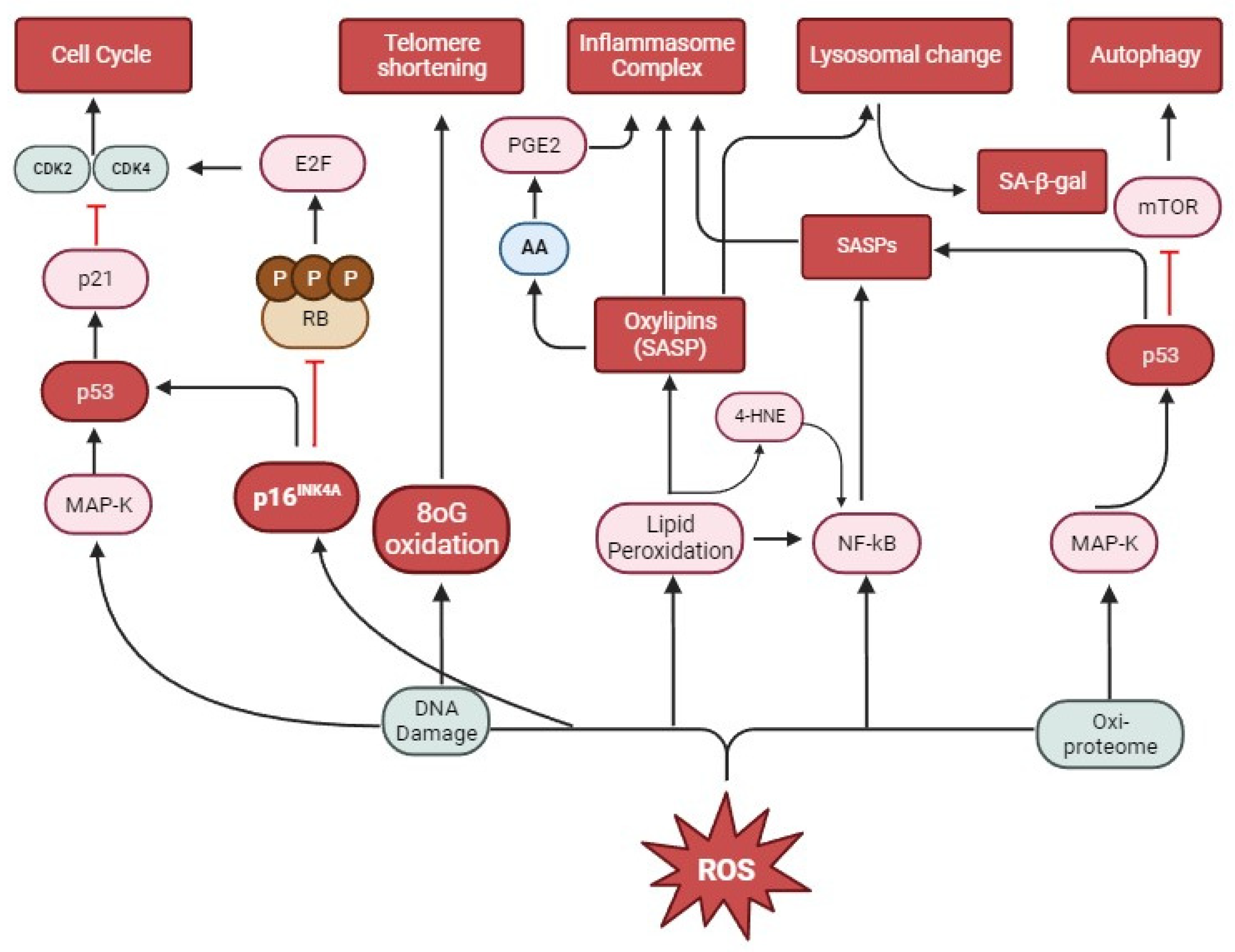
6. Oxidative Stress and Senescence

Due to the skin’s ability to regenerate, much of the damage caused by environmental stressors is transient, but continuous exposure associated with genetic factors and time activates the production of reactive oxygen species [119]. Since the skin is an external organ that requires constant cell renewal, the skin comprises different layers of metabolically active cells with high energy consumption [120]. In this context, mitochondria play a fundamental role in producing energy in the form of ATP for cells. However, this process results in increased ROS production [118,121,122].
These reactive molecules act on cells in the skin in response to different intrinsic and extrinsic stressors, activating the natural endogenous antioxidant defense [123]. Antioxidants are present in different layers of the skin. In the more external layer (epidermis), we find higher concentrations of antioxidants than in the innermost layer (dermis); however, in the stratum corneum of the epidermis, vitamins C and E (αγ and α-tocopherol), GSH and uric acid can be found [124]. However, with continuous stimuli, an imbalance occurs, and ROS production exceeds the synthesis of antioxidant enzymes, which becomes insufficient to neutralize the harmful effects of ROS. At this stage, tissue oxidative stress begins. The consumption or topical application of exogenous antioxidants helps to balance this effect [125,126,127]. Yet, the increase in ROS can activate different pathways via reacting with various compounds and participating in several redox reactions that drive cellular senescence in the skin. As a highly reactive species, ROS destroys cellular macromolecules such as proteins, lipids, and nucleic acids. Among the mechanisms modulated by ROS that induce senescence, the one that was most studied and, therefore, most consolidated is “DNA Damage” [128], probably because one of the main characteristics of senescence is the arrest of the cell cycle due to the shortening of telomeres or due to some damage to the DNA [128]. However, recent studies have identified that, before causing damage to the DNA, ROS targets the proteome first. During the oxidation of these proteins, the “oxi-proteome” prevents the repair of damaged DNA [129,130]. Around 35% of the proteins accumulated in senescent fibroblasts depend on oxidative processes [87], and this phenomenon has been associated with aging disorders [131]. The SIRT1 protein, for example, is an NAD+-dependent enzyme known as an autophagy initiator, and when suppressed by ROS, it induces senescence in keratinocytes [132].
Telomere shortening, a hallmark of senescence, also occurs through oxidative damage [133]. ROS can either directly damage mitochondrial DNA (mtDNA) or cause damage when in synergy with telomere damage [131]. This damage occurs due to a large amount of guanine present in telomeric DNA (8-hydroxyguanine) associated with the great affinity of ROS to oxidize guanine (8-oxoguanine (8oG)). Furthermore, ROS inhibits telomerase activity [134] and induces single-strand breaks (SSBs) on telomeres [133] (Figure 1). However, cellular senescence can be a great ally with anti-tumorigenic effects when it interrupts the cell cycle and, consequently, the proliferation of damaged or dysfunctional cells. Studies using murine models have highlighted the high prevalence of senescent cells in pre-malignant tumors, thus explaining the slow growth and low malignancy of these tumors [135]. In this model, senescence was associated with the activation of tumor suppressor proteins, such as p53, p16INK4A, and ARF, and their loss or inactivation led to tumor-malignant progression [136,137,138]. Furthermore, system cells rapidly eliminate senescent tumor cells, resulting in efficient tumor regression [135,139].
When cells activate DNA repair mechanisms, permanent structural and functional changes, such as senescence, trigger proteins with multiple functions, for example, the retinoblastoma (RB) family and the p53 proteins. Thus, the activation of tumor suppressors such as the RB protein and the p53 transcription factor via continuous ROS stimulation stimulates oncogene-induced senescence. In normal pathways, CDKs phosphorylate retinoblastoma proteins (RB1, RBL1, RBL2), reducing their ability to inactivate E2F, which is a protein necessary for cell cycle progression. However, during the senescence process, a CDK inhibitor known as p16INK4A prevents RB phosphorylation, repressing E2F and preventing cell cycle progression [140]. Furthermore, p16INK4A activates p53, which inhibits autophagy-inducing mTOR kinase (the mammalian target of rapamycin) [141]. However, although p16INK4A is a widely used marker for senescence, it may not be expressed by all senescent cells [104], thus revealing the importance of using multiple markers to detect senescence. p53, a protein encoded by the TP53 gene, also inhibits CDK4 and CDK2 kinases via p21, inducing cell cycle arrest [105]. When subjected to prolonged stress, excessive ROS production activates p53, suppressing the process of autophagy followed by apoptosis mediated by mTORC1, resulting in the accumulation of damaged cells [139] and the induction of the senescence process. A study carried out with murine models observed that, in adipose tissue, skeletal muscle, and eyes treated with a compound that removes senescent cells positive for p16INK4A, there was a delay in the onset of pathological phenotypes associated with aging. These findings indicate that cellular senescence is directly linked to age-related phenotypes and that the removal of senescent cells can prevent or delay aging-related pathologies and prolong life expectancy [15].
However, the many changes associated with the senescence process in the skin are directly related to mitochondria. The phosphorylation of the ATM, AKT, and mTOR complex increases mitochondrial biogenesis and, consequently, the DNA damage response (DDR) mediated by ROS. This promotes cell cycle arrest and SASP development and demonstrates the role of this organelle in both ox-inflammatory and DDR processes. SASPs, mainly characterized by pro-inflammatory cytokines, are transcriptionally regulated by NF-κB and CEBP-β in response to ROS-induced DNA damage, and their expression is maintained by autocrine feedback [141]. Thus, the activity of autophagy lysosomes and mitochondria are functionally interconnected during senescence. Under normal conditions, dysfunctional mitochondria undergo autophagy through lysosomal compartments [107]. However, the increase in ROS activates ataxia-telangiectasia mutation (ATM), a predominantly nuclear protein that is a member of the serine/threonine kinase family of proteins. This activated protein modulates p53-mediated DNA damage response targets [142] but also increases lysosomal acidity mediated by signaling extracellular signal-regulated kinase (Erk) activity, compromising the normal autophagy flow [143]. Thus, mitochondrial autophagy is hampered, and consequently, there is an accumulation of dysfunctional mitochondria and excessive production of ROS. The increase in lipid peroxidation during oxidative stress increases the content of intra-lysosomal lipofuscins in fibroblasts during senescence [19]. Furthermore, the increase in lysosomal-resident proteins such as β-galactosidase, associated with the delivery of autophagic proteins via the macro-autophagy (MA) and chaperone-mediated autophagy (CMA) pathways that occur in response to increased ROS [111], is also responsible for the change in lysosomal mass. It is noteworthy that the lysosomal β-galactosidase activity identified at pH 6.0 (SA-β-gal) is a byproduct generated via senescence and not by what causes cellular senescence [43].
ROS can also modulate skin senescence by activating SASPs. This complex mixture of factors can be produced as an ROS-stimulated DNA damage response [144]. Senescent cells can maintain the synthesis of SASP markers such as IL-6, TGF-β, VEGF, CCL2, and CCL20, which can reinforce senescence in an autocrine and paracrine manner [39]. Furthermore, it is reported that SASP factors that promote paracrine senescence, such as IL-1α and IL-1β, activate the inflammasome since IL-1β is only formed after activation via the inflammasome multiprotein complex. This complex is formed by caspase 1 and adapter molecules, such as NRLs and ASC [145,146,147]. Therefore, the synthesis of SASP factors is indisputably related to inflammatory diseases caused by senescent cells during the senescence and skin aging process. Within SASP, the 4-hydroxy-2-nonenal (4-HNE) is another molecule associated with inflammatory processes. 4-HNE is a highly reactive product of lipid peroxidation caused by the action of ROS, which can form adducts with cellular proteins and even DNA [148]. Human keratinocytes exposed to O3 inhibited cell proliferation and stimulated increased oxidative damage, which increased levels of 4-HNE and activated the NF-κB pathway, demonstrating that O3 is a potential inducer of skin inflammation [99]. The continuous action of this stressor has been associated with premature aging and senescence [149] (Figure 1).
Although the standard and alternative mechanisms that trigger senescence are well established, some gaps in the research still exist, and, as there is no single reliable marker for the detection of senescence in vivo, it is the consensus of the International Cell Senescence Association (ICSA) that at least one combination of more than two markers is used to identify a cell as senescent [40].

7. Oxylipins and Senescence

Lipids found in the skin are related to functions such as the epidermal lipid barrier, which prevents water loss and the entry of harmful substances [150], and also cell signaling, which ranges from membrane trafficking to the transduction signal [151]. This is in addition to the role of skin hydration through lipids produced in the sebaceous glands [152]. Therefore, some oxylipins are important for maintaining the biological functions of the skin, and knowing the molecules and pathways that activate the formation of these bioactive lipids can direct effective therapies. In this context, the role of Lipoxygenases (LOX), which are key enzymes in the synthesis of different oxylipins in various biological processes, has been investigated [153]. In the skin, for some decades now, LOX has been associated with the modulation and maintenance of the permeability barrier [154]. LOX oxidizes esterified structural lipids, such as ceramides, present in the corneal layer, and its deficiency negatively affects lipid levels essential for the barrier, as well as cornified envelope proteins [155].
However, in recent decades, the chemical or enzymatic modifications of lipids called epilipidoma [156] have been considered fundamental in understanding skin changes, including both pathological and common physiological changes in age. Furthermore, as already mentioned, 4-HNE is a widely used marker to characterize increased lipid peroxidation and the resulting accumulation of lipid-derived aldehydes in senescent cells [157] (Figure 1). In this session, we highlight the involvement of lipids resulting from oxidative stress, oxylipins [105], and their role in the senescence of skin cells.
Lipofuscin (LF) is a type of oxylipin found in lysosomes in pigments called the “age pigment” because they are frequently found during aging [158], promoting the senescence of skin cells [30]. LFs accumulate in lysosomes, leading to an increase in the lysosomal lumen. The presence of the already consolidated senescence marker SA-β-Gal and an increase in lysosomal mass have been associated with the presence of lipofuscin in senescent epithelial cells [159]. In this context, the interruption of the normal metabolic function of lysosomes, as well as the deficiency of the autophagy process, contribute to the maintenance of this cellular state [160].
Different oxylipins were identified in the composition of SASP, called SASP lipids [161]. These oxylipins, in addition to the characteristic pro-inflammatory role of SASPs, prevent senescent fibroblasts from being eliminated by the innate immune system, reducing the signaling mediated by Toll-like receptor (TLR) 2 and TLR6 in macrophages [162]. Arachidonic acid (AA), as well as prostaglandins (PGD2 and other variants), and leukotrienes are well-known lipid SASPs. Cyclooxygenase 2 (COX2) converts AA to PGD2, which activates a signaling cascade involving p53, which is a marker of cellular senescence [105,163]. Like prostaglandins, leukotrienes are synthesized from arachidonic acid cleaved during the oxidation of membrane phospholipids [164] and increase during cellular senescence [165]. Recent data demonstrate that increased lipid peroxidation drives increased metabolism, import, and lipid accumulation in senescent cells (SA-β-Gal-positive). Thus, oxidative imbalance promotes lipid peroxidation in skin cells, initiating a positive feedback cycle that maintains or triggers senescence, regardless of the persistence of other stressors [166].

8. Strategies to Prevent Skin Senescence

The search for understanding the different triggering factors and mechanisms involved in skin senescence has driven the investigation of different strategies to eliminate or prevent this cellular condition. These approaches are known as “senotherapy” or “senotherapeutic strategies” [167] and can be based on the following two distinct pathways: (1) senolytic pathway: the selective induction of cell death, used especially in cancer treatment (chemotherapy) [168] and (2) senomorphs, to control deleterious effects through SASP inhibition. However, a third strategy, “immune system enhancement”, has been proposed to minimize the damage of skin senescence [169]. It is worth mentioning that there are currently no officially clinically approved medications considered a “gold standard” in the treatment of skin senescence [30].
The clear relationship between ROS levels and senescence has sparked great interest in research into antioxidant compounds. Senotherapy based on antioxidants of natural or synthetic origin has shown promising results [170]. Synthetic flavonoids such as apigenin and kaempferol [171], as well as glucocorticoids and cortisone [172], regulate senescence through senomorphic mechanisms, inhibiting the activity of NF-κB-p65, which are activators of SASPs in skin cells. Ramrammycin, an mTOR inhibitor, also reduces the synthesis of SASP (IL-6) by suppressing the NF-κB pathway [173].
Resveratrol was investigated due to its potent antioxidant and anti-inflammatory capacity in senescent skin cells and induced a reduction in SASP by increasing the expression of AMPK-FOXO3 [174]. However, some studies have demonstrated that resveratrol also induces apoptosis and cellular senescence in primary and cancer cells through the activation of DNA damage response [175,176]. Other synthetic compounds that reduce SASPs are UR-13756 and BIRB 796 [177], which act by inhibiting the p38 MAPK pathway. Thus, the main signaling pathways that modulate SASP appear to converge on the NF-κB pathway, demonstrating the importance of this pathway for the study of senescence.
The investigation of synthetic compounds, such as ferulic acid [178] and quercetin [179], and natural compounds, such as nectrandrin B [180], used as senotherapy, demonstrates action that aims to maintain and progress the cell cycle, reversing senescence through a reduction in ROS and inhibition of the p16, p21, p27, p53 pathways. Nectrandrin B [180] is a natural compound composed of six herbs (Rehmannia glutinosa, Panax Ginseng, Asparagus cochinchinensis, Ophiopogon japonicus, Cortex Lycii., Poria cocos) [181] and Yang Yan Qing E Wan (YYQEW) [182].
In relation to the already-known effect of oxylipins on lysosomal compartments, induced autophagy was identified as an important therapeutic target in preventing cellular senescence [183]. In this context, caffeine was reported as a potent activator of signaling pathways for autophagy and the reduction in ROS levels; however, it may also benefit tumor cell growth [184]. The strategies presented in this session can be seen in Table 1.
In addition to the different strategies mentioned above, it is important to highlight that the skin’s antioxidant system is composed of enzymatic substances (glutathione peroxidase (GPx), catalase (CAT), and superoxide dismutase (SOD)) and non-enzymatic substances (such as estradiol and melatonin, vitamins E and C) [185]. In addition to the known action of SOD in neutralizing reactive molecules, there are reports that vitamin C positively modulates the expression of SOD and prevents collagen degradation [186]. It is already known that vitamins that act as skin antioxidants are mainly taken from food [93,187]. Furthermore, foods such as Ginseng [188], the seed of Prunus mume [189], in addition to the consumption of probiotics and prebiotics [190], have demonstrated efficiency in modulating the endogenous antioxidant capacity of the skin, reducing oxidative damage and, consequently, aging and/or senescence. These studies indicate that, in addition to the topical use of synthetic or natural formulas, a complementary strategy should be promoted for the consumption of functional foods to combat senescence and aging [191]. It is worth mentioning here that the consumption of synthetic antioxidants in high doses can harm the body and activate oxidative stress, and the safest way to ingest these antioxidants would be in the form of an adequate diet [191,192].
Thus, identifying new mechanisms as a therapeutic target, as well as identifying new bioactives that prevent telomere loss, cell cycle arrest, and increased ROS alongside inflammasome activation, is a new and exciting area of investigation that may well offer new treatments against skin senescence.

9. Conclusions

The redox imbalance promoted by the increase in ROS resulting from continuous stressful stimuli overloads the endogenous antioxidant enzymatic system, boosting the senescence process in the skin. ROS activate different pathways that culminate mainly in oxidative damage to telomeres and also in damage to DNA, activating pathways that block CDks, especially p16INK4A (retinoblastoma protein inhibitor) and p53, preventing cell cycle progression. Furthermore, lipid peroxidation, as a result of ROS, alters the content and, consequently, the lysosomal morphology of SA-β-gal. These events compromise the processes of autophagy and apoptosis by silencing proteins, such as mTOR and increasing the number of dysfunctional organelles in the cytoplasm such as mitochondria. The mechanistic basis of cutaneous inflammatory processes in senescent cells is directly related to the production of SASP and the formation of oxylipins, which, in a positive feedback process, activates the NF-κB pathway, promoting the formation of the inflammasome complex, releasing different pro-inflammatory factors and cytokines. However, these markers, in general, must be evaluated together, as they are not reliable when investigated in isolation.
Therapeutic strategies called senotherapy have investigated the action of different compounds. Yet, more studies are needed to verify their short and long-term effects, understand the molecular mechanisms that involve redox action in senescent cells, and provide new biomarkers that mainly identify therapeutic targets to combat-related pathologies.

Author Contributions

Conceptualization, M.M.S. and G.V.; methodology, M.M.S. and G.V.; validation, M.M.S., G.V. and R.V.G.; formal analysis, M.M.S.; investigation, M.M.S.; resources, G.V.; data curation, M.M.S., G.V. and R.V.G.; writing—original draft preparation, M.M.S.; writing—review and editing, G.V. and R.V.G.; visualization, M.M.S., G.V. and R.V.G.; supervision, G.V. and R.V.G. All authors have read and agreed to the published version of the manuscript.

Funding

This research received no external funding.

Conflicts of Interest

The authors declare no conflict of interest.

References

  1. de Bengy, A.-F.; Decorps, J.; Martin, L.S.; Pagnon, A.; Chevalier, F.P.; Sigaudo-Roussel, D.; Fromy, B. Alpha-Lipoic Acid Supplementation Restores Early Age-Related Sensory and Endothelial Dysfunction in the Skin. Biomedicines 2022, 10, 2887. [Google Scholar] [CrossRef]
  2. Csekes, E.; Račková, L. Skin Aging, Cellular Senescence and Natural Polyphenols. Int. J. Mol. Sci. 2021, 22, 12641. [Google Scholar] [CrossRef] [PubMed]
  3. Papaconstantinou, J. The Role of Signaling Pathways of Inflammation and Oxidative Stress in Development of Senescence and Aging Phenotypes in Cardiovascular Disease. Cells 2019, 8, 1383. [Google Scholar] [CrossRef]
  4. North, B.J.; Sinclair, D.A. The Intersection Between Aging and Cardiovascular Disease. Circ. Res. 2012, 110, 1097–1108. [Google Scholar] [CrossRef]
  5. Merkt, W.; Bueno, M.; Mora, A.L.; Lagares, D. Senotherapeutics: Targeting Senescence in Idiopathic Pulmonary Fibrosis. Semin. Cell Dev. Biol. 2020, 101, 104–110. [Google Scholar] [CrossRef]
  6. Tucker-Drob, E.M. Cognitive Aging and Dementia: A Life-Span Perspective. Annu. Rev. Dev. Psychol. 2019, 1, 177–196. [Google Scholar] [CrossRef]
  7. Lutshumba, J.; Nikolajczyk, B.S.; Bachstetter, A.D. Dysregulation of Systemic Immunity in Aging and Dementia. Front. Cell. Neurosci. 2021, 15, 652111. [Google Scholar] [CrossRef] [PubMed]
  8. Nanayakkara, N.; Curtis, A.J.; Heritier, S.; Gadowski, A.M.; Pavkov, M.E.; Kenealy, T.; Owens, D.R.; Thomas, R.L.; Song, S.; Wong, J.; et al. Impact of Age at Type 2 Diabetes Mellitus Diagnosis on Mortality and Vascular Complications: Systematic Review and Meta-Analyses. Diabetologia 2021, 64, 275–287. [Google Scholar] [CrossRef] [PubMed]
  9. Bellary, S.; Kyrou, I.; Brown, J.E.; Bailey, C.J. Type 2 Diabetes Mellitus in Older Adults: Clinical Considerations and Management. Nat. Rev. Endocrinol. 2021, 17, 534–548. [Google Scholar] [CrossRef] [PubMed]
  10. Pignolo, R.J.; Law, S.F.; Chandra, A. Bone Aging, Cellular Senescence, and Osteoporosis. JBMR Plus 2021, 5, e10488. [Google Scholar] [CrossRef] [PubMed]
  11. Nagira, K.; Ikuta, Y.; Shinohara, M.; Sanada, Y.; Omoto, T.; Kanaya, H.; Nakasa, T.; Ishikawa, M.; Adachi, N.; Miyaki, S.; et al. Histological Scoring System for Subchondral Bone Changes in Murine Models of Joint Aging and Osteoarthritis. Sci. Rep. 2020, 10, 10077. [Google Scholar] [CrossRef]
  12. Xia, C.; Dong, X.; Li, H.; Cao, M.; Sun, D.; He, S.; Yang, F.; Yan, X.; Zhang, S.; Li, N.; et al. Cancer Statistics in China and United States, 2022: Profiles, Trends, and Determinants. Chin. Med. J. 2022, 135, 584–590. [Google Scholar] [CrossRef] [PubMed]
  13. Jammal, A.A.; Berchuck, S.I.; Thompson, A.C.; Costa, V.P.; Medeiros, F.A. The Effect of Age on Increasing Susceptibility to Retinal Nerve Fiber Layer Loss in Glaucoma. Investig. Opthalmol. Vis. Sci. 2020, 61, 8. [Google Scholar] [CrossRef] [PubMed]
  14. Parrado, C.; Mercado-Saenz, S.; Perez-Davo, A.; Gilaberte, Y.; Gonzalez, S.; Juarranz, A. Environmental Stressors on Skin Aging. Mechanistic Insights. Front. Pharmacol. 2019, 10, 759. [Google Scholar] [CrossRef] [PubMed]
  15. Baker, D.J.; Wijshake, T.; Tchkonia, T.; LeBrasseur, N.K.; Childs, B.G.; van de Sluis, B.; Kirkland, J.L.; van Deursen, J.M. Clearance of p16INK4A -Positive Senescent Cells Delays Ageing-Associated Disorders. Nature 2011, 479, 232–236. [Google Scholar] [CrossRef] [PubMed]
  16. Martin, J.; Sheaff, M. The Pathology of Ageing: Concepts and Mechanisms. J. Pathol. 2007, 211, 111–113. [Google Scholar] [CrossRef]
  17. Duan, R.; Fu, Q.; Sun, Y.; Li, Q. Epigenetic Clock: A Promising Biomarker and Practical Tool in Aging. Ageing Res. Rev. 2022, 81, 101743. [Google Scholar] [CrossRef]
  18. Wagner, W. The Link Between Epigenetic Clocks for Aging and Senescence. Front. Genet. 2019, 10, 303. [Google Scholar] [CrossRef]
  19. McHugh, D.; Gil, J. Senescence and Aging: Causes, Consequences, and Therapeutic Avenues. J. Cell Biol. 2018, 217, 65–77. [Google Scholar] [CrossRef]
  20. Valacchi, G.; Virgili, F.; Cervellati, C.; Pecorelli, A. OxInflammation: From Subclinical Condition to Pathological Biomarker. Front. Physiol. 2018, 9, 858. [Google Scholar] [CrossRef]
  21. Ogrodnik, M.; Gladyshev, V.N. The Meaning of Adaptation in Aging: Insights from Cellular Senescence, Epigenetic Clocks and Stem Cell Alterations. Nat. Aging 2023, 3, 766–775. [Google Scholar] [CrossRef]
  22. López-Otín, C.; Blasco, M.A.; Partridge, L.; Serrano, M.; Kroemer, G. The Hallmarks of Aging. Cell 2013, 153, 1194–1217. [Google Scholar] [CrossRef] [PubMed]
  23. Di Micco, R.; Krizhanovsky, V.; Baker, D.; d’Adda di Fagagna, F. Cellular Senescence in Ageing: From Mechanisms to Therapeutic Opportunities. Nat. Rev. Mol. Cell Biol. 2021, 22, 75–95. [Google Scholar] [CrossRef] [PubMed]
  24. Schmeer, C.; Kretz, A.; Wengerodt, D.; Stojiljkovic, M.; Witte, O.W. Dissecting Aging and Senescence—Current Concepts and Open Lessons. Cells 2019, 8, 1446. [Google Scholar] [CrossRef] [PubMed]
  25. Davalli, P.; Mitic, T.; Caporali, A.; Lauriola, A.; D’Arca, D. ROS, Cell Senescence, and Novel Molecular Mechanisms in Aging and Age-Related Diseases. Oxidative Med. Cell. Longev. 2016, 2016, 3565127. [Google Scholar] [CrossRef] [PubMed]
  26. UN World Population Prospects 2022: Summary of Results Ten Key Messages; United Nations, Department of Economic and Social Affairs, Population Division: New York, NY, USA, 2022; pp. 2–3.
  27. Krutmann, J.; Schikowski, T.; Morita, A.; Berneburg, M. Environmentally-Induced (Extrinsic) Skin Aging: Exposomal Factors and Underlying Mechanisms. J. Investig. Dermatol. 2021, 141, 1096–1103. [Google Scholar] [CrossRef] [PubMed]
  28. Augello, F.R.; Lombardi, F.; Artone, S.; Ciafarone, A.; Altamura, S.; Di Marzio, L.; Cifone, M.G.; Palumbo, P.; Giuliani, M.; Cinque, B. Evaluation of the Effectiveness of an Innovative Polycomponent Formulation on Adult and Aged Human Dermal Fibroblasts. Biomedicines 2023, 11, 2410. [Google Scholar] [CrossRef] [PubMed]
  29. Hughes, B.K.; Bishop, C.L. Current Understanding of the Role of Senescent Melanocytes in Skin Ageing. Biomedicines 2022, 10, 3111. [Google Scholar] [CrossRef]
  30. Bulbiankova, D.; Díaz-Puertas, R.; Álvarez-Martínez, F.J.; Herranz-López, M.; Barrajón-Catalán, E.; Micol, V. Hallmarks and Biomarkers of Skin Senescence: An Updated Review of Skin Senotherapeutics. Antioxidants 2023, 12, 444. [Google Scholar] [CrossRef]
  31. Hayflick, L.; Moorhead, P.S. The Serial Cultivation of Human Diploid Cell Strains. Exp. Cell Res. 1961, 25, 585–621. [Google Scholar] [CrossRef]
  32. Wlaschek, M.; Maity, P.; Makrantonaki, E.; Scharffetter-Kochanek, K. Connective Tissue and Fibroblast Senescence in Skin Aging. J. Investig. Dermatol. 2021, 141, 985–992. [Google Scholar] [CrossRef]
  33. Epel, E.S.; Merkin, S.S.; Cawthon, R.; Blackburn, E.H.; Adler, N.E.; Pletcher, M.J.; Seeman, T.E. The Rate of Leukocyte Telomere Shortening Predicts Mortality from Cardiovascular Disease in Elderly Men: A Novel Demonstration. Aging 2008, 1, 81–88. [Google Scholar] [CrossRef]
  34. Aubert, G.; Lansdorp, P.M. Telomeres and Aging. Physiol. Rev. 2008, 88, 557–579. [Google Scholar] [CrossRef]
  35. Muñoz-Espín, D.; Serrano, M. Cellular Senescence: From Physiology to Pathology. Nat. Rev. Mol. Cell Biol. 2014, 15, 482–496. [Google Scholar] [CrossRef]
  36. Demaria, M.; Ohtani, N.; Youssef, S.A.; Rodier, F.; Toussaint, W.; Mitchell, J.R.; Laberge, R.-M.; Vijg, J.; Van Steeg, H.; Dollé, M.E.T.; et al. An Essential Role for Senescent Cells in Optimal Wound Healing through Secretion of PDGF-AA. Dev. Cell 2014, 31, 722–733. [Google Scholar] [CrossRef]
  37. Wiley, C.D.; Velarde, M.C.; Lecot, P.; Liu, S.; Sarnoski, E.A.; Freund, A.; Shirakawa, K.; Lim, H.W.; Davis, S.S.; Ramanathan, A.; et al. Mitochondrial Dysfunction Induces Senescence with a Distinct Secretory Phenotype. Cell Metab. 2016, 23, 303–314. [Google Scholar] [CrossRef] [PubMed]
  38. Petrova, N.V.; Velichko, A.K.; Razin, S.V.; Kantidze, O.L. Small Molecule Compounds That Induce Cellular Senescence. Aging Cell 2016, 15, 999–1017. [Google Scholar] [CrossRef]
  39. Acosta, J.C.; Banito, A.; Wuestefeld, T.; Georgilis, A.; Janich, P.; Morton, J.P.; Athineos, D.; Kang, T.-W.; Lasitschka, F.; Andrulis, M.; et al. A Complex Secretory Program Orchestrated by the Inflammasome Controls Paracrine Senescence. Nat. Cell Biol. 2013, 15, 978–990. [Google Scholar] [CrossRef] [PubMed]
  40. Gorgoulis, V.; Adams, P.D.; Alimonti, A.; Bennett, D.C.; Bischof, O.; Bishop, C.; Campisi, J.; Collado, M.; Evangelou, K.; Ferbeyre, G.; et al. Cellular Senescence: Defining a Path Forward. Cell 2019, 179, 813–827. [Google Scholar] [CrossRef] [PubMed]
  41. González-Gualda, E.; Baker, A.G.; Fruk, L.; Muñoz-Espín, D. A Guide to Assessing Cellular Senescence In Vitro and In Vivo. FEBS J. 2021, 288, 56–80. [Google Scholar] [CrossRef]
  42. Itahana, K.; Campisi, J.; Dimri, G.P. Methods to Detect Biomarkers of Cellular Senescence. Methods Mol. Biol. 2007, 371, 21–31. [Google Scholar]
  43. Lee, B.Y.; Han, J.A.; Im, J.S.; Morrone, A.; Johung, K.; Goodwin, E.C.; Kleijer, W.J.; DiMaio, D.; Hwang, E.S. Senescence-associated Β-galactosidase Is Lysosomal Β-galactosidase. Aging Cell 2006, 5, 187–195. [Google Scholar] [CrossRef] [PubMed]
  44. Gonzalez-Meljem, J.M.; Apps, J.R.; Fraser, H.C.; Martinez-Barbera, J.P. Paracrine Roles of Cellular Senescence in Promoting Tumourigenesis. Br. J. Cancer 2018, 118, 1283–1288. [Google Scholar] [CrossRef] [PubMed]
  45. Lee, Y.I.; Choi, S.; Roh, W.S.; Lee, J.H.; Kim, T.-G. Cellular Senescence and Inflammaging in the Skin Microenvironment. Int. J. Mol. Sci. 2021, 22, 3849. [Google Scholar] [CrossRef]
  46. Aioi, A. Inflammaging in Skin and Intrinsic Underlying Factors. Trends Immunother. 2021, 5, 44–53. [Google Scholar] [CrossRef]
  47. D’Arcangelo, D.; Tinaburri, L.; Dellambra, E. The Role of p16INK4A Pathway in Human Epidermal Stem Cell Self-Renewal, Aging and Cancer. Int. J. Mol. Sci. 2017, 18, 1591. [Google Scholar] [CrossRef] [PubMed]
  48. Gonzalez, A.C.d.O.; Costa, T.F.; Andrade, Z.d.A.; Medrado, A.R.A.P. Wound Healing—A Literature Review. An. Bras. Dermatol. 2016, 91, 614–620. [Google Scholar] [CrossRef]
  49. Pullar, J.; Carr, A.; Vissers, M. The Roles of Vitamin C in Skin Health. Nutrients 2017, 9, 866. [Google Scholar] [CrossRef]
  50. Ho, C.Y.; Dreesen, O. Faces of Cellular Senescence in Skin Aging. Mech. Ageing Dev. 2021, 198, 111525. [Google Scholar] [CrossRef]
  51. Chen, J.; Liu, Y.; Zhao, Z.; Qiu, J. Oxidative Stress in the Skin: Impact and Related Protection. Int. J. Cosmet. Sci. 2021, 43, 495–509. [Google Scholar] [CrossRef]
  52. Victorelli, S.; Lagnado, A.; Halim, J.; Moore, W.; Talbot, D.; Barrett, K.; Chapman, J.; Birch, J.; Ogrodnik, M.; Meves, A.; et al. Senescent Human Melanocytes Drive Skin Ageing via Paracrine Telomere Dysfunction. EMBO J. 2019, 38, e101982. [Google Scholar] [CrossRef]
  53. Krunic, D.; Moshir, S.; Greulich-Bode, K.M.; Figueroa, R.; Cerezo, A.; Stammer, H.; Stark, H.-J.; Gray, S.G.; Nielsen, K.V.; Hartschuh, W.; et al. Tissue Context-Activated Telomerase in Human Epidermis Correlates with Little Age-Dependent Telomere Loss. Biochim. Biophys. Acta (BBA) Mol. Basis Dis. 2009, 1792, 297–308. [Google Scholar] [CrossRef]
  54. Low, E.; Alimohammadiha, G.; Smith, L.A.; Costello, L.F.; Przyborski, S.A.; von Zglinicki, T.; Miwa, S. How Good Is the Evidence That Cellular Senescence Causes Skin Ageing? Ageing Res. Rev. 2021, 71, 101456. [Google Scholar] [CrossRef]
  55. Laethem, A.V.; Claerhout, S.; Garmyn, M.; Agostinis, P. The Sunburn Cell: Regulation of Death and Survival of the Keratinocyte. Int. J. Biochem. Cell Biol. 2005, 37, 1547–1553. [Google Scholar] [CrossRef]
  56. Velarde, M.C.; Flynn, J.M.; Day, N.U.; Melov, S.; Campisi, J. Mitochondrial Oxidative Stress Caused by Sod2 Deficiency Promotes Cellular Senescence and Aging Phenotypes in the Skin. Aging 2012, 4, 3–12. [Google Scholar] [CrossRef]
  57. Rübe, C.E.; Bäumert, C.; Schuler, N.; Isermann, A.; Schmal, Z.; Glanemann, M.; Mann, C.; Scherthan, H. Human Skin Aging Is Associated with Increased Expression of the Histone Variant H2A.J in the Epidermis. NPJ Aging Mech. Dis. 2021, 7, 7. [Google Scholar] [CrossRef]
  58. Jenkins, N.C.; Grossman, D. Role of Melanin in Melanocyte Dysregulation of Reactive Oxygen Species. BioMed Res. Int. 2013, 2013, 908797. [Google Scholar] [CrossRef]
  59. Jenkins, N.C.; Liu, T.; Cassidy, P.; Leachman, S.A.; Boucher, K.M.; Goodson, A.G.; Samadashwily, G.; Grossman, D. The p16INK4A Tumor Suppressor Regulates Cellular Oxidative Stress. Oncogene 2011, 30, 265–274. [Google Scholar] [CrossRef] [PubMed]
  60. D’Errico, M.; Lemma, T.; Calcagnile, A.; De Santis, L.P.; Dogliotti, E. Cell Type and DNA Damage Specific Response of Human Skin Cells to Environmental Agents. Mutat. Res. Fundam. Mol. Mech. Mutagen. 2007, 614, 37–47. [Google Scholar] [CrossRef] [PubMed]
  61. Tigges, J.; Krutmann, J.; Fritsche, E.; Haendeler, J.; Schaal, H.; Fischer, J.W.; Kalfalah, F.; Reinke, H.; Reifenberger, G.; Stühler, K.; et al. The Hallmarks of Fibroblast Ageing. Mech. Ageing Dev. 2014, 138, 26–44. [Google Scholar] [CrossRef] [PubMed]
  62. Rodier, F.; Coppé, J.-P.; Patil, C.K.; Hoeijmakers, W.A.M.; Muñoz, D.P.; Raza, S.R.; Freund, A.; Campeau, E.; Davalos, A.R.; Campisi, J. Persistent DNA Damage Signalling Triggers Senescence-Associated Inflammatory Cytokine Secretion. Nat. Cell Biol. 2009, 11, 973–979. [Google Scholar] [CrossRef]
  63. Gruber, F.; Kremslehner, C.; Eckhart, L.; Tschachler, E. Cell Aging and Cellular Senescence in Skin Aging—Recent Advances in Fibroblast and Keratinocyte Biology. Exp. Gerontol. 2020, 130, 110780. [Google Scholar] [CrossRef]
  64. Chung, C.L.; Lawrence, I.; Hoffman, M.; Elgindi, D.; Nadhan, K.; Potnis, M.; Jin, A.; Sershon, C.; Binnebose, R.; Lorenzini, A.; et al. Topical Rapamycin Reduces Markers of Senescence and Aging in Human Skin: An Exploratory, Prospective, Randomized Trial. GeroScience 2019, 41, 861–869. [Google Scholar] [CrossRef]
  65. McDaniel, D.; Farris, P.; Valacchi, G. Atmospheric Skin Aging—Contributors and Inhibitors. J. Cosmet. Dermatol. 2018, 17, 124–137. [Google Scholar] [CrossRef]
  66. Idda, M.L.; McClusky, W.G.; Lodde, V.; Munk, R.; Abdelmohsen, K.; Rossi, M.; Gorospe, M. Survey of Senescent Cell Markers with Age in Human Tissues. Aging 2020, 12, 4052–4066. [Google Scholar] [CrossRef]
  67. Farage, M.A.; Miller, K.W.; Elsner, P.; Maibach, H.I. Characteristics of the Aging Skin. Adv. Wound Care 2013, 2, 5–10. [Google Scholar] [CrossRef]
  68. Dimri, G.P.; Lee, X.; Basile, G.; Acosta, M.; Scott, G.; Roskelley, C.; Medrano, E.E.; Linskens, M.; Rubelj, I.; Pereira-Smith, O. A Biomarker That Identifies Senescent Human Cells in Culture and in Aging Skin in Vivo. Proc. Natl. Acad. Sci. USA 1995, 92, 9363–9367. [Google Scholar] [CrossRef]
  69. Ressler, S.; Bartkova, J.; Niederegger, H.; Bartek, J.; Scharffetter-Kochanek, K.; Jansen-Dürr, P.; Wlaschek, M. P16 INK4A Is a Robust in Vivo Biomarker of Cellular Aging in Human Skin. Aging Cell 2006, 5, 379–389. [Google Scholar] [CrossRef] [PubMed]
  70. Gonzales, K.A.U.; Fuchs, E. Skin and Its Regenerative Powers: An Alliance between Stem Cells and Their Niche. Dev. Cell 2017, 43, 387–401. [Google Scholar] [CrossRef] [PubMed]
  71. Lee, H.-J.; Kim, M. Skin Barrier Function and the Microbiome. Int. J. Mol. Sci. 2022, 23, 13071. [Google Scholar] [CrossRef] [PubMed]
  72. Lu, S.-Y.; Chang, K.-W.; Liu, C.-J.; Tseng, Y.-H.; Lu, H.-H.; Lee, S.-Y.; Lin, S.-C. Ripe Areca Nut Extract Induces G 1 Phase Arrests and Senescence-Associated Phenotypes in Normal Human Oral Keratinocyte. Carcinogenesis 2006, 27, 1273–1284. [Google Scholar] [CrossRef]
  73. Suzuki, Y.; Takaya, K.; Watanabe, S.; Otaki, M.; Kono, H.; Kishi, K. Evaluation of the Effect of Age of the Younger Mice on the Rejuvenation of the Older Mice by Heterochronic Parabiosis. Aging 2022, 14, 2507–2512. [Google Scholar] [CrossRef]
  74. Kim, J.C.; Park, T.J.; Kang, H.Y. Skin-Aging Pigmentation: Who Is the Real Enemy? Cells 2022, 11, 2541. [Google Scholar] [CrossRef]
  75. Martic, I.; Wedel, S.; Jansen-Dürr, P.; Cavinato, M. A New Model to Investigate UVB-Induced Cellular Senescence and Pigmentation in Melanocytes. Mech. Ageing Dev. 2020, 190, 111322. [Google Scholar] [CrossRef]
  76. Bandyopadhyay, D.M. Melanin Accumulation Accelerates Melanocyte Senescence by a Mechanism Involving P16 INK4a /CDK4/PRB and E2F1. Ann. N. Y. Acad. Sci. 2000, 908, 71–84. [Google Scholar] [CrossRef] [PubMed]
  77. Wang, A.S.; Dreesen, O. Biomarkers of Cellular Senescence and Skin Aging. Front. Genet. 2018, 9, 247. [Google Scholar] [CrossRef]
  78. Singh, K.; Maity, P.; Krug, L.; Meyer, P.; Treiber, N.; Lucas, T.; Basu, A.; Kochanek, S.; Wlaschek, M.; Geiger, H.; et al. Superoxide Anion Radicals Induce IGF-1 Resistance through Concomitant Activation of PTP1B and PTEN. EMBO Mol. Med. 2015, 7, 59–77. [Google Scholar] [CrossRef] [PubMed]
  79. Kovacs, D.; Bastonini, E.; Ottaviani, M.; Cota, C.; Migliano, E.; Dell’Anna, M.L.; Picardo, M. Vitiligo Skin: Exploring the Dermal Compartment. J. Investig. Dermatol. 2018, 138, 394–404. [Google Scholar] [CrossRef]
  80. Ciuciulete, A.R.; Stepan, A.E.; Badiu, A.M.; Andreiana, B.C.; Florescu, M.M.; Simionescu, C.E.; Vîlcea, A.M. E-Cadherin, Fibronectin and Slug Immunoexpression in Non-Melanoma Skin Cancers. Rom. J. Morphol. Embryol. 2021, 62, 705–712. [Google Scholar] [CrossRef] [PubMed]
  81. Malaquin, N.; Vercamer, C.; Bouali, F.; Martien, S.; Deruy, E.; Wernert, N.; Chwastyniak, M.; Pinet, F.; Abbadie, C.; Pourtier, A. Senescent Fibroblasts Enhance Early Skin Carcinogenic Events via a Paracrine MMP-PAR-1 Axis. PLoS ONE 2013, 8, e63607. [Google Scholar] [CrossRef]
  82. Quan, T.; Fisher, G.J. Role of Age-Associated Alterations of the Dermal Extracellular Matrix Microenvironment in Human Skin Aging: A Mini-Review. Gerontology 2015, 61, 427–434. [Google Scholar] [CrossRef] [PubMed]
  83. Fuks, K.B.; Woodby, B.; Valacchi, G. Skin Damage by Tropospheric Ozone. Der Hautarzt 2019, 70, 163–168. [Google Scholar] [CrossRef] [PubMed]
  84. Souyoul, S.A.; Saussy, K.P.; Lupo, M.P. Nutraceuticals: A Review. Dermatol. Ther. 2018, 8, 5–16. [Google Scholar] [CrossRef] [PubMed]
  85. Proksch, E. PH in Nature, Humans and Skin. J. Dermatol. 2018, 45, 1044–1052. [Google Scholar] [CrossRef] [PubMed]
  86. Petracca, B.; Rothen-Rutishauser, B.; Valacchi, G.; Eeman, M. Bench Approaches to Study the Detrimental Cutaneous Impact of Tropospheric Ozone. J. Exp. Sci. Environ. Epidemiol. 2021, 31, 137–148. [Google Scholar] [CrossRef] [PubMed]
  87. Holden, K.A.; Lee, A.R.; Hawcutt, D.B.; Sinha, I.P. The Impact of Poor Housing and Indoor Air Quality on Respiratory Health in Children. Breathe 2023, 19, 230058. [Google Scholar] [CrossRef] [PubMed]
  88. Farris, P.K.; Valacchi, G. Ultraviolet Light Protection: Is It Really Enough? Antioxidants 2022, 11, 1484. [Google Scholar] [CrossRef]
  89. Valacchi, G.; Sticozzi, C.; Pecorelli, A.; Cervellati, F.; Cervellati, C.; Maioli, E. Cutaneous Responses to Environmental Stressors. Ann. N. Y. Acad. Sci. 2012, 1271, 75–81. [Google Scholar] [CrossRef]
  90. Gerasymchuk, M.; Cherkasova, V.; Kovalchuk, O.; Kovalchuk, I. The Role of MicroRNAs in Organismal and Skin Aging. Int. J. Mol. Sci. 2020, 21, 5281. [Google Scholar] [CrossRef]
  91. Bauwens, E.; Parée, T.; Meurant, S.; Bouriez, I.; Hannart, C.; Wéra, A.-C.; Khelfi, A.; Fattaccioli, A.; Burteau, S.; Demazy, C.; et al. Senescence Induced by UVB in Keratinocytes Impairs Amino Acids Balance. J. Investig. Dermatol. 2023, 143, 554–565.e9. [Google Scholar] [CrossRef]
  92. Miyazawa, T.; Abe, C.; Burdeos, G.C.; Matsumoto, A.; Toda, M. Food Antioxidants and Aging: Theory, Current Evidence and Perspectives. Nutraceuticals 2022, 2, 181–204. [Google Scholar] [CrossRef]
  93. Cao, C.; Xiao, Z.; Wu, Y.; Ge, C. Diet and Skin Aging—From the Perspective of Food Nutrition. Nutrients 2020, 12, 870. [Google Scholar] [CrossRef]
  94. McMillan, T.J.; Leatherman, E.; Ridley, A.; Shorrocks, J.; Tobi, S.E.; Whiteside, J.R. Cellular Effects of Long Wavelength UV Light (UVA) in Mammalian Cells. J. Pharm. Pharmacol. 2010, 60, 969–976. [Google Scholar] [CrossRef]
  95. Scharffetter–Kochanek, K.; Brenneisen, P.; Wenk, J.; Herrmann, G.; Ma, W.; Kuhr, L.; Meewes, C.; Wlaschek, M. Photoaging of the Skin from Phenotype to Mechanisms. Exp. Gerontol. 2000, 35, 307–316. [Google Scholar] [CrossRef] [PubMed]
  96. Fitsiou, E.; Pulido, T.; Campisi, J.; Alimirah, F.; Demaria, M. Cellular Senescence and the Senescence-Associated Secretory Phenotype as Drivers of Skin Photoaging. J. Investig. Dermatol. 2021, 141, 1119–1126. [Google Scholar] [CrossRef] [PubMed]
  97. Kammeyer, A.; Luiten, R.M. Oxidation Events and Skin Aging. Ageing Res. Rev. 2015, 21, 16–29. [Google Scholar] [CrossRef] [PubMed]
  98. Valacchi, G.; Fortino, V.; Bocci, V. The Dual Action of Ozone on the Skin. Br. J. Dermatol. 2005, 153, 1096–1100. [Google Scholar] [CrossRef] [PubMed]
  99. Valacchi, G.; Sticozzi, C.; Belmonte, G.; Cervellati, F.; Demaude, J.; Chen, N.; Krol, Y.; Oresajo, C. Vitamin C Compound Mixtures Prevent Ozone-Induced Oxidative Damage in Human Keratinocytes as Initial Assessment of Pollution Protection. PLoS ONE 2015, 10, e0131097. [Google Scholar] [CrossRef]
  100. Kafoury, R.M.; Pryor, W.A.; Squadrito, G.L.; Salgo, M.G.; Zou, X.; Friedman, M. Lipid Ozonation Products Activate Phospholipases A2, C, and D. Toxicol. Appl. Pharmacol. 1998, 150, 338–349. [Google Scholar] [CrossRef]
  101. Reynolds, W.J.; Eje, N.; Christensen, P.; Li, W.; Daly, S.M.; Parsa, R.; Chavan, B.; Birch-Machin, M.A. Biological Effects of Air Pollution on the Function of Human Skin Equivalents. FASEB BioAdvances 2023, 5, 470–483. [Google Scholar] [CrossRef] [PubMed]
  102. Zdanov, S.; Bernard, D.; Debacqchainiaux, F.; Martien, S.; Gosselin, K.; Vercamer, C.; Chelli, F.; Toussaint, O.; Abbadie, C. Normal or Stress-Induced Fibroblast Senescence Involves COX-2 Activity. Exp. Cell Res. 2007, 313, 3046–3056. [Google Scholar] [CrossRef] [PubMed]
  103. Fuller, B. Role of PGE-2 and Other Inflammatory Mediators in Skin Aging and Their Inhibition by Topical Natural Anti-Inflammatories. Cosmetics 2019, 6, 6. [Google Scholar] [CrossRef]
  104. Karen-Ng, L.P.; Ahmad, U.S.; Gomes, L.; Hunter, K.D.; Wan, H.; Hagi-Pavli, E.; Parkinson, E.K. Extracellular Prostaglandins E1 and E2 and Inflammatory Cytokines Are Regulated by the Senescence Program in Potentially Premalignant Oral Keratinocytes. Cancers 2022, 14, 2636. [Google Scholar] [CrossRef] [PubMed]
  105. Wiley, C.D.; Sharma, R.; Davis, S.S.; Lopez-Dominguez, J.A.; Mitchell, K.P.; Wiley, S.; Alimirah, F.; Kim, D.E.; Payne, T.; Rosko, A.; et al. Oxylipin Biosynthesis Reinforces Cellular Senescence and Allows Detection of Senolysis. Cell Metab. 2021, 33, 1124–1136.e5. [Google Scholar] [CrossRef]
  106. Packer, L.; Valacchi, G. Antioxidants and the Response of Skin to Oxidative Stress: Vitamin E as a Key Indicator. Ski. Pharmacol. Physiol. 2002, 15, 282–290. [Google Scholar] [CrossRef] [PubMed]
  107. Ferrara, F.; Pambianchi, E.; Woodby, B.; Messano, N.; Therrien, J.-P.; Pecorelli, A.; Canella, R.; Valacchi, G. Evaluating the Effect of Ozone in UV Induced Skin Damage. Toxicol. Lett. 2021, 338, 40–50. [Google Scholar] [CrossRef]
  108. Xu, F.; Yan, S.; Wu, M.; Li, F.; Xu, X.; Song, W.; Zhao, J.; Xu, J.; Kan, H. Ambient Ozone Pollution as a Risk Factor for Skin Disorders. Br. J. Dermatol. 2011, 165, 224–225. [Google Scholar] [CrossRef]
  109. Valacchi, G.; Pecorelli, A.; Belmonte, G.; Pambianchi, E.; Cervellati, F.; Lynch, S.; Krol, Y.; Oresajo, C. Protective Effects of Topical Vitamin C Compound Mixtures against Ozone-Induced Damage in Human Skin. J. Investig. Dermatol. 2017, 137, 1373–1375. [Google Scholar] [CrossRef]
  110. Ryu, Y.S.; Kang, K.A.; Piao, M.J.; Ahn, M.J.; Yi, J.M.; Bossis, G.; Hyun, Y.-M.; Park, C.O.; Hyun, J.W. Particulate Matter-Induced Senescence of Skin Keratinocytes Involves Oxidative Stress-Dependent Epigenetic Modifications. Exp. Mol. Med. 2019, 51, 1–14. [Google Scholar] [CrossRef]
  111. Yang, G.; Zhang, C.; Liu, X.; Qian, G.; Deng, D. Effects of Cigarette Smoke Extracts on the Growth and Senescence of Skin Fibroblasts In Vitro. Int. J. Biol. Sci. 2013, 9, 613–623. [Google Scholar] [CrossRef]
  112. Arredondo, J.; Hall, L.L.; Ndoye, A.; Nguyen, V.T.; Chernyavsky, A.I.; Bercovich, D.; Orr-Urtreger, A.; Beaudet, A.L.; Grando, S.A. Central Role of Fibroblast A3 Nicotinic Acetylcholine Receptor in Mediating Cutaneous Effects of Nicotine. Lab. Investig. 2003, 83, 207–225. [Google Scholar] [CrossRef]
  113. Murakami, T.; Inagaki, N.; Kondoh, H. Cellular Senescence in Diabetes Mellitus: Distinct Senotherapeutic Strategies for Adipose Tissue and Pancreatic β Cells. Front. Endocrinol. 2022, 13, 869414. [Google Scholar] [CrossRef] [PubMed]
  114. Olivieri, F.; Prattichizzo, F.; Grillari, J.; Balistreri, C.R. Cellular Senescence and Inflammaging in Age-Related Diseases. Mediat. Inflamm. 2018, 2018, 9076485. [Google Scholar] [CrossRef]
  115. Keaney, T.C. Aging in the Male Face: Intrinsic and Extrinsic Factors. Dermatol. Surg. 2016, 42, 797–803. [Google Scholar] [CrossRef]
  116. Bellei, B.; Picardo, M. Premature Cell Senescence in Human Skin: Dual Face in Chronic Acquired Pigmentary Disorders. Ageing Res. Rev. 2020, 57, 100981. [Google Scholar] [CrossRef] [PubMed]
  117. Reilly, D.M.; Lozano, J. Skin Collagen through the Lifestages: Importance for Skin Health and Beauty. Plast. Aesthetic Res. 2021, 2021, 2347–9264. [Google Scholar] [CrossRef]
  118. Valko, M.; Leibfritz, D.; Moncol, J.; Cronin, M.T.D.; Mazur, M.; Telser, J. Free Radicals and Antioxidants in Normal Physiological Functions and Human Disease. Int. J. Biochem. Cell Biol. 2007, 39, 44–84. [Google Scholar] [CrossRef]
  119. Sörgel, C.A.; Cai, A.; Schmid, R.; Horch, R.E. Perspectives on the Current State of Bioprinted Skin Substitutes for Wound Healing. Biomedicines 2023, 11, 2678. [Google Scholar] [CrossRef]
  120. Smolina, N.; Bruton, J.; Kostareva, A.; Sejersen, T. Assaying Mitochondrial Respiration as an Indicator of Cellular Metabolism and Fitness. Cell Viability Assays Methods Protoc. 2017, 1601, 79–87. [Google Scholar]
  121. Fukai, T.; Ushio-Fukai, M. Superoxide Dismutases: Role in Redox Signaling, Vascular Function, and Diseases. Antioxid. Redox Signal. 2011, 15, 1583–1606. [Google Scholar] [CrossRef]
  122. Pitocco, D.; Zaccardi, F.; Di Stasio, E.; Romitelli, F.; Santini, S.A.; Zuppi, C.; Ghirlanda, G. Oxidative Stress, Nitric Oxide, and Diabetes. Rev. Diabet. Stud. RDS 2010, 7, 15–25. [Google Scholar] [CrossRef]
  123. Hebbar, S.; Knust, E. Reactive Oxygen Species (ROS) Constitute an Additional Player in Regulating Epithelial Development. BioEssays 2021, 43, 2100096. [Google Scholar] [CrossRef] [PubMed]
  124. Poljšak, B.; Dahmane, R. Free Radicals and Extrinsic Skin Aging. Dermatol. Res. Pract. 2012, 2012, 135206. [Google Scholar] [CrossRef]
  125. Ricciarelli, R.; Azzi, A.; Zingg, J. Reduction of Senescence-associated Beta-galactosidase Activity by Vitamin E in Human Fibroblasts Depends on Subjects’ Age and Cell Passage Number. BioFactors 2020, 46, 665–674. [Google Scholar] [CrossRef]
  126. Younus, H. Therapeutic Potentials of Superoxide Dismutase. Int. J. Health Sci. 2018, 12, 88–93. [Google Scholar]
  127. Pérez-Cano, F.; Castell, M. Flavonoids, Inflammation and Immune System. Nutrients 2016, 8, 659. [Google Scholar] [CrossRef] [PubMed]
  128. Shammas, M.A. Telomeres, Lifestyle, Cancer, and Aging. Curr. Opin. Clin. Nutr. Metab. Care 2011, 14, 28–34. [Google Scholar] [CrossRef] [PubMed]
  129. Karran, P.; Brem, R. Protein Oxidation, UVA and Human DNA Repair. DNA Repair 2016, 44, 178–185. [Google Scholar] [CrossRef] [PubMed]
  130. Hamon, M.; Ahmed, E.K.; Baraibar, M.A.; Friguet, B. Proteome Oxidative Modifications and Impairment of Specific Metabolic Pathways During Cellular Senescence and Aging. Proteomics 2020, 20, 421. [Google Scholar] [CrossRef]
  131. Garcia-Peterson, L.M.; Wilking-Busch, M.J.; Ndiaye, M.A.; Philippe, C.G.A.; Setaluri, V.; Ahmad, N. Sirtuins in Skin and Skin Cancers. Ski. Pharmacol. Physiol. 2017, 30, 216–224. [Google Scholar] [CrossRef]
  132. Morselli, E.; Mariño, G.; Bennetzen, M.V.; Eisenberg, T.; Megalou, E.; Schroeder, S.; Cabrera, S.; Bénit, P.; Rustin, P.; Criollo, A.; et al. Spermidine and Resveratrol Induce Autophagy by Distinct Pathways Converging on the Acetylproteome. J. Cell Biol. 2011, 192, 615–629. [Google Scholar] [CrossRef]
  133. Lin, J.; Epel, E. Stress and Telomere Shortening: Insights from Cellular Mechanisms. Ageing Res. Rev. 2022, 73, 101507. [Google Scholar] [CrossRef] [PubMed]
  134. Wu, Y.-F.; Ou, C.-C.; Chien, P.-J.; Chang, H.-Y.; Ko, J.-L.; Wang, B.-Y. Chidamide-Induced ROS Accumulation and MiR-129-3p-Dependent Cell Cycle Arrest in Non-Small Lung Cancer Cells. Phytomedicine 2019, 56, 94–102. [Google Scholar] [CrossRef] [PubMed]
  135. Wyld, L.; Bellantuono, I.; Tchkonia, T.; Morgan, J.; Turner, O.; Foss, F.; George, J.; Danson, S.; Kirkland, J.L. Senescence and Cancer: A Review of Clinical Implications of Senescence and Senotherapies. Cancers 2020, 12, 2134. [Google Scholar] [CrossRef]
  136. Akatli, A.N.; Ayva, E.S.; Bozdogan, O. P16 INK4a, and P14 ARF Expressions in Carcinogenesis of Squamous Cell Carcinoma of the Lip. Clin. Cancer Investig. J. 2022, 11, 1–8. [Google Scholar] [CrossRef]
  137. Ming, Z.; Lim, S.Y.; Rizos, H. Genetic Alterations in the INK4a/ARF Locus: Effects on Melanoma Development and Progression. Biomolecules 2020, 10, 1447. [Google Scholar] [CrossRef]
  138. Lagopati, N.; Belogiannis, K.; Angelopoulou, A.; Papaspyropoulos, A.; Gorgoulis, V. Non-Canonical Functions of the ARF Tumor Suppressor in Development and Tumorigenesis. Biomolecules 2021, 11, 86. [Google Scholar] [CrossRef]
  139. Young, A.R.; Narita, M. Connecting Autophagy to Senescence in Pathophysiology. Curr. Opin. Cell Biol. 2010, 22, 234–240. [Google Scholar] [CrossRef]
  140. Safwan-Zaiter, H.; Wagner, N.; Wagner, K.-D. p16INK4A—More Than a Senescence Marker. Life 2022, 12, 1332. [Google Scholar] [CrossRef]
  141. Correia-Melo, C.; Marques, F.D.; Anderson, R.; Hewitt, G.; Hewitt, R.; Cole, J.; Carroll, B.M.; Miwa, S.; Birch, J.; Merz, A.; et al. Mitochondria Are Required for Pro-ageing Features of the Senescent Phenotype. EMBO J. 2016, 35, 724–742. [Google Scholar] [CrossRef] [PubMed]
  142. Kurz, E.U.; Lees-Miller, S.P. DNA Damage-Induced Activation of ATM and ATM-Dependent Signaling Pathways. DNA Repair 2004, 3, 889–900. [Google Scholar] [CrossRef]
  143. Song, S.B.; Hwang, E.S. High Levels of ROS Impair Lysosomal Acidity and Autophagy Flux in Glucose-Deprived Fibroblasts by Activating ATM and Erk Pathways. Biomolecules 2020, 10, 761. [Google Scholar] [CrossRef] [PubMed]
  144. Pezone, A.; Olivieri, F.; Napoli, M.V.; Procopio, A.; Avvedimento, E.V.; Gabrielli, A. Inflammation and DNA Damage: Cause, Effect or Both. Nat. Rev. Rheumatol. 2023, 19, 200–211. [Google Scholar] [CrossRef] [PubMed]
  145. Vallese, A.; Cordone, V.; Pecorelli, A.; Valacchi, G. Ox-Inflammasome Involvement in Neuroinflammation. Free Radic. Biol. Med. 2023, 207, 161–177. [Google Scholar] [CrossRef] [PubMed]
  146. Rathinam, V.A.K.; Fitzgerald, K.A. Inflammasome Complexes: Emerging Mechanisms and Effector Functions. Cell 2016, 165, 792–800. [Google Scholar] [CrossRef]
  147. Ferrara, F.; Prieux, R.; Woodby, B.; Valacchi, G. Inflammasome Activation in Pollution-Induced Skin Conditions. Plast. Reconstr. Surg. 2021, 147, 15S–24S. [Google Scholar] [CrossRef]
  148. Pecorelli, A.; Woodby, B.; Prieux, R.; Valacchi, G. Involvement of 4-hydroxy-2-nonenal in Pollution-induced Skin Damage. BioFactors 2019, 45, 536–547. [Google Scholar] [CrossRef]
  149. Martic, I.; Jansen-Dürr, P.; Cavinato, M. Effects of Air Pollution on Cellular Senescence and Skin Aging. Cells 2022, 11, 2220. [Google Scholar] [CrossRef]
  150. Gruber, F.; Kremslehner, C.; Narzt, M.-S. The Impact of Recent Advances in Lipidomics and Redox Lipidomics on Dermatological Research. Free Radic. Biol. Med. 2019, 144, 256–265. [Google Scholar] [CrossRef]
  151. Jia, Y.; Gan, Y.; He, C.; Chen, Z.; Zhou, C. The Mechanism of Skin Lipids Influencing Skin Status. J. Dermatol. Sci. 2018, 89, 112–119. [Google Scholar] [CrossRef]
  152. Feingold, K.R. The Importance of Lipids in Cutaneous Function. J. Lipid Res. 2007, 48, 2529–2530. [Google Scholar] [CrossRef] [PubMed]
  153. Nugteren, D.H.; Kivits, G.A.A. Conversion of Linoleic Acid and Arachidonic Acid by Skin Epidermal Lipoxygenases. Biochim. Biophys. Acta (BBA) Lipids Lipid Metab. 1987, 921, 135–141. [Google Scholar] [CrossRef]
  154. Nugteren, D.H.; Christ-Hazelhof, E.; van der Beek, A.; Houtsmuller, U.M.T. Metabolism of Linoleic Acid and Other Essential Fatty Acids in the Epidermis of the Rat. Biochim. Biophys. Acta (BBA) Lipids Lipid Metab. 1985, 834, 429–436. [Google Scholar] [CrossRef]
  155. Krieg, P.; Fürstenberger, G. The Role of Lipoxygenases in Epidermis. Biochim. Biophys. Acta (BBA) Mol. Cell Biol. Lipids 2014, 1841, 390–400. [Google Scholar] [CrossRef]
  156. Ni, Z.; Goracci, L.; Cruciani, G.; Fedorova, M. Computational Solutions in Redox Lipidomics—Current Strategies and Future Perspectives. Free Radic. Biol. Med. 2019, 144, 110–123. [Google Scholar] [CrossRef]
  157. Flor, A.C.; Kron, S.J. Lipid-Derived Reactive Aldehydes Link Oxidative Stress to Cell Senescence. Cell Death Dis. 2016, 7, e2366. [Google Scholar] [CrossRef]
  158. Wolf, G. Lipofuscin, the Age Pigment. Nutr. Rev. 2009, 51, 205–206. [Google Scholar] [CrossRef]
  159. Tsuji, T.; Aoshiba, K.; Nagai, A. Cigarette Smoke Induces Senescence in Alveolar Epithelial Cells. Am. J. Respir. Cell Mol. Biol. 2004, 31, 643–649. [Google Scholar] [CrossRef] [PubMed]
  160. García-Prat, L.; Martínez-Vicente, M.; Perdiguero, E.; Ortet, L.; Rodríguez-Ubreva, J.; Rebollo, E.; Ruiz-Bonilla, V.; Gutarra, S.; Ballestar, E.; Serrano, A.L.; et al. Autophagy Maintains Stemness by Preventing Senescence. Nature 2016, 529, 37–42. [Google Scholar] [CrossRef]
  161. Hamsanathan, S.; Gurkar, A.U. Lipids as Regulators of Cellular Senescence. Front. Physiol. 2022, 13, 796850. [Google Scholar] [CrossRef]
  162. Narzt, M.-S.; Pils, V.; Kremslehner, C.; Nagelreiter, I.-M.; Schosserer, M.; Bessonova, E.; Bayer, A.; Reifschneider, R.; Terlecki-Zaniewicz, L.; Waidhofer-Söllner, P.; et al. Epilipidomics of Senescent Dermal Fibroblasts Identify Lysophosphatidylcholines as Pleiotropic Senescence-Associated Secretory Phenotype (SASP) Factors. J. Investig. Dermatol. 2021, 141, 993–1006.e15. [Google Scholar] [CrossRef]
  163. Yang, H.H.; Kim, C.; Jung, B.; Kim, K.S.; Kim, J.-R. Involvement of IGF Binding Protein 5 in Prostaglandin E2-Induced Cellular Senescence in Human Fibroblasts. Biogerontology 2011, 12, 239–252. [Google Scholar] [CrossRef] [PubMed]
  164. Wiley, C.D.; Brumwell, A.N.; Davis, S.S.; Jackson, J.R.; Valdovinos, A.; Calhoun, C.; Alimirah, F.; Castellanos, C.A.; Ruan, R.; Wei, Y.; et al. Secretion of Leukotrienes by Senescent Lung Fibroblasts Promotes Pulmonary Fibrosis. JCI Insight 2019, 4, e130056. [Google Scholar] [CrossRef] [PubMed]
  165. Lin, Y.; Xu, Z. Fibroblast Senescence in Idiopathic Pulmonary Fibrosis. Front. Cell Dev. Biol. 2020, 8, 593283. [Google Scholar] [CrossRef]
  166. Flor, A.C.; Wolfgeher, D.; Wu, D.; Kron, S.J. A Signature of Enhanced Lipid Metabolism, Lipid Peroxidation and Aldehyde Stress in Therapy-Induced Senescence. Cell Death Discov. 2017, 3, 17075. [Google Scholar] [CrossRef] [PubMed]
  167. Schmitt, R. Senotherapy: Growing Old and Staying Young? Pflügers Arch. Eur. J. Physiol. 2017, 469, 1051–1059. [Google Scholar] [CrossRef] [PubMed]
  168. Wang, L.; Lankhorst, L.; Bernards, R. Exploiting Senescence for the Treatment of Cancer. Nat. Rev. Cancer 2022, 22, 340–355. [Google Scholar] [CrossRef]
  169. Velarde, M.C.; Demaria, M. Targeting Senescent Cells: Possible Implications for Delaying Skin Aging: A Mini-Review. Gerontology 2016, 62, 513–518. [Google Scholar] [CrossRef] [PubMed]
  170. Domaszewska-Szostek, A.; Puzianowska-Kuźnicka, M.; Kuryłowicz, A. Flavonoids in Skin Senescence Prevention and Treatment. Int. J. Mol. Sci. 2021, 22, 6814. [Google Scholar] [CrossRef]
  171. Lim, H.; Park, H.; Kim, H.P. Effects of Flavonoids on Senescence-Associated Secretory Phenotype Formation from Bleomycin-Induced Senescence in BJ Fibroblasts. Biochem. Pharmacol. 2015, 96, 337–348. [Google Scholar] [CrossRef]
  172. Laberge, R.; Zhou, L.; Sarantos, M.R.; Rodier, F.; Freund, A.; de Keizer, P.L.J.; Liu, S.; Demaria, M.; Cong, Y.; Kapahi, P.; et al. Glucocorticoids Suppress Selected Components of the Senescence-associated Secretory Phenotype. Aging Cell 2012, 11, 569–578. [Google Scholar] [CrossRef]
  173. Laberge, R.-M.; Sun, Y.; Orjalo, A.V.; Patil, C.K.; Freund, A.; Zhou, L.; Curran, S.C.; Davalos, A.R.; Wilson-Edell, K.A.; Liu, S.; et al. MTOR Regulates the Pro-Tumorigenic Senescence-Associated Secretory Phenotype by Promoting IL1A Translation. Nat. Cell Biol. 2015, 17, 1049–1061. [Google Scholar] [CrossRef]
  174. Ido, Y.; Duranton, A.; Lan, F.; Weikel, K.A.; Breton, L.; Ruderman, N.B. Resveratrol Prevents Oxidative Stress-Induced Senescence and Proliferative Dysfunction by Activating the AMPK-FOXO3 Cascade in Cultured Primary Human Keratinocytes. PLoS ONE 2015, 10, e0115341. [Google Scholar] [CrossRef]
  175. Kilic Eren, M.; Kilincli, A.; Eren, Ö. Resveratrol Induced Premature Senescence Is Associated with DNA Damage Mediated SIRT1 and SIRT2 Down-Regulation. PLoS ONE 2015, 10, e0124837. [Google Scholar] [CrossRef] [PubMed]
  176. Faragher, R.G.A.; Burton, D.G.A.; Majecha, P.; Fong, N.S.Y.; Davis, T.; Sheerin, A.; Ostler, E.L. Resveratrol, but Not Dihydroresveratrol, Induces Premature Senescence in Primary Human Fibroblasts. AGE 2011, 33, 555–564. [Google Scholar] [CrossRef]
  177. Alimbetov, D.; Davis, T.; Brook, A.J.C.; Cox, L.S.; Faragher, R.G.A.; Nurgozhin, T.; Zhumadilov, Z.; Kipling, D. Suppression of the Senescence-Associated Secretory Phenotype (SASP) in Human Fibroblasts Using Small Molecule Inhibitors of P38 MAP Kinase and MK2. Biogerontology 2016, 17, 305–315. [Google Scholar] [CrossRef] [PubMed]
  178. Hahn, H.J.; Kim, K.B.; Bae, S.; Choi, B.G.; An, S.; Ahn, K.J.; Kim, S.Y. Pretreatment of Ferulic Acid Protects Human Dermal Fibroblasts against Ultraviolet A Irradiation. Ann. Dermatol. 2016, 28, 740. [Google Scholar] [CrossRef]
  179. Sohn, E.-J.; Kim, J.M.; Kang, S.-H.; Kwon, J.; An, H.J.; Sung, J.-S.; Cho, K.A.; Jang, I.-S.; Choi, J.-S. Restoring Effects of Natural Anti-Oxidant Quercetin on Cellular Senescent Human Dermal Fibroblasts. Am. J. Chin. Med. 2018, 46, 853–873. [Google Scholar] [CrossRef]
  180. Jang, H.-J.; Yang, K.E.; Oh, W.K.; Lee, S.-I.; Hwang, I.-H.; Ban, K.-T.; Yoo, H.-S.; Choi, J.-S.; Yeo, E.-J.; Jang, I.-S. Nectandrin B-Mediated Activation of the AMPK Pathway Prevents Cellular Senescence in Human Diploid Fibroblasts by Reducing Intracellular ROS Levels. Aging 2019, 11, 3731–3749. [Google Scholar] [CrossRef] [PubMed]
  181. Zhu, S.; Jia, L.; Wang, X.; Liu, T.; Qin, W.; Ma, H.; Lv, Y.; Hu, J.; Guo, Q.; Tan, S.; et al. Anti-Aging Formula Protects Skin from Oxidative Stress-Induced Senescence through the Inhibition of CXCR2 Expression. J. Ethnopharmacol. 2024, 318, 116996. [Google Scholar] [CrossRef]
  182. Tian, L.-M.; Peng, Y.; Ke, D.; Li, H.; Chen, L.; Zhang, C.; Sen, L.; Tian, D.-Z.; Zhou, M.-S.; Ai, X.-S.; et al. The Effect of Yang Yan Qing E Wan on Senescent Phenotypes and the Expression of β-Catenin and p16INK4A in Human Skin Fibroblasts. J. Tissue Viability 2020, 29, 354–358. [Google Scholar] [CrossRef]
  183. Song, X.; Narzt, M.S.; Nagelreiter, I.M.; Hohensinner, P.; Terlecki-Zaniewicz, L.; Tschachler, E.; Grillari, J.; Gruber, F. Autophagy Deficient Keratinocytes Display Increased DNA Damage, Senescence and Aberrant Lipid Composition after Oxidative Stress In Vitro and In Vivo. Redox Biol. 2017, 11, 219–230. [Google Scholar] [CrossRef] [PubMed]
  184. Li, Y.-F.; Ouyang, S.-H.; Tu, L.-F.; Wang, X.; Yuan, W.-L.; Wang, G.-E.; Wu, Y.-P.; Duan, W.-J.; Yu, H.-M.; Fang, Z.-Z.; et al. Caffeine Protects Skin from Oxidative Stress-Induced Senescence through the Activation of Autophagy. Theranostics 2018, 8, 5713–5730. [Google Scholar] [CrossRef]
  185. Kvam, E.; Dahle, J. Pigmented Melanocytes Are Protected Against Ultraviolet-A-Induced Membrane Damage. J. Investig. Dermatol. 2003, 121, 564–569. [Google Scholar] [CrossRef]
  186. Handbook of Diet, Nutrition and the Skin; Preedy, V.R. (Ed.) Brill|Wageningen Academic: Wageningen, The Netherlands, 2012; ISBN 9789086867295. [Google Scholar]
  187. Evans, J.R.; Lawrenson, J.G. Antioxidant Vitamin and Mineral Supplements for Slowing the Progression of Age-Related Macular Degeneration. Cochrane Database Syst. Rev. 2017, 2017, CD000254. [Google Scholar] [CrossRef]
  188. Su, J.; Su, Q.; Hu, S.; Ruan, X.; Ouyang, S. Research Progress on the Anti-Aging Potential of the Active Components of Ginseng. Nutrients 2023, 15, 3286. [Google Scholar] [CrossRef] [PubMed]
  189. Son, H.-U.; Choi, H.-J.; Alam, M.B.; Jeong, C.G.; Lee, H.I.; Kim, S.L.; Zhao, P.; Kim, T.-H.; Lee, S.-H. Prunus Mume Seed Exhibits Inhibitory Effect on Skin Senescence via SIRT1 and MMP-1 Regulation. Oxidative Med. Cell. Longev. 2021, 2021, 5528795. [Google Scholar] [CrossRef] [PubMed]
  190. Boyajian, J.L.; Ghebretatios, M.; Schaly, S.; Islam, P.; Prakash, S. Microbiome and Human Aging: Probiotic and Prebiotic Potentials in Longevity, Skin Health and Cellular Senescence. Nutrients 2021, 13, 4550. [Google Scholar] [CrossRef]
  191. Schagen, S.K.; Zampeli, V.A.; Makrantonaki, E.; Zouboulis, C.C. Discovering the Link between Nutrition and Skin Aging. Derm. Endocrinol. 2012, 4, 298–307. [Google Scholar] [CrossRef]
  192. Gašperlin, M.; Gosenca, M. Main Approaches for Delivering Antioxidant Vitamins through the Skin to Prevent Skin Ageing. Expert Opin. Drug Deliv. 2011, 8, 905–919. [Google Scholar] [CrossRef]
Figure 1. Main mechanisms involved in skin cellular senescence by oxidative stress.
Biomedicines 12 00348 g001
Table 1. Main strategies and therapeutic targets for skin cell senescence.
ReferencesSkin CellMain Senescence MarkersSenotherapeutic StrategyIntervention OriginMain Senotherapeutic TargetMechanism of Action
[171]Human fibroblastBrdU and
SA β-gal
Flavonoids (apigenin and kaempferol)SyntheticReduced SASP inhibited NF-κB p65 activityReduced inflammation
[172]Human fibroblastBrdU and
SA β-gal
Glucocorticoids cortisol and corticosteroneSyntheticReduced SASP inhibited NF-κB p65 activity and suppressed IL1AInhibited the ability of SASP to stimulate tumor cell invasion
[173]Human fibroblastSASP componentsRapamycinSyntheticReduced SASP inhibited NF-κBInhibited cell proliferative activity
[174]Human KeratinocytesSA β-galResveratrolSyntheticReduced SASP increased Activation of AMPK-FOXO3Inhibited cell proliferative activity
[184]Human KeratinocytesROSCaffeineSyntheticA2AR/SIRT3/AMPK-mediated autophagy and ROS reductionReduced ROS increased proliferative activity
[177]Human fibroblastBrdU, SA β-gal and SASP componentsUR-13756 and BIRB 796SyntheticReduced SASP inhibited p38 MAPK and MAPKAPK2Reduced inflammation
[178]Human fibroblastROS and
SA β-gal
Ferulic acidSyntheticIncreased proliferation and cell cycle progressionIncreased proliferative activity and antioxidant effects
[180]Human diploid fibroblastsROS and
SA β-gal
Nectandrin BNaturalCell cycle progression, reduction in p16, p21, p27, p53Increased proliferative activity and antioxidant effects
[181]Human fibroblastSAHF and
SA β-gal
Rehmannia glutinosa, Panax Ginseng, Asparagus cochinchinensis, Ophiopogon japonicus, Cortex Lycii., Poria cocosNaturalCell cycle progression, reduction in p16, p21Improved skin morphological parameters
[182]Human fibroblastROS, SA β-gal and p16INK4aYang Yan Qing E Wan (YYQEW)NaturalCell cycle progression, reduction in p16 INK4a, and reduced ROSIncreased antioxidant effects
[179]Human fibroblastROS and
SA β-gal
QuercetinSyntheticCell cycle progression, reduction in p16, p53, and reduced ROSIncreased antioxidant effects
SA β-gal: senescence β-galactosidase; SAHF: senescence-associated heterochromatin foci; ROS: reactive oxygen species.
Disclaimer/Publisher’s Note: The statements, opinions and data contained in all publications are solely those of the individual author(s) and contributor(s) and not of MDPI and/or the editor(s). MDPI and/or the editor(s) disclaim responsibility for any injury to people or property resulting from any ideas, methods, instructions or products referred to in the content.

