
Real-Life Performance of F-18-FDG PET/CT in Patients with Cervical Lymph Node Metastasis of Unknown Primary Tumor
 ,
,
Abstract
:1. Introduction
2. Materials and Methods
2.1. Patients
2.2. Investigation Protocol
2.3. Analysis
3. Results
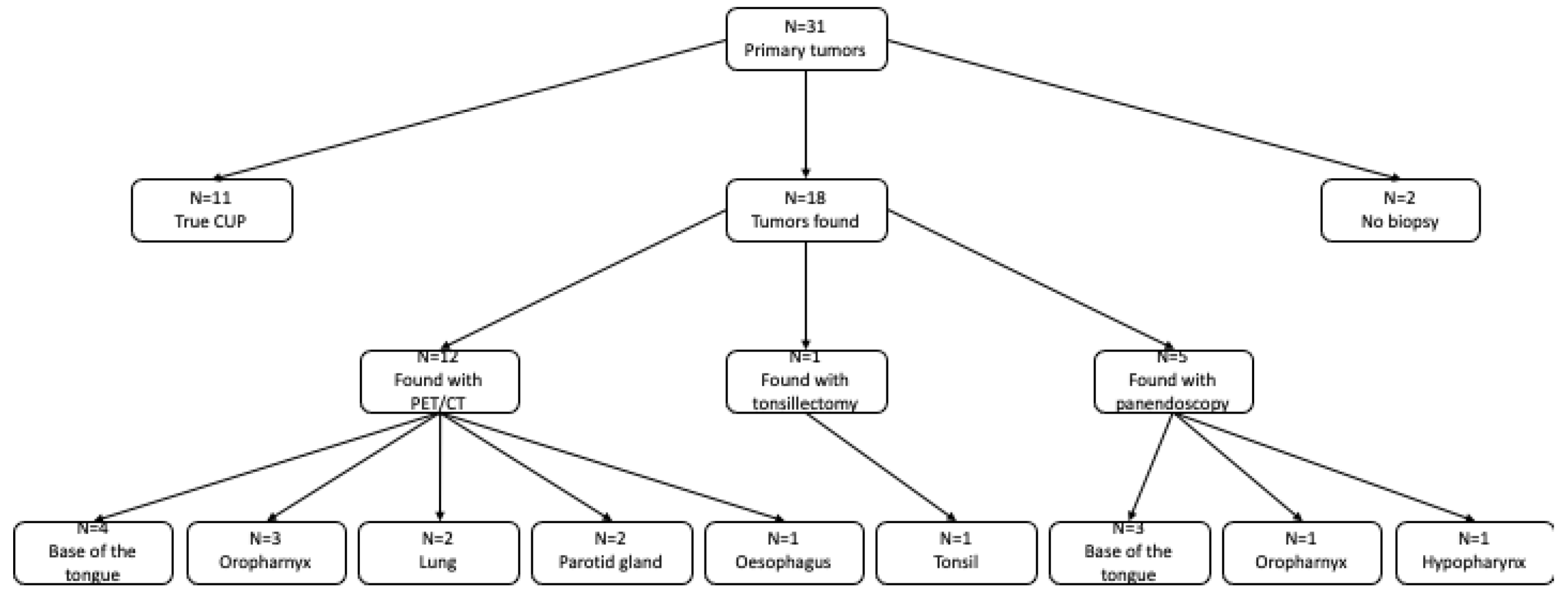
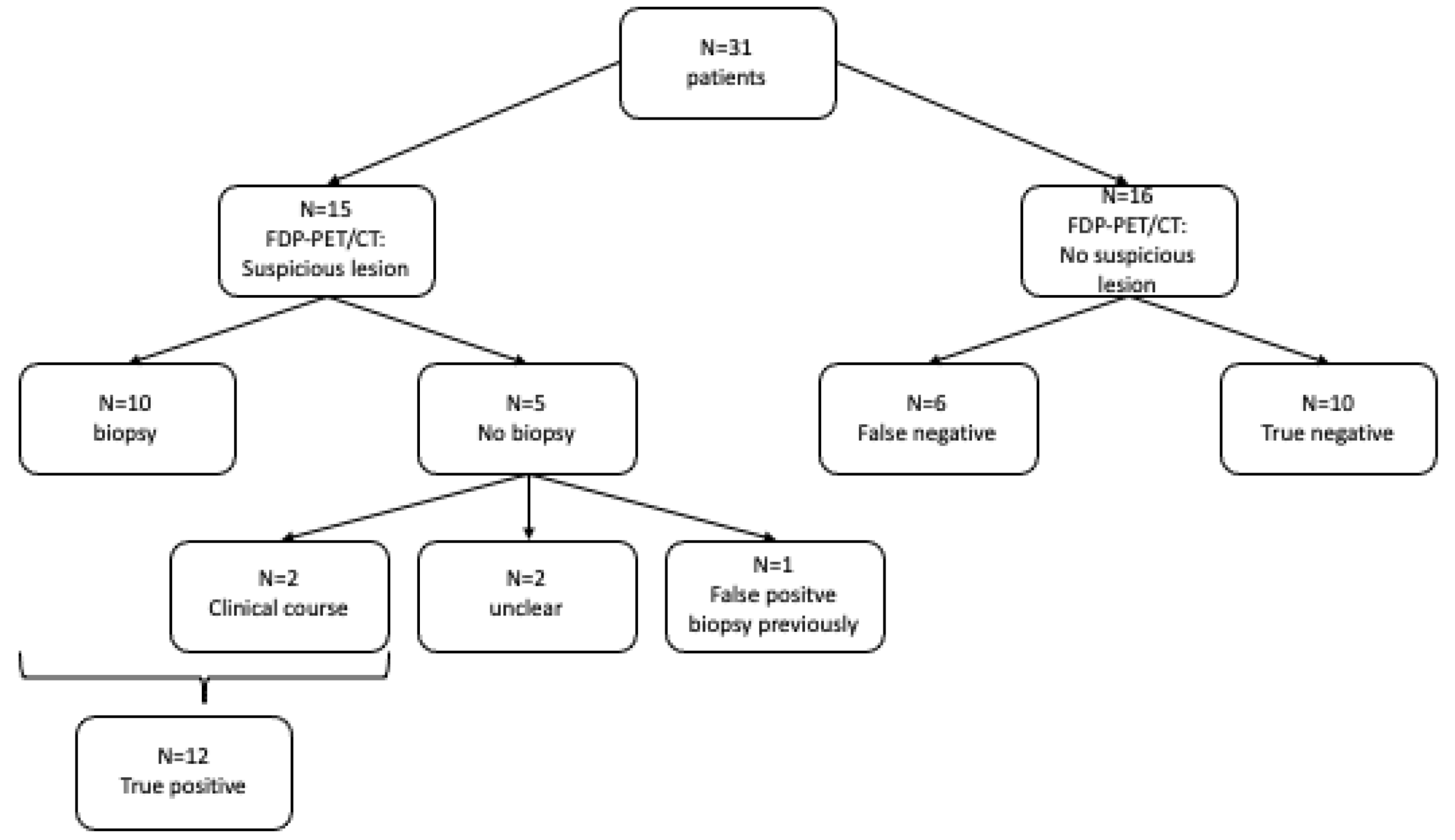
3.1. Primary Tumor Lesion
3.2. Prior Diagnostics
3.3. Distant Metastasis
3.4. Secondary Carcinoma
3.5. Summary
4. Discussion
5. Conclusions
Author Contributions
Funding
Institutional Review Board Statement
Informed Consent Statement
Data Availability Statement
Conflicts of Interest
References
- Fries, F. Cervical CUP syndrome. Radiologe 2020, 60, 1047–1051. [Google Scholar]
- Mozet, C.; Wichmann, G.; Stumpp, P. Cervical CUP syndrome. Onkologe 2013, 19, 44–51. [Google Scholar]
- Golusinski, P.; Di Maio, P.; Pehlivan, B. Evidence for the approach to the diagnostic evaluation of squamous cell carcinoma occult primary tumors of the head and neck. Oral Oncol. 2019, 88, 145–152. [Google Scholar]
- Gutzeit, A.; Antoch, G.; Kuhl, H.; Egelhof, T.; Fischer, M.; Hauth, E.; Goehde, S.; Bockisch, A.; Debatin, J.; Freudenberg, L. Unknown primary tumors: Detection with dual-modality PET/CT—Initial experience. Radiology 2005, 234, 227–234. [Google Scholar] [CrossRef]
- Nassenstein, K.; Veit-Haibach, P.; Stergar, H.; Gutzeit, A.; Freudenberg, L.; Kuehl, H.; Fischer, M.; Barkhausen, J.; Bockisch, A.; Antoch, G. Cervical lymph node metastases of unknown origin: Primary tumor detection with whole-body positron emission tomography/computed tomography. Acta. Radiol. 2007, 48, 1101–1108. [Google Scholar] [CrossRef]
- Roh, J.-L.; Kim, J.S.; Lee, J.H.; Cho, K.-J.; Choi, S.-H.; Nam, S.Y.; Kim, S.Y. Utility of combined 18F-fluorodeoxyglucose-positron emission tomography and computed tomography in patients with cervical metastases from unknown primary tumors. Oral Oncol. 2009, 45, 218–224. [Google Scholar] [CrossRef]
- Keller, F.; Psychogios, G.; Linke, R.; Lell, M.; Kuwert, T.; Iro, H.; Zenk, J. Carcinoma of unknown primary in the head and neck: Comparison between positron emission tomography (PET) and PET/CT. Head Neck 2010, 33, 1569–1575. [Google Scholar] [CrossRef]
- Wong, W.; Sonoda, L.; Gharpurhy, A.; Gollub, F.; Wellsted, D.; Goodchild, K.; Lemon, C.; Farrell, R.; Saunders, M. 18F-fluorodeoxyglucose positron emission tomography/computed tomography in the assessment of occult primary head and neck cancers—An audit and review of published studies. Clin. Oncol. R. Coll. Radiol. 2012, 24, 190–195. [Google Scholar] [CrossRef]
- Lee, J.R.; Kim, J.S.; Roh, J.-L.; Lee, J.H.; Baek, J.H.; Cho, K.-J.; Choi, S.-H.; Nam, S.Y.; Kim, S.Y. Detection of occult primary tumors in patients with cervical metastases of unknown primary tumors: Comparison of 18F FDG PET/CT with contrast-enhanced CT or CT/MR imaging—Prospective study. Radiology 2015, 274, 764–771. [Google Scholar] [CrossRef]
- Sprave, T.; Rühle, A.; Hees, K. Radiotherapeutic management of cervical lymph node metastases from an unknown primary site—Experiences from a large cohort treated with modern radiation techniques. Radiat. Oncol. 2020, 15, 80. [Google Scholar]
- Rusthoven, K.E.; Koshy, M.; Paulino, A.C. The role of fluorodeoxyglucose positron emission tomography in cervical lymph node metastases from an unknown primary tumor. Cancer 2004, 101, 2641–2649. [Google Scholar] [CrossRef]
- Nikolova, P.N.; Hadzhiyska, V.H.; Mladenov, K.B.; Ilcheva, M.G.; Veneva, S.; Grudeva, V.V.; Dineva, S.E.; Asenov, Y.N. The impact of 18F-FDG PET/CT in the clinical management of patients with lymph node metastasis of unknown primary origin. Neoplasma 2021, 68, 180–189. [Google Scholar] [CrossRef]
- Yabuki, K.; Tsukuda, M.; Horichui, C. Role of 18F-FDG pet in detecting primary site in the patient with primary unknown carcinoma. Eur. Arch. Otorhinolaryngol. 2010, 267, 1785–1792. [Google Scholar]
- Burglin, S.A.; Hess, S.; Høilund-Carlsen, P.F.; Gerke, O. 18F-FDG PET/CT for detection of the primary tumor in adults with extracervical metastases from cancer of unknown primary. Medicine 2017, 96, e6713. [Google Scholar] [CrossRef]
- Kwee, T.C.; Kwee, R.M. Combined FDG-PET/CT for the detection of unknown primary tumors: Systematic review and meta-analysis. Eur. Radiol. 2009, 19, 731–744. [Google Scholar] [CrossRef] [Green Version]
- Antoch, G.; Vogt, F.M.; Freudenberg, L.S.; Nazaradeh, F.; Goehde, S.C.; Barkhausen, J.; Dahmen, G.; Bockisch, A.; Debatin, J.F.; Ruehm, S.G. Whole-body dual-modality PET/CT and whole-body MRI for tumor staging in oncology. JAMA 2003, 290, 3199. [Google Scholar] [CrossRef]
- Kyzas, P.A.; Evangelou, E.; Denaxa-Kyza, D.; Ioannidis, J.P.A. 18F-fluorodeoxyglucose positron emission tomography to evaluate cervical node metastases in patients with head and neck squamous cell carcinoma: A meta-analysis. JNCI J. Natl. Cancer Inst. 2008, 100, 712–720. [Google Scholar] [CrossRef]
- Gődény, M.; Lengyel, Z.; Polony, G. Impact of 3T multiparametric MRI and FDG-PET-CT in the evaluation of occult primary cancer with cervical node metastasis. Cancer Imaging 2016, 16, 38. [Google Scholar] [CrossRef]
- Evangelista, L.; Cervino, A.R.; Chondrogiannis, S. Comparison between anatomical cross-sectional imaging and 18F-FDG PET/CT in the staging, restaging, treatment response, and long-term surveillance of squamous cell head and neck cancer: A systematic literature overview. Nucl. Med. Commun. 2014, 35, 123–134. [Google Scholar]
- Al Kadah, B.; Papaspyrou, G.; Linxweiler, M.; Schick, B.; Rübe, C.; Büchler, B.S.; Niewald, M. Cancer of unknown primary (CUP) of the head and neck: Retrospective analysis of 81 patients. Eur. Arch. Otorhinolaryngol. 2017, 274, 2557–2566. [Google Scholar] [CrossRef]
- Chuang, S.C.; Scelo, G.; Tonita, J.M. Risk of second primary cancer among patients with head and neck cancers: A pooled analysis of 13 cancer registries. Int. J. 2008, 123, 2390–2396. [Google Scholar]
- Matthias, C.; Harréus, U.; Strange, R. Influential factors on tumor recurrence in head and neck cancer patients. Eur. Arch. Otorhinolaryngol. 2006, 263, 37–42. [Google Scholar] [CrossRef]
- Rettig, E.M.; D’Souza, G. Epidemiology of head and neck cancer. Surg. Oncol. Clin. N. Am. 2015, 24, 379–396. [Google Scholar]
- Castaldi, P.; Leccisotti, L.; Bussu, F.; Miccichè, F.; Rufini, V. Role of 18F-FDG PET-CT in head and neck squamous cell carcinoma. Acta Otorhinolaryngol. Ital. 2013, 33, 1–8. [Google Scholar]
- Strobel, K.; Haerle, S.K.; Stoeckli, S.J.; Schrank, M.; Soyka, J.D.; Veit-Haibach, P.; Hany, T.F. Head and neck squamous cell carcinoma (HNSCC)—Detection of synchronous primaries with 18F-FDG-PET/CT. Eur. J. Nucl. Med. Moll. Imaging 2009, 36, 919–927. [Google Scholar] [CrossRef]
- Dietl, B.; Marienhagen, J.; Kühnel, T.; Schreyer, A.; Kölbl, O. The impact of FDG-PET/CT on the management of head and neck tumours: The radiotherapist’s perspective. Oral Oncol. 2008, 44, 504–508. [Google Scholar] [CrossRef]
- Xu, G.Z.; Guan, D.E.; He, Z.Y. 18FDG-PET/CT for detecting distant metastases and second primary cancersin patients with head and neck cancer. A meta-analysis. Oral Oncol. 2011, 47, 560–565. [Google Scholar]
- Send, T.; Kreppel, B.; Gaertner, F.C.; Bundschuh, R.A.; Strunk, H.; Bootz, F.; Essler, M. PET-CT in head and neck cancer. HNO 2017, 65, 504–513. [Google Scholar]
- Johansen, J.; Buus, S.; Loft, A. Prospective study of 18FDG-PET in the detection and management of patients with lymph node metastases to the neck from unknown primary tumor. Results form the DAHANCA-13 study. Head Neck 2008, 30, 471–478. [Google Scholar]


| Sex | Female | 5 (16%) |
| Male | 26 (84%) | |
| Age (years) | 40–49 | 3 |
| 50–59 | 9 | |
| 60–69 | 11 | |
| 70–79 | 7 | |
| 80–89 | 1 | |
| Use of alcohol | Yes | 12 |
| No | 1 | |
| No information | 18 | |
| Use of nicotine | Yes | 11 |
| No | 3 | |
| No information | 17 | |
| Lymph node metastasis | Squamous cell carcinoma | 20 (65%) |
| Dedifferentiated/Not further characterized carcinoma | 8 (26%) | |
| Melanoma | 1 (3%) | |
| Poorly differentiated carcinoma | 1 (3%) | |
| Adenocell carcinoma | 1 (3%) | |
| Previous diagnostics | Main diagnostic | 15 (48%) |
| Minor diagnostic | 4 (13%) | |
| No previous diagnostic | 9 (29%) | |
| No information | 3 (10%) | |
| Previous therapy | Radiotherapy | 1 |
| Radiotherapy and chemotherapy | 4 | |
| No therapy | 26 | |
| Timing of PET/CT in relation to diagnosis | Within 1 month | 20 |
| Within 1 year | 3 | |
| >1 year | 2 | |
| No information | 6 | |
| Median time of PET/CT after diagnosis | 8 days |
| T and N and HPV Status of Histologically Proven ENT Tumors * | ||
|---|---|---|
| T | X | 11 |
| 1 | 9 | |
| 2 | 4 | |
| 3 | 0 | |
| 4 | 2 | |
| N | 1 | 6 |
| 2 | 18 | |
| 3 | 2 | |
| HPV | Positive | 2 |
| Negative | 3 | |
| Unknown | 21 | |
| No Previous Diagnostic | Previous Diagnostic | Patients Excluded with Previous Therapy and Time from Diagnosis to PET > 1 Year | Total Collective | |
|---|---|---|---|---|
| Sensitivity | 75% (40.1–93.7%) | 63% (30.4–86.5%) | 71% (46.6–87.0%) | 67% (43.6–83.9%) |
| Specificity | 100% (16.7–100%) | 89% (54.3–100%) | 86% (46.7–99.5%) | 91% (60.1–100%) |
| Positive predictive value | 100% (55.7–100%) | 83% (41.8–98.9%) | 92% (64.6–100%) | 92% (64.6–100%) |
| Negative predictive value | 33% (5.6–79.8%) | 73% (42.9–90.8%) | 55% (28.0–78.7%) | 63% (38.5–81.6%) |
| Accuracy | 87.5% | 76% | 78.5% | 79% |
| F-18-FDG-PET/CT | 12/31 | Primary tumor lesion |
| 10/31 | True negative (=True CUP syndrome) | |
| 7/31 | Diagnosis of the previously refuted diagnostics | |
| 5/31 | Unknown metastasis | |
| 1/31 | Second primary tumor |
| Patients (n) | Sensitivity | Specificity | Positive Predictive Value | Negative Predictive Value | |
|---|---|---|---|---|---|
| Gutzeit et al. 2005 [4] | 18 | 35% | - | 86% | - |
| Nassenstein et al. 2007 [5] | 39 | 31% | - | 73% | - |
| Roh et al. 2009 [6] | 44 | 88% | 82% | 74% | 92% |
| Keller et al. 2011 [7] | 38 | 78% | 95% | 93% | 83% |
| Wong et al. 2012 [8] | 78 | 100% | 67% | 65% | 100% |
| Lee et al. 2015 [9] | 56 | 69% | 88% | 88% | 69% |
| Eilsberger et al. 2022 (total collective) | 29 | 67% | 91% | 92% | 63% |
| Eilsberger et al. 2022 (modified group) | 24 | 71% | 86% | 92% | 55% |
Publisher’s Note: MDPI stays neutral with regard to jurisdictional claims in published maps and institutional affiliations. |
© 2022 by the authors. Licensee MDPI, Basel, Switzerland. This article is an open access article distributed under the terms and conditions of the Creative Commons Attribution (CC BY) license (https://creativecommons.org/licenses/by/4.0/).
Share and Cite
Eilsberger, F.; Noltenius, F.E.; Librizzi, D.; Wessendorf, J.; Luster, M.; Hoch, S.; Pfestroff, A. Real-Life Performance of F-18-FDG PET/CT in Patients with Cervical Lymph Node Metastasis of Unknown Primary Tumor. Biomedicines 2022, 10, 2095. https://doi.org/10.3390/biomedicines10092095
Eilsberger F, Noltenius FE, Librizzi D, Wessendorf J, Luster M, Hoch S, Pfestroff A. Real-Life Performance of F-18-FDG PET/CT in Patients with Cervical Lymph Node Metastasis of Unknown Primary Tumor. Biomedicines. 2022; 10(9):2095. https://doi.org/10.3390/biomedicines10092095
Chicago/Turabian StyleEilsberger, Friederike, Friederike Elisabeth Noltenius, Damiano Librizzi, Joel Wessendorf, Markus Luster, Stephan Hoch, and Andreas Pfestroff. 2022. "Real-Life Performance of F-18-FDG PET/CT in Patients with Cervical Lymph Node Metastasis of Unknown Primary Tumor" Biomedicines 10, no. 9: 2095. https://doi.org/10.3390/biomedicines10092095
APA StyleEilsberger, F., Noltenius, F. E., Librizzi, D., Wessendorf, J., Luster, M., Hoch, S., & Pfestroff, A. (2022). Real-Life Performance of F-18-FDG PET/CT in Patients with Cervical Lymph Node Metastasis of Unknown Primary Tumor. Biomedicines, 10(9), 2095. https://doi.org/10.3390/biomedicines10092095