Abstract
Purpose: To evaluate the actual variability of the mean difference between chronological and dental age using the Cameriere method of open apices and to test its accuracy in variable age groups. Method: We selected studies that contained data about the mean, standard deviation, and number of cases for chronological age, dental age and gender. We used a random-effects model. Statistical significance was estimated, at a p < 0.05, using prediction intervals. For the analysis of publication bias we used the funnel plot and Egger’s regression test for plot asymmetry. I2 was used to test the presence of heterogeneity between studies. The Z test was used to test for statistical differences between subgroups, with p < 0.05 being considered statistically significant. We also used 95% for confidence intervals and prediction intervals. Results: In boys, the average difference between chronological and dental age was 0.44 (0.26–0.63) years, while in girls the average difference between chronological and dental age was 0.34 (0.19–0.49) years. In the 6–7 years age group and in the 14–15 years age group, there was a statistically significant difference between dental and chronological age. Our study shows that the Cameriere method is useful for estimating the chronological age, with errors of less than one year. Conclusions: The Cameriere method of evaluating dental age using open apices is sufficiently accurate for forensic practice, at least in the 7–14 age-interval.
1. Introduction
Age estimation is one of the most important objectives of forensic anthropology, with wide applicability for cadavers and skeletons, but also for living individuals (to establish age of majority, for adoption, illegal migration, sexual exploitation, or to determine criminal liability) [1,2]. Teeth are especially useful in this regard, mostly in children and adolescents, where the developmental stages are well known and characterized, and they have a decreased variability depending on environmental and genetic factors [3,4,5]. Radiological analysis of dental development is widely used to this intent, being preferred over the morphological analysis of dental eruption, which is more prone to errors (increased variability due to malnutrition, crowding, teeth decay or premature loss [1,3,6]).
In 2006, Cameriere et al. published a method of age estimation based on the measurement of the ratio between the length of the projection of the open apices and the length of the tooth axis major (known as the open apices method). Briefly, the method uses the seven left mandibular teeth. The first step is to identify the ones with closed apices, which are counted, and the sum is abbreviated N0. For the rest of the teeth with open apices, the distance between the inner sides of the open apex (for single-root teeth), or the sum of distances between the inner sides of the open apices (for multi-root teeth) is measured. These measurements are abbreviated to Ai, i = 1–7) and are then divided by the tooth length (Li, i = 1–7) in order to obtain the normalized measurements for the seven teeth (xi = Ai/Li). Then, a variable entitled s is computed, which is equal to N0 + sum(xi). This value is included in the formula: Age = 8.971 + 0.375g (gender, with 1 for boys and 0 for girls) + 1.631 × 5 + 0.674N0 − 1.034s − 0.176sN0 [7]. The method was updated in 2007 to Age = 8.387 + 0.282g − 1.692 × 5 + 0.835N0 − 0.116s − 0.139sN0 [8].
Many authors have devised, based on this methodology, methods that have been applied in specific populations (yielding different regression equations [9,10,11,12]), leading to the identification of new areas of interest [13,14,15]. For example, Angelakopoulos et al. developed a new formula—based on the Cameriere approach—using Bayesian methods, in South African children [16]. Additionally, numerous authors have shown this method to be more accurate in estimating age compared to others such as Demirjian, Nolla, Haavikko, or Willems [17,18]. However, the mean difference between chronological and dental age, as estimated by this method, has been shown to be variable in different studies, with some of them having been performed on similar populations [17,18]. Moreover, some studies—which evaluated mean differences in different age intervals—showed variable levels of accuracy depending on the age of the subjects [19,20,21]. However, the statistical significance of these results was not always determined, mainly due to a decreased number of cases per age group.
The purpose of this study is to evaluate the actual variability of the mean difference between chronological and dental age using the Cameriere method of open apices (both the original and the European formula), and to test the accuracy of the method at variable age groups.
2. Materials and Methods
We performed the study according to the PRISMA (Preferred Reporting Items for Systematic Reviews and Meta-Analyses) and MOOSE (Meta-analysis of Observational Studies in Epidemiology) guidelines for reporting systematic reviews and meta-analyses of observational studies in epidemiology [22,23].
2.1. Selection Criteria
Inclusion criteria: studies that contained data about the mean, standard deviation, and number of cases for chronological age, dental age, and gender (for the overall values).
We used the following exclusion criteria: (1) the absence of relevant information to obtain the data needed for the analysis; (2) studies with less than 20 subjects; (3) case series/case reports without a specification of the study population from which the cases were drawn upon and without a specific detection algorithm; (4) calculations made using methods other than the original and European formulas developed by Cameriere [7,8]. Two authors independently performed this search. In cases of disagreement between these two authors regarding inclusion of paper in the study and/or the presence of relevant exclusion criteria, a third reviewer was involved and the article was discussed until a consensus was reached.
2.2. Search Method
We analyzed the results obtained from Pubmed and Web of Science using the following keyword: “Cameriere”, with a timeframe that ranged from 2005 to 2019. We preferred not to use additional, restrictive criteria (e.g., article type) as other forms of publication (letters, case presentations, reviews) could potentially add relevant data to the meta-analysis (secondary references, discussions, finding other appropriate articles). The reference list of each relevant article was scrutinized for other, potentially relevant studies to be included in the meta-analysis. The references, abstract, and full text were imported in Paperpile.
2.3. Data Collection and Analysis
For each study, two reviewers extracted the data separately and included it in Excel Datasheets. We summarized the following information: study, name of the authors, year, total number of cases, country, mean, standard deviation and number of cases per gender/age group, inclusion and exclusion criteria.
2.4. Quality Assessment and Risk of Bias
Quality assessment was performed using the checklist from the STROBE (STrengthening the Reporting of OBservational studies in Epidemiology) Statement Checklist for observational studies [24]. This is a 22-items checklist, and the studies were considered to be of low quality if the sum was below 15, average quality if the sum was between 15.01 and 18, and high quality if the sum was between 18.01 and 22. The analysis was performed by two reviewers and the final score was obtained by averaging the results. Risk of bias was included as a point in the above-mentioned checklist and was quantified for each study.
2.5. Statistical Analysis
We used Jamovi (1.2, Sydney, Australia) and Microsoft Excel 365 (Microsoft, Redmond, WA, USA) for Mac for statistical analyses. We used a random effects model with the DerSimonian–Laird estimator and raw mean difference for effect size model measures (except for comparing results between groups, where in such cases we used standardized mean differences). Statistical significance was estimated, at a p < 0.05 using prediction intervals. For the analysis of publication bias we used the funnel plot and Egger’s regression test for plot asymmetry. I2 was used to test the presence heterogeneity between studies, using the following thresholds: 0–35%—most likely not important, 36–55%—moderate heterogeneity, 56–85%—most likely substantial heterogeneity, and 86–100%—significant heterogeneity (average values based on [25]). The Z test was used to test statistical differences between subgroups, with a p < 0.05 being considered statistically significant. We used 95% confidence intervals and prediction intervals.
3. Results
3.1. Search Synthesis
During the initial database research, we obtained 116 articles from Pubmed and 82 articles from Web of Science from which, after deleting duplicates and irrelevant studies, we selected 91 to be further scrutinized. By analyzing their references, we found another 10 potentially relevant articles that were also downloaded. From the 101 articles, 15 were included in the final analysis of prevalence. Details about the search synthesis are presented in Figure 1 [26]. The papers contained in the meta-analysis are detailed in Table 1.
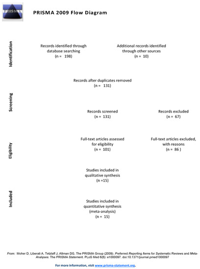
Figure 1.
PRISMA flow-chart. Reprinted with permission from ref. [26]. Copyright 2009 the Creative Commons Attribution License. (www.prisma-statement.org (accessed on 17 February 2021)).

Table 1.
Summary of studies included in the meta-analysis.
3.2. Quality Assessment and Risk of Bias
Quality assessment scores were between 13.5 and 20. The score for each study is included in Table 1. No studies showed any significant bias.
3.3. Accuracy of Cameriere Formulas Depending on Gender
In boys, the average difference between chronological and dental age was 0.44 years (0.26–0.63), as shown in Figure 2. The heterogeneity was significant (I2 = 94.33%). Publication bias was statistically significant (Z = 2.591, p = 0.01). In studies using the original formula [11,17,27,29,30,34], the average difference was 0.53 (0.28–0.78). The heterogeneity was most likely significant (I2 = 75.02%). Publication bias was not statistically significant (Z = 1.008, p = 0.314). In studies using the European formula [19,20,28,31,32,33,35,36,37], the average difference between chronological and dental age was 0.38 (0.13–0.62). The heterogeneity was significant (I2 = 87.6%). Publication bias was statistically significant (Z = 2.609, p = 0.009). The prediction interval overlapped with the value 0 in all three analyses, rendering the difference between dental and chronological ages not statistically significant. The results obtained using the two formulas were not statistically different (Z = 1.01, p = 0.27).
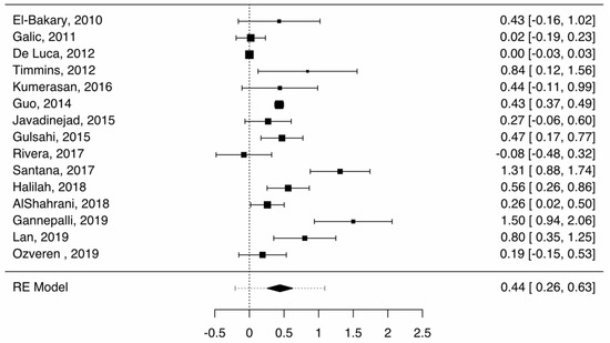
Figure 2.
Forest plot—Cameriere, boys, overall (boys).
In girls, the average difference between chronological and dental age was 0.34 (0.19–0.49), as shown in Figure 3. The heterogeneity was significant (I2 = 88.83%). Publication bias was statistically significant (Z = 6.464, p < 0.001). In studies using the original formula [11,17,27,29,30,34], the average difference was 0.44 (0.11–0.77). The heterogeneity was most likely significant (I2 = 84.3%). The publication bias was statistically significant (Z = 2.611, p = 0.009). In studies using the European formula [19,20,28,31,32,33,35,36,37], the average difference was 0.34 (0.10–0.58). The heterogeneity was significant (I2 = 89.03%). Publication bias was statistically significant (Z = 4.308, p < 0.001). The prediction interval overlapped with the value 0 in all three analyses, rendering the difference between dental and chronological ages not statistically significant. The differences between these two methods were not statistically significant (Z = 0.87, p = 0.38).
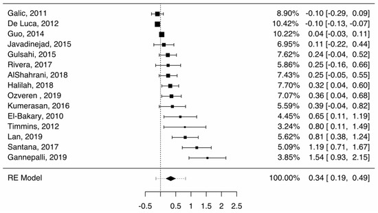
Figure 3.
Forest plot—Cameriere, boys, overall (girls).
Using the original formula, the difference between boys and girls was not statistically significant (Z = 1.47, p = 0.14). Using the European formula, the difference between boys and girls was also not statistically significant (Z = 0.31, p = 0.076). As the differences between gender and age formulas were not statistically significant, for the evaluation of the age group accuracy we used combined data.
3.4. Accuracy of Cameriere Formulas in Age Groups
The results of the analyses performed on age groups are presented in Table 2. In the 6–7 years age group there was a statistically significant difference between dental and chronological age (−0.81, with the prediction interval between −0.05 and −0.71). In the 14–15 years age group there was a statistically significant difference between dental and chronological age (0.87, with the prediction interval between 0.35 and 1.4). The difference between dental and chronological age was not statistically different in the other age groups, but there was an obvious trend, with higher dental ages compared to chronological ages in the younger, and higher chronological ages compared to dental ages in the older subadults (see Figure 4).
Table 2.
Standardized age difference in different age groups.
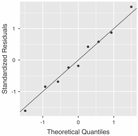
Figure 4.
Q-Q Plot. Average differences between age groups.
4. Discussion
Methods based on the analysis of the open apices in teeth have been used extensively in the last fifteen years in order to assess dental age in subadults, but also to evaluate age of majority (based on the analysis of the third molar) [1,2,20] due to their ease of use, high reproducibility and accuracy.
These methods have been evaluated, comparatively, with other dental and skeletal methods of age estimation, and have been shown to have significant advantages. For example, Kumaresan et al. showed that Cameriere’s method was more precise and accurate when estimating dental age in a Malaysian population sample when compared to Demirjian, Nolla, Haavikko and Willems [32]. Javadinejad et al. showed, in an Iranian population aged 3 to 15, that Cameriere’s method was less accurate compared to Smith’s, but more accurate when compared to Willems’ and Demirjian’s [17]. Our study has shown the Cameriere method to be useful for estimating chronological age, with errors of less than a year (and significantly smaller around the 8–11 year range, where the differences between dental and chronological age were minimal). This method has been revised numerous times, usually in order to make it more accurate in specific populations. Rai et al., for example, applied the European formula to an Indian population, showing that the European formula is a significant predictor for age. However, due to geographic differences between different regions from India, they proposed another formula, namely Age = 9.402 − 0.879C (C = 0 for the center and north of India and 1 for the south) + 0.663N0 − 0.711s − 0.106sN0, which explained 89.7% of the total variance [12]. Cugati et al. developed a specific formula for Malaysian populations, namely, Age = 11.368 − 0.345g + 0.553N0 − 1.096s − 0.380sN0, which yielded an R2 value of 0.871 [18]. Cameriere developed two main formulas to assess dental age based on open apices. The equations are very similar, and they have been used by many authors to evaluate dental age in different populations. Our study has shown that these formulas give comparable results, and this represents the reason why we used studies that were performed using either formula to evaluate the accuracy of age estimation based on the open apices.
In addition to the studies included in our analysis, there have been other studies that used the Cameriere formulas, but the data from such articles was not sufficient to fulfill all the inclusion criteria for this analysis. For example, Cameriere et al., in a study assessing Italian, Spanish and Croatian children aged between 5 and 15, showed the Cameriere method to yield a mean prediction error of 0.407 for girls and a slightly lower value (0.38) for boys [38]. However, the article did not contain sufficient data to estimate the necessary values to warrant inclusion in this meta-analysis.
Wolf et al. compared the usefulness of both the Cameriere and Demirjian methods in a 6–14 year old German population; they gave details about dental age but not chronological age (they only presented the mean difference in various age groups) [39]. The results from this study are in line with ours, showing lower dental compared to chronological ages in the younger age group and a trend reversal after eleven years [39]. Apaydin and Yasar, showed the same trend in a Turkish population, with the Cameriere method overestimating age in younger age-groups (the differences being statistically significant for the age groups 5–6 and 6–7), and underestimating age in older groups (with the differences between dental and chronological ages being statistically significant over eleven years), while the Willems method showed the most precise results. One reason for the age underestimation represents the difficulty in evaluating the small apex opening, which is almost closed/closed in older groups [40]. Timmins et al. showed that the Cameriere method is especially useful up to around 14 years of age (at which point all seven teeth reach maturity), making the Demirjian method more useful in older adolescents (up to sixteen years of age) [29]. Our study showed a similar trend—the only age groups in which the difference between chronological and dental age were statistically significant were the 6–7 and 14–15 age groups. It should be noted that we only evaluated the usefulness of the Cameriere method between 6 and 15 years of age, as the number of studies containing data for younger and older age groups was below four.
The usage of radiological methods for dental assessment in children/adolescents must, however, be used cautiously, as they are considered highly intrusive, due to the risks associated with the procedure, which are not counterbalanced by medical benefits for the patients [41]. Normally, a medical intervention that has more than minimal risks should not be allowed, unless there is a significant medical benefit for the subject and informed consent for the procedure has been obtained. The goal of the procedure is often not for the medical benefit of the patient, but rather for the benefit of a third party, or even maleficent for him/her [42] who can, for example, be deported, or be jailed as a direct consequence of this expertise. Moreover, the issue of consent is highly debatable in many instances—the potential aims of the procedure may be to establish age of majority or criminal competence, as these can be correlated—from a legal point of view—with age. If the subject signs the consent and he/she is not considered legally competent afterwards, to what degree is the consent valid? Similarly, if she/he does not sign the consent, and instead it is signed by a legal guardian, and she/he is considered legally competent, was the procedure not performed without the approval of the person who should accept it [43,44,45]?
Recently, the usefulness of 3D radiological methods for dental age determination have been evaluated, with promising results. Cone-Beam Computed Tomography has been found to be more accurate than orthopantograms for dental evaluation, including dental anatomy and forensics, as shown in a recent systematic review [46]; therefore, its usage might increase the accuracy of the Cameriere method, which could lead to increased usage of the method for dental age assessments.
Limitations
The first major limitation of this study is the fact that we used two different formulas for estimating the dental age based on open apices. We preferred this approach as the differences between them were minor, and as the initial analysis showed no statistically significant difference between the results of these two studies. However, this approach most likely increased the heterogeneity of the results. Another major limitation is represented by the fact that we were unable to include all studies that were conducted using these two formulas, as the data from some articles were incomplete. The addition of further studies could have yielded more precise results. Additionally, we preferred to use a standardized method for developing the random-effects model (DerSimonian–Laird); this method underestimates the true heterogeneity when the τ2 is large and the number of studies is small, but the main advantage of this approach involves the possibility for other meta-analyses—performed with similar purposes—to be compared more easily. The optimal method is that suggested by Hunter and Schmidt (which weights the inverse of the within-study variance), but this is rarely used, and the results would have been harder to compare. Moreover, our initial evaluation (not presented here) showed that the results were highly similar with the published approach. For statistical significance purposes we preferred to use prediction intervals, which are larger than confidence intervals, which are usually used in random-effects models. Our approach decreases the possibility to catch statistically significant differences, however, it is the correct approach to be used from a methodological point of view. Briefly, in a meta-analysis using random-effects models, researchers present the summary effect size and the confidence interval; based on these methods, one can estimate the mean effect size and the precision, but not the distribution of the true effects around the summary estimates [47], which is only correctly depicted by the prediction interval. The age group 3–5 was not included in the analysis due to the very low amount of subject data available. The quality criteria for clinical meta-analyses does not reflect the quality of studies on age estimation. The extent to which the method has been applied correctly or incorrectly in the individual studies was not evaluated. This is particularly important for methods where a large amount of manual measurements are required.
5. Conclusions
The Cameriere method of evaluating dental age on open apices is accurate enough for clinical practice, at least in the 7–14 age-interval. It should not be used outside this age range. Its actual usefulness, compared to that of other methods, should be assessed by comparing meta-analyses for each method, using a reproducible methodology. Dental age remains a reliable age estimation with important criminal and civil consequences.
Author Contributions
S.H. designed the study, performed statistical analysis, drafted the manuscript and the revision, approved the final version, and agreed to be held accountable for all aspects of the article; I.D. performed data gathering and analysis, revised the manuscript, approved the final version, and agreed to be held accountable for all aspects of the article; M.C.R. performed data gathering and analysis, revised the manuscript, approved the final version, and agreed to be held accountable for all aspects of the article; I.N. performed data gathering and analysis, statistical analysis of the results, revised the manuscript, approved the final version, and agreed to be held accountable for all aspects of the article. All authors have read and agreed to the published version of the manuscript.
Funding
This research received no external funding.
Institutional Review Board Statement
None relevant.
Informed Consent Statement
None relevant.
Data Availability Statement
None relevant.
Conflicts of Interest
The authors declare no conflict of interest.
References
- Cavrić, J.; Galić, I.; Vodanović, M.; Brkić, H.; Gregov, J.; Viva, S.; Rey, L.; Cameriere, R. Third molar maturity index (I3M) for assessing age of majority in a black African population in Botswana. Int. J. Leg. Med. 2016, 130, 1109–1120. [Google Scholar] [CrossRef]
- Cameriere, R.; Pacifici, A.; Viva, S.; Carbone, D.; Pacifici, L.; Polimeni, A. Adult or not? Accuracy of Cameriere’s cut-off value for third molar in assessing 18 years of age for legal purposes. Minerva Stomatol. 2014, 63, 283–294. [Google Scholar] [PubMed]
- Hostiuc, S.; Teodoru, D.; Isailă, O. Tratat de Medicină Legală Odontostomatologică; Hostiuc, S., Ed.; All: București, Romania, 2020. [Google Scholar]
- Sharma, P.; Wadhwan, V.; Prakash, R.; Goel, S.; Aggarwal, P. Age estimation in children by measurement of open apices in teeth: A study in North Indian population. Aust. J. Forensic Sci. 2016, 48, 592–600. [Google Scholar] [CrossRef]
- Franklin, D.; Karkhanis, S.; Flavel, A.; Collini, F.; DeLuca, S.; Cameriere, R. Accuracy of a cut-off value based on the third molar index: Validation in an Australian population. Forensic Sci. Int. 2016, 266, 575.e1–575.e6. [Google Scholar] [CrossRef]
- Cameriere, R.; Pacifici, A.; Polimeni, L.; Federici, F.; Cingolani, M.; Ferrante, L. Age estimation in children by measurement of open apices in teeth with Bayesian calibration approach. Forensic Sci. Int. 2016, 258, 50–54. [Google Scholar] [CrossRef] [PubMed]
- Cameriere, R.; Ferrante, L.; Cingolani, M. Age estimation in children by measurement of open apices in teeth. Int. J. Leg. Med. 2006, 120, 49–52. [Google Scholar] [CrossRef]
- Cameriere, R.; De Angelis, D.; Ferrante, L.; Scarpino, F.; Cingolani, M. Age estimation in children by measurement of open apices in teeth: A European formula. Int. J. Leg. Med. 2007, 121, 449–453. [Google Scholar] [CrossRef]
- Balla, S.B.; Lingam, S.; Kotra, A.; Hima, R.P.; Karuna, P.; Madhuri, N.N.; Cameriere, R. New regression models for dental age estimation in children using third molar maturity index: A preliminary analysis testing its usefulness as reliable age marker. Leg. Med. 2019, 39, 35–40. [Google Scholar] [CrossRef]
- Pratyusha, K.; Prasad, M.G.; Radhakrishna, A.N.; Saujanya, K.; Raviteja, N.V.K.; Chandrasekhar, S. Applicability of Demirjian’s Method and Modified Cameriere’s Methods for Dental Age Assessment in Children. J. Clin. Diagn. Res. 2017, 11, ZC40–ZC43. [Google Scholar] [CrossRef] [PubMed]
- Santana, S.A.; Bethard, J.D.; Moore, T.L. Accuracy of Dental Age in Nonadults: A Comparison of Two Methods for Age Estimation Using Radiographs of Developing Teeth. J. Forensic Sci. 2017, 62, 1320–1325. [Google Scholar] [CrossRef]
- Rai, B.; Kaur, J.; Cingolani, M.; Ferrante, L.; Cameriere, R. Age estimation in children by measurement of open apices in teeth: An Indian formula. Int. J. Leg. Med. 2010, 124, 237–241. [Google Scholar] [CrossRef]
- De Luca, S.; Alemán, I.; Bertoldi, F.; Ferrante, L.; Mastrangelo, P.; Cingolani, M.; Cameriere, R. Age estimation by tooth/pulp ratio in canines by peri-apical X-rays: Reliability in age determination of Spanish and Italian medieval skeletal remains. J. Archaeol. Sci. 2010, 37, 3048–3058. [Google Scholar] [CrossRef]
- Lauc, T.; Nakaš, E.; Latić-Dautović, M.; Džemidžić, V.; Tiro, A.; Rupić, I.; Kostić, M.; Galić, I. Dental Age in Orthodontic Patients with Different Skeletal Patterns. BioMed Res. Int. 2017, 2017, 1–7. [Google Scholar] [CrossRef] [PubMed]
- De Luca, S.; Bautista, J.; Alemán, I.; Cameriere, R. Age-at-Death Estimation by Pulp/Tooth Area Ratio in Canines: Study of a 20th-Century Mexican Sample of Prisoners to Test Cameriere’s Method. J. Forensic Sci. 2011, 56, 1302–1309. [Google Scholar] [CrossRef]
- Angelakopoulos, N.; De Luca, S.; Palacio, L.A.V.; Coccia, E.; Ferrante, L.; Pinchi, V.; Cameriere, R. Age estimation by measuring open apices in teeth: A new formula for two samples of South African black and white children. Int. J. Leg. Med. 2019, 133, 1529–1536. [Google Scholar] [CrossRef]
- Javadinejad, S.; Sekhavati, H.; Ghafari, R. A Comparison of the Accuracy of Four Age Estimation Methods Based on Panoramic Radiography of Developing Teeth. J. Dent. Res. Dent. Clin. Dent. Prospect. 2015, 9, 72–78. [Google Scholar] [CrossRef] [PubMed]
- Cugati, N.; Kumaresan, R.; Srinivasan, B.; Karthikeyan, P. Dental age estimation of growing children by measurement of open apices: A Malaysian formula. J. Forensic Dent. Sci. 2015, 7, 227–231. [Google Scholar] [CrossRef]
- De Luca, S.; de Giorgio, S.; Butti, A.C.; Biagi, R.; Cingolani, M.; Cameriere, R. Age estimation in children by measurement of open apices in tooth roots: Study of a Mexican sample. Forensic Sci. Int. 2012, 221, 155.e1–155.e7. [Google Scholar] [CrossRef]
- Rivera, M.; De Luca, S.; Aguilar, L.; Palacio, L.A.V.; Galić, I.; Cameriere, R. Measurement of open apices in tooth roots in Colombian children as a tool for human identification in asylum and criminal proceedings. J. Forensic Leg. Med. 2017, 48, 9–14. [Google Scholar] [CrossRef]
- Ozveren, N.; Serindere, G. Comparison of the applicability of Demirjian and Willems methods for dental age estimation in children from the Thrace region, Turkey. Forensic Sci. Int. 2018, 285, 38–43. [Google Scholar] [CrossRef]
- Moher, D.; Altman, D.G.; Liberati, A.; Tetzlaff, J. PRISMA Statement. Epidemiology 2011, 22, 128. [Google Scholar] [CrossRef] [PubMed]
- Liberati, A.; Altman, D.G.; Tetzlaff, J.; Mulrow, C.; Gøtzsche, P.C.; Ioannidis, J.P.A.; Clarke, M.; Devereaux, P.J.; Kleijnen, J.; Moher, D. The PRISMA statement for reporting systematic reviews and meta-analyses of studies that evaluate health care interventions: Explanation and elaboration. J. Clin. Epidemiol. 2009, 62, e1–e34. [Google Scholar] [CrossRef]
- Von Elm, E.; Altman, D.G.; Egger, M.; Pocock, S.J.; Gøtzsche, P.C.; Vandenbroucke, J.P.; Initiative, F.T.S. The Strengthening the Reporting of Observational Studies in Epidemiology (STROBE) Statement: Guidelines for Reporting Observational Studies. Ann. Intern. Med. 2007, 147, 573–577. [Google Scholar] [CrossRef] [PubMed]
- Deeks, J.; Higgins, J.; Altman, D. Chapter 10: Analysing Data and Undertaking Meta-analyses. In Cochrane Handbook for Systematic Reviews of Interventions, 6th ed.; Higgins, J., Thomas, J., Eds.; John Wiley and Sons: Chicester, UK, 2019; pp. 241–284. [Google Scholar]
- Moher, D.; Liberati, A.; Tetzlaff, J.; Altman, D.G.; PRISMA Group. Preferred reporting items for systematic reviews and meta-analyses: The PRISMA statement. PLoS Med. 2009, 6, e1000097. [Google Scholar] [CrossRef] [PubMed]
- El-Bakary, A.A.; Hammad, S.M.; Mohammed, F. Dental age estimation in Egyptian children, comparison between two methods. J. Forensic Leg. Med. 2010, 17, 363–367. [Google Scholar] [CrossRef]
- Galić, I.; Vodanović, M.; Cameriere, R.; Nakaš, E.; Galić, E.; Selimović, E.; Brkić, H. Accuracy of Cameriere, Haavikko, and Willems radiographic methods on age estimation on Bosnian–Herzegovian children age groups 6–13. Int. J. Leg. Med. 2011, 125, 315–321. [Google Scholar] [CrossRef] [PubMed]
- Timmins, K.; Liversidge, H.; Farella, M.; Herbison, P.; Kieser, J. The usefulness of dental and cervical maturation stages in New Zealand children for Disaster Victim Identification. Forensic Sci. Med. Pathol. 2011, 8, 101–108. [Google Scholar] [CrossRef][Green Version]
- Guo, Y.-C.; Yan, C.-X.; Lin, X.-W.; Zhou, H.; Li, J.-P.; Pan, F.; Zhang, Z.-Y.; Wei, L.; Tang, Z.; Chen, T. Age estimation in northern Chinese children by measurement of open apices in tooth roots. Int. J. Leg. Med. 2014, 129, 179–186. [Google Scholar] [CrossRef] [PubMed]
- Gulsahi, A.; Tirali, R.E.; Cehreli, S.B.; De Luca, S.; Ferrante, L.; Cameriere, R. The reliability of Cameriere’s method in Turkish children: A preliminary report. Forensic Sci. Int. 2015, 249, 319.e1–319.e5. [Google Scholar] [CrossRef]
- Kumaresan, R.; Cugati, N.; Chandrasekaran, B.; Karthikeyan, P. Reliability and validity of five radiographic dental-age estimation methods in a population of Malaysian children. J. Investig. Clin. Dent. 2014, 7, 102–109. [Google Scholar] [CrossRef]
- Halilah, T.; Khdairi, N.; Jost-Brinkmann, P.-G.; Bartzela, T. Age estimation in 5–16-year-old children by measurement of open apices: North German formula. Forensic Sci. Int. 2018, 293, 103.e1–103.e8. [Google Scholar] [CrossRef]
- Alshahrani, I.; Yassin, S.M.; Togoo, R.A.; Tikare, S.; Khader, M.A.; Alkahtani, Z.M. Age estimation by measurement of open apices in tooth roots: Study using Saudi Arabian samples. J. Forensic Leg. Med. 2019, 62, 63–68. [Google Scholar] [CrossRef]
- Gannepalli, A.; Balla, S.B.; Pacha, V.B.; Babu, D.B.G.; Vinay, B.H.; Perkari, S. Applicability of Cameriere European formula for age estimation of 10–15 years legal threshold in South Indian population. J. Forensic Dent. Sci. 2019, 11, 78–83. [Google Scholar] [CrossRef] [PubMed]
- Lan, L.M.; Yang, Z.D.; Sun, S.L.; Wen, D.; Kureshi, A.; Zeye, M.M.J.; Zha, L.; Li, M. Application of Demirjian’s and Cameriere’s Method in Dental Age Estimation of 8–16 Year Old Adolescents from Hunan Han Nationality. J. Forensic Med. 2019, 35, 406–410. [Google Scholar]
- Ozveren, N.; Serindere, G.; Meric, P.; Cameriere, R. A comparison of the accuracy of Willems’ and Cameriere’s methods based on panoramic radiography. Forensic Sci. Int. 2019, 302, 109912. [Google Scholar] [CrossRef] [PubMed]
- Cameriere, R.; Ferrante, L.; Liversidge, H.; Prieto, J.; Brkic, H. Accuracy of age estimation in children using radiograph of developing teeth. Forensic Sci. Int. 2008, 176, 173–177. [Google Scholar] [CrossRef]
- Wolf, T.G.; Briseño-Marroquín, B.; Callaway, A.; Patyna, M.; Müller, V.T.; Willershausen, I.; Ehlers, V.; Willershausen, B. Dental age assessment in 6- to 14-year old German children: Comparison of Cameriere and Demirjian methods. BMC Oral Health 2016, 16, 1–8. [Google Scholar] [CrossRef] [PubMed]
- Apaydin, B.K.; Yasar, F. Accuracy of the demirjian, willems and cameriere methods of estimating dental age on turkish children. Niger. J. Clin. Pract. 2018, 21, 257–263. [Google Scholar]
- De Micco, F.; Martino, F.; Campobasso, C.P. Ethical issues in age assessment by the third molar development. Aust. J. Forensic Sci. 2020, 1–12. [Google Scholar] [CrossRef]
- Malmqvist, E.; Furberg, E.; Sandman, L. Ethical aspects of medical age assessment in the asylum process: A Swedish perspective. Int. J. Leg. Med. 2018, 132, 815–823. [Google Scholar] [CrossRef]
- Kangaude, G.D.; Skelton, A. (De)Criminalizing Adolescent Sex: A Rights-Based Assessment of Age of Consent Laws in Eastern and Southern Africa. SAGE Open 2018, 8, 215824401880603. [Google Scholar] [CrossRef]
- Focardi, M.; Pinchi, V.; De Luca, F.; Norelli, G.-A. Age estimation for forensic purposes in Italy: Ethical issues. Int. J. Leg. Med. 2014, 128, 515–522. [Google Scholar] [CrossRef] [PubMed]
- Rudolf, E. Comments to Focardi et al., Age estimation for forensic purposes in Italy: Ethical issues. Int. J. Leg. Med. 2014, 129, 1271–1273. [Google Scholar] [CrossRef] [PubMed]
- Dalessandri, D.; Tonni, I.; Laffranchi, L.; Migliorati, M.; Isola, G.; Visconti, L.; Bonetti, S.; Paganelli, C. 2D vs. 3D Radiological Methods for Dental Age Determination around 18 Years: A Systematic Review. Appl. Sci. 2020, 10, 3094. [Google Scholar] [CrossRef]
- Borenstein, M.; Hedges, L.V.; Higgins, J.P.T.; Rothstein, H.R. Prediction intervals. In Introduction to Meta-Analysis; Borenstein, M., Ed.; John Wiley & Sons: Chichester, UK, 2009; pp. 127–133. [Google Scholar]
Publisher’s Note: MDPI stays neutral with regard to jurisdictional claims in published maps and institutional affiliations. |
© 2021 by the authors. Licensee MDPI, Basel, Switzerland. This article is an open access article distributed under the terms and conditions of the Creative Commons Attribution (CC BY) license (http://creativecommons.org/licenses/by/4.0/).