Review

Calcium-Permeable Channels Cooperation for Rheumatoid Arthritis: Therapeutic Opportunities

1
The Second School of Clinical Medicine, Anhui Medical University, Hefei 230032, China
2
Department of Clinical Pharmacology, The Second Hospital of Anhui Medical University, Hefei 230601, China
3
The Key Laboratory of Anti-Inflammatory and Immune Medicine, Anhui Medical University, Hefei 230032, China
*
Authors to whom correspondence should be addressed.
Biomolecules 2022, 12(10), 1383; https://doi.org/10.3390/biom12101383
Submission received: 29 July 2022 / Revised: 18 September 2022 / Accepted: 20 September 2022 / Published: 27 September 2022

Abstract

:
Rheumatoid arthritis is a common autoimmune disease that results from the deposition of antibodies–autoantigens in the joints, leading to long-lasting inflammation. The main features of RA include cartilage damage, synovial invasion and flare-ups of intra-articular inflammation, and these pathological processes significantly reduce patients’ quality of life. To date, there is still no drug target that can act in rheumatoid arthritis. Therefore, the search for novel drug targets has become urgent. Due to their unique physicochemical properties, calcium ions play an important role in all cellular activities and the body has evolved a rigorous calcium signaling system. Calcium-permeable channels, as the main operators of calcium signaling, are widely distributed in cell membranes, endoplasmic reticulum membranes and mitochondrial membranes, and mediate the efflux and entry of Ca2+. Over the last century, more and more calcium-permeable channels have been identified in human cells, and the role of this large family of calcium-permeable channels in rheumatoid arthritis has gradually become clear. In this review, we briefly introduce the major calcium-permeable channels involved in the pathogenesis of RA (e.g., acid-sensitive ion channel (ASIC), transient receptor potential (TRP) channel and P2X receptor) and explain the specific roles and mechanisms of these calcium-permeable channels in the pathogenesis of RA, providing more comprehensive ideas and targets for the treatment of RA.

1. Introduction

Rheumatoid arthritis (RA) is an autoimmune disease of unknown cause that affects several organs and tissues, including synovial joints and can involve multiple systems throughout the body. RA affects approximately 1% of the population and has a male-to-female ratio of 2.5:1 [1]. From the pathological perspective, the pathogenesis of RA mainly includes three aspects: synovitis, structure destruction and inflammation infiltration. Synovial and bone changes have been reported in some patients as early as four months after the onset of the disease. In terms of clinical features, patients often experience joint deformity, morning stiffness and pain, swelling and hyperplasia of the synovial membrane, increased C-reactive protein in the blood and weakness due to impaired motor function [1,2,3,4,5]. In addition to affecting the locomotor system, novel clinical signs of RA have also been identified, such as skin and respiratory involvement [6,7]. In terms of treatment, there has been considerable progress in the drug treatment of RA. The classical RA treatment drugs include non-steroidal anti-inflammatory drugs (NSAIDs) such as aspirin, glucocorticoids such as dexamethasone, and disease-modifying antirheumatic drugs (DMARDs) such as methotrexate. These drugs can relieve the condition and the pain of RA, but, due to the multiple adverse reactions to NSAIDS [8,9] and adverse effects of DMARDs that have been clinically reported [10,11], it has become more to explore new therapeutic targets for RA.
Calcium ions (Ca2+) affect almost every aspect of cellular life. As an important second messenger for cellular signaling, there is a huge difference in concentration between intracellular (~100 nM free) and extracellular (mM) Ca2+ levels, and the cell devotes a large amount of energy to maintaining this difference in concentration [12]. Clearly, the large difference in intracellular and extracellular Ca2+ concentration provides the basis for rapid changes in [Ca2+]i, which conduct the intracellular signal transduction [12,13]. Ca2+ is a central aspect of inflammation as well as inflammatory diseases. In fact, Ca2+ is thought to be closely associated with the release and metabolism of inflammatory substances such as histamine, prostaglandins and leukotrienes by mast cells during the outbreak of inflammation [14]. It has been suggested that Ca2+ can regulate the metabolism of arachidonic acid in T cells and mediate synovial inflammation in RA patients during the inflammatory response [15]. In addition, there is a large body of data showing that Ca2+ mediates the infiltration of large numbers of immune cells during the development of RA, leading to an uncontrolled inflammatory response [16]. These results indicate that Ca2+ itself plays a core role in the inflammatory response, particularly in RA. Ca2+ channels, as a main driver of calcium signaling transduction that are widely expressed on the surface of cell membranes, endoplasmic reticulum membranes and mitochondrial membranes, mediate the influx of extracellular Ca2+ or the release of Ca2+ from Ca2+ stores. Based on the gating mechanism of these channels, Ca2+ channels can be classified as: (1) voltage-gated Ca2+ channels, (2) ligand-gated channels, (3) store-operated channels (SOC), (4) second messenger-operated channels, (5) acid-sensing ion channels, and (6) mechano-gated channels [17].
Ca2+ channels, therefore, are particularly important in all life activities. Indeed, Ca2+ channels have long been a therapeutic target for various diseases. It is well-established that the moderate inhibition of Ca2+ channels in cardiac and vascular smooth muscle cells can inhibit myocardial contraction and vasodilation, thereby relieving tachycardia and angina pectoris [18,19,20]. In respiratory diseases such as asthma, the blockade of Ca2+ channels can inhibit bronchial ground constriction [17,21]. In addition, there has been increasing evidence in recent years that calcium-permeable channels play an important role in joint diseases such as RA. In RA, the modulation of different families of Ca2+ channels has been shown to regulate the progression of RA. For example, our previous study found that the inhibition of acid-sensitive ion channels (a calcium-permeable channel) can slow arthritic rat chondrocyte apoptosis [22,23]. Here, we briefly describe the pathogenesis of RA, review the Ca2+-permeable channels that play an important role in RA, and detail the mechanisms of Ca2+-permeable channels in the pathogenesis of RA, providing novel therapeutic targets for RA (Figure 1).

2. Pathology of Rheumatoid Arthritis (RA)

RA is defined as a systemic autoimmune disease associated with a chronic inflammatory process. RA can damage joints and extra-articular organs, including the heart, kidneys, lungs, digestive system, eyes, skin, and nervous system. The main joint manifestations can be divided into three areas, including synovitis, structural damage to joints, and the activation of the inflammatory response [24].
Inflammation of the synovium due to reflex immune activation in RA joint-swelling is characterized by the infiltration of leukocytes into the normally sparse synovium [25]. The aggregation of leukocytes is not due to proliferation but is the result of the chemotaxis of circulating leukocytes. There are many possible reasons for the leukocyte infiltration that occurs in the synovium in RA, including: (1) adaptive immune pathways, (2) activation of the innate immune system, (3) cytokines and intracellular signaling pathways, and (4) mesenchymal tissue response [26,27,28]. Within a short time after onset, the synovium can thicken to 10 cells deep and is mainly composed of type A (macrophage-like) and type B (fibroblast-like) synovial cells (FLSs) [1]. The infiltrating immune cells produce a variety of cytokines that activate FLSs. Under the action of various mediators, activated FLSs apoptosis is reduced, while it can proliferate in large numbers. The exact mechanism regarding synovial cell proliferation may be related to the induction of certain growth factors such as TGF-β or to oncogenes [29,30]. Recently, the process of interaction between synovial macrophages and FLS has also been revealed, which may suggest the existence of a mechanism for the development of chronic inflammation in RA [31], but the exact mechanism still needs to be further explored. In this way, the inflammation of the synovial membrane, as well as the proliferation of the landscape, is well-described.
The erosion of cartilage and bone is the main sign of structural damage in RA, and the quantification of bone and cartilage damage has been an important tool in the clinical diagnosis of the disease and to monitor the efficacy of drug therapy [32]. However, two different mechanisms are involved in the erosion of bone and cartilage [30,32]. Bone erosion tends to occur in the part of the synovial membrane that is in contact with the bone, i.e., the bone around the joint is more susceptible to erosion [32,33]. Indeed, after the onset of RA, synovial inflammation can lead to local hypoxic conditions and cytokine-induced neovascularization, increasing the microvasculature in the interstitial space [34]. The microvasculature located in these interstitial spaces contributes to the homing of osteoclasts. Osteoclasts consist of multinucleated giant cells containing from 2 to 50 closely packed nuclei, mainly on the surface of the bone, around the vascular channels in the bone. Osteoclasts mainly perform bone resorption functions and functionally correspond to osteoblasts. The high expression of anti-tartaric acid phosphatase and histone K are the main markers of osteoclasts. Current research on osteoclast activation has focused on the regulation of the RANK/RANKL/OPG axis. Osteoclast formation is regulated by a variety of factors, and NFATc1, which was first identified in T cells, is one of the most critical and widely studied transcription factors [35]. When osteoclasts come into contact with bone and receive the relevant molecular signals, they are activated and osteoclast-mediated bone resorption leads to the loss of cortical bone [36]. The high-resolution CT of joints in RA patients also demonstrates the bone resorption role played by osteoclasts [37,38,39,40,41]. Articular cartilage consists of a non-mineralized superficial layer and a deep mineralized layer close to the bone. The disintegration of undifferentiated cartilage includes two aspects. One is that chondrocytes can secrete various mediators such as ADAMTS (a metalloproteinase with a thrombin reactive protein motif) and matrix metalloproteinase (MMP) after inflammatory stimulation, leading to the degradation of the perichondral matrix. Secondly, FLSs and infiltrating immune cells can also secrete MMPs and other inflammatory cytokines to degrade the surrounding cartilage [30]. In addition, the synovium invades the periarticular bone at the cartilage–bone junction due to a persistent chronic inflammatory state, leading to bone erosion and cartilage degeneration. Recently, a number of in vivo studies have demonstrated that the modulation of FLSs can alleviate cartilage damage in RA and suggested a number of potential drug targets [42,43].
A sustained inflammatory response occurs when autoantigen–antibody complexes are deposited in the joint and become difficult to remove. The onset of inflammation is often the main driver of the clinical manifestations of RA. Localized inflammation in the joints often leads to redness, swelling and pain, and a high level of inflammatory cytokines can be detected in the joint cavity. At this point, large numbers of immune cells such as dendritic cells, neutrophils, lymphocytes, T cells, and mast cells infiltrate the joint and release large amounts of inflammatory factors. Dendritic cells often play the role of “sentinel” when inflammation occurs, which can activate the immune system, processing and presenting self-antigens to naive T cells. During the development of RA, the migration of dendritic cells towards the site of inflammation is mediated by the chemokine CCR6 and increased CCR6 expression on the surface of DCs during RA pathogenesis, whose ligand CCL20 is highly expressed in synovial tissue. Once attracted to the joint, mature DCs produce cytokines IL-12 and IL-23, which promote antigen-specific Th17 responses, leading to an imbalance between Th1, Th2, and Th17 responses and the release of inflammatory factors [24,44]. This initiates the outbreak of inflammatory cytokines at the joint. In fact, synovial proliferation and cartilage erosion are both based on inflammatory eruptions, and the release of inflammatory factors can promote synovial tissue proliferation and cartilage destruction; the three are not completely independent, but are linked.

3. Acid-Sensitive Ion Channels (ASIC)

Why can acid induce pain? Why can neurons sense changes in extracellular pH? These questions drove the search for acid sensors in the body. The first proton receptors located on nerve cell membranes were discovered in 1980s [45]. Subsequently, R. Waldmann et al. cloned one of these proton receptors for the first time and named it an acid-sensitive ion channel (ASIC) [46]. ASICs are degenerin/epithelial sodium channel superfamily and, to date, seven subtypes of ASIC channels have been identified. These include ASIC1a, ASIC1b, ASIC1b2, ASIC2a, ASIC2b, ASIC3, and ASIC4. When activated under acidic conditions, they all mediate the inward flow of a variety of cations, including Ca2+.

3.1. ASIC1a

As an acid-sensitive channel, ASIC1a is mainly located in the axon terminals of the nervous system. In recent years, it has also been found in tumors, bone tissues and other non-nervous systems. ASIC can be activated by a variety of acidifying pathological environments, including inflammation, tumor and ischemia and other processes, and ASIC1a plays an important role in a variety of pathophysiological processes. In the inflammatory response, local acidification due to the accumulation of inflammatory metabolites in the microenvironment leads to the activation of ASIC channels. In addition, in ischemic diseases such as ischemic stroke, ASIC1a can also mediate acid toxicity, leading to nerve damage [47].
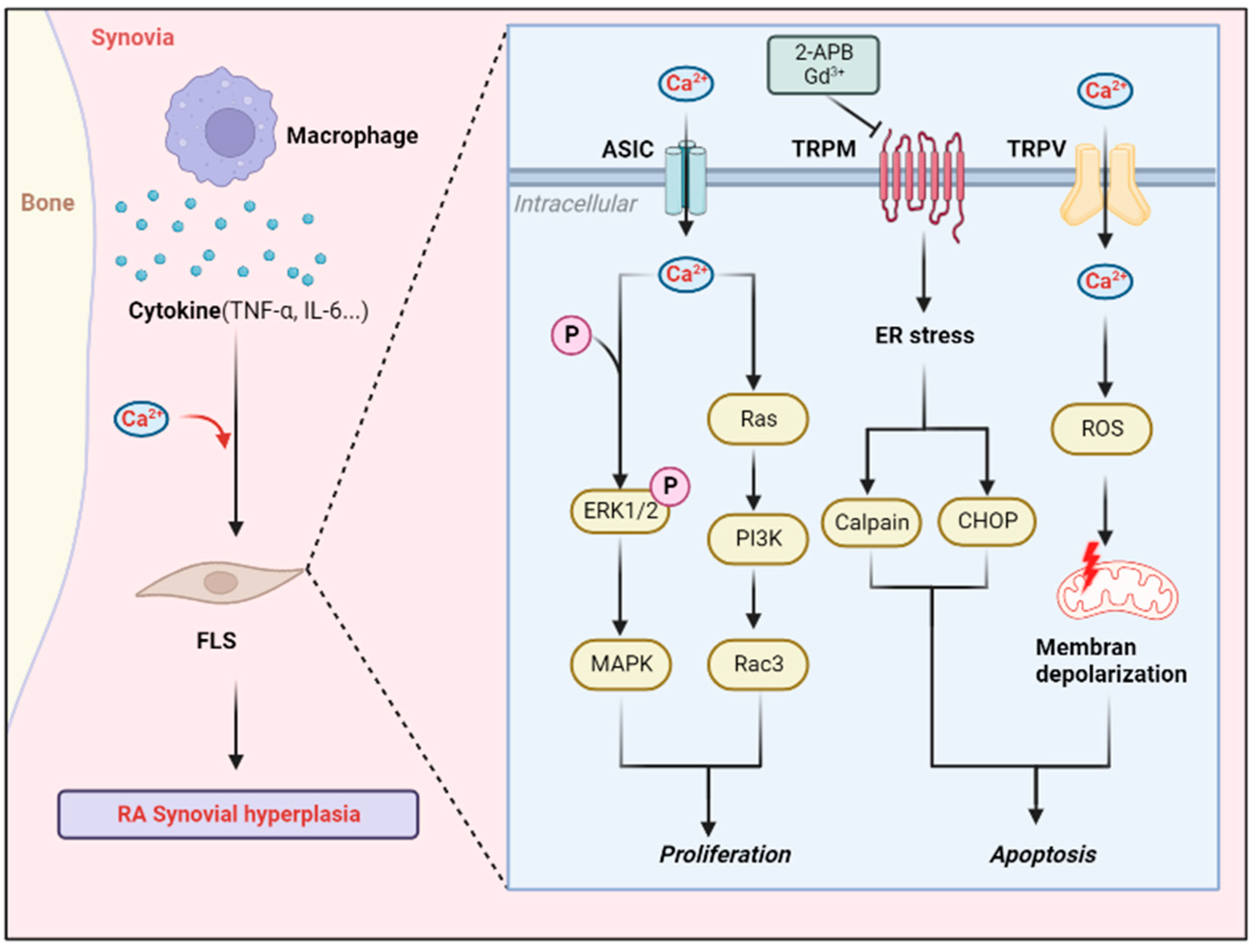
The typical RA lesion is synovial inflammation, which can strike in the early stages of RA. Synovitis often leads to hyperplasia and tissue acidification of the synovial membrane. This provides a pathological basis for the role of the ASIC1a channel in RA synovitis. Indeed, it has been shown that the opening of ASIC1a channels activates Ras-related C3 botulinum toxin substrate 1 and promotes RA synovial cell invasion [48]. In addition, recent evidence has also demonstrated that ASIC1a promotes the proliferation of RA FLS cells through the ERK/MAPK and NFAT signaling [49,50]. This available evidence suggests that ASIC1a plays a toxic role in synovial inflammation, and the proper blockade of ASIC1a channels in the synovium may slow the progression of RA synovitis.
As described in the previous section, cartilage damage during RA is mainly attributed to the apoptosis of chondrocytes and degradation of the extracellular matrix. Ca2+, due to their unique physicochemical properties, are one of the most important signals in apoptosis and, therefore, calcium-permeable channels, which are responsible for their transport, are an important target when controlling cartilage damage. In the various pathological processes of RA, inflammatory bursts in the tissues lead to a decrease in pH within the local cartilage tissue [51]. ASIC1a has been shown to play an important role as an acid sensor in RA cartilage injury. Chen et al. first proposed that the inhibition of ASIC channels using amiloride reduced cartilage damage as well as chondrocyte apoptosis in rats with adjuvant arthritis (AA, an animal model of RA), identifying the chondroprotective effects of ASIC channel inhibitors [52]. However, due to the low selectivity of amiloride, it was difficult to identify the specific subtype of ASIC, although it is certain that this reveals a direct role of ASIC channels in chondrocyte apoptosis and creates a path for subsequent studies. In vitro studies have demonstrated that when ASIC1a is activated by extracellular acidification, chondrocyte extracellular matrix expression is reduced, and it is proposed that this may be due to a strong increase in [Ca2+]i activation downstream of MAPK/ERK signaling as a result of ASIC1a activation [53]. This reveals the mechanism through which ASIC1a affects chondrocytes and provides new insights into cartilage damage in RA. Since then, a number of studies have identified a role for the ASIC1a channel in promoting apoptosis in chondrocytes as well as FLSs, possibly through the mitochondrial pathway [54], and the phosphorylation of extracellular-signal-regulated kinases [55]. In addition to apoptosis, pyroptosis is also a form of programmed cell death, characterized by the activation of caspase-1 and the secretion of the pro-inflammatory cytokine IL-1β/18. Our recent study reports that the ASIC1a-mediated elevation of [Ca2+]i may lead to the pyroptosis of rat chondrocytes through the activation of calpain-2/calcineurin [56,57]. These studies have enriched the mechanisms by which the ASIC1a channel mediates cartilage damage, but relevant in vivo experiments are still lacking. However, our previous studies have shown that IL-6 significantly upregulates ASIC1a levels in chondrocytes of rats with adjuvant arthritis in a dose-dependent manner and significantly mediates chondrocyte apoptosis via ASIC1a [58]. In addition to IL-6, IL-1β and TNF-α also upregulated chondrocyte apoptosis in an NF-κB-dependent manner through the upregulation of ASIC1a [23]. These studies reveal the role that joint inflammation plays in cartilage damage, which is bridged by ASIC1a. In any case, the inhibition of ASIC1a channels at the pharmacological level using reasonable inhibitors has the potential to reduce chondrocyte apoptosis or scorch death. This provides new targets and options for RA treatment.
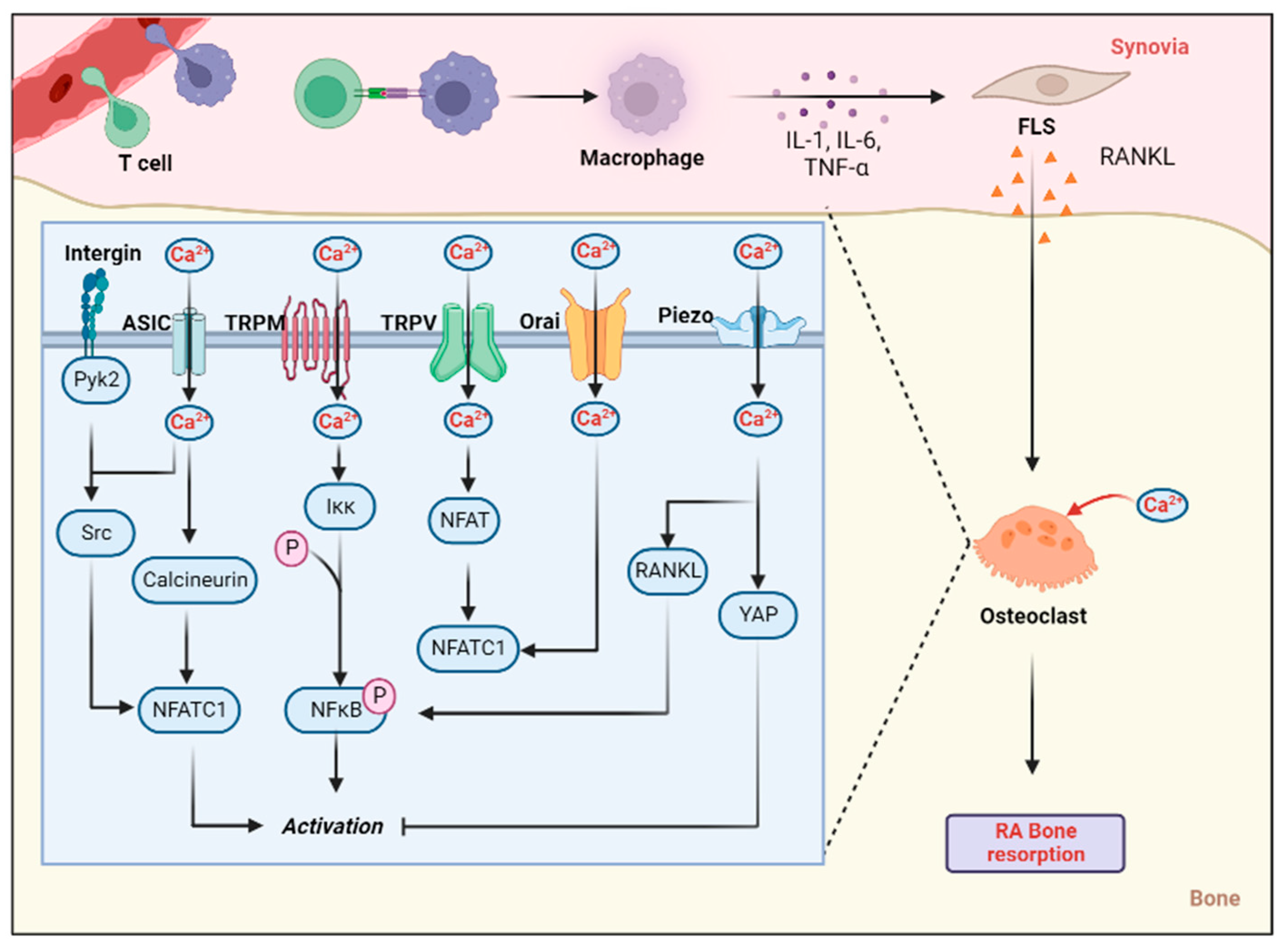
In adults, bone tissue is balanced by the continuous resorption of osteoclasts and the production of osteoblasts. The enhanced osteogenesis has a mitigating effect on the RA bone damage. Osteoclasts originate from monocytes and are found in the blood and circulation. The process of differentiation for osteoclasts is mainly regulated by the macrophage colony-stimulating factor and NF-κB receptor activator ligand (RANKL) [59]. Studies have shown that extracellular acidosis increases the activation and formation of osteoclast precursor cells and multinucleated osteoclast-like cells [60]. As a pH receptor, ASIC plays an important role in osteoclast activation induced by extracellular acidification. It has been reported that acidosis activates ASIC1a in osteoclasts as well as osteoclast precursor cells and promotes osteoclast migration and adhesion via integrin/Pyk2/Src signaling [61]. Cell activation and migration are important aspects of osteoclast-mediated bone resorption, and this study suggests an important role for ASIC1a in osteoclast-mediated bone destruction. However, the limitation is that the study was only performed under a low pH condition (pH = 6.0) and it was difficult to determine the effect of the pH gradient on osteoclast migration. In addition, there is a lack of data on the effect of low pH on the migration and adhesion of osteoclast precursor cells in vivo. Elevated [Ca2+]i can also regulate osteoclast differentiation through a variety of pathways, such as nuclear factor of activated T cells c1 (NFATc1), etc. [62]. The elevated [Ca2+]i mediated by ASIC1a activation is well known. Indeed, Li et al. showed that ASIC1a activation mediates osteoclast genesis via the NFATc1 pathway and that can be inhibited by the ASIC1a antagonist PcTX1 [63]. These studies point to an important role for ASIC1a in acid-induced osteoclast genesis, activation and migration, providing an important therapeutic target in osteoclast activation-related diseases such as RA and osteoporosis.
Local inflammation during RA is often characterized by swelling of the joint, pain and an outbreak of inflammatory factors in the synovial cavity. It has been shown that in RA synovial tissue, activation of ASIC1a mediates an increase in [Ca2+]i and promotes the release of more inflammatory cytokines such as MIP-1a and IL-8 from FLSs via the NFATc3/RANTES pathway [49]. Although the role of ASIC1a in the release of inflammatory factors in RA is well established, the mechanism remains unclear and the evidence is still insufficient.
In conclusion, the toxic role of ASIC1a during RA is well established, i.e., overexpressed or activated ASIC1a promotes inflammation, structural damage and the release of inflammatory factors in RA synovium during the development of RA. Appropriate blockade of ASIC1a at the pharmacological level may be a novel strategy for the targeted treatment of RA, but the clinical trials that currently scarce. Therefore, further clinical evidence of the therapeutic significance of ASIC1a in RA and the need to develop more specific blocking agents are needed in the future.

3.2. ASIC2a

ASIC2a is mainly localized in the central nervous system and peripheral nervous system, and its pH50 is 4.5–4.9, similar to ASIC1a, and it also plays a certain role in the inflammatory response and the acid toxicity of the nervous system. Although the role of ASIC1a and ASIC3 in cartilage damage during RA is well established, interestingly, we found that overexpression of ASIC2a enhanced the chondroprotective effects of PcTx1 (an ASIC1a-specific inhibitor) and APETx2 (an ASIC3-specific inhibitor) and inhibited the ERK1/2 MAPK signaling pathway [64]. This is distinct from the other ASIC isoforms (ASIC1a/3) in cartilage injury and may be due to some unique physicochemical properties of ASIC2a, but in any case, this is the first suggestion of a role for ASIC2a in cartilage injury.

3.3. ASIC3

ASIC3 was mainly localized in the peripheral nervous system but was also detected in the trigeminal ganglion in the central nervous system. ASIC plays an important role in pain transmission due to its unique acid-sensitive current and its high expression in the nervous system. In fact, ASIC3-mediated currents were first detected in cardiac afferents and demonstrated that ASIC3 might be involved in pain transmission [65]. This also opened the research on ASIC3. At the physiological level, compared with other ASIC channels, ASIC3 can be opened at a pH closer to physiological conditions (pH50 = 6.4–6.6), and the transient inward current triggered by protons lasts longer [66]. This suggests that ASIC3 may play an important role in a variety of long-term chronic diseases, such as RA.
The main cause of synovial invasion in RA is the proliferation of intra-articular synovial cells, especially FLSs. Therefore, induction of apoptosis or inhibition of synovial cell proliferation can be a means to alleviate synovial invasion in RA. Hyaluronic acid strengthens the stability of synovial cells and prevents the spread of inflammatory mediators, thus reducing synovial inflammation. Sluka et al. identified ASIC3 channels in mouse knee synovial cells for the first time during their study of the role that ASIC3 plays in nociceptive sensitization in mouse joint inflammation [67], and their additional work revealed a role for ASIC3 in regulating the release of hyaluronic acid from FLSs [68]. This suggests an important role for ASIC channels in synovial tissue. In fact, the different subunits of the ASIC family play inconsistent roles in RA synovial invasion. It has been suggested that ASIC3 activation mediates a rise in [Ca2+]i and induces apoptosis in the FLSs of RA patients [55]. Furthermore, in the collagen-induced arthritis model (an animal model that mimics RA in vivo), ASIC3 mediates the apoptosis of FLSs in response to intra-articular inflammatory mediators (e.g., IL-1β), and ASIC3−/− mice exhibit marked synovial inflammation and invasion [69]. These reports suggest that ASIC3 channels may alleviate the synovial invasive process of RA by mediating calcium toxicity or inflammation, and thus inducing apoptosis in RA synovial cells. This differs from the previous toxic effects of ASIC1a in synovial inflammation, and there are many reasons for this contradiction, the most important of which may be the differences between ASIC isoforms that lead to this effect, the different cell types used, etc. To better understand the therapeutic opportunities that the ASIC3 channel provide in RA, a number of studies will be needed in the future to validate the impact of different modalities of modulation on RA.
In cartilage damage, it has been suggested for a decade that ASIC3 is expressed in FLSs as well as the chondrocytes and that it activates FLSs to release hyaluronic acid when extracellular pH is reduced (~5.5) [68]. This is the first indication of a role for ASIC family channels in the regulation of the extracellular matrix of articular cartilage, although the exact mechanism has not been revealed.
Indeed, early studies have shown that ASIC3 channels are activated during joint inflammation and that their activation is involved in the development of inflammatory pain [67,70,71,72]. Furthermore, in carrageenan-induced arthritis in mice (an acute arthritis model), ASIC3 expression in DRG neurons in joint nerve fibers was significantly upregulated (an approximately 50% increase) [70]. These results all suggest that a decrease in pH during joint inflammation may activate the ASIC3 channels expressed on innervated neurons, thereby transmitting pain signals to the center, manifesting as nociceptive hyperalgesia. In 2012, it was first suggested that, in mice with collagen-induced arthritis, ASIC3 knockout led to significant synovial inflammation and increased FLSs release of IL-6 and MMPs; however, interestingly, the mice showed less painful behavior [69]. This phenomenon is explained by the fact that ASIC3, which is localized at the nerve endings, plays an important role in pain transmission in RA, whereas ASIC3, which is located in synovial cells, plays a certain anti-inflammatory role. Therefore, when ASIC3 is knoced out, nociceptive transmission is reduced and inflammation in the synovial membrane is increased. This study led to an investigation of the role of ASIC channels in RA joint inflammation and also enriched the mechanism by which ASIC3 induces apoptosis in articular chondrocytes, possibly through the upregulation of FLSs to release MMPs. There may be some inhibitory effects of ASIC3 activation or overexpression on synovial invasion as well as an inflammatory burst in RA, but the role of other aspects of RA pathogenesis needs to be further explored. Due to its role in pain transmission and inflammation control, the potential significance of ASIC3 as a therapeutic target for RA is highlighted. Different modulations may have different effects, and more targeted drugs need to be developed in the future to precisely control ASIC3 channels during RA (Figure 2).

4. Transient Receptor Potential (TRP) Channels

In the last century, it was discovered that the deletion of a particular gene in fruit flies causes the onset of temporary visual impairment, so the researchers named this particular gene “transient receptor potential” (TRP) [73]. In 1993, TRP was recognized as a new superfamily of ion channels and its role in Ca2+ permeability was identified [74]. The first case of a human TRP homolog (TRPC1) was reported in 1995 [75]. To date, seven TRP families have been identified: TRPC, TRPV, TRPM, TRPA, TRPP, TRPML, and TRPN. An eighth TRP family was recently identified in yeast and named TRPY (Y for yeast) [76]. In this review, we focus on the TRPM, C and V subfamilies. It is worth mentioning that a fragment with α-kinase activity was fused to the C-terminus of TRPM6 and TRPM7, which is rare in other ion channel families. The research on its α-kinase function is limited.

4.1. TRPM7 Channel

The TRPM channel mainly consists of eight members, TRPM1-8. Interestingly, TRPM6 and TRPM7 form an additional serine/threonine-rich kinase domain at the C-terminus and an α-kinase domain that is homologous to cytoplasmic kinases. TRPM channels are distributed in various mammalian tissues, predominantly the brain and heart. Notably, however, all TRPM channels except TRPM7 were not expressed in bone and cartilage tissues [77]. The TRPM7 channel has received a lot of attention in recent years due to its essential role in various cellular activities. Studies have shown that TRPM7 plays an integral role in cell proliferation, blood vessel development, and cancer progression [16].
TRPM channel is a widely expressed Ca2+ channel, and a potential correlation between TRPM channels and RA FLSs has been demonstrated [78]. Similar to ASIC, the role of the TRPM family subtypes in RA synovial invasion was inconsistent, with TRPM8 being shown to mediate menthol-induced apoptosis in RA rat FLSs [79]. Another member of the TRPM family, TRPM7, is thought to have an inhibitory effect on synovial proliferation. It was noted that TRPM7 can mediate apoptosis in RA FLSs via endoplasmic reticulum stress, and FLSs apoptosis can be inhibited by 2-APB- or Gd3+ [80]. Although this difference is acceptable, many experiments are needed to validate 2-APB due to its low selectivity.
During chondrogenesis, the secretion of alkaline phosphatase leads to calcification of the cartilage matrix and chondrocytes apoptosis, causing cartilage traps. Previous studies have shown that silencing TRPM7 reduces collagen expression in cartilage ADTC5 cells and confirms that TRPM7 might be involved in ionic homeostasis during chondrocyte hypertrophy, promoting endochondral ossification [81]. This explains the mechanism of cartilage damage from another perspective, but relevant in vivo experiments are still lacking. Our recent study revealed the role of TRPM7 in cartilage injury in RA. By inhibiting TRPM7 using 2-APB or si-RNA, we demonstrated in vivo and in vitro, respectively, that the inhibition of TRPM7 reduces the extent of cartilage injury in AA rats by modulating Ihh signaling [82]. Interestingly, TRPM7 can not only mediate programmed apoptosis in chondrocytes. Our recent study also revealed that TRPM7 could promote chondrocyte ferroptosis through the regulation of calcium ions, and thus through the PKCα–NOX4 axis. This suggests that the genetic or pharmacological inhibition of TRPM7 could alleviate RA cartilage destruction by reducing chondrocyte ferroptosis [83]. In conclusion, TRPM7 is an effective therapeutic target for RA and the development of more specific drugs will be necessary in future.
Intracellular Ca2+ and Mg2+ are essential to the proliferation of a wide range of cells. TRPM7, a Ca2+ and Mg2+ channel, was first shown to be expressed in osteoblasts in 2007 [84]. It was noted that TRPM7 in osteoblasts could be increased in compensation in the absence of extracellular Ca2+ and Mg2+, and that the specific si-RNA blockade of TRPM7 inhibits osteoblast proliferation [84]. This study reveals an important role for TRPM7 in intracellular ion homeostasis in osteoblasts and suggests that TRPM7 may be an important factor in osteoblast proliferation. Subsequent studies also identified that PDGF mediates osteoblast proliferation and migration via TRPM7-Mg2+ influx [85], but interestingly, since intracellular Mg2+ plays a regulatory role in TRPM7, interference with TRPM7 in different Mg2+ environments could not be excluded. In addition to PDGF, gallium ions can also upregulate osteoblast activity via TRPM7 and promote the expression of osteogenic-related proteins [86]. In recent years, the role of TRPM7 kinase has also received increasing attention. A recent study has clarified the role of TRPM7 kinase in bone regeneration. The research points show that, in the early stages of inflammation, Mg2+ can flow into macrophages via TRPM7, leading to the cleavage and nuclear accumulation of TRPM7 kinase and promoting the formation of an immune microenvironment for osteoblasts. In late inflammation, Mg2+ promotes the activation of NF-κB in macrophages and upregulates the number of osteoclasts, exerting a negative effect on osteogenesis [87]. Overall, the negative effects of Mg2+ on osteogenesis can outweigh its early positive effects. This reveals a diverse role of Mg2+ in osteogenesis via TRPM7 kinase. In any case, TRPM7, as a transducer of Mg2+ and Ca2+, plays an important role in all life activities of osteoblasts and osteoclasts, offering new therapeutic opportunities for diseases related to bone loss.
The signaling pathways regulated by TRPM channels also play an important role in calcium signaling and leukocyte physiology, affecting immune processes such as phagocytosis, degranulation, cytokine expression, chemotaxis and invasiveness. The role that TRPM channels play in the regulation of inflammation in a variety of pro-inflammatory diseases, including RA, has become increasingly clear in recent years [16,88]. Neutrophils, as the most abundant leukocytes, play an important role in immune responses such as inflammation. In RA patients, CD147 has been reported to upregulate neutrophil chemotaxis, adhesiveness and invasiveness, and this role may be due to the influx of Ca2+, which is mediated by the activation of TRPM7 [89]. This research reveals the potential role of TRPM7 in the inflammatory response in RA, using promyelocytic leukemia cells and peripheral blood or synovial fluid neutrophils from RA patients with high conviction. In addition, the role of TRPM7 intracellular kinase in the immune response has gained attention in recent years due to the specificity of its structure. Thanks to the development of specific kinase inhibitors, the role of TRPM7 kinase in the inflammatory process of RA has been defined. Studies have shown that neutrophils treated with the TRPM7-specific inhibitor TG100-115 or kinase-activated gene-ablated mouse neutrophils have a significantly reduced ability to respond to Gram-negative bacterial lipopolysaccharide LPS or CXCL8 chemokine gradient migration, and suggest that this effect may be due to the Akt/mTOR pathway [90]. All the above studies suggest that the TRPM7 activity of RA neutrophils is critical to their ability to migrate. In the future, more specific drugs could be developed to target this role and inhibit the inflammatory response in RA.
TRPM7 plays a somewhat deleterious role in RA, and the proper blockade of TRPM7 may slow down the synovitis, as well as the process of cartilage destruction during RA. Unfortunately, however, there is still a lack of clinically relevant drugs that can translate the existing basic research.

4.2. TRPC Channel

The TRPC subfamily includes seven members, TRPC1-7. TRPC2 was identified in mice but is a pseudogene in humans [91]. In previous years, the potential role of TRPC in osteoclast genesis has been explored. It was shown that osteoclast formation was increased in I-MFA−/− (MyoD family inhibitor) mice, whereas osteoclast formation was inhibited in mice that were deficient in both TRPC1 and I-MFA genes. Interestingly, they identified a new TRPC1 spliceosome, TRPC1α and TRPC1ε, and revealed that I-MFA might inhibit ICRAC and promote osteoclast formation via TRPC1α and TRPC1ε [92]. In addition to TRPC1, TRPC3 and TRPC6 play important roles in osteoclast regulation. Osteoclast activity is increased in TRPC6-deficient osteoclasts, which exhibit an osteoporotic-like phenotype. In contrast, TRPC3 expression was increased in TRPC6-deficient osteoclasts compared to wild-type (WT), and the inhibition of TRPC3 with Pyr3 reduced bone resorption and differentiation in TRPC6-deficient osteoclasts [93]. This suggests that TRPC6 may play a negative role in osteoclast activity and that the overexpression of TRPC3 promotes bone resorption in osteoclasts. This diverse role has not been independently verified: does reduced osteoclast activity occur in TRPC3−/− mice?
In contrast to TRPM7, TRPC channels were identified as having some anti-inflammatory effects on RA. Studies have shown that mice treated with ML204 (a TRPC5 antagonist) or TRPC5 knockout exhibit greater pain and swlling of joint tissue [94]. However, this study only indicated the anti-inflammatory effect that TRPC5 has on the joints of mice but did not reveal the exact mechanism. More research is needed to validate the role of TRPC channels in the inflammatory response to RA to develop more effective drugs.

4.3. TRPV Channel

How do people perceive heat and the stimulation of capsaicin? This question drives the search for tracking temperature receptors under physiological conditions. Finally, in 1997, the founding member of the TRPV family, TRPV1, was discovered to be a heat- and capsaicin-sensitive receptor [95]. In the last 20 years, studies have shown that heat-sensitive TRPVs (TRPV1–4) are widely expressed in various tissues and organs, but are mainly localized to neurons for sensory transmission. Among the TRPVs, TRPV1 is the most representative class of TRPV channels, and its expression in bone tissue has been demonstrated in recent years [96]. In addition, TRPV2 and TRPV4 have been shown to be expressed in synovial tissue [97]. Thermosensitive TRPVs are a class of nonselective ion channels that play important roles in neuropathic pain, inflammation, immunity, neuronal development, diabetes, cardiovascular disease, and cancer by regulating intracellular and extracellular ion homeostasis [98].

4.3.1. TRPV1

TRPV1 was first demonstrated to be significantly more expressed in the FLSs of RA patients than controls in 2007 [99], suggesting an important role for TRPV1 in synovial disorder. Indeed, it was shown that, in a collagen-induced arthritis model, the activation of TRPV1 channels with capsaicin led to the apoptosis of synovial cells through the upregulation of [Ca2+]i, ROS and mitochondrial membrane depolarization [100].
Mechanical stimulation has long been recognized as a key factor in osteosynthesis, and the role of TRPV channels as a mechanosensitive channels in osteoblasts and osteoclasts has been widely reported. Studies have shown that capsazepine, a TRPV1 ion channel antagonist, inhibits osteoclast bone resorption, osteoblast activity and bone formation, and, in vivo, capsazepine inhibits ovariectomy-induced bone loss in mice [101]. This is the first report on the effect of TRPV1 channels on osteoclasts, showing that the inhibition of TRPV1 has a significantly lower effect on osteoblasts than on osteoclasts. At the overall level, the inhibition of TRPV1 led to a reduction in bone loss, but the exact mechanism behind this has yet to be investigated. This was subsequently demonstrated by Rossi et al., using TRPV1−/− mice [102]. In addition to capsazepine, there are also upstream substances that exert some regulatory effects on osteoclasts via TRPV1. For example, 6-gingerol stimulates TRPV1, promotes the influx of Ca2+, and thus stimulates osteoclast differentiation [103]. Sirtuin 1, also known as NAD-dependent deacetylase, has also recently been shown to directly inhibit TNF-α-induced osteoclast genesis in mice by inhibiting TRPV1 [104]. Interestingly, by co-studying the cannabinoid 2 (CB2) receptor on the surface of osteoclasts, it was found that the stimulation of TRPV1 in osteoclasts inhibited osteogenic activity [105,106]. This suggests that the uncertainty in the role of TRPV1 in osteoclasts is due to crosstalk between CB2 receptors and TRPV1. In fact, it has been shown that TRPV1 deficiencies lead to reduced mRNA and protein expression of Runx2 and ALP in bone marrow stromal cells, and reduces osteoclast genesis and osteogenesis [107]. However, this is limited by the fact that the effect of TRPV1 on osteogenic activity has not been studied in osteoblasts. In any case, the important role that TRPV1 plays in osteoclasts has been established, but more research is still needed to investigate the mechanisms behind this and the specific role that TRPV1 plays in osteoblasts in order to provide a therapeutic bullet for RA.
It was noted that knockout of the TRPV1 channel significantly alleviated pain behavior in mice with fully Freund’s adjuvant-induced adjuvant arthritis [108]. Furthermore, a recent drug-controlled trial indicated that the anti-inflammatory effect of using the TRPV1 blocker APHC3 in adjuvant mice could be equal or superior to that of NSAIDs (e.g., diclofenac ibuprofen and meloxicam) [109].
In conclusion, the role of TRPV1 channels in RA is gradually becoming clearer. The activation of TRPV1 may alleviate the synovial invasion process in RA, while the inhibition of TRPV1 may reduce the bone loss process in RA. However, the overall effect on RA is still unclear, and more in vivo experiments will be conducted to verify the therapeutic significance of TRPV1 channel in the regulation of RA.

4.3.2. TRPV2

As a kind of TRP channel, TRPV2 has been shown to be related to the activation of RA FLS in recent years. The data suggest that the specific activation of TRPV2 in FLS can reduce the content of Rac1 in RA FLS and inhibit FLS invasiveness [110]. Further, in vitro experiments have determined that the TRPV2-specific agonist LER13 reduces the invasiveness of FLSs in RA mice, thereby alleviating synovial invasion during RA [97]. These results provide significant evidence for the inhibitory role of TRPV2 in RA synovial membrane invasiveness. In addition to FLSs, there was also a link between TRPV2 and osteoclasts. TRPV2 expression was significantly increased in RAW264.7 cells (preosteoclasts) after RANKL treatment, and the inhibition of TRPV2 significantly reduces the frequency of Ca2+ oscillations and osteoclast genesis, revealing an important role for TRPV2 in osteoclast genesis [111]. Later, Bai et al. revealed that TRPV2 regulates osteoclast differentiation through the Ca2+-calcineurin-NFAT signaling pathway in multiple myeloma [112]. Further studies in other models, as well as in vivo, are needed to determine the important role that TRPV2 plays in osteoclast differentiation in future. The above studies suggest that TRPV2 may be an essential factor in the differentiation of osteoclasts in RA, and that reducing osteoclast production may alleviate bone loss in RA.
Unlike TRPV1, TRPV2 has been identified as a suppressor of arthritis inflammation. The specific activation of TRPV2 was shown to inhibit the IL-1β-induced expression of MMP-2 and MMP-3 and to suppress the aggressiveness of FLSs [97]. This evidence was also validated by Gulko et al. in FLS cell lines from RA patients [110]. Although the role of TRPV2 in joint inflammation is relatively clear, a lot of research is still needed to explore its specific mechanism in the future.

4.3.3. TRPV4

In addition to TRPV1 and 2, TRPV4 also plays an important role in RA synovitis. It is reported that the activation of TRPV4 following hypotonicity promoted the release of ATP, ROS and cell proliferation from FLSs in RA rats [113]. This suggests that the activation of TRPV4 may aggravate synovial hyperplasia, which is different from TRPV1 and TRPV2, and the reason for this deserves further investigation.
In addition to the synovial invasion, TRPV4 has also been shown to mediate Ca2+ influx and chondrocyte apoptosis in osteoarthritis models [114]. In addition to TRPV5, TRPV4 has also been shown to impact the differentiation and activity of osteoclasts [115,116]. It was shown that TRPV4 mediates Ca2+ influx into rat osteoclasts and provides sufficient [Ca2+]i to maintain RANKL-induced osteoclast differentiation [117]. This study revealed for the first time the important role of TRPV4 in osteoclast genesis and gave rise to the study of TRPV4 in osteoclasts. A recent study also indicated that the knockdown of TRPV4 could inhibit osteoclast differentiation and alleviate osteoporosis in mice by inhibiting cellular autophagy as well as regulating NFATc1 [118]. In any case, the important role of TRPV4 in osteoclast differentiation has been identified and more treatments for osteoclast abnormalities, such as RA or osteoporosis, could be developed, targeting TRPV4 in the future. Furthermore, in vivo double knockout TRPV1 and TRPV4 models also showed reduced osteoclast activity [116], suggesting a role for TRPV1 in osteoclasts. However the researchers did not state whether the effects of TRPV1 and TRPV4 are synergistic or antagonistic. Both genetic and pharmacological inhibition have shown therapeutic potential to alleviate bone loss and structural damage in RA, and more specific drugs targeting TRPV4 will be needed in the future.
TRPV, as a mechanically gated channel, is important in the mechanical nociceptive transmission of the inflammatory process in RA. IL-17, an important inflammatory mediator in the RA process, is thought to upregulate TRPV4 in DRG neurons and mediate nociceptive hyperalgesia [119]. Although the results demonstrate that TRPV4 can play an important role in IL-17-mediated pain, this has not been validated in RA models.
Overall, the activation of TRPV4 may play a role in overall toxicity during RA, including promoting synovitis, mediating osteoclast activation leading to bone loss, and promoting inflammatory bursts. The appropriate inhibition of TRPV4 may alleviate the RA process. However, it is a potential drug target for the treatment of RA.

4.3.4. TRPV5

Ca2+ influx is indispensable in chondrocyte apoptosis, necrosis, and matrix degradation. TRPV5, as a Ca2+ channel, can mediate Ca2+ influx and activate the downstream MAPK and Akt/mTOR pathways, thereby promoting chondrocyte apoptosis [120]. However, the in vivo effect has not been observed using the RA model. Indeed, the role of TRPV channels in mediating cartilage damage in osteoarthritis has been extensively reported [121,122,123], but there is still a lack of RA animal models in which TRPV is the target.
The role of TRPV channels in osteoclast bone resorption was first identified by Eerden BC et al. with TRPV5. It was suggested that TRPV5 channels were expressed in both human and murine osteoblasts and that TRPV5-/- mice showed higher osteoclast numbers and osteoclast volumes compared to WT.; however, bone resorption was virtually absent in TRPV5−/− osteoclast cultures in vitro [124]. This establishes an important role for TRPV5 in osteoclast bone resorption but is limited by the fact that the exact mechanism is not explained. To verify the role of TRPV5 in osteoclast bone resorption, the calcium kinetic regulator econazole was proposed to downregulate TRPV5 levels in rat osteoclasts in a dose-dependent manner and inhibit bone resorption [125]. However, the investigators found an increase in [Ca2+]i in osteoclasts after TRPV5 inhibition, suggesting that there may be another intracellular compensatory mechanism that compensates for TRPV5 downregulation, making the results inconclusive. In fact, the important role of TRPV5 in osteoclast bone resorption was questioned in 2008. The data showed that TRPV5−/− mice still underwent significant bone resorption and that treatment with alendronate normalized bone thickness in these mice [126]. Subsequently, it was noted that TRPV5 inactivation completely inhibited the RANKL-induced increase in osteoclast [Ca2+]i and was accompanied by a significant activation of bone resorption [127]. This suggests that TRPV5 may be an important pathway, mediating RANKL-induced [Ca2+]i elevation in osteoclasts. A role for TRPV5 in osteoclast calcium homeostasis was also confirmed [128]. Interestingly, studies have shown that estrogen inhibits RANKL-induced osteoclast differentiation and bone resorption activity by increasing the expression of TRPV5 channels [129]. Further research revealed that estrogen E2 activates NF-κB via the estrogen receptor, which binds to the 286 nt~−277 nt fragment of the TRPV5 promoter region, upregulates TRPV5 and inhibits osteoclast genesis [130]. The above studies suggest that TRPV5 overexpression may play an inhibitory role in osteoclast genesis and bone resorption, contrary to the results of earlier studies, which may be due to errors caused by objective conditions, such as the use of different animal or cellular models and differences in experimental conditions. In any case, the current research advances suggest that the appropriate stimulation of TRPV5 may reduce bone loss and joint structural damage in late RA.

5. P2X7 Receptor (P2X7R)

Adenosine triphosphate (ATP) was originally thought to be the universal energy currency of the cell and has also been identified as an extracellular, non-adrenergic, non-cholinergic (NANC) neurotransmitter in smooth muscle organs [131]. Purinergic signaling is a highly evolutionarily conserved signaling system with extracellular ATP, related nucleotides and adenosine, which serve as transmitter molecules that play a crucial role in many physiological processes and pathological responses [132,133,134]. The receptors stimulated by these purine nucleotides are classified into two types: ligand-gated cation channels, called P2X receptors (P2XRs) (seven mammalian subtypes: P2X1-7), and G protein-coupled P2Y receptors (P2YRs) (eight mammalian subtypes: P2Y1, 2, 4, 6, 11-14) [135,136]. P2XR mediates the inward flow of extracellular cations, including Na+, Ca2+ and K+, after binding to ligands.
It has been shown that P2X7R plays a potential role in the regulation of RA development and progression [137,138]. At the genetic level, studies have shown that polymorphisms at positions 1068 and 1513 of the P2X7R gene may be susceptibility loci for RA [139]. Comparable research has demonstrated that the 489C>T SNP in the P2X7R gene potentiates the function of P2X7R in RA patients and may be engaged in the development of RA [140]. In addition, the significantly elevated expression of P2X7R in the peripheral blood mononuclear cells of RA patients has been observed in several studies [141,142]. In other cases, elevated P2XR levels in synovial tissue have been found in patients with RA, which has a high diagnostic value for RA [133,143]. The above studies suggest that P2X7R plays an important role in the pathogenesis of RA. In cartilage injury, it has been suggested that the over-activation of P2X7R can mediate apoptosis in osteoarthritic articular chondrocytes through the IRE1-mTOR-PERK axis [144], but can this result be extended to RA? A significant number of in vitro and in vivo studies are needed to demonstrate the specific mechanisms of P2XR in articular cartilage damage in RA.
ATP was shown earlier to be a potent stimulator of osteoclast formation, and P2XR, a purinergic signaling receptor, and was also found to play an important role in osteoclasts in recent years [145]. Osteoblasts and osteoclasts express a range of P2X purinergic receptors. However, until recently, only the P2X7R subtype’s role in the bone metabolism has been documented, and fewer studies have addressed the role of the remaining six P2X receptor subtypes [146]. The role of P2X7R in osteoblasts is still controversial. To mimic osteoporosis after estrogen withdrawal, the use of ovariectomized mice revealed more severe bone loss in P2X7R-deficient mice, and histological examination showed an increased number of osteoclasts in P2X7R−/− mice [147]. This is the first in vivo demonstration of the activating effect of inhibiting P2X7R on osteoclasts and is consistent with previous studies at the genetic level [148,149]. In addition, appropriate concentrations of ATP were shown to activate P2X7R and mediate Ca2+ inward flow in rabbit osteoclasts, possibly leading to the inhibition of bone resorption [150]. The above studies suggest that P2X7R activation exerts a negative regulation on bone resorption. Surprisingly, blocking P2X7R on osteoclast precursors with blocking antibodies inhibited the formation of multinucleated osteoclasts in vitro, while in vivo P2X7R knockout mice maintained the ability to form multinucleated osteoclasts [151]. This discrepancy may be due to differences in experimental models or to the complexity of the system. Further studies have shown that the P2X7R antagonists AZ15d, KN62 and OATP all reduce RANKL and M-CSF-induced osteoclast genesis in vitro [152]. However, it is worth noting that the P2X7R antagonists may activate other mechanisms, such as AZ15d, which can affect some pore structures, activated by trichothecene [153]. A recent report also showed that surface expression of P2X7Rs was downregulated when RAW 264.7 cells were chronically exposed to high levels of ATP, thereby preventing precursor osteoclast fusion [154]. In conclusion, the present findings demonstrate that either the activation or inhibition of P2X7R can have an inhibitory effect on osteoclast genesis, which may be the result of the combined action of multiple P2XR isoforms due to the low experimental selectivity. In fact, previous studies suggest that low concentrations of ATP can activate P2XRs other than P2X7 receptors, such as P2X2R and P2X3R, thereby increasing bone resorption [145]. It was shown that the selective non-nucleotide antagonist of the P2X2/3 receptor, A317491, reduced RANKL expression in mice in an unloading model with tail suspension [155], and, in addition, 1 µM Minodronic acid (an antagonist of P2X2R and P2X3R) treatment of mice resulted in t hedetachment of osteoclasts from the bone surface, inhibiting bone resorption [156]. These studies reveal a potential role for P2X2/3R in osteoclast formation and bone resorption, and more research is needed in the future to confirm the mechanisms behind this.
As a purinergic receptor, P2XR is involved in a variety of autoimmune diseases and plays an important role in the regulation of immune processes. In the regulation of inflammation by P2XR, it has been suggested that P2X7R enhances the release of pro-inflammatory factors by macrophages [157]. In addition to macrophages, IL-17-producing T helper (Th17) cells are the main effector cells in the pathogenesis of RA, and recent studies have shown that the pharmacological inhibition of P2X7R in a model of collagen-induced experimental arthritis exhibits a significant reduction in Th17-cell-promoting factors (e.g., IL-1β and IL-6) [158]. This clarifies that P2X7R plays an important role in the release of inflammatory factors in collagen-induced arthritis and in T cell differentiation, and suggests a new approach to P2X7R-mediated inflammation. In fact, in early years, researchers suggested that there may be a cytokine-independent pro-inflammatory mechanism of the P2X7R that mediates the release of histone proteases from macrophages and suggested that this mechanism may be relevant to joint disease [159]. Although the role of P2X7R in the regulation of peripheral inflammation in RA is relatively well-established, its specific mechanisms need to be further explored and may involve multiple cellular interactions.
Overall, P2X7R plays an important role in the structural damage of RA, and plays a certain toxic role in cartilage apoptosis, suggesting that the inhibition of P2X7R can alleviate RA to a certain extent. However, relevant clinical research is still lacking at present, and some translational research could be appropriately carried out in the future (Figure 3).

6. Other Channels

6.1. Orai1 Channel

Upon depletion of intracellular Ca2+, store-operated calcium (SOC) channels mediate inward flux of extracellular Ca2+ to replenish the Ca2+ pools of the endoplasmic reticulum. This phenomenon was proposed more than 30 years ago by Putney et al. [160]. The most calcium-selective of the SOC channel family is the Ca2+ release-activated-Ca2+ (CRAC) channel, which mainly consists of Orai channels in the cytoplasmic membrane and stromal interaction molecules (STIM) in the endoplasmic reticulum membrane [161]. There are three mammalian members of the Orai channel (Orai1-3), which mainly consists of a hexametric complex forming a selective calcium-permeable channel in the cytosol, of which Orai1 is the most intensively studied, and a 33 kDa protein consisting of four transmembrane regions, with both the N and C termini located intracellularly [162]. Orai1 channels activated by STIM1 play an important role in a variety of pathological and physiological processes. In the central nervous system, mice knocked out of Orai1 or its activator STIM1 exhibited stronger seizures, as well as convulsive mortality [163]. Orai1-STIM1 also plays an important role in tumors. For instance, the downregulation of Orai1 reduces the invasion of glioblastoma cells and downregulates the proliferation of breast cancer cell lines [164].
In 2014, the Orai1 channel was shown to be genetically polymorphic in the Orai1 gene and associated with RA susceptibility in the population, linking Orai1 etiologically to RA [165]. Furthermore, the efficacy of using si-RNA intra-articular injections in RA has been established [166,167]. Although Orai1 has been shown to be associated with susceptibility to RA, information on its role in RA synovial invasion remains relatively limited. A recent report suggests that Orai1 channels can reverse Fas apoptotic signaling in RA FLSs [168]. It has been suggested that Orai channels may play a synovial protective role in RA, and further exploration of the role that CRAC channels play in RA synovial invasion is needed in the future to develop more promising therapeutic targets. In addition to its effect on synovial inflammation, the role that Orai1 channels play in osteoclasts is becoming increasingly evident, suggesting its place in RA joint damage [169]. In the previous decade, it was shown that Ca2+ oscillations in pro-osteoclasts were triggered by the RANKL-dependent activation of TRPV2 and SOCE and intracellular Ca2+ release [111]. Furthermore, STIM1 and TRPV4 have also been shown to regulate shear-induced calcium oscillations during osteoblast differentiation [170], suggesting a potential role for Orai1 in intracellular calcium homeostasis in osteoclasts. Later, by observing shRNA silencing of Orai1 in RAW264.7 cells, NFATc1 transduction was found to be inhibited, with impaired osteoclast synthesis [171]. This is direct evidence of an important role for Orai1 in osteoclasts and suggests that this may be due to the silencing of Orai1 leading to the inhibition of SOCE, and consequently affecting the transcription of RANKL signaling. Interestingly, another study found reduced cortical ossification and trabecular thinning in Orai1−/− animals compared to controls on microcomputed tomography, and subsequent in vitro studies confirmed that the inhibition of Orai1 activity not only impairs the differentiation of human osteoclasts, but of osteoblasts and their function [172]. It is hypothesized that the loss of Orai1 in osteoblasts leads to calcium disorders; therefore, silencing Orai1 can have an effect on both osteoblasts and osteoclasts. Could this mechanism be extrapolated to chondrocytes? More research will be needed in the future to confirm the role of Orai1 in chondrocytes.
Overall, research on Orai1 in RA is lacking at present, although it has shown some results in terms of RA synovial protection and bone loss (Figure 4).

6.2. Piezo1 Channel

In mammals, mechanical signal transduction mainly involves physiological responses to touch, pressure, airflow, sound, blood pressure, etc. Mechanical transduction often requires mechano-transduction proteins to convert mechanical signals into biochemical signals. The discovery of a new group of mammalians, mechanically activated channels, Piezo channels, by Patapoutian et al. in 2010 attracted considerable attention from researchers around the world and opened the field of research into eukaryotic mechanically activated channels [173]. There are two Piezo proteins (Piezo1–2) in mammals and, along with their massive structures, they are the largest cytosolic ion channel complexes reported to date [174]. Human Piezo1 channels are permeable to monovalent ions such as K+, Na+ and Cs+, divalent ions such as Ba2+, Ca2+ and Mn2+, and organic cations such as tetramethylammonium and tetraethylammonium.
The potential role that Piezo channels play in chondrocytes was first proposed by Lee et al. [175]. Subsequently, the Piezo1 protein was identified as having an apoptosis-inducing effect on human primary chondrocytes, possibly via the MAPK/ERK signaling pathway [176]. This study clarifies the mechanism by which mechanical loading leads to chondrocyte apoptosis and lays the foundation for explorations of the role of Piezo1 channels in degenerative cartilage diseases. Although the mechanism by which the Piezo1 channel mediates chondrocyte apoptosis has been well-established in models of osteoarthritis [177], studies in RA animal models have been very limited. In addition to its effect on chondrocyte apoptosis, a recent study found that targeting Piezo1 deficiency in chondrocytes resulted in an osteoporotic phenotype and further revealed that Piezo1 deficiencies impaired the process of chondrocyte ossification, leading to impaired bone trabecula synthesis [178]. This suggests that although Piezo1 mediates chondrocyte apoptosis, its in vivo deletion can also lead to impaired chondrocyte ossification and consequently increased bone loss. This paradoxical effect has led to difficulties in drug development and more in vivo experiments are needed to investigate the combined effects of Piezo1 on articular cartilage in vivo to better develop therapeutic agents for RA.
Piezo1 is expressed as a mechanosensitive channel in a variety of non-sensory tissues, and mechanical forces are also thought to be an effective stimulus for bone reconstruction; therefore, is there a role for Piezo1 in osteosynthesis? A genetic impact mapping of human osteoporosis identified several Piezo1 single-nucleotide polymorphisms associated with osteoporosis and fracture [179], suggesting a potential role for Piezo1 in osteosynthesis. In addition, in human primary periodontal ligament cells (hPDLC) cultured in vitro, Piezo1 expression increased after mechanical stress stimulation and persisted for 12h, and osteoclast-activation-related genes, such as RANKL and COX2, were activated, whereas RANKL and COX2 were inhibited after the use of a Piezo1 inhibitor (GsMTx4). To verify the effect of Piezo1 modulation on osteoclasts in hPDLC, the co-culture of hPDLC and RAW264.7 showed that the RAW264.7’s ability to differentiate into osteoclasts was reduced after GsMTx4 treatment [180]. The results of this study suggest another important role for Piezo1 in osteoclast synthesis, but the current studies are limited to in vitro studies, and the exact mechanism is not yet clear. It is worth noting that a recent in vivo study reported increased bone resorption in Piezo1 conditional knockout mice, mechanistically suggesting that Piezo1 deficiency impairs Col-II and Col-IX production by reducing YAP nuclear localization, leading to increased osteoclast activity [181]. This seems to be the opposite conclusion to the previous in vitro experiments, but the in vivo experiments are more convincing as there are many reasons for the in vitro errors, such as a lack of high selectivity in the GsMTx4, failure to consider the complexity of the internal environment, and the fact that Jin et al. did not directly demonstrate the role of Piezo1 in osteoclasts. Further research is needed to explore the other effects of Piezo1 on osteoclasts and its mechanisms to better treat osteoclast abnormalities in diseases such as RA (Table 1).

7. Concluding Remarks

In this review, we briefly describe the main pathological features of RA, including cartilage damage, synovial invasion and the onset of joint inflammation, and present a short description of the calcium-permeable channels, such as ASIC, TRP, P2XR and Piezo, which are involved in the pathogenesis of RA. Finally, we explain the role of these calcium-permeable channels in the pathogenesis of RA. At the overall level, the activation of the vast majority of Ca2+-permeable channels can exacerbate RA synovial inflammation by promoting synovial cell hyperproliferation, as well as other signaling pathways. However, a small number of channels can also promote synovial cell apoptosis. In addition, at the level of structural damage in RA, the activation of Ca2+-permeable channels often leads to chondrocyte death and osteoclast activation through different signaling pathways. This subsequently leads to increased cartilage damage and bone erosion in RA. Although the specific mechanisms of some of these channels in the pathogenesis of RA are still unclear, there are sufficient studies supporting their role in RA. The description of the role of calcium-permeable channels in various aspects of RA provides a more comprehensive and in-depth review of the therapeutic targets in RA.

Author Contributions

H.-Y.L. and H.-X.Y. searched literatures. H.-Y.L. and Y.C. drafted the manuscript. Y.-J.Z. and S.-F.L. critically discussed and revised the manuscript. W.H. and R.-P.Z. generated themes and ideas. All authors have read and agreed to the published version of the manuscript.

Funding

This work was supported by grants from the National Natural Science Foundation of China (82272450, 81902182, 82071591), the Natural Science Foundation Incubation Program of The Second Hospital of Anhui Medical University (2021GMFY06).

Institutional Review Board Statement

Not applicable.

Informed Consent Statement

Not applicable.

Data Availability Statement

Not applicable.

Conflicts of Interest

The authors declare that they have no conflict of interest.

References

  1. Lee, D.M.; Weinblatt, M.E. Rheumatoid arthritis. Lancet 2001, 358, 903–911. [Google Scholar] [CrossRef]
  2. McQueen, F.M.; Stewart, N.; Crabbe, J.; Robinson, E.; Yeoman, S.; Tan, P.L.; McLean, L. Magnetic resonance imaging of the wrist in early rheumatoid arthritis reveals a high prevalence of erosions at four months after symptom onset. Ann. Rheum. Dis. 1998, 57, 350–356. [Google Scholar] [CrossRef]
  3. McGonagle, D.; Conaghan, P.G.; O’Connor, P.; Gibbon, W.; Green, M.; Wakefield, R.; Ridgway, J.; Emery, P. The relationship between synovitis and bone changes in early untreated rheumatoid arthritis: A controlled magnetic resonance imaging study. Arthritis Rheum. 1999, 42, 1706–1711. [Google Scholar] [CrossRef]
  4. Brzustewicz, E.; Henc, I.; Daca, A.; Szarecka, M.; Sochocka-Bykowska, M.; Witkowski, J.; Bryl, E. Autoantibodies, C-reactive protein, erythrocyte sedimentation rate and serum cytokine profiling in monitoring of early treatment. Cent. Eur. J. Immunol. 2017, 42, 259–268. [Google Scholar] [CrossRef] [PubMed]
  5. Aletaha, D.; Smolen, J.S. Diagnosis and Management of Rheumatoid Arthritis: A Review. JAMA 2018, 320, 1360–1372. [Google Scholar] [CrossRef] [PubMed]
  6. Sayah, A.; English, J.C., 3rd. Rheumatoid arthritis: A review of the cutaneous manifestations. J. Am. Acad. Dermatol. 2005, 53, 191–209; quiz 210-192. [Google Scholar] [CrossRef]
  7. Alunno, A.; Gerli, R.; Giacomelli, R.; Carubbi, F. Clinical, Epidemiological, and Histopathological Features of Respiratory Involvement in Rheumatoid Arthritis. Biomed. Res. Int. 2017, 2017, 7915340. [Google Scholar] [CrossRef]
  8. Schjerning, A.M.; McGettigan, P.; Gislason, G. Cardiovascular effects and safety of (non-aspirin) NSAIDs. Nat. Rev. Cardiol. 2020, 17, 574–584. [Google Scholar] [CrossRef]
  9. Harirforoosh, S.; Asghar, W.; Jamali, F. Adverse effects of nonsteroidal antiinflammatory drugs: An update of gastrointestinal, cardiovascular and renal complications. J. Pharm. Pharm. Sci. 2013, 16, 821–847. [Google Scholar] [CrossRef]
  10. Solomon, D.H.; Glynn, R.J.; Karlson, E.W.; Lu, F.; Corrigan, C.; Colls, J.; Xu, C.; MacFadyen, J.; Barbhaiya, M.; Berliner, N.; et al. Adverse Effects of Low-Dose Methotrexate: A Randomized Trial. Ann. Intern. Med. 2020, 172, 369–380. [Google Scholar] [CrossRef]
  11. Morand, E.F.; McCloud, P.I.; Littlejohn, G.O. Life table analysis of 879 treatment episodes with slow acting antirheumatic drugs in community rheumatology practice. J. Rheumatol. 1992, 19, 704–708. [Google Scholar] [PubMed]
  12. Clapham, D.E. Calcium signaling. Cell 2007, 131, 1047–1058. [Google Scholar] [CrossRef] [PubMed]
  13. Islam, M.S. Calcium Signaling: From Basic to Bedside. Adv. Exp. Med. Biol. 2020, 1131, 1–6. [Google Scholar] [CrossRef] [PubMed]
  14. Alm, E.; Bloom, G.D. Cyclic nucleotide involvement in histamine release from mast cells—a reevaluation. Life Sci. 1982, 30, 213–218. [Google Scholar] [CrossRef]
  15. Ye, Z.; Shen, Y.; Jin, K.; Qiu, J.; Hu, B.; Jadhav, R.R.; Sheth, K.; Weyand, C.M.; Goronzy, J.J. Arachidonic acid-regulated calcium signaling in T cells from patients with rheumatoid arthritis promotes synovial inflammation. Nat. Commun. 2021, 12, 907. [Google Scholar] [CrossRef]
  16. Liang, H.Y.; Chen, Y.; Wei, X.; Ma, G.G.; Ding, J.; Lu, C.; Zhou, R.P.; Hu, W. Immunomodulatory functions of TRPM7 and its implications in autoimmune diseases. Immunology 2021, 165, 3–21. [Google Scholar] [CrossRef]
  17. So, S.Y.; Ip, M.; Lam, W.K. Calcium channel blockers and asthma. Lung 1986, 164, 1–16. [Google Scholar] [CrossRef]
  18. Sueta, D.; Tabata, N.; Hokimoto, S. Clinical roles of calcium channel blockers in ischemic heart diseases. Hypertens. Res. 2017, 40, 423–428. [Google Scholar] [CrossRef]
  19. Landstrom, A.P.; Dobrev, D.; Wehrens, X.H.T. Calcium Signaling and Cardiac Arrhythmias. Circ. Res. 2017, 120, 1969–1993. [Google Scholar] [CrossRef]
  20. Clem, J.R. Pharmacotherapy of ischemic heart disease. AACN Clin. Issues 1995, 6, 404–417; quiz 493-404. [Google Scholar] [CrossRef]
  21. Lee, S.; Jo, S.; Talbot, S.; Zhang, H.B.; Kotoda, M.; Andrews, N.A.; Puopolo, M.; Liu, P.W.; Jacquemont, T.; Pascal, M.; et al. Novel charged sodium and calcium channel inhibitor active against neurogenic inflammation. Elife 2019, 8, e48118. [Google Scholar] [CrossRef] [PubMed]
  22. Chen, Y.; Zhu, C.J.; Zhu, F.; Dai, B.B.; Song, S.J.; Wang, Z.Q.; Feng, Y.B.; Ge, J.F.; Zhou, R.P.; Chen, F.H. Necrostatin-1 ameliorates adjuvant arthritis rat articular chondrocyte injury via inhibiting ASIC1a-mediated necroptosis. Biochem. Biophys. Res. Commun. 2018, 504, 843–850. [Google Scholar] [CrossRef] [PubMed]
  23. Zhou, R.P.; Dai, B.B.; Xie, Y.Y.; Wu, X.S.; Wang, Z.S.; Li, Y.; Wang, Z.Q.; Zu, S.Q.; Ge, J.F.; Chen, F.H. Interleukin-1beta and tumor necrosis factor-alpha augment acidosis-induced rat articular chondrocyte apoptosis via nuclear factor-kappaB-dependent upregulation of ASIC1a channel. Biochim. Biophys. Acta Mol. Basis Dis. 2018, 1864, 162–177. [Google Scholar] [CrossRef] [PubMed]
  24. Lin, Y.J.; Anzaghe, M.; Schulke, S. Update on the Pathomechanism, Diagnosis, and Treatment Options for Rheumatoid Arthritis. Cells 2020, 9, 880. [Google Scholar] [CrossRef]
  25. Henderson, B.; Revell, P.A.; Edwards, J.C. Synovial lining cell hyperplasia in rheumatoid arthritis: Dogma and fact. Ann. Rheum. Dis. 1988, 47, 348–349. [Google Scholar] [CrossRef]
  26. McInnes, I.B.; Schett, G. The pathogenesis of rheumatoid arthritis. N. Engl. J. Med. 2011, 365, 2205–2219. [Google Scholar] [CrossRef]
  27. Suto, T.; Tosevska, A.; Dalwigk, K.; Kugler, M.; Dellinger, M.; Stanic, I.; Platzer, A.; Niederreiter, B.; Sevelda, F.; Bonelli, M.; et al. TNFR2 is critical for TNF-induced rheumatoid arthritis fibroblast-like synoviocyte inflammation. Rheumatology (Oxford) 2022. [Google Scholar] [CrossRef]
  28. Luz-Crawford, P.; Hernandez, J.; Djouad, F.; Luque-Campos, N.; Caicedo, A.; Carrere-Kremer, S.; Brondello, J.M.; Vignais, M.L.; Pene, J.; Jorgensen, C. Mesenchymal stem cell repression of Th17 cells is triggered by mitochondrial transfer. Stem Cell Res. Ther. 2019, 10, 232. [Google Scholar] [CrossRef]
  29. Pap, T.; Muller-Ladner, U.; Gay, R.E.; Gay, S. Fibroblast biology. Role of synovial fibroblasts in the pathogenesis of rheumatoid arthritis. Arthritis Res. 2000, 2, 361–367. [Google Scholar] [CrossRef]
  30. McInnes, I.B.; Schett, G. Cytokines in the pathogenesis of rheumatoid arthritis. Nat. Rev. Immunol. 2007, 7, 429–442. [Google Scholar] [CrossRef]
  31. Saeki, N.; Imai, Y. Reprogramming of synovial macrophage metabolism by synovial fibroblasts under inflammatory conditions. Cell Commun. Signal. 2020, 18, 188. [Google Scholar] [CrossRef] [PubMed]
  32. Schett, G.; Gravallese, E. Bone erosion in rheumatoid arthritis: Mechanisms, diagnosis and treatment. Nat. Rev. Rheumatol. 2012, 8, 656–664. [Google Scholar] [CrossRef]
  33. McGonagle, D.; Tan, A.L.; Moller Dohn, U.; Ostergaard, M.; Benjamin, M. Microanatomic studies to define predictive factors for the topography of periarticular erosion formation in inflammatory arthritis. Arthritis Rheum. 2009, 60, 1042–1051. [Google Scholar] [CrossRef]
  34. Hayer, S.; Redlich, K.; Korb, A.; Hermann, S.; Smolen, J.; Schett, G. Tenosynovitis and osteoclast formation as the initial preclinical changes in a murine model of inflammatory arthritis. Arthritis Rheum. 2007, 56, 79–88. [Google Scholar] [CrossRef]
  35. Teitelbaum, S.L. Bone resorption by osteoclasts. Science 2000, 289, 1504–1508. [Google Scholar] [CrossRef] [PubMed]
  36. Schett, G.; Stolina, M.; Bolon, B.; Middleton, S.; Adlam, M.; Brown, H.; Zhu, L.; Feige, U.; Zack, D.J. Analysis of the kinetics of osteoclastogenesis in arthritic rats. Arthritis Rheum. 2005, 52, 3192–3201. [Google Scholar] [CrossRef] [PubMed]
  37. Aeberli, D.; Eser, P.; Bonel, H.; Widmer, J.; Caliezi, G.; Varisco, P.A.; Moller, B.; Villiger, P.M. Reduced trabecular bone mineral density and cortical thickness accompanied by increased outer bone circumference in metacarpal bone of rheumatoid arthritis patients: A cross-sectional study. Arthritis Res. Ther. 2010, 12, R119. [Google Scholar] [CrossRef]
  38. Sharp, J.T.; Tsuji, W.; Ory, P.; Harper-Barek, C.; Wang, H.; Newmark, R. Denosumab prevents metacarpal shaft cortical bone loss in patients with erosive rheumatoid arthritis. Arthritis Care Res. 2010, 62, 537–544. [Google Scholar] [CrossRef] [PubMed]
  39. Tournis, S.; Samdanis, V.; Psarelis, S.; Liakou, C.; Antoniou, J.; Georgoulas, T.; Dontas, I.; Papaioannou, N.; Gazi, S.; Lyritis, G.P. Effect of rheumatoid arthritis on volumetric bone mineral density and bone geometry, assessed by peripheral quantitative computed tomography in postmenopausal women treated with bisphosphonates. J. Rheumatol. 2012, 39, 1215–1220. [Google Scholar] [CrossRef]
  40. Hoff, M.; Haugeberg, G.; Odegard, S.; Syversen, S.; Landewe, R.; van der Heijde, D.; Kvien, T.K. Cortical hand bone loss after 1 year in early rheumatoid arthritis predicts radiographic hand joint damage at 5-year and 10-year follow-up. Ann. Rheum. Dis. 2009, 68, 324–329. [Google Scholar] [CrossRef]
  41. Haugeberg, G.; Lodder, M.C.; Lems, W.F.; Uhlig, T.; Orstavik, R.E.; Dijkmans, B.A.; Kvien, T.K.; Woolf, A.D. Hand cortical bone mass and its associations with radiographic joint damage and fractures in 50-70 year old female patients with rheumatoid arthritis: Cross sectional Oslo-Truro-Amsterdam (OSTRA) collaborative study. Ann. Rheum. Dis. 2004, 63, 1331–1334. [Google Scholar] [CrossRef]
  42. Wang, R.; Liu, J.; Wang, Z.; Wu, X.; Guo, H.; Jiao, X.; Zhang, H.; Qi, C.; Li, X. Mangiferin exert protective effects on joints of adjuvant-induced arthritis rats by regulating the MAPKs/NF-kappaB pathway of fibroblast-like synoviocytes. Int. Immunopharmacol. 2021, 101, 108352. [Google Scholar] [CrossRef]
  43. Wang, W.; Deng, Z.; Liu, G.; Yang, J.; Zhou, W.; Zhang, C.; Shen, W.; Zhang, Y. Platelet-derived extracellular vesicles promote the migration and invasion of rheumatoid arthritis fibroblast-like synoviocytes via CXCR2 signaling. Exp. Ther. Med. 2021, 22, 1120. [Google Scholar] [CrossRef]
  44. Page, G.; Miossec, P. Paired synovium and lymph nodes from rheumatoid arthritis patients differ in dendritic cell and chemokine expression. J. Pathol. 2004, 204, 28–38. [Google Scholar] [CrossRef]
  45. Krishtal, O.A.; Pidoplichko, V.I. A receptor for protons in the nerve cell membrane. Neuroscience 1980, 5, 2325–2327. [Google Scholar] [CrossRef]
  46. Waldmann, R.; Champigny, G.; Bassilana, F.; Heurteaux, C.; Lazdunski, M. A proton-gated cation channel involved in acid-sensing. Nature 1997, 386, 173–177. [Google Scholar] [CrossRef]
  47. Zhou, R.P.; Wu, X.S.; Wang, Z.S.; Xie, Y.Y.; Ge, J.F.; Chen, F.H. Novel Insights into Acid-Sensing Ion Channels: Implications for Degenerative Diseases. Aging Dis. 2016, 7, 491–501. [Google Scholar] [CrossRef] [PubMed]
  48. Niu, R.; Hang, X.; Feng, Y.; Zhang, Y.; Qian, X.; Song, S.; Wang, C.; Tao, J.; Peng, X.; Chen, F. ASIC1a promotes synovial invasion of rheumatoid arthritis via Ca(2+)/Rac1 pathway. Int. Immunopharmacol. 2020, 79, 106089. [Google Scholar] [CrossRef]
  49. Zhang, Y.; Qian, X.; Yang, X.; Niu, R.; Song, S.; Zhu, F.; Zhu, C.; Peng, X.; Chen, F. ASIC1a induces synovial inflammation via the Ca(2+)/NFATc3/ RANTES pathway. Theranostics 2020, 10, 247–264. [Google Scholar] [CrossRef]
  50. Tao, J.; Lu, Z.; Su, J.; Qian, X.; Zhang, Y.; Xu, Y.; Song, S.; Hang, X.; Peng, X.; Chen, F. ASIC1a promotes the proliferation of synovial fibroblasts via the ERK/MAPK pathway. Lab. Invest. 2021, 101, 1353–1362. [Google Scholar] [CrossRef]
  51. Geborek, P.; Saxne, T.; Pettersson, H.; Wollheim, F.A. Synovial fluid acidosis correlates with radiological joint destruction in rheumatoid arthritis knee joints. J. Rheumatol. 1989, 16, 468–472. [Google Scholar]
  52. Yuan, F.L.; Chen, F.H.; Lu, W.G.; Li, X.; Li, J.P.; Li, C.W.; Xu, R.S.; Wu, F.R.; Hu, W.; Zhang, T.Y. Inhibition of acid-sensing ion channels in articular chondrocytes by amiloride attenuates articular cartilage destruction in rats with adjuvant arthritis. Inflamm. Res. 2010, 59, 939–947. [Google Scholar] [CrossRef] [PubMed]
  53. Sun, C.; Wang, S.; Hu, W. Acid-sensing ion channel 1a mediates acid-induced inhibition of matrix metabolism of rat articular chondrocytes via the MAPK signaling pathway. Mol. Cell Biochem. 2018, 443, 81–91. [Google Scholar] [CrossRef] [PubMed]
  54. Rong, C.; Chen, F.H.; Jiang, S.; Hu, W.; Wu, F.R.; Chen, T.Y.; Yuan, F.L. Inhibition of acid-sensing ion channels by amiloride protects rat articular chondrocytes from acid-induced apoptosis via a mitochondrial-mediated pathway. Cell Biol. Int. 2012, 36, 635–641. [Google Scholar] [CrossRef]
  55. Gong, W.; Kolker, S.J.; Usachev, Y.; Walder, R.Y.; Boyle, D.L.; Firestein, G.S.; Sluka, K.A. Acid-sensing ion channel 3 decreases phosphorylation of extracellular signal-regulated kinases and induces synoviocyte cell death by increasing intracellular calcium. Arthritis Res. Ther. 2014, 16, R121. [Google Scholar] [CrossRef]
  56. Zu, S.Q.; Feng, Y.B.; Zhu, C.J.; Wu, X.S.; Zhou, R.P.; Li, G.; Dai, B.B.; Wang, Z.S.; Xie, Y.Y.; Li, Y.; et al. Acid-sensing ion channel 1a mediates acid-induced pyroptosis through calpain-2/calcineurin pathway in rat articular chondrocytes. Cell Biol. Int. 2020, 44, 2140–2152. [Google Scholar] [CrossRef]
  57. Wu, X.; Ren, G.; Zhou, R.; Ge, J.; Chen, F.H. The role of Ca(2+) in acid-sensing ion channel 1a-mediated chondrocyte pyroptosis in rat adjuvant arthritis. Lab. Invest. 2019, 99, 499–513. [Google Scholar] [CrossRef]
  58. Zhou, R.; Wu, X.; Wang, Z.; Ge, J.; Chen, F. Interleukin-6 enhances acid-induced apoptosis via upregulating acid-sensing ion channel 1a expression and function in rat articular chondrocytes. Int. Immunopharmacol. 2015, 29, 748–760. [Google Scholar] [CrossRef]
  59. Udagawa, N.; Takahashi, N.; Jimi, E.; Matsuzaki, K.; Tsurukai, T.; Itoh, K.; Nakagawa, N.; Yasuda, H.; Goto, M.; Tsuda, E.; et al. Osteoblasts/stromal cells stimulate osteoclast activation through expression of osteoclast differentiation factor/RANKL but not macrophage colony-stimulating factor: Receptor activator of NF-kappa B ligand. Bone 1999, 25, 517–523. [Google Scholar] [CrossRef]
  60. Komarova, S.V.; Pereverzev, A.; Shum, J.W.; Sims, S.M.; Dixon, S.J. Convergent signaling by acidosis and receptor activator of NF-kappaB ligand (RANKL) on the calcium/calcineurin/NFAT pathway in osteoclasts. Proc. Natl. Acad. Sci. USA 2005, 102, 2643–2648. [Google Scholar] [CrossRef]
  61. Li, X.; Ye, J.X.; Xu, M.H.; Zhao, M.D.; Yuan, F.L. Evidence that activation of ASIC1a by acidosis increases osteoclast migration and adhesion by modulating integrin/Pyk2/Src signaling pathway. Osteoporos. Int. 2017, 28, 2221–2231. [Google Scholar] [CrossRef]
  62. Negishi-Koga, T.; Takayanagi, H. Ca2+-NFATc1 signaling is an essential axis of osteoclast differentiation. Immunol. Rev. 2009, 231, 241–256. [Google Scholar] [CrossRef]
  63. Li, X.; Xu, R.S.; Jiang, D.L.; He, X.L.; Jin, C.; Lu, W.G.; Su, Q.; Yuan, F.L. Acid-sensing ion channel 1a is involved in acid-induced osteoclastogenesis by regulating activation of the transcription factor NFATc1. FEBS Lett. 2013, 587, 3236–3242. [Google Scholar] [CrossRef]
  64. Zhou, R.P.; Ni, W.L.; Dai, B.B.; Wu, X.S.; Wang, Z.S.; Xie, Y.Y.; Wang, Z.Q.; Yang, W.J.; Ge, J.F.; Hu, W.; et al. ASIC2a overexpression enhances the protective effect of PcTx1 and APETx2 against acidosis-induced articular chondrocyte apoptosis and cytotoxicity. Gene 2018, 642, 230–240. [Google Scholar] [CrossRef]
  65. Wemmie, J.A.; Price, M.P.; Welsh, M.J. Acid-sensing ion channels: Advances, questions and therapeutic opportunities. Trends Neurosci. 2006, 29, 578–586. [Google Scholar] [CrossRef]
  66. Wemmie, J.A.; Taugher, R.J.; Kreple, C.J. Acid-sensing ion channels in pain and disease. Nat. Rev. Neurosci. 2013, 14, 461–471. [Google Scholar] [CrossRef]
  67. Ikeuchi, M.; Kolker, S.J.; Burnes, L.A.; Walder, R.Y.; Sluka, K.A. Role of ASIC3 in the primary and secondary hyperalgesia produced by joint inflammation in mice. Pain 2008, 137, 662–669. [Google Scholar] [CrossRef]
  68. Kolker, S.J.; Walder, R.Y.; Usachev, Y.; Hillman, J.; Boyle, D.L.; Firestein, G.S.; Sluka, K.A. Acid-sensing ion channel 3 expressed in type B synoviocytes and chondrocytes modulates hyaluronan expression and release. Ann. Rheum. Dis. 2010, 69, 903–909. [Google Scholar] [CrossRef]
  69. Sluka, K.A.; Rasmussen, L.A.; Edgar, M.M.; O’Donnell, J.M.; Walder, R.Y.; Kolker, S.J.; Boyle, D.L.; Firestein, G.S. Acid-sensing ion channel 3 deficiency increases inflammation but decreases pain behavior in murine arthritis. Arthritis Rheum. 2013, 65, 1194–1202. [Google Scholar] [CrossRef]
  70. Ikeuchi, M.; Kolker, S.J.; Sluka, K.A. Acid-sensing ion channel 3 expression in mouse knee joint afferents and effects of carrageenan-induced arthritis. J. Pain 2009, 10, 336–342. [Google Scholar] [CrossRef]
  71. Yuan, F.L.; Chen, F.H.; Lu, W.G.; Li, X. Acid-sensing ion channels 3: A potential therapeutic target for pain treatment in arthritis. Mol. Biol. Rep. 2010, 37, 3233–3238. [Google Scholar] [CrossRef]
  72. Yu, G.M.; Liu, D.; Yuan, N.; Liu, B.H. Dual role of acid-sensing ion channels 3 in rheumatoid arthritis: Destruction or protection? Immunopharmacol. Immunotoxicol. 2018, 40, 273–277. [Google Scholar] [CrossRef]
  73. Cosens, D.J.; Manning, A. Abnormal electroretinogram from a Drosophila mutant. Nature 1969, 224, 285–287. [Google Scholar] [CrossRef]
  74. Hardie, R.C.; Minke, B. Novel Ca2+ channels underlying transduction in Drosophila photoreceptors: Implications for phosphoinositide-mediated Ca2+ mobilization. Trends Neurosci. 1993, 16, 371–376. [Google Scholar] [CrossRef]
  75. Wes, P.D.; Chevesich, J.; Jeromin, A.; Rosenberg, C.; Stetten, G.; Montell, C. TRPC1, a human homolog of a Drosophila store-operated channel. Proc. Natl. Acad. Sci. USA 1995, 92, 9652–9656. [Google Scholar] [CrossRef]
  76. Li, H. TRP Channel Classification. Adv. Exp. Med. Biol. 2017, 976, 1–8. [Google Scholar] [CrossRef]
  77. Meng, S.; Alanazi, R.; Ji, D.; Bandura, J.; Luo, Z.W.; Fleig, A.; Feng, Z.P.; Sun, H.S. Role of TRPM7 kinase in cancer. Cell Calcium 2021, 96, 102400. [Google Scholar] [CrossRef]
  78. Ciurtin, C.; Majeed, Y.; Naylor, J.; Sukumar, P.; English, A.A.; Emery, P.; Beech, D.J. TRPM3 channel stimulated by pregnenolone sulphate in synovial fibroblasts and negatively coupled to hyaluronan. BMC Musculoskelet. Disord. 2010, 11, 111. [Google Scholar] [CrossRef]
  79. Zhu, S.; Wang, Y.; Pan, L.; Yang, S.; Sun, Y.; Wang, X.; Hu, F. Involvement of transient receptor potential melastatin-8 (TRPM8) in menthol-induced calcium entry, reactive oxygen species production and cell death in rheumatoid arthritis rat synovial fibroblasts. Eur. J. Pharmacol. 2014, 725, 1–9. [Google Scholar] [CrossRef]
  80. Li, X.; Wang, X.; Wang, Y.; Li, X.; Huang, C.; Li, J. Inhibition of transient receptor potential melastatin 7 (TRPM7) channel induces RA FLSs apoptosis through endoplasmic reticulum (ER) stress. Clin. Rheumatol. 2014, 33, 1565–1574. [Google Scholar] [CrossRef]
  81. Lu, D.; Qu, J.; Sun, L.; Li, Q.; Ling, H.; Yang, N.; Ma, T.; Wang, Q.; Li, M.; Zhang, K.; et al. Ca2+/Mg2+ homeostasisrelated TRPM7 channel mediates chondrocyte hypertrophy via regulation of the PI3KAkt signaling pathway. Mol. Med. Rep. 2017, 16, 5699–5705. [Google Scholar] [CrossRef] [Green Version]
  82. Ma, G.; Yang, Y.; Chen, Y.; Wei, X.; Ding, J.; Zhou, R.P.; Hu, W. Blockade of TRPM7 Alleviates Chondrocyte Apoptosis and Articular Cartilage Damage in the Adjuvant Arthritis Rat Model Through Regulation of the Indian Hedgehog Signaling Pathway. Front. Pharmacol. 2021, 12, 655551. [Google Scholar] [CrossRef]
  83. Zhou, R.; Chen, Y.; Li, S.; Wei, X.; Hu, W.; Tang, S.; Ding, J.; Fu, W.; Zhang, H.; Chen, F. TRPM7 channel inhibition attenuates rheumatoid arthritis articular chondrocyte ferroptosis by suppression of the PKCalpha-NOX4 axis. Redox Biol. 2022, 55, 102411. [Google Scholar] [CrossRef]
  84. Abed, E.; Moreau, R. Importance of melastatin-like transient receptor potential 7 and cations (magnesium, calcium) in human osteoblast-like cell proliferation. Cell Prolif. 2007, 40, 849–865. [Google Scholar] [CrossRef]
  85. Abed, E.; Moreau, R. Importance of melastatin-like transient receptor potential 7 and magnesium in the stimulation of osteoblast proliferation and migration by platelet-derived growth factor. Am. J. Physiol. Cell Physiol. 2009, 297, C360–C368. [Google Scholar] [CrossRef]
  86. Yu, M.; Wang, Y.; Zhang, Y.; Cui, D.; Gu, G.; Zhao, D. Gallium ions promote osteoinduction of human and mouse osteoblasts via the TRPM7/Akt signaling pathway. Mol. Med. Rep. 2020, 22, 2741–2752. [Google Scholar] [CrossRef]
  87. Qiao, W.; Wong, K.H.M.; Shen, J.; Wang, W.; Wu, J.; Li, J.; Lin, Z.; Chen, Z.; Matinlinna, J.P.; Zheng, Y.; et al. TRPM7 kinase-mediated immunomodulation in macrophage plays a central role in magnesium ion-induced bone regeneration. Nat. Commun. 2021, 12, 2885. [Google Scholar] [CrossRef]
  88. Zierler, S.; Hampe, S.; Nadolni, W. TRPM channels as potential therapeutic targets against pro-inflammatory diseases. Cell Calcium 2017, 67, 105–115. [Google Scholar] [CrossRef]
  89. Wang, C.H.; Rong, M.Y.; Wang, L.; Ren, Z.; Chen, L.N.; Jia, J.F.; Li, X.Y.; Wu, Z.B.; Chen, Z.N.; Zhu, P. CD147 up-regulates calcium-induced chemotaxis, adhesion ability and invasiveness of human neutrophils via a TRPM-7-mediated mechanism. Rheumatology 2014, 53, 2288–2296. [Google Scholar] [CrossRef]
  90. Nadolni, W.; Immler, R.; Hoelting, K.; Fraticelli, M.; Ripphahn, M.; Rothmiller, S.; Matsushita, M.; Boekhoff, I.; Gudermann, T.; Sperandio, M.; et al. TRPM7 Kinase Is Essential for Neutrophil Recruitment and Function via Regulation of Akt/mTOR Signaling. Front. Immunol. 2020, 11, 606893. [Google Scholar] [CrossRef]
  91. He, Z. TRPC Channel Downstream Signaling Cascades. Adv. Exp. Med. Biol. 2017, 976, 25–33. [Google Scholar] [CrossRef]
  92. Ong, E.C.; Nesin, V.; Long, C.L.; Bai, C.X.; Guz, J.L.; Ivanov, I.P.; Abramowitz, J.; Birnbaumer, L.; Humphrey, M.B.; Tsiokas, L. A TRPC1 protein-dependent pathway regulates osteoclast formation and function. J. Biol Chem. 2013, 288, 22219–22232. [Google Scholar] [CrossRef]
  93. Klein, S.; Mentrup, B.; Timmen, M.; Sherwood, J.; Lindemann, O.; Fobker, M.; Kronenberg, D.; Pap, T.; Raschke, M.J.; Stange, R. Modulation of Transient Receptor Potential Channels 3 and 6 Regulates Osteoclast Function with Impact on Trabecular Bone Loss. Calcif. Tissue Int. 2020, 106, 655–664. [Google Scholar] [CrossRef]
  94. Alawi, K.M.; Russell, F.A.; Aubdool, A.A.; Srivastava, S.; Riffo-Vasquez, Y.; Baldissera, L., Jr.; Thakore, P.; Saleque, N.; Fernandes, E.S.; Walsh, D.A.; et al. Transient receptor potential canonical 5 (TRPC5) protects against pain and vascular inflammation in arthritis and joint inflammation. Ann. Rheum. Dis. 2017, 76, 252–260. [Google Scholar] [CrossRef]
  95. Caterina, M.J.; Schumacher, M.A.; Tominaga, M.; Rosen, T.A.; Levine, J.D.; Julius, D. The capsaicin receptor: A heat-activated ion channel in the pain pathway. Nature 1997, 389, 816–824. [Google Scholar] [CrossRef]
  96. Kameda, T.; Zvick, J.; Vuk, M.; Sadowska, A.; Tam, W.K.; Leung, V.Y.; Bolcskei, K.; Helyes, Z.; Applegate, L.A.; Hausmann, O.N. Expression and Activity of TRPA1 and TRPV1 in the Intervertebral Disc: Association with Inflammation and Matrix Remodeling. Int. J. Mol. Sci. 2019, 20, 1767. [Google Scholar] [CrossRef]
  97. Laragione, T.; Cheng, K.F.; Tanner, M.R.; He, M.; Beeton, C.; Al-Abed, Y.; Gulko, P.S. The cation channel Trpv2 is a new suppressor of arthritis severity, joint damage, and synovial fibroblast invasion. Clin. Immunol. 2015, 158, 183–192. [Google Scholar] [CrossRef]
  98. Montell, C. The TRP superfamily of cation channels. Sci. STKE 2005, 2005, re3. [Google Scholar] [CrossRef]
  99. Engler, A.; Aeschlimann, A.; Simmen, B.R.; Michel, B.A.; Gay, R.E.; Gay, S.; Sprott, H. Expression of transient receptor potential vanilloid 1 (TRPV1) in synovial fibroblasts from patients with osteoarthritis and rheumatoid arthritis. Biochem. Biophys. Res. Commun. 2007, 359, 884–888. [Google Scholar] [CrossRef]
  100. Hu, F.; Sun, W.W.; Zhao, X.T.; Cui, Z.J.; Yang, W.X. TRPV1 mediates cell death in rat synovial fibroblasts through calcium entry-dependent ROS production and mitochondrial depolarization. Biochem. Biophys. Res. Commun. 2008, 369, 989–993. [Google Scholar] [CrossRef]
  101. Idris, A.I.; Landao-Bassonga, E.; Ralston, S.H. The TRPV1 ion channel antagonist capsazepine inhibits osteoclast and osteoblast differentiation in vitro and ovariectomy induced bone loss in vivo. Bone 2010, 46, 1089–1099. [Google Scholar] [CrossRef] [PubMed]
  102. Rossi, F.; Bellini, G.; Torella, M.; Tortora, C.; Manzo, I.; Giordano, C.; Guida, F.; Luongo, L.; Papale, F.; Rosso, F.; et al. The genetic ablation or pharmacological inhibition of TRPV1 signalling is beneficial for the restoration of quiescent osteoclast activity in ovariectomized mice. Br. J. Pharmacol. 2014, 171, 2621–2630. [Google Scholar] [CrossRef] [PubMed]
  103. Khan, K.; Singh, A.; Mittal, M.; Sharan, K.; Singh, N.; Dixit, P.; Sanyal, S.; Maurya, R.; Chattopadhyay, N. [6]-Gingerol induces bone loss in ovary intact adult mice and augments osteoclast function via the transient receptor potential vanilloid 1 channel. Mol. Nutr. Food Res. 2012, 56, 1860–1873. [Google Scholar] [CrossRef]
  104. Yan, S.; Miao, L.; Lu, Y.; Wang, L. Sirtuin 1 inhibits TNF-alpha-mediated osteoclastogenesis of bone marrow-derived macrophages through both ROS generation and TRPV1 activation. Mol. Cell Biochem. 2019, 455, 135–145. [Google Scholar] [CrossRef]
  105. Rossi, F.; Bellini, G.; Tortora, C.; Bernardo, M.E.; Luongo, L.; Conforti, A.; Starc, N.; Manzo, I.; Nobili, B.; Locatelli, F.; et al. CB(2) and TRPV(1) receptors oppositely modulate in vitro human osteoblast activity. Pharmacol. Res. 2015, 99, 194–201. [Google Scholar] [CrossRef]
  106. Bellini, G.; Torella, M.; Manzo, I.; Tortora, C.; Luongo, L.; Punzo, F.; Colacurci, N.; Nobili, B.; Maione, S.; Rossi, F. PKCbetaII-mediated cross-talk of TRPV1/CB2 modulates the glucocorticoid-induced osteoclast overactivity. Pharmacol. Res. 2017, 115, 267–274. [Google Scholar] [CrossRef]
  107. He, L.H.; Liu, M.; He, Y.; Xiao, E.; Zhao, L.; Zhang, T.; Yang, H.Q.; Zhang, Y. TRPV1 deletion impaired fracture healing and inhibited osteoclast and osteoblast differentiation. Sci. Rep. 2017, 7, 42385. [Google Scholar] [CrossRef]
  108. Hsieh, W.S.; Kung, C.C.; Huang, S.L.; Lin, S.C.; Sun, W.H. TDAG8, TRPV1, and ASIC3 involved in establishing hyperalgesic priming in experimental rheumatoid arthritis. Sci. Rep. 2017, 7, 8870. [Google Scholar] [CrossRef]
  109. Logashina, Y.A.; Palikova, Y.A.; Palikov, V.A.; Kazakov, V.A.; Smolskaya, S.V.; Dyachenko, I.A.; Tarasova, N.V.; Andreev, Y.A. Anti-Inflammatory and Analgesic Effects of TRPV1 Polypeptide Modulator APHC3 in Models of Osteo- and Rheumatoid Arthritis. Mar. Drugs 2021, 19, 39. [Google Scholar] [CrossRef]
  110. Laragione, T.; Harris, C.; Gulko, P.S. TRPV2 suppresses Rac1 and RhoA activation and invasion in rheumatoid arthritis fibroblast-like synoviocytes. Int. Immunopharmacol. 2019, 70, 268–273. [Google Scholar] [CrossRef]
  111. Kajiya, H.; Okamoto, F.; Nemoto, T.; Kimachi, K.; Toh-Goto, K.; Nakayana, S.; Okabe, K. RANKL-induced TRPV2 expression regulates osteoclastogenesis via calcium oscillations. Cell Calcium 2010, 48, 260–269. [Google Scholar] [CrossRef]
  112. Bai, H.; Zhu, H.; Yan, Q.; Shen, X.; Lu, X.; Wang, J.; Li, J.; Chen, L. TRPV2-induced Ca(2+)-calcineurin-NFAT signaling regulates differentiation of osteoclast in multiple myeloma. Cell Commun. Signal. 2018, 16, 68. [Google Scholar] [CrossRef] [PubMed]
  113. Hu, F.; Hui, Z.; Wei, W.; Yang, J.; Chen, Z.; Guo, B.; Xing, F.; Zhang, X.; Pan, L.; Xu, J. Hypotonic stress promotes ATP release, reactive oxygen species production and cell proliferation via TRPV4 activation in rheumatoid arthritis rat synovial fibroblasts. Biochem Biophys Res. Commun. 2017, 486, 108–115. [Google Scholar] [CrossRef] [PubMed]
  114. Xu, B.; Xing, R.; Huang, Z.; Yin, S.; Li, X.; Zhang, L.; Ding, L.; Wang, P. Excessive mechanical stress induces chondrocyte apoptosis through TRPV4 in an anterior cruciate ligament-transected rat osteoarthritis model. Life Sci. 2019, 228, 158–166. [Google Scholar] [CrossRef]
  115. McNulty, A.L.; Leddy, H.A.; Liedtke, W.; Guilak, F. TRPV4 as a therapeutic target for joint diseases. Naunyn. Schmiedebergs Arch. Pharmacol. 2015, 388, 437–450. [Google Scholar] [CrossRef]
  116. Nishimura, H.; Kawasaki, M.; Tsukamoto, M.; Menuki, K.; Suzuki, H.; Matsuura, T.; Baba, K.; Motojima, Y.; Fujitani, T.; Ohnishi, H.; et al. Transient receptor potential vanilloid 1 and 4 double knockout leads to increased bone mass in mice. Bone Rep. 2020, 12, 100268. [Google Scholar] [CrossRef]
  117. Masuyama, R.; Vriens, J.; Voets, T.; Karashima, Y.; Owsianik, G.; Vennekens, R.; Lieben, L.; Torrekens, S.; Moermans, K.; Vanden Bosch, A.; et al. TRPV4-mediated calcium influx regulates terminal differentiation of osteoclasts. Cell Metab. 2008, 8, 257–265. [Google Scholar] [CrossRef]
  118. Cao, B.; Dai, X.; Wang, W. Knockdown of TRPV4 suppresses osteoclast differentiation and osteoporosis by inhibiting autophagy through Ca(2+) -calcineurin-NFATc1 pathway. J. Cell Physiol. 2019, 234, 6831–6841. [Google Scholar] [CrossRef]
  119. Segond von Banchet, G.; Boettger, M.K.; Konig, C.; Iwakura, Y.; Brauer, R.; Schaible, H.G. Neuronal IL-17 receptor upregulates TRPV4 but not TRPV1 receptors in DRG neurons and mediates mechanical but not thermal hyperalgesia. Mol. Cell Neurosci. 2013, 52, 152–160. [Google Scholar] [CrossRef]
  120. Wei, Y.; Jin, Z.; Zhang, H.; Piao, S.; Lu, J.; Bai, L. The Transient Receptor Potential Channel, Vanilloid 5, Induces Chondrocyte Apoptosis via Ca2+ CaMKII-Dependent MAPK and Akt/ mTOR Pathways in a Rat Osteoarthritis Model. Cell Physiol. Biochem. 2018, 51, 2309–2323. [Google Scholar] [CrossRef]
  121. Atobe, M. Activation of Transient Receptor Potential Vanilloid (TRPV) 4 as a Therapeutic Strategy in Osteoarthritis. Curr. Top. Med. Chem. 2019, 19, 2254–2267. [Google Scholar] [CrossRef] [PubMed]
  122. Nakamoto, H.; Katanosaka, Y.; Chijimatsu, R.; Mori, D.; Xuan, F.; Yano, F.; Omata, Y.; Maenohara, Y.; Murahashi, Y.; Kawaguchi, K.; et al. Involvement of Transient Receptor Potential Vanilloid Channel 2 in the Induction of Lubricin and Suppression of Ectopic Endochondral Ossification in Mouse Articular Cartilage. Arthritis Rheumatol. 2021, 73, 1441–1450. [Google Scholar] [CrossRef] [PubMed]
  123. O’Conor, C.J.; Ramalingam, S.; Zelenski, N.A.; Benefield, H.C.; Rigo, I.; Little, D.; Wu, C.L.; Chen, D.; Liedtke, W.; McNulty, A.L.; et al. Cartilage-Specific Knockout of the Mechanosensory Ion Channel TRPV4 Decreases Age-Related Osteoarthritis. Sci. Rep. 2016, 6, 29053. [Google Scholar] [CrossRef]
  124. van der Eerden, B.C.; Hoenderop, J.G.; de Vries, T.J.; Schoenmaker, T.; Buurman, C.J.; Uitterlinden, A.G.; Pols, H.A.; Bindels, R.J.; van Leeuwen, J.P. The epithelial Ca2+ channel TRPV5 is essential for proper osteoclastic bone resorption. Proc. Natl. Acad. Sci. USA 2005, 102, 17507–17512. [Google Scholar] [CrossRef]
  125. Yan, P.; Li, T.; Bo, M.; Die, L.; Xing, L. Inhibition of bone resorption by econazole in rat osteoclast-like cells through suppressing TRPV5. Arch. Pharm. Res. 2011, 34, 1007–1013. [Google Scholar] [CrossRef]
  126. Nijenhuis, T.; van der Eerden, B.C.; Hoenderop, J.G.; Weinans, H.; van Leeuwen, J.P.; Bindels, R.J. Bone resorption inhibitor alendronate normalizes the reduced bone thickness of TRPV5(-/-) mice. J. Bone Miner. Res. 2008, 23, 1815–1824. [Google Scholar] [CrossRef]
  127. Chamoux, E.; Bisson, M.; Payet, M.D.; Roux, S. TRPV-5 mediates a receptor activator of NF-kappaB (RANK) ligand-induced increase in cytosolic Ca2+ in human osteoclasts and down-regulates bone resorption. J. Biol. Chem. 2010, 285, 25354–25362. [Google Scholar] [CrossRef]
  128. van der Eerden, B.C.; Koek, W.N.; Roschger, P.; Zillikens, M.C.; Waarsing, J.H.; van der Kemp, A.; Schreuders-Koedam, M.; Fratzl-Zelman, N.; Leenen, P.J.; Hoenderop, J.G.; et al. Lifelong challenge of calcium homeostasis in male mice lacking TRPV5 leads to changes in bone and calcium metabolism. Oncotarget 2016, 7, 24928–24941. [Google Scholar] [CrossRef]
  129. Chen, F.; Ouyang, Y.; Ye, T.; Ni, B.; Chen, A. Estrogen inhibits RANKL-induced osteoclastic differentiation by increasing the expression of TRPV5 channel. J. Cell Biochem. 2014, 115, 651–658. [Google Scholar] [CrossRef]
  130. Song, T.; Lin, T.; Ma, J.; Guo, L.; Zhang, L.; Zhou, X.; Ye, T. Regulation of TRPV5 transcription and expression by E2/ERalpha signalling contributes to inhibition of osteoclastogenesis. J. Cell Mol. Med. 2018, 22, 4738–4750. [Google Scholar] [CrossRef]
  131. Illes, P.; Muller, C.E.; Jacobson, K.A.; Grutter, T.; Nicke, A.; Fountain, S.J.; Kennedy, C.; Schmalzing, G.; Jarvis, M.F.; Stojilkovic, S.S.; et al. Update of P2X receptor properties and their pharmacology: IUPHAR Review 30. Br. J. Pharmacol. 2021, 178, 489–514. [Google Scholar] [CrossRef] [PubMed]
  132. Verkhratsky, A.; Burnstock, G. Biology of purinergic signalling: Its ancient evolutionary roots, its omnipresence and its multiple functional significance. Bioessays 2014, 36, 697–705. [Google Scholar] [CrossRef] [PubMed]
  133. Cao, F.; Hu, L.Q.; Yao, S.R.; Hu, Y.; Wang, D.G.; Fan, Y.G.; Pan, G.X.; Tao, S.S.; Zhang, Q.; Pan, H.F.; et al. P2X7 receptor: A potential therapeutic target for autoimmune diseases. Autoimmun. Rev. 2019, 18, 767–777. [Google Scholar] [CrossRef] [PubMed]
  134. Zhang, T.; Tsutsuki, H.; Islam, W.; Ono, K.; Takeda, K.; Akaike, T.; Sawa, T. ATP exposure stimulates glutathione efflux as a necessary switch for NLRP3 inflammasome activation. Redox Biol. 2021, 41, 101930. [Google Scholar] [CrossRef] [PubMed]
  135. Khakh, B.S.; Burnstock, G.; Kennedy, C.; King, B.F.; North, R.A.; Seguela, P.; Voigt, M.; Humphrey, P.P. International union of pharmacology. XXIV. Current status of the nomenclature and properties of P2X receptors and their subunits. Pharmacol. Rev. 2001, 53, 107–118. [Google Scholar]
  136. Abbracchio, M.P.; Burnstock, G.; Boeynaems, J.M.; Barnard, E.A.; Boyer, J.L.; Kennedy, C.; Knight, G.E.; Fumagalli, M.; Gachet, C.; Jacobson, K.A.; et al. International Union of Pharmacology LVIII: Update on the P2Y G protein-coupled nucleotide receptors: From molecular mechanisms and pathophysiology to therapy. Pharmacol. Rev. 2006, 58, 281–341. [Google Scholar] [CrossRef]
  137. Baroja-Mazo, A.; Pelegrin, P. Modulating P2X7 Receptor Signaling during Rheumatoid Arthritis: New Therapeutic Approaches for Bisphosphonates. J. Osteoporos. 2012, 2012, 408242. [Google Scholar] [CrossRef]
  138. Burnstock, G.; Kennedy, C. P2X receptors in health and disease. Adv. Pharmacol. 2011, 61, 333–372. [Google Scholar] [CrossRef]
  139. Al-Shukaili, A.; Al-Kaabi, J.; Hassan, B.; Al-Araimi, T.; Al-Tobi, M.; Al-Kindi, M.; Al-Maniri, A.; Al-Gheilani, A.; Al-Ansari, A. P2X7 receptor gene polymorphism analysis in rheumatoid arthritis. Int. J. Immunogenet. 2011, 38, 389–396. [Google Scholar] [CrossRef]
  140. Portales-Cervantes, L.; Nino-Moreno, P.; Salgado-Bustamante, M.; Garcia-Hernandez, M.H.; Baranda-Candido, L.; Reynaga-Hernandez, E.; Barajas-Lopez, C.; Gonzalez-Amaro, R.; Portales-Perez, D.P. The His155Tyr (489C>T) single nucleotide polymorphism of P2RX7 gene confers an enhanced function of P2X7 receptor in immune cells from patients with rheumatoid arthritis. Cell Immunol. 2012, 276, 168–175. [Google Scholar] [CrossRef]
  141. Al-Shukaili, A.; Al-Kaabi, J.; Hassan, B. A comparative study of interleukin-1beta production and p2x7 expression after ATP stimulation by peripheral blood mononuclear cells isolated from rheumatoid arthritis patients and normal healthy controls. Inflammation 2008, 31, 84–90. [Google Scholar] [CrossRef] [PubMed]
  142. Portales-Cervantes, L.; Nino-Moreno, P.; Doniz-Padilla, L.; Baranda-Candido, L.; Garcia-Hernandez, M.; Salgado-Bustamante, M.; Gonzalez-Amaro, R.; Portales-Perez, D. Expression and function of the P2X(7) purinergic receptor in patients with systemic lupus erythematosus and rheumatoid arthritis. Hum. Immunol. 2010, 71, 818–825. [Google Scholar] [CrossRef] [PubMed]
  143. Chen, Y.; Su, B.; Shang, M. [Diagnostic value of P2X7 receptor and its role in inflammatory reaction in rheumatoid arthritis]. Nan Fang Yi Ke Da Xue Xue Bao 2018, 38, 1453–1458. [Google Scholar] [CrossRef] [PubMed]
  144. Li, Z.; Huang, Z.; Zhang, H.; Lu, J.; Wei, Y.; Yang, Y.; Bai, L. IRE1-mTOR-PERK Axis Coordinates Autophagy and ER Stress-Apoptosis Induced by P2X7-Mediated Ca(2+) Influx in Osteoarthritis. Front. Cell Dev. Biol. 2021, 9, 695041. [Google Scholar] [CrossRef]
  145. Morrison, M.S.; Turin, L.; King, B.F.; Burnstock, G.; Arnett, T.R. ATP is a potent stimulator of the activation and formation of rodent osteoclasts. J. Physiol. 1998, 511 Pt 2, 495–500. [Google Scholar] [CrossRef]
  146. Jorgensen, N.R. Role of the purinergic P2X receptors in osteoclast pathophysiology. Curr. Opin. Pharmacol. 2019, 47, 97–101. [Google Scholar] [CrossRef]
  147. Wang, N.; Agrawal, A.; Jorgensen, N.R.; Gartland, A. P2X7 receptor regulates osteoclast function and bone loss in a mouse model of osteoporosis. Sci. Rep. 2018, 8, 3507. [Google Scholar] [CrossRef]
  148. Jorgensen, N.R.; Husted, L.B.; Skarratt, K.K.; Stokes, L.; Tofteng, C.L.; Kvist, T.; Jensen, J.E.; Eiken, P.; Brixen, K.; Fuller, S.; et al. Single-nucleotide polymorphisms in the P2X7 receptor gene are associated with post-menopausal bone loss and vertebral fractures. Eur. J. Hum. Genet. 2012, 20, 675–681. [Google Scholar] [CrossRef]
  149. Syberg, S.; Schwarz, P.; Petersen, S.; Steinberg, T.H.; Jensen, J.E.; Teilmann, J.; Jorgensen, N.R. Association between P2X7 Receptor Polymorphisms and Bone Status in Mice. J. Osteoporos. 2012, 2012, 637986. [Google Scholar] [CrossRef]
  150. Naemsch, L.N.; Dixon, S.J.; Sims, S.M. Activity-dependent development of P2X7 current and Ca2+ entry in rabbit osteoclasts. J. Biol. Chem. 2001, 276, 39107–39114. [Google Scholar] [CrossRef]
  151. Gartland, A.; Buckley, K.A.; Hipskind, R.A.; Perry, M.J.; Tobias, J.H.; Buell, G.; Chessell, I.; Bowler, W.B.; Gallagher, J.A. Multinucleated osteoclast formation in vivo and in vitro by P2X7 receptor-deficient mice. Crit. Rev. Eukaryot. Gene Expr. 2003, 13, 243–253. [Google Scholar] [CrossRef] [PubMed]
  152. Agrawal, A.; Buckley, K.A.; Bowers, K.; Furber, M.; Gallagher, J.A.; Gartland, A. The effects of P2X7 receptor antagonists on the formation and function of human osteoclasts in vitro. Purinergic. Signal. 2010, 6, 307–315. [Google Scholar] [CrossRef] [Green Version]
  153. Schilling, W.P.; Wasylyna, T.; Dubyak, G.R.; Humphreys, B.D.; Sinkins, W.G. Maitotoxin and P2Z/P2X(7) purinergic receptor stimulation activate a common cytolytic pore. Am. J. Physiol. 1999, 277, C766–C776. [Google Scholar] [CrossRef] [PubMed]
  154. Steinberg, T.H.; Hiken, J.F. P2 receptors in macrophage fusion and osteoclast formation. Purinergic. Signal. 2007, 3, 53–57. [Google Scholar] [CrossRef] [PubMed]
  155. Hanaka, M.; Iba, K.; Dohke, T.; Kanaya, K.; Okazaki, S.; Yamashita, T. Antagonists to TRPV1, ASICs and P2X have a potential role to prevent the triggering of regional bone metabolic disorder and pain-like behavior in tail-suspended mice. Bone 2018, 110, 284–294. [Google Scholar] [CrossRef]
  156. Tanaka, M.; Hosoya, A.; Mori, H.; Kayasuga, R.; Nakamura, H.; Ozawa, H. Minodronic acid induces morphological changes in osteoclasts at bone resorption sites and reaches a level required for antagonism of purinergic P2X2/3 receptors. J. Bone Miner. Metab. 2018, 36, 54–63. [Google Scholar] [CrossRef]
  157. Wewers, M.D.; Sarkar, A. P2X(7) receptor and macrophage function. Purinergic. Signal. 2009, 5, 189–195. [Google Scholar] [CrossRef]
  158. Fan, Z.D.; Zhang, Y.Y.; Guo, Y.H.; Huang, N.; Ma, H.H.; Huang, H.; Yu, H.G. Involvement of P2X7 receptor signaling on regulating the differentiation of Th17 cells and type II collagen-induced arthritis in mice. Sci. Rep. 2016, 6, 35804. [Google Scholar] [CrossRef]
  159. Lopez-Castejon, G.; Theaker, J.; Pelegrin, P.; Clifton, A.D.; Braddock, M.; Surprenant, A. P2X(7) receptor-mediated release of cathepsins from macrophages is a cytokine-independent mechanism potentially involved in joint diseases. J. Immunol. 2010, 185, 2611–2619. [Google Scholar] [CrossRef]
  160. Putney, J.W., Jr. A model for receptor-regulated calcium entry. Cell Calcium 1986, 7, 1–12. [Google Scholar] [CrossRef]
  161. Bhuvaneshwari, S.; Sankaranarayanan, K. Structural and Mechanistic Insights of CRAC Channel as a Drug Target in Autoimmune Disorder. Curr. Drug Targets 2020, 21, 55–75. [Google Scholar] [CrossRef] [PubMed]
  162. Lunz, V.; Romanin, C.; Frischauf, I. STIM1 activation of Orai1. Cell Calcium 2019, 77, 29–38. [Google Scholar] [CrossRef] [PubMed]
  163. Hori, K.; Tsujikawa, S.; Novakovic, M.M.; Yamashita, M.; Prakriya, M. Regulation of chemoconvulsant-induced seizures by store-operated Orai1 channels. J. Physiol. 2020, 598, 5391–5409. [Google Scholar] [CrossRef] [PubMed]
  164. Jardin, I.; Diez-Bello, R.; Lopez, J.J.; Redondo, P.C.; Salido, G.M.; Smani, T.; Rosado, J.A. TRPC6 Channels Are Required for Proliferation, Migration and Invasion of Breast Cancer Cell Lines by Modulation of Orai1 and Orai3 Surface Exposure. Cancers 2018, 10, 331. [Google Scholar] [CrossRef]
  165. Yen, J.H.; Chang, C.M.; Hsu, Y.W.; Lee, C.H.; Wu, M.S.; Hwang, D.Y.; Chen, B.K.; Liao, H.T.; Wu, M.T.; Chang, W.C. A polymorphism of ORAI1 rs7135617, is associated with susceptibility to rheumatoid arthritis. Mediators Inflamm. 2014, 2014, 834831. [Google Scholar] [CrossRef]
  166. Lu, J.; Yang, J.; Zheng, Y.; Fang, S.; Chen, X. Resveratrol reduces store-operated Ca(2+) entry and enhances the apoptosis of fibroblast-like synoviocytes in adjuvant arthritis rats model via targeting ORAI1-STIM1 complex. Biol. Res. 2019, 52, 45. [Google Scholar] [CrossRef]
  167. Liu, S.; Kiyoi, T.; Takemasa, E.; Maeyama, K. Systemic lentivirus-mediated delivery of short hairpin RNA targeting calcium release-activated calcium channel 3 as gene therapy for collagen-induced arthritis. J. Immunol. 2015, 194, 76–83. [Google Scholar] [CrossRef]
  168. Janczi, T.; Bohm, B.B.; Fehrl, Y.; DeGiacomo, P.; Kinne, R.W.; Burkhardt, H. ADAM15 in Apoptosis Resistance of Synovial Fibroblasts: Converting Fas/CD95 Death Signals Into the Activation of Prosurvival Pathways by Calmodulin Recruitment. Arthritis Rheumatol. 2019, 71, 63–72. [Google Scholar] [CrossRef]
  169. Robinson, L.J.; Soboloff, J.; Tourkova, I.L.; Larrouture, Q.C.; Witt, M.R.; Gross, S.; Hooper, R.; Samakai, E.; Worley, P.F.; Barnett, J.B.; et al. The function of the calcium channel Orai1 in osteoclast development. FASEB J. 2021, 35, e21653. [Google Scholar] [CrossRef]
  170. Li, P.; Bian, X.; Liu, C.; Wang, S.; Guo, M.; Tao, Y.; Huo, B. STIM1 and TRPV4 regulate fluid flow-induced calcium oscillation at early and late stages of osteoclast differentiation. Cell Calcium 2018, 71, 45–52. [Google Scholar] [CrossRef]
  171. Hwang, S.Y.; Putney, J.W. Orai1-mediated calcium entry plays a critical role in osteoclast differentiation and function by regulating activation of the transcription factor NFATc1. FASEB J. 2012, 26, 1484–1492. [Google Scholar] [CrossRef] [PubMed]
  172. Robinson, L.J.; Mancarella, S.; Songsawad, D.; Tourkova, I.L.; Barnett, J.B.; Gill, D.L.; Soboloff, J.; Blair, H.C. Gene disruption of the calcium channel Orai1 results in inhibition of osteoclast and osteoblast differentiation and impairs skeletal development. Lab. Invest. 2012, 92, 1071–1083. [Google Scholar] [CrossRef] [PubMed] [Green Version]
  173. Coste, B.; Mathur, J.; Schmidt, M.; Earley, T.J.; Ranade, S.; Petrus, M.J.; Dubin, A.E.; Patapoutian, A. Piezo1 and Piezo2 are essential components of distinct mechanically activated cation channels. Science 2010, 330, 55–60. [Google Scholar] [CrossRef] [PubMed]
  174. Coste, B.; Xiao, B.; Santos, J.S.; Syeda, R.; Grandl, J.; Spencer, K.S.; Kim, S.E.; Schmidt, M.; Mathur, J.; Dubin, A.E.; et al. Piezo proteins are pore-forming subunits of mechanically activated channels. Nature 2012, 483, 176–181. [Google Scholar] [CrossRef] [PubMed]
  175. Lee, W.; Leddy, H.A.; Chen, Y.; Lee, S.H.; Zelenski, N.A.; McNulty, A.L.; Wu, J.; Beicker, K.N.; Coles, J.; Zauscher, S.; et al. Synergy between Piezo1 and Piezo2 channels confers high-strain mechanosensitivity to articular cartilage. Proc. Natl. Acad. Sci. USA 2014, 111, E5114–E5122. [Google Scholar] [CrossRef]
  176. Li, X.F.; Zhang, Z.; Li, X.D.; Wang, T.B.; Zhang, H.N. [Mechanism of the Piezo1 protein-induced apoptosis of the chondrocytes through the MAPK/ERK1/2 signal pathway]. Zhonghua Yi Xue Za Zhi 2016, 96, 2472–2477. [Google Scholar] [CrossRef]
  177. Sun, Y.; Leng, P.; Guo, P.; Gao, H.; Liu, Y.; Li, C.; Li, Z.; Zhang, H. G protein coupled estrogen receptor attenuates mechanical stress-mediated apoptosis of chondrocyte in osteoarthritis via suppression of Piezo1. Mol. Med. 2021, 27, 96. [Google Scholar] [CrossRef]
  178. Hendrickx, G.; Fischer, V.; Liedert, A.; von Kroge, S.; Haffner-Luntzer, M.; Brylka, L.; Pawlus, E.; Schweizer, M.; Yorgan, T.; Baranowsky, A.; et al. Piezo1 Inactivation in Chondrocytes Impairs Trabecular Bone Formation. J. Bone Miner. Res. 2021, 36, 369–384. [Google Scholar] [CrossRef]
  179. Morris, J.A.; Kemp, J.P.; Youlten, S.E.; Laurent, L.; Logan, J.G.; Chai, R.C.; Vulpescu, N.A.; Forgetta, V.; Kleinman, A.; Mohanty, S.T.; et al. An atlas of genetic influences on osteoporosis in humans and mice. Nat. Genet. 2019, 51, 258–266. [Google Scholar] [CrossRef]
  180. Jin, Y.; Li, J.; Wang, Y.; Ye, R.; Feng, X.; Jing, Z.; Zhao, Z. Functional role of mechanosensitive ion channel Piezo1 in human periodontal ligament cells. Angle Orthod. 2015, 85, 87–94. [Google Scholar] [CrossRef]
  181. Wang, L.; You, X.; Lotinun, S.; Zhang, L.; Wu, N.; Zou, W. Mechanical sensing protein PIEZO1 regulates bone homeostasis via osteoblast-osteoclast crosstalk. Nat. Commun. 2020, 11, 282. [Google Scholar] [CrossRef] [PubMed] [Green Version]
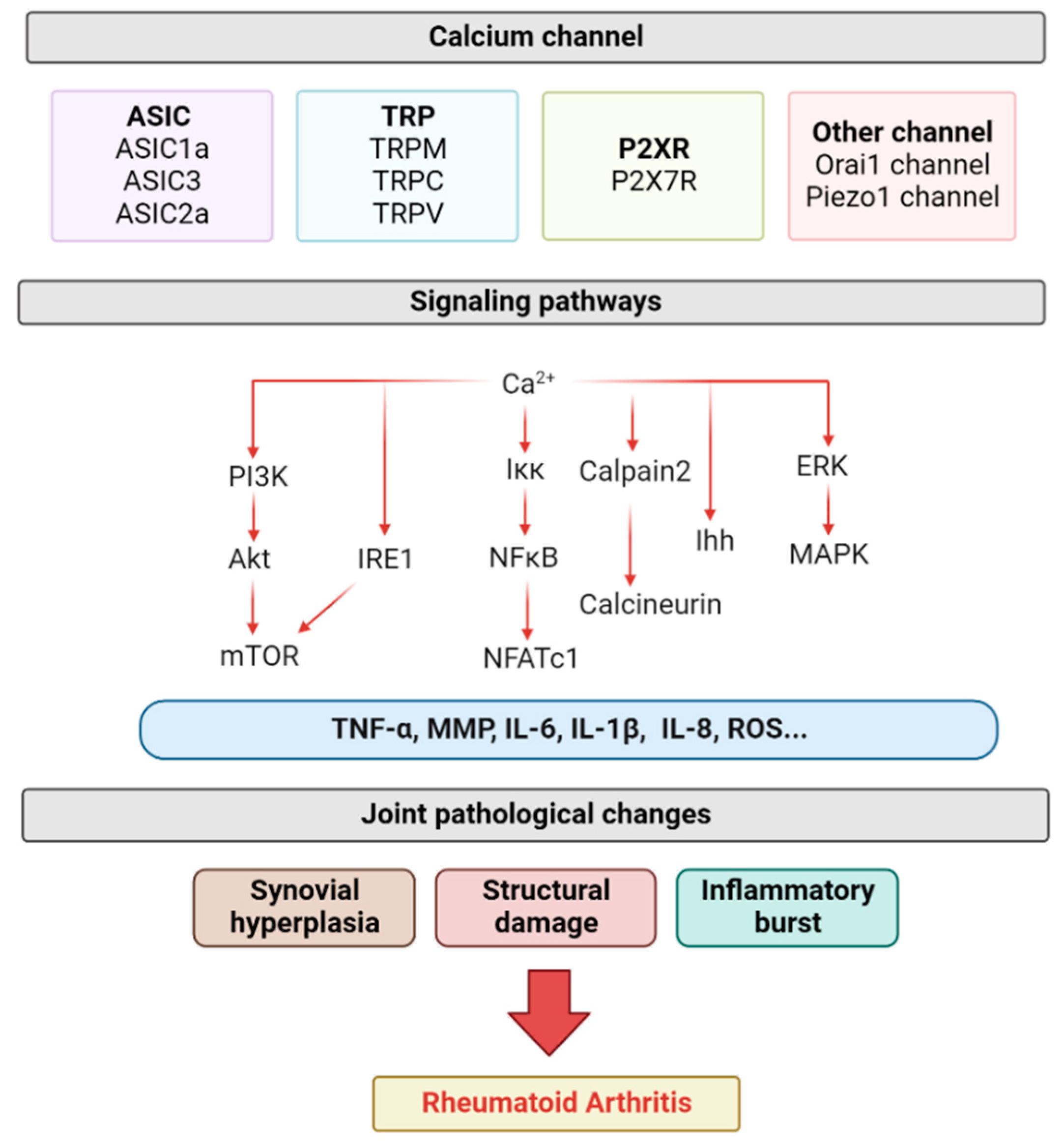
Figure 1. The role of calcium-permeable channels in the pathogenesis of RA. ASIC, TRP, P2X7R, Orai1 and Piezo1 channels regulate a variety of calcium-ion-related signaling pathways, which, in turn, lead to changes in related inflammatory factors, ultimately leading to synovitis, structural damage, and inflammatory outbreaks (created with Biorender.com, access date 29 July 2022).
Biomolecules 12 01383 g001
Figure 2. Role of calcium-permeable channel in synovitis. Macrophages can secrete a variety of cytokines to stimulate FLSs in the synovium, leading to a large influx of calcium ions in FLSs cells and triggering related cascade reactions, eventually leading to excessive apoptosis or proliferation of FLSs, and regulating synovial inflammation (created with Biorender.com).
Biomolecules 12 01383 g002
Figure 3. Role of calcium-permeable channel in cartilage destruction. Stimulated synovial cells can promote chondrocyte apoptosis by secreting inflammatory factors. At the same time, a large influx of calcium ions in chondrocytes triggers the activation of intracellular calcium ion signaling pathways (NF-κB pathway, PI3K-Akt pathway, etc.). This eventually leads to the degradation of cartilage matrix and apoptosis, pyroptosis and ferroptosis of chondrocytes (created with Biorender.com).
Biomolecules 12 01383 g003
Figure 4. Role of calcium-permeable channel in bone resorption. Synovitis-activated FLS cells can release a number of factors, such as RANKL, leading to an influx of osteoclast calcium ions in the bone. Through signal transmission, this eventually leads to the proliferation and activation of osteoclasts, which intensifies bone resorption (created with Biorender.com).
Biomolecules 12 01383 g004
Table 1. Role of activating the calcium-permeable channel in RA.
Synovial HyperplasiaCartilage DestructionBone DamageInflammation
ASICASIC1aRA synovial invasion, Ras-associated C3 botulinum toxin substrate1 [48];
RA FLSs proliferation, ERK/MAPK [49,50]
ECM expression [53];
chondrocyte pyroptosis, calpain-2/calcineurin [56,57];
chondrocyte apoptosis, NF-κB [23]
osteoclast migration, integrin/Pyk2/Src [61];
osteoclast genesis, NFATc1 [63]
MIP-1a, IL-8, NFATc3/RANTES [49]
ASIC2a chondroprotective effects of PcTx1 and APETx2, ERK/MAPK [64]
ASIC3FLS apoptosis, [Ca2+]i [55];
Sensitivity of FLS to inflammatory mediators [69]
hyaluronic acid [68] IL-6, MMP [69];
Pain behavior [70]
TRPMTRPM7FLS apoptosis [80]Ihh, chondrocyte damage [82];
collagen X, PI3K/Akt [81];
ferroptosis, PKCα-NOX4 [83]
osteoclast genesis [85];
osteoblast proliferation [84]
neutrophil chemotaxis, adhesion, invasiveness, Ca2+ influx [89];
Akt/mTOR [90]
TRPM8FLS apoptosis [79]
TRPCTRPC1 osteoclast genesis [92]
TRPC5 joint pain and swelling [94]
TRPC6 osteoclast activity [93]
TRPVTRPV1FLS apoptosis, [Ca2+]i, ROS mitochondrial membrane depolarization [100] osteoblast differentiation [103];
bone resorption [101]
painful behavior [109]
TRPV2aggressiveness of FLS [97] osteoclast differentiation, Ca2+-NFAT signaling [111]MMP-2, MMP-3 [97]
TRPV4proliferation of FLSs [113]chondrocyte apoptosis [114]osteoclast differentiation, RANKL [117];
NFATc1 transcription, cellular autophagy [118]
TRPV5 chondrocyte apoptosis, MAPK and Akt/mTOR pathways [120]osteoclast genesis [130];
osteoclast [Ca2+]i, bone resorption [129]
P2XRP2X7R chondrocyte apoptosis, IRE1-mTOR-PERK [144]osteoclasts activation [150];
number of osteoclasts [147]
histone proteases [159];
IL-1β, IL-6 [158]
Other channelOrai1Fas signaling [168] osteoclast synthesis, NFATc1 transduction [171];
osteoblast function, bone trabeculae [172]
Piezo1 chondrocyte apoptosis, MAPK/ERK [177]RANKL, RUNX2, osteoclasts differentiation [180];
osteoclast activity, bone resorption [181]
i: intracellular.
Publisher’s Note: MDPI stays neutral with regard to jurisdictional claims in published maps and institutional affiliations.