Review

Yersinia pestis Plasminogen Activator

by
Florent Sebbane
1,*,
Vladimir N. Uversky
2,3,* and
Andrey P. Anisimov
4,*
1
Université de Lille, Inserm, CNRS, CHU Lille, Institut Pasteur de Lille, U1019—UMR9017—CIIL—Center for Infection and Immunity of Lille, F-59000 Lille, France
2
Department of Molecular Medicine and USF Health Byrd Alzheimer’s Research Institute, Morsani College of Medicine, University of South Florida, Tampa, FL 33612, USA
3
Laboratory of New Methods in Biology, Federal Research Center “Pushchino Scientific Center for Biological Research of the Russian Academy of Sciences”, Institute for Biological Instrumentation of the Russian Academy of Sciences, 142290 Pushchino, Moscow Region, Russia
4
Laboratory for Plague Microbiology, State Research Center for Applied Microbiology and Biotechnology, Especially Dangerous Infections Department, 142279 Obolensk, Moscow Region, Russia
*
Authors to whom correspondence should be addressed.
Biomolecules 2020, 10(11), 1554; https://doi.org/10.3390/biom10111554
Submission received: 23 October 2020 / Revised: 12 November 2020 / Accepted: 12 November 2020 / Published: 14 November 2020
(This article belongs to the Collection Yersinia pestis Biomolecules)

Abstract

:
The Gram-negative bacterium Yersinia pestis causes plague, a fatal flea-borne anthropozoonosis, which can progress to aerosol-transmitted pneumonia. Y. pestis overcomes the innate immunity of its host thanks to many pathogenicity factors, including plasminogen activator, Pla. This factor is a broad-spectrum outer membrane protease also acting as adhesin and invasin. Y. pestis uses Pla adhesion and proteolytic capacity to manipulate the fibrinolytic cascade and immune system to produce bacteremia necessary for pathogen transmission via fleabite or aerosols. Because of microevolution, Y. pestis invasiveness has increased significantly after a single amino-acid substitution (I259T) in Pla of one of the oldest Y. pestis phylogenetic groups. This mutation caused a better ability to activate plasminogen. In paradox with its fibrinolytic activity, Pla cleaves and inactivates the tissue factor pathway inhibitor (TFPI), a key inhibitor of the coagulation cascade. This function in the plague remains enigmatic. Pla (or pla) had been used as a specific marker of Y. pestis, but its solitary detection is no longer valid as this gene is present in other species of Enterobacteriaceae. Though recovering hosts generate anti-Pla antibodies, Pla is not a good subunit vaccine. However, its deletion increases the safety of attenuated Y. pestis strains, providing a means to generate a safe live plague vaccine.

1. Discovery, History, Genetic Control, Biosynthesis Conditions, Isolation, Purification, and Physicochemical Properties of Plasminogen Activator

1.1. The Discovery

Plague is a fatal disease transmitted by fleas, but it can also be contracted through inhalation of contaminated aerosols and the ingestion of contaminated food. It is in 1894 that Alexandre Yersin isolated its causal agent, the Gram-negative bacterium named Yersinia pestis [1,2]. In 1936, R.R. Madison revealed that Y. pestis displays a fibrinolytic activity (i.e., the capability to break down the product of blood coagulation, a fibrin clot) [3]. However, this activity varies with the source of fibrin. In fact, a comparison of the fibrinolytic activity using fibrin from several mammal species indicated that the activity against human and guinea pig fibrins was about six times lower than that against rat fibrin. Furthermore, gopher, cat, rabbit, cow, and monkey fibrins were even more resistant to lysis. Lastly, no lysis of horse, ram, and pig fibrins was noted. Eight years after the discovery of fibrinolysin by R.R. Madison, E. Jawetz, and K.F. Meyer discovered that Y. pestis has also the ability to coagulate plasma [4]. However, this coagulase activity has been demonstrated in the rabbit plasma clotting assay [5], but could not be observed using human, mouse, or rat plasma [6]. Almost 20 years after Jawetz and Meyer’s discovery, Domaradsky reported that the fibrinoyltic and the coagulase activities of Y. pestis are interrelated [7,8]. He also indicated that these traits differentiate Y. pestis from its most recent ancestor species, Yersinia pseudotuberculosis, an enteropathogenic bacterium causing mild bowel disease in humans, which is not transmitted by fleas or aerosols. In the same period of time as I.V. Domaradsky, R.R. Brubaker et al. reported that fibrinoyltic and coagulase activities are linked to the production of a bacteriocin (named “pesticin”), which is active against Y. pseudotuberculosis but not Y. pestis [9,10]. It turned out that genes responsible for fibrinoytic/coagulase activity and bacteriocin production are all harbored by a small plasmid named pPst (pPla, pPCP1, or pYP) [11,12] that contains at least three genes including pla. This latter was rapidly found to encode the enzyme (plasminogen activator, Pla) responsible for both fibrinolytic and plasma-coagulase activities of Y. pestis [13,14] and had adhesive properties [15]. Lastly, we must mention that some strains, including one clinical isolate recovered from the blood of a fatal human plague case, was shown to contain the pPst dimer [16]. This dimeric form of the plasmid is stably inherited and also stably coexists with the monomeric form of the pPst. More recently, the simultaneous presence of two different pPst plasmids, one with and one without the pla locus, was found in most of the sequenced genomes from the end of the second-plague-pandemic burials, but not in the Y. pestis strains circulating during the Black Death peak. It has been suggested that the proliferation of Pla-depleted strains could, among other reasons, contribute to the disappearance of the second plague pandemic in eighteenth-century Europe [17]. Furthermore, we also have to emphasize here that not all the Y. pestis strains harbor the pPst plasmid and, accordingly, any of its genes, including pla.

1.2. Pla, Location, Processing, and Conformation

Pla is a member of the omptin family, which is composed of outer membrane proteins classified as bacterial aspartate proteases [18,19,20,21]. Hence, Pla shares high homology with four surface enterobacterial proteases: PgtE (Salmonella enterica), OmpT (Escherichia coli), OmpP (E. coli), and SopA (Shigella flexneri) [22,23,24,25]. Pla is composed of 312 amino acid residues (no cysteine residues) and includes the N-terminal signal sequence (i.e., the mature protein is 292 amino acid residue-long) [13]. Upon removal of the signal peptide (during protein exportation), an α-Pla (37 kDa) protein is formed. Being an integral outer membrane protease (omptin), the plasminogen activator Pla from Y. pestis is characterized by a highly conserved β-barrel structure spanning the membrane. Figure 1 shows that Pla forms a narrow β-barrel with an elliptical cross-section. This rather extended, ~70 Å long barrel consists of 10 antiparallel β-strands connected by short periplasmic turns and five extracellular loops that define the substrate specificity of this bacterial fibrinolysin [18,19]. The export of α-Pla yields to the production of two other forms, β-Pla (35 kDa) and γ-Pla (31 kDa), which are associated with the outer membrane as well [26,27]. V.V. Kutyrev et al. reported the existence of the σ-variant (23 kDa) of Pla [26]. However, there was no confirmation of this information by other researchers. β-Pla is formed as a result of the autoprocessing of the C-terminal surface loop of the α-Pla molecule at amino acid residue Lys262 [28]. The α- and β-forms do not differ in their fibrinolytic activity [28]. Although γ-Pla displays lower molecular weight than α-Pla, it is generally accepted that this form of the protein is an alternative form of α-Pla folding [28]. Indeed, the boiling of samples in the presence of SDS led to the disappearance of γ-Pla [29].
Similar to other omptins, Pla is an aspartate protease with the catalytic site residues Asp84, Asp86, Asp206, and His208 located at the extracellular opening of the β-barrel. Akin to other aspartate proteases, Pla uses a water molecule for cleavage of target peptides [30]. In fact, in the active Pla molecule, catalytic residues are grouped into Asp84/Asp86 and Asp206/His208 couples located at opposite sides of the barrel, with the minimal distance between these active site couples being ~4.7 Å. This distance is between Asp84 in one couple and His208 in another couple, with the catalytic nucleophile water molecule being located between the carboxyl group of Asp84 and the Nɛ2 atom of His208 [30].
It was pointed out that in a crystal structure of an active form of Pla, residues Glu252–Ser269 in loop L5 are missing (this region of mature Pla corresponds to the residues 273–290 in the preprocessed protein, UniProt ID: P17811), likely due to the Pla autocatalytic activity [30]. As sites of the proteolytic attack are preferentially located within flexible regions or intrinsically disordered protein regions (IDPRs) [31,32,33,34], this loop is likely characterized by high structural plasticity. Despite such conformational flexibility, loop L5 seems to play an important role in controlling Pla activity. This notion is supported by an interesting observation that in the ancestral Y. pestis lineages Microtus and Angola, this loop L5 carries a single amino acid change Thr259Ile that affects the fibrinolytic activity of Pla [35]. In fact, Pla from the Microtus lineage was more efficient than the wild-type Pla in α2-antiplasmin inactivation, suggesting that this ancestral form of Pla evolved into a more efficient plasminogen activator in the pandemic Y. pestis lineages [35].
To see if loop 5 of Y. pestis is indeed disordered/flexible and to evaluate the intrinsic disorder status of the transmembrane protein Pla, we used predictors of intrinsic disorder to study the intrinsic disorder profile of Pla pestis and of several other omptin family outer membrane proteases (PgtE from S. enterica, OmpT and OmpR from E. coli, and SopA/IcsP from S. flexneri) (Figure 2). Although sequence identities of these different protease ranges from 72.44% (between Pla and PgtE) to 39.41% (between Pla and SopA/IcsP) or even 38.76% (between PgtE and SopA/IcsP), their disorder profiles are characterized by remarkable similarity, showing conserved positioning of major disordered and flexible regions. More particularly, Pla (despite being an integral transmembrane protein) is predicted to have several IDPRs (i.e., regions with the disorder scores exceeding the threshold of 0.5) and several flexible regions (i.e., regions possessing disorder scores between 0.2 and 0.5) (Figure 2A). They also show that loop L5 is predicted to be disordered. Furthermore, other extracellular loops (residues 53–57 (L1), 108–116 (L2), 165–185 (L3), and 228–232 (L4)) are also predicted to be disordered by at last one of the computational tools used here. Signal peptide, which is removed during maturation of Pla is also an IDPR. These observations suggest that intrinsic disorder and conformational flexibility are important for the functionality of Pla. Further support to the idea of potential functional importance of disordered and flexible regions of Pla is given by their evolutionary conservation.

1.3. Regulation of Pla

It has been initially reported that pla is regulated neither at the transcriptional nor at the translational level, suggesting that the temperature dependence of the fibrinolytic activity may be due to temperature-induced modifications or conformational changes in the Pla protein [36]. This was somewhat consistent with the presence of −10 and −35 core promoter elements recognized by sigma factor 70 located upstream of the transcription start sites of pla. However, comparative transcriptomic analysis using bacteria cultured in vitro in artificial media indicated a modest overexpression of pla (two-fold changed) after temperature shift from ≤28 °C to 37 °C [37]. This transcriptional activation was further correlated with a subsequent qRT-PCR study [38] and a comparative proteomic analysis [39], as well as western-blot analysis [40]. However, transcriptome analysis of Y. pestis showed no induction of pla at 37 °C in human plasma [41]. Furthermore, pla expression levels in the liver and lungs of mice sacrificed 4 days after intravenous challenge were about 10-fold lower than those seen in vitro at 37 °C [38]. We must point out though that these results were obtained using a strain of Y. pestis lacking the pigmentation locus (pgm) which may encode a factor affecting pla expression and/or activity [42]. Indeed, WT strain displays far greater fibrinolytic activity than pgm-negative strain. In any case, the regulation of pla in vivo appears complex. Being one of the genes the most highly expressed in the bubo of moribund rat [43], pla was found to be repressed in the lung of mice 48 h after intranasal inoculation [44,45] prior to being over-expressed >10-fold in the lungs of animals displaying a terminal stage of infection [45]. Based on in vitro molecular studies [46,47,48] and an investigation of mice infected by the intranasal route, it has been proposed that the in vivo expression pattern of pla in infected lungs is driven by the glucose concentration and depends on the transcriptional regulator cyclic AMP (cAMP) receptor protein (CRP). In this model, Y. pestis entering the lung encounters an environment, where the concentration of glucose is high enough to maintain an intracellular concentration of cAMP low to avoid CRP activation. Consequently, CRP is unable to bind to a site upstream of pla between bp −70 and −52 to activate the pla transcription [47]. During the infection, Y. pestis consumes the glucose present in the lungs. Therefore, at the terminal stage of infection, the environmental glucose concentration is so low that the intracellular concentration of cAMP is high enough to activate CRP, and thereby promote the CRP-dependent pla expression. In conclusion, pla is expressed thanks to –70 dependent promoter, whose activity is enhanced in the absence of glucose due to the direct binding of CRP to the Pla promoter. Temperature > 25 °C can induce pla according to a mechanism that remains to be elucidated.

1.4. Role of Temperature and Lipooligosaccharide (Los) on Pla Activities

The modest transcriptional activation of pla during a shift from ≤28 °C to 37 °C alone cannot explain the dramatically higher fibrinolytic activity of the bacterium Y. pestis cultured at 37 °C vs. ≤28 °C [36]. Furthermore, this apparent regulation does not find an explanation in the amount of Pla per cell. In fact, comparative proteomic analysis and western-blot analysis showed a 2-fold increase in the Pla production, when bacteria are shifted from ≤28 °C to 37 °C [39,40]. Accordingly, E. Ruback et al., who compared the ability of Y. pestis grown at 37 °C or ≤28 °C to activate plasminogen, reported that the higher enzymatic activity of “37 °C" bacteria was accompanied by a 1.6-fold increase in the Pla content in the outer-membrane preparations, and a slight increase in the specific activity of “37 °C” Pla [29]. According to immunoblotting, the β-form of Pla was detected only at 37 °C. In other words, the Pla proteolytic activity (at least autoproteolysis and plasminogen activation) depends on a post-translation event. The role of the interaction of Pla with the outer membrane was incriminated. Indeed, for isolation and purification of Pla, gel filtration of cell membrane extracts using Sephadex G-200 was previously used. In the process of isolation, the preparations retained fibrinolytic but lost their coagulase activity [5]. More recently it was found that the product of pla, renatured in the presence of LOS, retains all the studied properties of a partially purified molecule, presumably because the interaction of Pla with LPS affects the structure of the protein catalytic site, generating its correct conformation [18,19].
Although the interaction of the outer membrane with Pla was suspected to play a role in plasminogen activation, differences in presence of O-antigen was rapidly excluded, since Y. pestis produces a LOS. However, it is interesting to note that the fibrinolytic activity of Pla, but not its auto-processing, requires the absence of O-side polysaccharide chains synthesized by numerous bacterial species (such as Yersinia pseudotuberculosis and S. enterica) [40,49,50,51]. In other words, Pla is functional, when it interacts with the LOS naturally produced by the Y. pestis cells [51].
Since O-antigen modifications cannot explain the difference of Pla activity at the different growth temperature, temperature-dependent change in the structure of lipid A was suspected for the enzymatic activity of Pla. At 37 °C, Y. pestis expresses tetra-acylated LOS, whereas the LOS is mainly hexa-acylated when the bacterium is grown at ≤28 °C, incriminating the role of low acylation of lipid A in Pla activity [51]. On the one hand, the “acylation” idea was supported by the experiments using E. coli expressing Pla and producing a rough LPS with lower acylation [40]. On the other hand, Pla activities remained unchanged in strains of Y. pestis engineered to produce only low or high lipid A acylation regardless of the growth temperature [51,52,53,54].
Lastly, the role of the LOS core in the temperature-dependent activities of Pla was also examined. It was found that the shortening of the core part of the LOS of Y. pestis or the LPS of E. coli producing Pla negatively impacts Pla activity [40]. Furthermore, gradual shortening of the core was correlated with the decrease in the Pla activity, and variants of the core oligosaccharide containing four or less monosaccharide residues loss the fibrinolytic and coagulase activities of Pla [55]. In other words, the data indicate that the presence of a complete core but not its precise carbohydrate structure is critical for Pla activity.
A number of prokaryotic and eukaryotic proteins that bind to LPS have conserved three-dimensional structures formed with the participation of two positively charged arginine residues interacting with the negatively charged phosphate groups of lipid A [11]. In the Pla molecule, these arginines are located at positions 138 and 171. The Arg171Glu amino acid substitution significantly reduced the enzymatic activity of the Pla preparation, refolding of which was carried out in the presence of LOS. At the same time, the Arg138Glu replacement or the double Arg138Glu + Arg171Glu replacement led to a complete loss of the Pla ability to activate plasminogen [29]. Variants of Pla with the Arg138Glu orArg171Glu substitutions formed a significantly lower amount of β-Pla compared to the wild-type molecules. The double Arg138Glu + Arg171Glu mutant was not able to form β- and γ-Pla at all, which indicates the necessity of binding to LOS for autoprocessing and correct folding of Pla [29]. Overall, these data led to the idea that the requirement of LOS for Pla activity could represent a means to keep the protease inactive during the translocation from the cytoplasm into the outer membrane [19].

2. Pla as a Broad-Spectrum Protease with Adhesion Properties

2.1. The Multiple Substrates Processed by Pla

2.1.1. The Substrates Related to Hemostasis

Coagulation and fibrinolysis are complex biological phenomena minutely balanced by the intervention of numerous factors playing either an activator or a repressor role (Figure 3), and where an inhibited target can impede its own inhibitor [56,57,58]. Following trauma or injury, a cascade of enzymatic cleavages causes prothrombin to be cleaved into thrombin, which splits fibrinogen. This results in the release of fibrin, which forms a mesh that traps blood cells and is therefore serves as an essential element in the formation of a blood clot. The fibrin clot can also be degraded by the action of the peptidase named plasmin, which is a product of the degradation of plasminogen by the action of tissue plasminogen activator (tPA) and the urokinase-type plasminogen activator (uPA), with the latter being primarily involved in cell migration and tissue remodeling linked to the inflammation and bacterial infections. The proteolytic activity of tPA and uPA can be balanced by the plasminogen activator inhibitor-1 and -2 (PAI-1 and -2), whereas α2-antiplasmin, α-2-macroglobuline, as well as the thrombin-activatable fibrinolysis inhibitor (TAFI) impede the plasmin activity. PAI (stabilized when complexed with vitronectin [Vn]) is a competitive inhibitor composed of a sequence (“a bait”), whose integration into the catalytic site of uPA or tPA leads to the formation of an irreversible covalent bond between the inhibitor and the enzymes. The α2-antiplasmin (A2AP) is a serine protease inhibitor (serpin) that degrades plasmin. As for the activated TAFI, it decreases plasmin production and fibrinolysis by removing lysine residues from partially degraded fibrin, which reduces the binding of t-PA and plasminogen to the fibrin clot. Interestingly, activated TAFI (TAFIa), as well as TAFI, are subjected to destructive proteolysis by plasmin. It is worth noting that both fibrinolysis and coagulation are linked to an immune response. For instance, PAI-1 participates in immune cell migration, binding to cell surface receptors, and cytokine production.
Thanks to its proteolytic activity, Pla disturbs several factors belonging to both coagulation and fibrinolytic pathways. Notably, Pla seems to induce coagulation by inactivating the tissue factor pathway inhibitor (TFPI), a protease that inhibits the factor (F) VII initiating one of the first steps of blood coagulation (Figure 3) [59]. This induction of coagulation could be further enhanced by the activation by Pla, albeit not very efficiently, of FVII [59]. It is worth noting that no hydrolysis was found towards the FV and FX [59].
Paradoxically and in the opposite way to the coagulation activation, Pla induces fibrinolysis. The direct cleavage of the plasminogen by Pla, at the same site with uPA (i.e., R561-V562), was the first mechanism identified that explains how Pla induces fibrinolysis [6,60]. Subsequent studies that have reviewed the different actors in the fibrinolytic cascade sensitive to Pla have revealed that plasmin production is amplified both by activation of uPA [61] and also by the degradation of several fibrinolysis inhibitors such as PAI-1 [30,62,63], A2AP, and TAFI. Pla cleaves PAI-I in the inhibitory “bait”, between R346 and M347, resulting in the inability of the inhibitor to covalently obstruct the catalytic site of uPA [30]. The inactivation of PAI-1 is further accentuated by the hydrolysis of its stabilizer Vn to different pieces by Pla [30,63]. However, efficient degradation of Vn requires the presence of the adhesin Ail that confers binding to Vn [63]. The precise site of cleavage is unknown for A2AP and TAFI, although, Pla is known to cleave TAFI at a Ct region [64]. As aforementioned, plasmin inactivates TAFI and TAFIa, indicating that Pla, directly and indirectly, lowers the potential inhibitory effects of TAFIa on fibrinolysis. In conclusion, Pla acts, directly and indirectly, to produce plasmin from plasminogen. When it acts indirectly, it activates plasminogen activators and degrades the inhibitors of these activators as well as plasmin. Altogether, it is clear that the different actions of Pla lead to high-level production of plasmin. Since plasmin degrades several components of the extracellular matrix in addition to fibrin, it appears reasonable to consider that the fibrinolytic activity of Pla can lead to uncontrolled proteolysis. However, as described later on, this uncontrolled proteolysis does not necessarily play a role or a role of the same importance in every site colonized by Y. pestis.
Furthermore, we would like to emphasize here that the ability of Pla to degrade some components of the fibrinolytic activity in vitro is not necessarily observed in vivo. Besides, when a substrate is proven in vivo, the totality of Pla’s substrate synthesized by the host is not necessarily cleaved and the proof of cleavage does not preclude a role in pathogenesis [65,66].

2.1.2. Immune-Related Targets

Several investigations indicated that the proteolytic activity of Pla interferes directly with the host immune response in addition to fibrinolytic and coagulation pathways. Convincing evidence came from peptide array-based approach and comparative proteomic analysis using cell-free bronchoalveolar lavage fluid (BALF) samples aiming at discovering putative host substrates of Pla [66,67]. These studies unveiled a little over a dozen Pla targets, including several previously known targets. Interestingly, the majority of the putative substrates identified in this study have a role in immune system processes or proteolysis, suggesting that Pla is dedicated to inducing overwhelming proteolysis but also to affect the immune response. In this regard, it is important to remember that the fibrinolytic/coagulation pathways are intimately associated with the host immune response, suggesting Pla may disrupt the host defense from different sides. Among the proteins related to the immune response and confirmed to be substrates for Pla are the previously identified complement components 3 and 4b, the Peroxiredoxin-6 (Prdx6), and the Fas ligand (FasL) [6,66,67]. Although C3 and C4b play a central role in the activation of the complement cascade, their degradation by Pla neither confers any advantage regarding serum resistance nor seems to interfere with the opsonophagocytosis associated with the complement cascade [6,60]. PrdX6 is a glutathione peroxidase carrying phospholipase A2 activity that plays an antioxidant role in the lungs, notably under various stress conditions [68,69,70,71]. Pla cleaves this protein at sites Lis173/Arg174, Lys201/Leu202, and at the undefined site located in the C-terminal region, leading to the disruption of both enzymatic activities of PrdX6 [66]. FasL is a type-II transmembrane protein that induces apoptosis upon interaction with the Fas receptor. This induced cell death program, which implies the caspase-8 followed by the caspase-3/7 pathway, plays an important role in the regulation of the immune system, avoiding excessive immune response and protecting against various infections [72,73,74]. Hence, the proteolysis at multiple sites located within the extracellular domain of FasL by Pla annihilates the capacity of FasL to induce apoptosis. In addition to cleaving proteins, Pla was proposed to cleave antimicrobial peptides. This idea originated from an experiment aiming at understanding how Y. pestis can resist the action of the antimicrobial peptides contained in the rat BALF [75]. After showing that antimicrobial peptides containing in the BALF kill Y. pestis, the proteolytic activity of Pla was assumed to confer some protection against antimicrobial peptides displaying a putative cleavage site for Pla (i.e., two consecutive basic amino acid residues), such as LL37 [75,76]. However, the role of Pla was unveiled only when Y. pestis was unable to produce the pseudocapsule F1, and formal proof of peptide degradation was not given [75]. In other words, it is possible that Y. pestis proteins processed by Pla could sensitize the bacillus to antimicrobial peptides. Indeed, several chromosomally encoded proteins were found to be cleaved by Pla [77]. To date, in addition to Pla, 4 of these proteins were identified. They are named KatY, YapA, YapE, and YpaG.

2.1.3. Ypestis Proteins Processed by Pla

YapA, YapE, and YapG belong to the type V autotransporter family [78,79]. The proteins of this family possess all the domains needed for their own translocation across the bacterial membranes [80]. Notably, a central domain harboring the function of the mature protein (referred to as the passenger domain) is located between the N-terminal signal peptide (allowing the secretion through the inner membrane via the Sec system) and the C-terminal domain that mediates translocation of the passenger domain across the outer membrane. Once translocated, the three Yap autotranspoters are cleaved by Pla [78,79,81]. Notably, Pla processes YapG at multiple sites (Lys512, Lys548/Lys549, Lys594/Lys595, Lys558, and Lys604) and YapE at two sites (Lys232 and Lys338 but preferentially at Lys232) (see Table 1).
Consequently, different portions of the passenger domain of YapG and YapE are released into the environment, but a significant portion of the YapE passenger domain remains associated with the bacteria. Interestingly, this portion is required for autoaggregation and adherence to eukaryotic cells, which presumably explains why YapE is important for bubonic plague [78,79,81,84]. Therefore, processing of the surface Y. pestis proteins by Pla can unveil functions important for pathogenesis. In addition to the ability to degrade chromosomally encoded proteins, Pla was also reported to cleave in vitro a set of plasmid-encoded proteins, the Yersinia outer membrane proteins (Yops) [14,26,38,47,86,87]. However, the degradation of Yops does not occur in a condition mimicking the phagolysosome [77]. The Yops are exotoxins secreted by a type three secretion system from the bacterial cytoplasm into the host (mostly into phagocytes) to disrupt the mammalian immune system, by inhibiting phagocytosis and production of pro-inflammatory cytokines and inducing apoptosis and section anti-inflammatory cytokines [88].

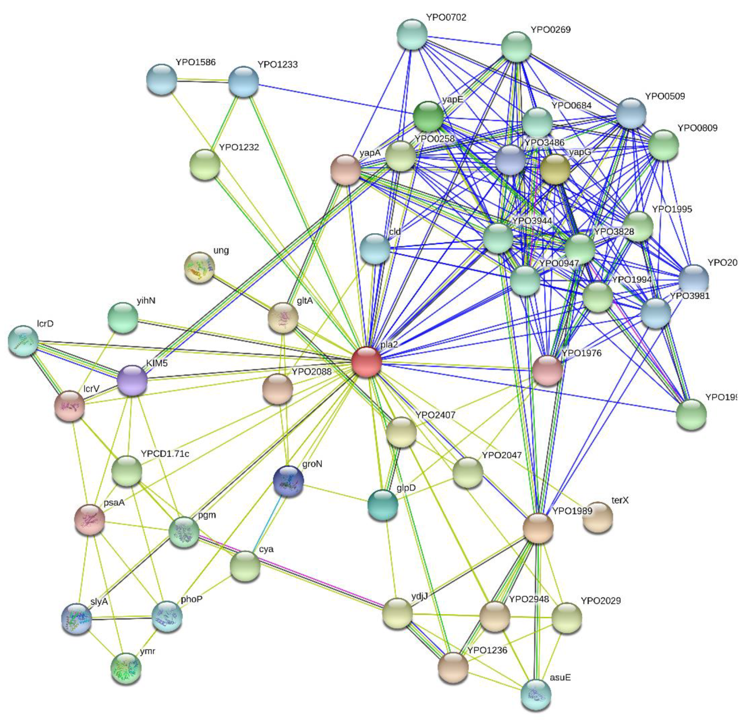
2.2. Overall Interactibility of Pla

Pla is a multifunctional protein, which in addition to its role in the cleavage (activation) of plasminogen to generate plasmin [5,89] is also important for suppression and evasion of the innate immune responses [6,90]. Furthermore, Pla can control the development of primary pneumonic plague [91,92], can degrade several potentially important mammalian proteins [93,94], and even act as both an adhesin and invasin, mediating tight binding to fibronectin and promoting the uptake of the bacteria by non-phagocytic cells [94,95]. Often, protein multifunctionality is determined by the presence of IDPRs, which are capable of promiscuous binding to multiple unrelated partners, which enables them to function in regulation, signaling, and control, where they are commonly engaged in one-to-many and many-to-one interactions [96,97,98,99,100,101,102,103,104,105,106,107].
To further illustrate this idea, Figure 4 represents the results of the in silico analysis of the interactability of Pla from Y. pestis. The resulting Pla-centered network contains 49 nodes (proteins) connected by 247 edges (protein-protein interactions). In this network, the average node degree is 10.1 (in other words, on average, each protein in this network is involved in an interaction with at least 10 other members of the network). Furthermore, the average local clustering coefficient (which defines how close its neighbors are to being a complete clique) is 0.792; a coefficient of 1 means that every neighbor connected to a given node Ni is also connected to every other node within the neighborhood, and a zero value indicates an absence of connection between the nodes. The number of interactions cited above is unexpectedly high since the number of interactions among proteins in a similar size set of proteins randomly selected from the human proteome is equal to 102. In other words, the Pla-centered PPPI network has significantly more interactions than expected, being characterized by a PPI enrichment p-value of <10−16. Therefore, Pla could be considered as a hub protein capable to degrade or interact with many Y. pestis proteins.

2.3. Structure and Catalytic Mechanisms

In contrast to OmpT from E. coli, Pla cleaves α2-antiplasmin, and has a stronger ability to activate plasminogen [28]. The tertiary structure of Pla and OmpT is represented by ten transmembrane β-strands and five loops located on the cell surface (L1–L5) (see Figure 1). This preservation of the structure makes it possible to generate Pla-OmpT hybrids to identify the key elements that explain the difference in the enzymatic activities of these two omptins. Analysis of hybrid Pla-OmpT proteins constructed by exchanging surface loops between Pla and OmpT showed that L3 and L4 loops are responsible for the substrate specificity of Pla with respect to α2-antiplasmin [28]. Consistently, the crystallographic analysis revealed that loops L3 and L4 form the entrance to the active site [18]. Replacement of 25 surface-located amino acid residues made it possible to establish that residues His101, His208, Asp84, Asp86, Asp206, and Ser99 are necessary for the proteolytic activity of Pla [28]. Furthermore, unidentified residues in the L3 loop and the residue Arg211 located in the L4 loop and pointed inward to the active site play a role in the cleavage of α2-antiplasmin and activation of plasminogen [18]. Interestingly, the conversion of the OmpT protein to a protease similar to Pla in its enzymatic activity was achieved by removing residues Asp214 and Pro215, replacing Lys217 with Arg217 in the L4 OmpT loop, and replacing the entire L3 loop with the analogous one from Pla [28]. Surprisingly, only 2 out of the known 7 residues required for plasminogen activation are needed for activation of the host plasminogen uPA [61]. Furthermore, these mutations did not abrogate the cleavage activity against the inactive form of uPa [61]. Therefore, Pla could have different active sites, which could explain why Pla is such a broad-spectrum protease. Figure 5 shows the zoomed-in active site of Pla from Y. pestis and illustrates peculiarities of the local environment of four catalytic residues of this protein, Asp84, Asp86, Asp206, and His208.

2.4. Pla, an Adhesin with Multiple Binding Substrates

Although Pla was initially identified as a protease, a study that screened a cosmid library for E. coli clones containing Y. pestis genes that confers the ability to bind type IV collagen, identified Pla as an adhesin [15]. Its adhesiveness has been documented with human, but not with murine collagen, however [15,109].
Further investigations that assessed the adhesiveness of purified Pla or Y. pestis or E. coli expressing pla to different substrates revealed that the protein binds weakly to fibronectin, glycosylated type I, type IV, and type V collagens and strongly to laminin (one of the major extracellular matrix component) [109,110]. Consequently, Pla has been shown to be important for binding to different types of epithelial and endothelial cells, as well as diverse types of macrophages [38,50,95,111,112]. Interestingly, binding to extracellular matrix components and cells is independent of the proteolytic activity but subject to environmental conditions (pH and temperature) [38,111]. Pla binds to laminin more efficiently if Y. pestis is cultured at 37 °C rather than ≤28 °C, especially if the bacteria are incubated at pH 7 rather than pH 6 [38,113]. Thanks to the phage display and alanine-scanning mutagenesis, the motif Leu65-Thr66-Leu67 as well as the residues Gly178 and Leu179 were found to play a role in the interaction with laminin [113]. However, the presence of these residues in the region of Pla embedded in the membrane suggests that they are not directly involved in binding. Lastly, Pla may also be an invasin (i.e., a protein that promotes the entry into host cells) because its presence, under specific conditions, promotes invasion of HeLa cells and human type I pneumocytes [95]. Pla also promotes invasion of alveolar macrophages via the C-type lectin receptor, DEC-205 (CD205), which has an important role in the antigen-presenting process present on many APCs in both human and mice [114]. Lastly, like the adhesiveness property, the invasiveness capacity of Pla is independent of its proteolytic activity, since a substitution mutation at the residues Ser99 or Asp206 that abrogates the Pla’s proteolytic activity does not impact the capacity of E. coli expressing Pla to invade human endothelial cells [111]. Hence, a link between Pla-mediated adhesion and invasion likely exists.

3. The Biological Role of Plasminogen Activator in Pathogenesis

3.1. The Proven or Unproven Role of Pla

Thanks to its proteolytic and adhesin/invasin activities, Pla is a Swiss army knife of Y. pestis’ life cycle. However, Y. pestis uses Pla to infect the mammals, but not the fleas [6,115]. Furthermore, our knowledge of the Pla-mediated molecular mechanism used by Y. pestis to produce infection remains limited, notably for bubonic plague. We do not know whether the Pla-invasin mediated activity is important for the plague. In contrast, Y. pestis has been demonstrated to use the Pla-mediated adhesion and proteolytic activities to establish an infection in mammals. However, the adhesin activity was studied only in pneumonic plague [116]. It is also worth noting that not all of Pla’s targets identified in vitro seem to be processed in vivo. For instance, the α2-antiplasmin does not appear to be a substrate during pneumonic plague [65]. In fact, PAI-1 and Fas ligand are the two only substrates for Pla confirmed in vivo [62,67]. Lastly, indirect evidence suggests that Pla degrades Prx6 and YapE in vivo. Notably, Pla may cleave Prdx6 during the pneumonic plague, because the amount of extracellular Prdx6 is lower in the lungs of mice infected with wild-type strain than in animals infected with a Δpla mutant [66]. Whether this cleavage occurs really or not, it does not play any obvious role in the pathogenesis of pneumonic plague. As for YapE, it might be cleaved at least in the skin and/or in the course of lymph node colonization. In fact, a ΔyapE mutant displays similar dissemination defect as a Δpla mutant after subcutaneous inoculation of bacteria [78].

3.2. The Role of Pla during Bubonic Plague

Initially, it was suggested that Y. pestis uses the coagulase and fibrinolytic activity of Pla to produce a transmissible infection in flea and to initiate the host colonization at the fleabite site, respectively [6,117]. In particular, Y. pestis would have used the coagulase activity (Pla is active at ≤28 °C) to produce a fibrin clot that obstructs the digestive tract of the flea. This process increases the likelihood of Y. pestis transmission by fleas [118]. Once transmitted to a new mammalian host, Y. pestis would have used the fibrinolytic activity of Pla (active at ≥37 °C) to prevent the formation of a blood clot produced at the insect’s suction point; this clot normally traps bacteria and prevent them from spreading. Hence, Y. pestis would use Pla to degrade fibrin clot, extracellular matrix, and basement membrane (i.e., the main obstacle to the spread of microorganisms) to escape from the skin and reach the drainage lymph node, where its active replication leads to the production of an enlarged painful lymph node, the bubo (the hallmark of bubonic plague). In such a model, Y. pestis cannot produce a transmissible infection in flea or a flea-borne infection in the absence of Pla. However, experiments performed in the Xenopsylla cheopis rat flea rejected the potential Pla’s role in this insect [36,115]. Conversely, the role of Pla in mammals has been confirmed on multiple occasions [6,60,90]. Its deletion results in a one million-fold reduction in the bacterial virulence in mice inoculated by a subcutaneous route. Histological analysis indicates that a Δpla mutant does not reach deep tissue when injected at a dose equivalent to that of regurgitated by fleas [6,60,90,119]. Virulence studies utilizing plasminogen-deficient mice supported the idea that Y. pestis uses Pla to degrade the plasminogen, and so the uncontrolled production of active plasmin, to produce bubonic plague [89]. Indeed, plasminogen-deficient mice are resistant to Y. pestis. However, a pla-negative Y. pestis mutant remains avirulent in plasminogen-deficient mice. Therefore, whether Pla cleaves the plasminogen during the bubonic plague, this cleavage can only partially explain the role of Pla in this form of the disease. The proteolytic activity of Pla against multiple complement regulatory proteins could have been incriminated to explain why a Pla-negative Y. pestis remains avirulent in plasminogen deficient mice. However, Y. pestis does not use Pla for serum resistance [6]. Furthermore, virulence studies in congenic C5A+ and C5A- mice revealed that Pla-associated virulence is not mediated by interference with the phagocytic chemoattractant C5a [60]. Lastly, it is worth reminding that Y. pestis cleaves YapE thanks to Pla, and a ΔyapE mutant displays a similar dissemination defect as a Δpla mutant after subcutaneous inoculation of bacteria [78]. Hence, Y. pestis may use Pla to cleave its own proteins rather than the host proteins to produce bubonic plague. Overall, it is clear that the molecular mechanisms leading to bubonic plague and involving Pla remain to be elucidated.
Based on the above-cited virulence data, it was believed for many years that flea-borne plague is impossible if the bacillus lacks Pla. However, this view is somewhat challenged by the presence of natural isolates that lack Pla (0.PE2 or bv. Caucasica) and are fully virulent [120,121]. In addition, mice challenged with fleas infected with a mutant Δpla die of primary septicemic plague because fleas seem to regurgitate bacteria in the extravascular part of the dermis in 70–90% of the cases and the intravascular part of the dermis in 10–30% [90,122,123]. In a natural context of infection (i.e., fleabite), a Δpla mutant can sometimes spread to regional lymph nodes, as others have observed after subcutaneous or intradermal inoculation. However, the infection does not progress to severe lymphadenitis and bubonic plague. Pla, therefore, increases the incidence of plague by allowing the production of the bubonic form of the disease. In contrast to the model suggesting an essential role of Pla for the spread of Y. pestis from the skin, one study suggested that Pla is not needed for infiltration and destruction of the draining lymph node [124]. Indeed, the Δpla mutant colonized and destroyed this organ in a similar manner to the wild strain when it was injected at a relatively high dose (5000 CFU) into the pinna of the mouse ear. In fact, Pla was proposed to be a key genetic component necessary to survive and multiply in the draining lymph node. This idea was partly based on immunostaining which revealed an abnormal bacterial morphology. However, the use of an anti-Y. pestis antiserum and not a specific antibody precludes to definitively conclude about this idea. Furthermore, one may question why the mutant will be destroyed in the lymph node but not in the skin and deep tissues. In this context, it is worth noting that discrete clumps of Δpla bacteria were present only in the marginal sinus of the lymph node of moribund mice infected by fleabite [90]. Furthermore, these bacterial clumps were in close contact with fibrin deposit and inflammatory cells, suggesting that Y. pestis uses Pla to escape from fibrin clots in the lymph node after fleabite. Therefore, the role of Pla in a natural vs. artificial context of infection could be different. Alternatively, the low number of lymph nodes analyzed in mice that developed fatal plague after fleabite may have revealed just one of Pla’s multiple roles in the pathogenesis.

3.3. The Role of Pla during Primary Pneumonic Plague

Although Pla is essential for producing bubonic plague after a fleabite, it is not crucial for producing a fatal infection after inhalation of Y. pestis [92]. Its loss slightly delays the mortality of mice inoculated by the intranasal route. However, mice that die after such an inoculation succumb to septicemia and not to pneumonia. In other words, Pla is dispensable for dissemination into the bloodstream from the lungs but is essential for the outgrowth of Y. pestis in the lungs, which leads to edema, severe tissue damage, and immune cell influx, i.e., primary pneumonic plague. Hence, Pla appears essential for aerosol transmission of the disease, i.e., human-to-human transmission. A series of elegant investigations suggest that Pla plays a role during all stages leading to pneumonic plague [44,62,67,112,116,125,126,127]. During the early stages, Y. pestis appears to use the adhesin and the proteolytic activity of Pla to temporize the immune response, thus to establish the colonization [112,116]. Notably, Y. pestis binds to alveolar macrophages thanks to Pla. This Pla-mediated adhesion aids Y. pestis to translocate the Yop exotoxins into the cytoplasm of alveolar macrophages, and thus inhibit the production of an inflammatory response and recruitment of PMNs. However, since the inhibition is never complete, some PMNs are still recruited. Y. pestis resists these recruited cells via the proteolytic activity of Pla. The Pla’s substrate leading to this resistance is unknown. One may suggest that they are some of the extracellular effectors known to be secreted by PMNs in infected tissues [43,75,76,128]. Regardless of the exact mechanism of how Y. pestis resists the low influx of PMNs in the lungs, this resistance, combined with Pla-mediated adhesion, creates a niche characterized by relatively low inflammation, where Y. pestis rapidly proliferates.
As a rule, the initial suppression of the host’s immune response by Y. pestis is never complete; so eventually the active replication of bacteria is recognized by the immune system. Hence, the inflammation is amplified and the host initiates a strong immune response. During this high inflammation stage, Y. pestis transcribed more actively pla (via CRP) in response to the reduction of glucose concentration in the lungs [48] and uses Pla to cleave FasL and PAI-I (whose level increased in response to the presence of Y. pestis in the lungs) [62,67]. The cleavage of FasL disrupts the Fas-FasL signaling, which normally induces cellular apoptosis dependent on the caspase-3/7-dependent pathway to mount an efficient innate immune response. Hence, the cleavage of FasL alters cytokine production, immune cell recruitment, and lung permeability, which altogether would allow a high proinflammatory response and tissue damage leading to pneumonia. In contrast to the inactivation of FasL, the inactivation of PAI-I by Pla does not seem to play a major role in pneumonic plague [62,67]. Indeed, the kinetics of death in PAI-1 KO and WT mice infected by the intranasal route with the Δpla mutant was similar; i.e., the loss of PAI-1 did not restore a wild-type phenotype of Δpla mutant. Furthermore, wild-type Y. pestis grows similarly in the lungs of WT and PAI-1 KO mice and causes less severe lung damage in PAI-1 KO than WT mice, presumably due to lower ability to recruit PMNs in PAI-1 KO mice. This later data was unexpected as the inactivation of PAI-1 by Pla is thought to enhance plasmin generation, thus facilitating bacterial outgrowth and dissemination. Paradoxically with the less severe lung injury and reduction of immune cell recruitment observed in PAI-1 KO mice, the cleavage of PAI-1 by Pla has been associated with increased production of proinflammatory cytokines. This suggests that the cleavage of PAI-1 by Pla may play a role during lung colonization and/or pneumonic plague production that is hidden by a major process, such as that caused by the Pla-mediated cleavage of FasL.

4. The Evolution Aspect

Comparative genomic analysis provided deep insight into the emergence of Y. pestis and its microevolution [129,130]. The different strains of Y. pestis can be grouped into two distinct subspecies. One subspecies, named microti, includes the oldest among currently living strains, which can be further grouped into distinct biovars: Altaica (0.PE4), Qinghaiensis (0.PE4ab), Xilingolensis (0.PE4cd), Talassica (0.PE4), Hissarica (0.PE4), and Ulegeica (0.PE5) (Figure 6). These strains circulate in populations of various species of voles (Microtus spp.) and are highly virulent for their main hosts and laboratory mice. However, as a rule, such strains are avirulent for guinea pigs and do not cause epidemic outbreaks in humans [131]. The second Y. pestis subspecies, named pestis, is composed of strains grouped into four distinct biovars: Antiqua (0.ANT-4.ANT), Medievalis (2.MED), Orientalis (1.ORI), and Intermedium (1.IN). These strains can infect a large variety of mammalian species and some of them were associated with the three plague pandemics [2].
Interestingly, some microti strains, namely those belonging to the biovar Caucasica (0.PE2 cluster), lack pla (Figure 6) [120,135,136]. Furthermore, Haiko et al. showed that some microti stains, representatives of biovars Angola and Xilingolensis, synthesize a Pla displaying isoleucine at position 259 instead of the threonine present in Pla from all “modern” isolates [35,137]. The absence of pla and the presence of the I259 isoform of Pla was further confirmed in a survey of 100 microti strains belonging to the 0.PE cluster [131]. This later study confirmed that all stains of the biovar Caucasica (0.PE2) lack pla whereas all the stains of the biovars Altaica (0.PE4), Qinghaiensis (0.PE4ab), Xilingolensis (0.PE4cd), Talassica (0.PE4), Hissarica (0.PE4), and Ulegeica (0.PE5) express the I259 isoform. The Bronze-Age (0.PRE1, 0.PRE2) SNP-based Y. pestis genotypes [138], as well as the Neolithic-lineage strain [133], were also found to exhibit the ancestral pla allele. Hence, comparative genomic studies suggest that I259 Pla is an ancient isoform, and this later form gave rise to the T259 isoform. Interestingly, the I259T substitution is associated with a better ability to activate plasminogen, as well as a decrease in the ability to inactivate α2-antiplasmin [35].
Altogether, in silico analysis and our knowledge regarding the host spectrum of the different Y. pestis subspecies provided a strong argument in favor of the decisive importance of the acquisition of Pla and/or the I259T substitution for an increased incidence of plague and/or the transition of the plague microbe from a “narrow” to a "broad" mammalian host spectrum. Consistent with this idea, flea-borne transmission of Y. pestis leads to the production of fatal primary septicemic plague (septicemia without bubo) and fatal bubonic plague (septicemia with bubo) and only this later form was pla-dependent [90,122,123]. Therefore, the acquisition of pla appears to increase the incidence of flea-borne plague by allowing the bacteria to overwhelm the lymph node draining the fleabite site. Although important for the production of fatal bubonic plague, the acquisition of Pla was not essential for the infection by the intranasal route. However, its acquisition would have resulted in the production of pneumonia characteristic of pneumonic plague, i.e., production of a rapidly fatal disease that can be transmitted by aerosol [139]. Consistent with the idea that the I259T substitution provided a selective advantage, virulence testing in mice infected with variants of the Pestoides F (Pla, microti lineage) or CO92 (T259 Pla, pestis lineage) strains, differing in the presence or absence of one of the Pla isoforms, concluded that the presence of the T259 Pla isoform significantly enhances the invasive ability of Y. pestis during bubonic plague [139]. However, a different conclusion was drawn from an investigation using Pestoides F analog. The expression of the T259 Pla isoform in this microti strain does not change its invasiveness or its ability to kill mice and guinea pigs [140]. However, both studies agreed that the I259T substitution reduces the time to death in mice, indicating that substitution accelerates the transmission rate of the bacillus at least in some host species. However, the hypothesized role of Pla and its various forms in the emergence of plague are somewhat weakened by the fact that the deletion of pPst in (at least several) modern subsp. pestis strains did not impact the virulence in mice and guinea pig infected by cutaneous and by aerosol route [136,141]. Furthermore, DNA of the oldest Y. pestis lineages containing the ancient allele of the pla gene is found in the bones of people who died in the Bronze and Neolithic Ages [132].
It should be noted that Pla is not necessarily essential for all strains of Y. pestis. Indeed, the 0.PE2 natural isolates do not have the pPst (harboring Pla). Furthermore, a number of strains cured in the laboratory of the pPst plasmid retain virulence at the level of wild-type strains. This dependency of Pla according to the strain considered could be due to the presence of another pathogenicity factor encoded by additional plasmids or inserted in the chromosome. Alternatively, a mutation could compensate for the absence of Pla. However, 0.PE2 natural isolates, as well as strain 231 (0.ANT3), lack additional cryptic plasmids, and the amino acid sequences of the Y. pestis Pla-like protein (YcoA) is 100% identical to that of strains for which Pla-negative derivatives practically completely lost their virulence. It, therefore, remains to be elucidated why some strains of Y. pestis depend on Pla and others do not.

5. Pla: Not a Vaccine Antigen, Maybe an Antibacterial Target, but a Diagnosis Tool

One may suggest that Pla antigen may be relevant to plague resistance because convalescent animals or patient victims of plague or vaccinated with a vaccine strain developed antibody raised against this outer membrane protein [142,143,144,145]. However, anti-Pla titers were relatively low and early antibiotic treatment as well as the antibiotic used for treatment reduced the percent of animals mounting an anti-Pla response, indicating that Pla may not be a protective antigen [143]. Consistently, the immunization with Pla (purified from recombinant E. coli) is barely immunogenic and unable to confer protection against the different forms of plague in different animal models [143,144,146]. Furthermore, a Pla-based DNA vaccine was also unable to confer protection against the disease, even after optimization which increases the DNA vaccine immunogenicity [146]. Yet, DNA can be highly effective in the induction of cell and humoral mediated responses. Thus, Pla does not appear to be a protective antigen against plague. This conclusion does not mean that interest in pla, or rather its absence to produce an anti-plague vaccine, is meaningless. Strains attenuated in virulence due to the absence of pla replicate in the skin after subcutaneous inoculation. Furthermore, they show a transient replication at peripheral sites when injected at high doses. Thus, a Pla mutant might stimulate a vigorous immune response. This information added with the fact that Pla is not an effective immunogen and is not a protective antigen in mice suggest that the absence of pla should not decrease the efficacy of a live plague vaccine while increasing its safety. Consistent with this idea, different vaccine strains were engineered [147,148,149,150]. One of them, lacking the Braun lipoprotein, the lipid A biosynthesis myristoyltransferase and Pla (i.e., a Δlpp ΔmsbB Δpla strain), confers long-term humoral and cell-mediated immune responses and full protection against pneumonic plague in rats [149].
Considering its critical role in the pathogenesis of plague, Pla has been proposed to be an interesting protein to target for treatment. Notably, it has been suggested that the inhibition of pla would expand the window during which treatment of primary pneumonic plague could be successfully administered to treat the disease because the deletion of pla increases the time to death [92]. As a proof of concept, the repression of pla transcription during the early stage of primary pneumonic plague in mice was shown to extend significantly the time to death. As Pla plays a dual critical role in establishing lung infection during the early stages of disease [116], it shall now be interesting to test whether inhibition of Pla at later time points (i.e., after prophylaxis time or just at the time of curative treatment) can really improve survival rate.
Although a therapeutic approach based on Pla has yet to be demonstrated, the use of Pla and its product as a means of identifying the presence of the bacterium has proven to be effective. The pla gene was proposed to be an interesting target to detect the presence of Y. pestis. Indeed, this gene is important for infection, present in high copy number (as high as 186 per bacterium) and it was believed for a long time that it is absent from closely related Yersinia species [6,151]. Hence, PCR assay was proposed to be a more rapid and sensitive alternative to the laboratory diagnosis of plague based on pathogen isolation and identification. PCR assay was also more rapid and sensitive than a previous approach based on DNA probe hybridization [152]. Multiple PCR assays were developed to detect the presence of single pla or in combination with other genes (i.e., simplex or multiplex PCR). The initial PCR-based approach implied the detection of amplified fragment after migration of the amplicon in an agarose gel followed by gel staining [153,154,155,156]. Although useful, the PCR assay suffers from its reduced ability to perform high throughput testing and is associated with the potential for contamination between samples handled. In other words, this diagnostic method is particularly not well suited to epidemics. Real-time PCR alleviated this issue. Based on fluorescence emission, this later technology allows simultaneous amplification and detection without the need for an agarose gel and reduces the risk of contamination [157,158,159]. “Simple” and real-time PCR assays had been and are used around the world to detect the presence of Y. pestis in the environment, several rodent species, fleas, primate, and human samples [153,156,159,160,161,162,163,164,165,166,167,168,169,170]. Fascinatingly, PCR targeting pla was used to diagnose septicemia caused by Y. pestis in humans that died of plague during ancient pandemics, and so unveiled the history of plague [171,172,173].
PCR is a great tool but the risk of false-positive is inherent to this technology. This is why the branched DNA technology (bDNA), a non-PCR-based detection of sequence, was proposed as an interesting alternative [174,175,176]. This technic implies a cascade of target-specific probes that hybridize with other probes, resulting in the amplification of a signal that can be quantified. The use of bDNA targeting Pla was shown to be very sensitive [177]. Yet, this approach was not developed further.
In addition to the pla gene, the detection of the enzyme itself was also proposed to identify Y. pestis. Thanks to a library of monoclonal antibodies (MAbs) directed against different Pla epitopes, specific antibodies raised against the proteins from Y. pestis was generated [178,179,180]. The selected MAbs detect successfully natural Y. pestis isolates from the sputum of pneumonic plague patients and the liver and spleen of rodents [179,180]. Furthermore, the selected MAbs are part of a rapid immunoassay test, usable under field conditions [180]. However, the assay suffers from its limit of detection (106 bacteria).
A study of the immune response unveiled that Pla is a predominant antigen in laboratory and wild animals and in humans that survived plague or which were vaccinated with live plague vaccine [142,143,145,181]. These data suggest that a serodiagnosis based on Pla could be of interest after epidemics. Consistently, a report mentioned that some of several captured animals are serologically positive for Y. pestis whereas the fleas associated with serologically positive and negative captured animals are free of Y. pestis [182]. However, a comparison of immune responses in human volunteers vaccinated or not with live plague vaccine showed that response to Pla can be aspecific. On the other hand, mapping of the cross-reactive regions of Pla identified one specific peptide (PNAKVFAEFTYSKY) that reacts with 50% of sera from vaccinated individuals [183]. Furthermore, unlike the unvaccinated, the vaccinated individuals produced IgA, IgE, IgG3 against Pla, and their PBMCs release IL-17A upon stimulation with Pla antigen. Thus, a Pla-based serodiagnosis could be developed but its development does not seem trivial to put in place.
It is undeniable that pla and its product may be of interest for detecting the past or present presence of Y. pestis in the environment, in sick or convalescent animals or humans. However, basing a diagnosis, whatever its nature, solely on the Pla seems foolhardy. Indeed, as we stressed above, Pla is absent in some Y. pestis strains [120,135,136]. Furthermore, Pla is not essential to produce a lethal infection by aerosol and flea bites [44,45]. Worse, Pla is not specific to Y. pestis. Indeed, positive PCR and evening sequencing revealed the presence of Pla in European rats and archaeological samples [184,185]. Furthermore, this gene occurs in some Citrobacter koseri and E. coli strains [185,186]. In this context, it seems essential that diagnostic tests should involve other targets in addition to the pla and its product. Hence, the multiplex PCR assays already set up make sense [156,159,162,169,187,188].

6. Concluding Remark

In conclusion, a considerable number of investigations on pla and its product have provided a nice example of how gene acquisition, gene loss, and point mutation shaped a bacterial genome for the emergence of a pathogen such as Y. pestis. Overall, they have also provided a better understanding of the role of Pla in the physiopathology and the molecular mechanisms leading to bubonic and pneumonic plague. However, we still need to perform further investigations in vivo to understand the exact role of the “ancient and modern” Pla proteins (as a protease and adhesin/invasin and its multiple substrates) during the different stages and forms of plague. This is even truer for the progression of bubonic plague in a natural context of infection as flea saliva and Pla both interfere with the coagulation pathway. However, a better understanding of the role of Pla in pathogenesis could arise only if the different investigators compare and apply the same standard operating procedures; to avoid discrepancy conclusions in the literature. Besides a better understanding of plague and Y. pestis, a tremendous amount of work also highlighted pla and its product as a target of medical importance, notably for diagnosis and putative treatment. However, we have to take this interesting highlight with caution in light of the genetic diversity of the different Y. pestis strains and the real medical world; i.e., with regards to our available and efficient antibiotics arsenal for prophylaxis vs. curative treatment, and palliative care that is used by the intensive care unit.

Author Contributions

Conceptualization, F.S. and A.P.A.; literature collection and analysis, F.S., V.N.U., and A.P.A.; writing—original draft preparation, F.S., V.N.U., and A.P.A.; writing—review and editing, F.S., V.N.U., and A.P.A.; visualization, F.S., V.N.U., and A.P.A. All authors have read and agreed to the published version of the manuscript.

Funding

Publication of this article and A.P.A. were supported by the Russian Science Foundation (grant number 19-15-00072).

Conflicts of Interest

The authors declare no conflict of interest.

References

  1. Yersin, A. Bubonic plague in Hong Kong. 1894. Rev. Med. Suisse Romande 1994, 114, 393–395. [Google Scholar] [PubMed]
  2. Butler, T. Plague and Other Yersinia Infections; Plenum Medical Book Company: New York, NY, USA, 1983. [Google Scholar]
  3. Madison, R.R. Fibrinolytic specificity of bacillus pestis. Proc. Soc. Exptl. Biol. Med. 1936, 34, 301–302. [Google Scholar] [CrossRef]
  4. Jawetz, E.; Meyer, K.F. Studies on plague immunity in experimental animals. II. Some factors of the immunity mechanism in bubonic plague. J. Immunol. 1944, 49, 15–30. [Google Scholar]
  5. Beesley, E.D.; Brubaker, R.R.; Janssen, W.A.; Surgalla, M.J. Pesticins. 3. Expression of coagulase and mechanism of fibrinolysis. J. Bacteriol. 1967, 94, 19–26. [Google Scholar] [CrossRef] [PubMed] [Green Version]
  6. Sodeinde, O.A.; Subrahmanyam, Y.V.; Stark, K.; Quan, T.; Bao, Y.; Goguen, J.D. A surface protease and the invasive character of plague. Science 1992, 258, 1004–1007. [Google Scholar] [CrossRef]
  7. Domaradskiy, I.V. Plague; Meditsina Press: Moscow, Russia, 1998; p. 176. [Google Scholar]
  8. Domaradskiy, I.V. Outlines of Plague Pathogenesis; Meditsina Press: Moscow, Russia, 1966; p. 272. [Google Scholar]
  9. Brubaker, R.R.; Beesley, E.D.; Surgalla, M.J. Pasteurella pestis: Role of Pesticin I and Iron in Experimental Plague. Science 1965, 149, 422–424. [Google Scholar] [CrossRef] [Green Version]
  10. Brubaker, R.R.; Surgalla, M.J.; Beesley, E.D. Pesticinogeny and bacterial virulence. Zentr. Bakteriol. Parasitenk. Abt. I Orig. 1965, 196, 302–312. [Google Scholar]
  11. Ferber, D.M.; Brubaker, R.R. Plasmids in Yersinia pestis. Infect. Immun. 1981, 31, 839–841. [Google Scholar] [CrossRef] [Green Version]
  12. Protsenko, O.; Pi, A.; Mozharov, O.; Konnov, N.; Popov, I. Detection and characterization of the plasmids of the plague microbe which determine the synthesis of pesticin 1, fraction 1 antigen and “mouse” toxin exotoxin. Genetika 1983, 19, 1081–1090. [Google Scholar]
  13. Sodeinde, O.A.; Goguen, J.D. Nucleotide sequence of the plasminogen activator gene of Yersinia pestis: Relationship to ompT of Escherichia coli and gene E of Salmonella typhimurium. Infect. Immun. 1989, 57, 1517–1523. [Google Scholar] [CrossRef] [Green Version]
  14. Sodeinde, O.A.; Sample, A.K.; Brubaker, R.R.; Goguen, J.D. Plasminogen activator/coagulase gene of Yersinia pestis is responsible for degradation of plasmid-encoded outer membrane proteins. Infect. Immun. 1988, 56, 2749–2752. [Google Scholar] [CrossRef] [PubMed] [Green Version]
  15. Kienle, Z.; Emody, L.; Svanborg, C.; O′Toole, P.W. Adhesive properties conferred by the plasminogen activator of Yersinia pestis. J. Gen. Microbiol. 1992, 138 Pt 8, 1679–1687. [Google Scholar] [CrossRef] [Green Version]
  16. Chu, M.C.; Dong, X.Q.; Zhou, X.; Garon, C.F. A cryptic 19-kilobase plasmid associated with U.S. isolates of Yersinia pestis: A dimer of the 9.5-kilobase plasmid. Am. J. Trop. Med. Hyg. 1998, 59, 679–686. [Google Scholar] [CrossRef] [PubMed] [Green Version]
  17. Susat, J.; Bonczarowska, J.H.; Petersone-Gordina, E.; Immel, A.; Nebel, A.; Gerhards, G.; Krause-Kyora, B. Yersinia pestis strains from Latvia show depletion of the pla virulence gene at the end of the second plague pandemic. Sci. Rep. 2020, 10, 14628. [Google Scholar] [CrossRef] [PubMed]
  18. Eren, E.; Murphy, M.; Goguen, J.; van den Berg, B. An active site water network in the plasminogen activator pla from Yersinia pestis. Structure 2010, 18, 809–818. [Google Scholar] [CrossRef] [PubMed] [Green Version]
  19. Eren, E.; van den Berg, B. Structural basis for activation of an integral membrane protease by lipopolysaccharide. J. Biol. Chem. 2012, 287, 23971–23976. [Google Scholar] [CrossRef] [Green Version]
  20. Kukkonen, M.; Korhonen, T.K. The omptin family of enterobacterial surface proteases/adhesins: From housekeeping in Escherichia coli to systemic spread of Yersinia pestis. Int. J. Med. Microbiol. 2004, 294, 7–14. [Google Scholar] [CrossRef]
  21. Mangel, W.F.; Toledo, D.L.; Brown, M.T.; Worzalla, K.; Lee, M.; Dunn, J.J. Omptin: An Escherichia coli outer membrane proteinase that activates plasminogen. Methods Enzymol. 1994, 244, 384–399. [Google Scholar] [CrossRef]
  22. Grodberg, J.; Dunn, J.J. ompT encodes the Escherichia coli outer membrane protease that cleaves T7 RNA polymerase during purification. J. Bacteriol. 1988, 170, 1245–1253. [Google Scholar] [CrossRef] [Green Version]
  23. Guina, T.; Yi, E.C.; Wang, H.; Hackett, M.; Miller, S.I. A PhoP-regulated outer membrane protease of Salmonella enterica serovar typhimurium promotes resistance to alpha-helical antimicrobial peptides. J. Bacteriol. 2000, 182, 4077–4086. [Google Scholar] [CrossRef] [Green Version]
  24. Egile, C.; d′Hauteville, H.; Parsot, C.; Sansonetti, P.J. SopA, the outer membrane protease responsible for polar localization of IcsA in Shigella flexneri. Mol. Microbiol. 1997, 23, 1063–1073. [Google Scholar] [CrossRef] [PubMed]
  25. Kaufmann, A.; Stierhof, Y.D.; Henning, U. New outer membrane-associated protease of Escherichia coli K-12. J. Bacteriol. 1994, 176, 359–367. [Google Scholar] [CrossRef] [PubMed] [Green Version]
  26. Kutyrev, V.; Mehigh, R.J.; Motin, V.L.; Pokrovskaya, M.S.; Smirnov, G.B.; Brubaker, R.R. Expression of the plague plasminogen activator in Yersinia pseudotuberculosis and Escherichia coli. Infect. Immun. 1999, 67, 1359–1367. [Google Scholar] [CrossRef] [PubMed] [Green Version]
  27. Sodeinde, O.A.; Goguen, J.D. Genetic analysis of the 9.5-kilobase virulence plasmid of Yersinia pestis. Infect. Immun. 1988, 56, 2743–2748. [Google Scholar] [CrossRef] [Green Version]
  28. Kukkonen, M.; Lahteenmaki, K.; Suomalainen, M.; Kalkkinen, N.; Emody, L.; Lang, H.; Korhonen, T.K. Protein regions important for plasminogen activation and inactivation of alpha2-antiplasmin in the surface protease Pla of Yersinia pestis. Mol. Microbiol. 2001, 40, 1097–1111. [Google Scholar] [CrossRef]
  29. Ruback, E.; Lobo, L.A.; Franca, T.C.; Pascutti, P.G. Structural analysis of Pla protein from the biological warfare agent Yersinia pestis: Docking and molecular dynamics of interactions with the mammalian plasminogen system. J. Biomol. Struct. Dyn. 2013, 31, 477–484. [Google Scholar] [CrossRef]
  30. Haiko, J.; Laakkonen, L.; Juuti, K.; Kalkkinen, N.; Korhonen, T.K. The omptins of Yersinia pestis and Salmonella enterica cleave the reactive center loop of plasminogen activator inhibitor 1. J. Bacteriol. 2010, 192, 4553–4561. [Google Scholar] [CrossRef] [Green Version]
  31. Iakoucheva, L.M.; Kimzey, A.L.; Masselon, C.D.; Bruce, J.E.; Garner, E.C.; Brown, C.J.; Dunker, A.K.; Smith, R.D.; Ackerman, E.J. Identification of intrinsic order and disorder in the DNA repair protein XPA. Protein Sci. 2001, 10, 560–571. [Google Scholar] [CrossRef] [Green Version]
  32. Receveur-Brechot, V.; Bourhis, J.M.; Uversky, V.N.; Canard, B.; Longhi, S. Assessing protein disorder and induced folding. Proteins 2006, 62, 24–45. [Google Scholar] [CrossRef]
  33. Uversky, V.N.; Dunker, A.K. Multiparametric analysis of intrinsically disordered proteins: Looking at intrinsic disorder through compound eyes. Anal. Chem. 2012, 84, 2096–2104. [Google Scholar] [CrossRef]
  34. Uversky, V.N. Biophysical Methods to Investigate Intrinsically Disordered Proteins: Avoiding an “Elephant and Blind Men” Situation. Adv. Exp. Med. Biol. 2015, 870, 215–260. [Google Scholar] [CrossRef] [PubMed]
  35. Haiko, J.; Kukkonen, M.; Ravantti, J.J.; Westerlund-Wikstrom, B.; Korhonen, T.K. The single substitution I259T, conserved in the plasminogen activator Pla of pandemic Yersinia pestis branches, enhances fibrinolytic activity. J. Bacteriol. 2009, 191, 4758–4766. [Google Scholar] [CrossRef] [PubMed] [Green Version]
  36. McDonough, K.A.; Falkow, S. A Yersinia pestis-specific DNA fragment encodes temperature-dependent coagulase and fibrinolysin-associated phenotypes. Mol. Microbiol. 1989, 3, 767–775. [Google Scholar] [CrossRef] [PubMed]
  37. Motin, V.L.; Georgescu, A.M.; Fitch, J.P.; Gu, P.P.; Nelson, D.O.; Mabery, S.L.; Garnham, J.B.; Sokhansanj, B.A.; Ott, L.L.; Coleman, M.A.; et al. Temporal global changes in gene expression during temperature transition in Yersinia pestis. J. Bacteriol. 2004, 186, 6298–6305. [Google Scholar] [CrossRef] [PubMed] [Green Version]
  38. Felek, S.; Tsang, T.M.; Krukonis, E.S. Three Yersinia pestis adhesins facilitate Yop delivery to eukaryotic cells and contribute to plague virulence. Infect. Immun. 2010, 78, 4134–4150. [Google Scholar] [CrossRef] [PubMed] [Green Version]
  39. Chromy, B.A.; Choi, M.W.; Murphy, G.A.; Gonzales, A.D.; Corzett, C.H.; Chang, B.C.; Fitch, J.P.; McCutchen-Maloney, S.L. Proteomic characterization of Yersinia pestis virulence. J. Bacteriol. 2005, 187, 8172–8180. [Google Scholar] [CrossRef] [Green Version]
  40. Suomalainen, M.; Lobo, L.A.; Brandenburg, K.; Lindner, B.; Virkola, R.; Knirel, Y.A.; Anisimov, A.P.; Holst, O.; Korhonen, T.K. Temperature-induced changes in the lipopolysaccharide of Yersinia pestis affect plasminogen activation by the pla surface protease. Infect. Immun. 2010, 78, 2644–2652. [Google Scholar] [CrossRef] [Green Version]
  41. Chauvaux, S.; Rosso, M.L.; Frangeul, L.; Lacroix, C.; Labarre, L.; Schiavo, A.; Marceau, M.; Dillies, M.A.; Foulon, J.; Coppee, J.Y.; et al. Transcriptome analysis of Yersinia pestis in human plasma: An approach for discovering bacterial genes involved in septicaemic plague. Microbiology 2007, 153, 3112–3124. [Google Scholar] [CrossRef] [Green Version]
  42. Smiley, S.T.; Szaba, F.M.; Kummer, L.W.; Duso, D.K.; Lin, J.S. Yersinia pestis Pla Protein Thwarts T Cell Defense against Plague. Infect. Immun. 2019, 87. [Google Scholar] [CrossRef] [Green Version]
  43. Sebbane, F.; Lemaitre, N.; Sturdevant, D.E.; Rebeil, R.; Virtaneva, K.; Porcella, S.F.; Hinnebusch, B.J. Adaptive response of Yersinia pestis to extracellular effectors of innate immunity during bubonic plague. Proc. Natl. Acad. Sci. USA 2006, 103, 11766–11771. [Google Scholar] [CrossRef] [Green Version]
  44. Lathem, W.W.; Crosby, S.D.; Miller, V.L.; Goldman, W.E. Progression of primary pneumonic plague: A mouse model of infection, pathology, and bacterial transcriptional activity. Proc. Natl. Acad. Sci. USA 2005, 102, 17786–17791. [Google Scholar] [CrossRef] [PubMed] [Green Version]
  45. Ritzert, J.T.; Lathem, W.W. Depletion of Glucose Activates Catabolite Repression during Pneumonic Plague. J. Bacteriol. 2018, 200. [Google Scholar] [CrossRef] [PubMed] [Green Version]
  46. Zhan, L.; Han, Y.; Yang, L.; Geng, J.; Li, Y.; Gao, H.; Guo, Z.; Fan, W.; Li, G.; Zhang, L.; et al. The cyclic AMP receptor protein, CRP, is required for both virulence and expression of the minimal CRP regulon in Yersinia pestis biovar microtus. Infect. Immun. 2008, 76, 5028–5037. [Google Scholar] [CrossRef] [PubMed] [Green Version]
  47. Kim, T.J.; Chauhan, S.; Motin, V.L.; Goh, E.B.; Igo, M.M.; Young, G.M. Direct transcriptional control of the plasminogen activator gene of Yersinia pestis by the cyclic AMP receptor protein. J. Bacteriol. 2007, 189, 8890–8900. [Google Scholar] [CrossRef] [Green Version]
  48. Lathem, W.W.; Schroeder, J.A.; Bellows, L.E.; Ritzert, J.T.; Koo, J.T.; Price, P.A.; Caulfield, A.J.; Goldman, W.E. Posttranscriptional regulation of the Yersinia pestis cyclic AMP receptor protein Crp and impact on virulence. mBio 2014, 5, e01038-13. [Google Scholar] [CrossRef] [Green Version]
  49. Pouillot, F.; Derbise, A.; Kukkonen, M.; Foulon, J.; Korhonen, T.K.; Carniel, E. Evaluation of O-antigen inactivation on Pla activity and virulence of Yersinia pseudotuberculosis harbouring the pPla plasmid. Microbiology 2005, 151, 3759–3768. [Google Scholar] [CrossRef] [Green Version]
  50. Kukkonen, M.; Suomalainen, M.; Kyllonen, P.; Lahteenmaki, K.; Lang, H.; Virkola, R.; Helander, I.M.; Holst, O.; Korhonen, T.K. Lack of O-antigen is essential for plasminogen activation by Yersinia pestis and Salmonella enterica. Mol. Microbiol. 2004, 51, 215–225. [Google Scholar] [CrossRef] [Green Version]
  51. Rebeil, R.; Ernst, R.K.; Gowen, B.B.; Miller, S.I.; Hinnebusch, B.J. Variation in lipid A structure in the pathogenic yersiniae. Mol. Microbiol. 2004, 52, 1363–1373. [Google Scholar] [CrossRef]
  52. Dentovskaya, S.V.; Platonov, M.E.; Bakhteeva, I.V.; Anisimov, A.P. Presence of the full lipopolysaccharide core structure is necessary for activation of plasminogen by Yersinia pestis. Probl. Part. Danger. Infect. 2007, 93, 49–51. [Google Scholar]
  53. Montminy, S.W.; Khan, N.; McGrath, S.; Walkowicz, M.J.; Sharp, F.; Conlon, J.E.; Fukase, K.; Kusumoto, S.; Sweet, C.; Miyake, K.; et al. Virulence factors of Yersinia pestis are overcome by a strong lipopolysaccharide response. Nat. Immunol. 2006, 7, 1066–1073. [Google Scholar] [CrossRef]
  54. Knirel, Y.A.; Anisimov, A.P. Lipopolysaccharide of Yersinia pestis, the Cause of Plague: Structure, Genetics, Biological Properties. Acta Nat. 2012, 4, 46–58. [Google Scholar] [CrossRef] [Green Version]
  55. Dentovskaya, S.V.; Anisimov, A.P.; Kondakova, A.N.; Lindner, B.; Bystrova, O.V.; Svetoch, T.E.; Shaikhutdinova, R.Z.; Ivanov, S.A.; Bakhteeva, I.V.; Titareva, G.M.; et al. Functional characterization and biological significance of Yersinia pestis lipopolysaccharide biosynthesis genes. Biochem. (Mosc.) 2011, 76, 808–822. [Google Scholar] [CrossRef] [PubMed]
  56. Hoffman, M.; Monroe, D.M., 3rd. A cell-based model of hemostasis. Thromb. Haemost. 2001, 85, 958–965. [Google Scholar] [PubMed]
  57. Swieringa, F.; Spronk, H.M.H.; Heemskerk, J.W.M.; van der Meijden, P.E.J. Integrating platelet and coagulation activation in fibrin clot formation. Res. Pract. Thromb. Haemost. 2018, 2, 450–460. [Google Scholar] [CrossRef]
  58. Bagoly, Z.; Szegedi, I.; Kalmandi, R.; Toth, N.K.; Csiba, L. Markers of Coagulation and Fibrinolysis Predicting the Outcome of Acute Ischemic Stroke Thrombolysis Treatment: A Review of the Literature. Front. Neurol. 2019, 10, 513. [Google Scholar] [CrossRef] [PubMed]
  59. Yun, T.H.; Cott, J.E.; Tapping, R.I.; Slauch, J.M.; Morrissey, J.H. Proteolytic inactivation of tissue factor pathway inhibitor by bacterial omptins. Blood 2009, 113, 1139–1148. [Google Scholar] [CrossRef] [PubMed] [Green Version]
  60. Welkos, S.L.; Friedlander, A.M.; Davis, K.J. Studies on the role of plasminogen activator in systemic infection by virulent Yersinia pestis strain C092. Microb. Pathog. 1997, 23, 211–223. [Google Scholar] [CrossRef] [PubMed]
  61. Jarvinen, H.M.; Laakkonen, L.; Haiko, J.; Johansson, T.; Juuti, K.; Suomalainen, M.; Buchrieser, C.; Kalkkinen, N.; Korhonen, T.K. Human single-chain urokinase is activated by the omptins PgtE of Salmonella enterica and Pla of Yersinia pestis despite mutations of active site residues. Mol. Microbiol. 2013, 89, 507–517. [Google Scholar] [CrossRef]
  62. Eddy, J.L.; Schroeder, J.A.; Zimbler, D.L.; Caulfield, A.J.; Lathem, W.W. Proteolysis of plasminogen activator inhibitor-1 by Yersinia pestis remodulates the host environment to promote virulence. J. Thromb. Haemost. 2016, 14, 1833–1843. [Google Scholar] [CrossRef]
  63. Bartra, S.S.; Ding, Y.; Miya Fujimoto, L.; Ring, J.G.; Jain, V.; Ram, S.; Marassi, F.M.; Plano, G.V. Yersinia pestis uses the Ail outer membrane protein to recruit vitronectin. Microbiology 2015, 161, 2174–2183. [Google Scholar] [CrossRef]
  64. Valls Seron, M.; Haiko, J.; PG, D.E.G.; Korhonen, T.K.; Meijers, J.C. Thrombin-activatable fibrinolysis inhibitor is degraded by Salmonella enterica and Yersinia pestis. J. Thromb. Haemost. 2010, 8, 2232–2240. [Google Scholar] [CrossRef] [PubMed]
  65. Eddy, J.L.; Schroeder, J.A.; Zimbler, D.L.; Bellows, L.E.; Lathem, W.W. Impact of the Pla protease substrate alpha2-antiplasmin on the progression of primary pneumonic plague. Infect. Immun. 2015, 83, 4837–4847. [Google Scholar] [CrossRef] [PubMed] [Green Version]
  66. Zimbler, D.L.; Eddy, J.L.; Schroeder, J.A.; Lathem, W.W. Inactivation of Peroxiredoxin 6 by the Pla Protease of Yersinia pestis. Infect. Immun. 2016, 84, 365–374. [Google Scholar] [CrossRef] [PubMed] [Green Version]
  67. Caulfield, A.J.; Walker, M.E.; Gielda, L.M.; Lathem, W.W. The Pla protease of Yersinia pestis degrades fas ligand to manipulate host cell death and inflammation. Cell Host Microbe 2014, 15, 424–434. [Google Scholar] [CrossRef] [PubMed] [Green Version]
  68. Fisher, A.B. Peroxiredoxin 6: A bifunctional enzyme with glutathione peroxidase and phospholipase A(2) activities. Antioxid Redox Signal 2011, 15, 831–844. [Google Scholar] [CrossRef] [Green Version]
  69. Sharapov, M.G.; Novoselov, V.I.; Gudkov, S.V. Radioprotective Role of Peroxiredoxin 6. Antioxidants 2019, 8, 15. [Google Scholar] [CrossRef] [Green Version]
  70. Wang, X.; Phelan, S.A.; Forsman-Semb, K.; Taylor, E.F.; Petros, C.; Brown, A.; Lerner, C.P.; Paigen, B. Mice with targeted mutation of peroxiredoxin 6 develop normally but are susceptible to oxidative stress. J. Biol. Chem. 2003, 278, 25179–25190. [Google Scholar] [CrossRef] [PubMed] [Green Version]
  71. Wang, Y.; Phelan, S.A.; Manevich, Y.; Feinstein, S.I.; Fisher, A.B. Transgenic mice overexpressing peroxiredoxin 6 show increased resistance to lung injury in hyperoxia. Am. J. Respir. Cell Mol. Biol. 2006, 34, 481–486. [Google Scholar] [CrossRef] [Green Version]
  72. Nagata, S. Apoptosis by death factor. Cell 1997, 88, 355–365. [Google Scholar] [CrossRef] [Green Version]
  73. Caulfield, A.J.; Lathem, W.W. Disruption of fas-fas ligand signaling, apoptosis, and innate immunity by bacterial pathogens. PLoS Pathog. 2014, 10, e1004252. [Google Scholar] [CrossRef]
  74. Yamada, A.; Arakaki, R.; Saito, M.; Kudo, Y.; Ishimaru, N. Dual Role of Fas/FasL-Mediated Signal in Peripheral Immune Tolerance. Front. Immunol. 2017, 8, 403. [Google Scholar] [CrossRef] [PubMed] [Green Version]
  75. Galvan, E.M.; Lasaro, M.A.; Schifferli, D.M. Capsular antigen fraction 1 and Pla modulate the susceptibility of Yersinia pestis to pulmonary antimicrobial peptides such as cathelicidin. Infect. Immun. 2008, 76, 1456–1464. [Google Scholar] [CrossRef] [PubMed] [Green Version]
  76. Agarkov, A.; Chauhan, S.; Lory, P.J.; Gilbertson, S.R.; Motin, V.L. Substrate specificity and screening of the integral membrane protease Pla. Bioorg. Med. Chem. Lett. 2008, 18, 427–431. [Google Scholar] [CrossRef] [PubMed] [Green Version]
  77. Feodorova, V.A.; Devdariani, Z.L. Expression of acid-stable proteins and modified lipopolysaccharide of Yersinia pestis in acidic growth medium. J. Med. Microbiol. 2001, 50, 979–985. [Google Scholar] [CrossRef] [Green Version]
  78. Lawrenz, M.B.; Lenz, J.D.; Miller, V.L. A novel autotransporter adhesin is required for efficient colonization during bubonic plague. Infect. Immun. 2009, 77, 317–326. [Google Scholar] [CrossRef] [Green Version]
  79. Lenz, J.D.; Lawrenz, M.B.; Cotter, D.G.; Lane, M.C.; Gonzalez, R.J.; Palacios, M.; Miller, V.L. Expression during host infection and localization of Yersinia pestis autotransporter proteins. J. Bacteriol. 2011, 193, 5936–5949. [Google Scholar] [CrossRef] [Green Version]
  80. Henderson, I.R.; Navarro-Garcia, F.; Desvaux, M.; Fernandez, R.C.; Ala’Aldeen, D. Type V protein secretion pathway: The autotransporter story. Microbiol. Mol. Biol. Rev. 2004, 68, 692–744. [Google Scholar] [CrossRef] [Green Version]
  81. Lane, M.C.; Lenz, J.D.; Miller, V.L. Proteolytic processing of the Yersinia pestis YapG autotransporter by the omptin protease Pla and the contribution of YapG to murine plague pathogenesis. J. Med. Microbiol. 2013, 62, 1124–1134. [Google Scholar] [CrossRef]
  82. Caulfield, A.J.; Lathem, W.W. Substrates of the plasminogen activator protease of Yersinia pestis. Adv. Exp. Med. Biol. 2012, 954, 253–260. [Google Scholar] [CrossRef] [Green Version]
  83. Yen, Y.T.; Karkal, A.; Bhattacharya, M.; Fernandez, R.C.; Stathopoulos, C. Identification and characterization of autotransporter proteins of Yersinia pestis KIM. Mol. Membr. Biol. 2007, 24, 28–40. [Google Scholar] [CrossRef]
  84. Lawrenz, M.B.; Pennington, J.; Miller, V.L. Acquisition of omptin reveals cryptic virulence function of autotransporter YapE in Yersinia pestis. Mol. Microbiol. 2013, 89, 276–287. [Google Scholar] [CrossRef] [PubMed] [Green Version]
  85. Garcia, E.; Nedialkov, Y.A.; Elliott, J.; Motin, V.L.; Brubaker, R.R. Molecular characterization of KatY (antigen 5), a thermoregulated chromosomally encoded catalase-peroxidase of Yersinia pestis. J. Bacteriol. 1999, 181, 3114–3122. [Google Scholar] [CrossRef] [PubMed] [Green Version]
  86. Mehigh, R.J.; Sample, A.K.; Brubaker, R.R. Expression of the low calcium response in Yersinia pestis. Microb. Pathog. 1989, 6, 203–217. [Google Scholar] [CrossRef]
  87. Plano, G.V.; Straley, S.C. Multiple effects of lcrD mutations in Yersinia pestis. J. Bacteriol. 1993, 175, 3536–3545. [Google Scholar] [CrossRef] [PubMed] [Green Version]
  88. Plano, G.V.; Schesser, K. The Yersinia pestis type III secretion system: Expression, assembly and role in the evasion of host defenses. Immunol. Res. 2013, 57, 237–245. [Google Scholar] [CrossRef] [PubMed]
  89. Degen, J.L.; Bugge, T.H.; Goguen, J.D. Fibrin and fibrinolysis in infection and host defense. J. Thromb. Haemost. 2007, 5 (Suppl. S1), 24–31. [Google Scholar] [CrossRef]
  90. Sebbane, F.; Jarrett, C.O.; Gardner, D.; Long, D.; Hinnebusch, B.J. Role of the Yersinia pestis plasminogen activator in the incidence of distinct septicemic and bubonic forms of flea-borne plague. Proc. Natl. Acad. Sci. USA 2006, 103, 5526–5530. [Google Scholar] [CrossRef] [Green Version]
  91. Agar, S.L.; Sha, J.; Foltz, S.M.; Erova, T.E.; Walberg, K.G.; Baze, W.B.; Suarez, G.; Peterson, J.W.; Chopra, A.K. Characterization of the rat pneumonic plague model: Infection kinetics following aerosolization of Yersinia pestis CO92. Microbes Infect. 2009, 11, 205–214. [Google Scholar] [CrossRef]
  92. Lathem, W.W.; Price, P.A.; Miller, V.L.; Goldman, W.E. A plasminogen-activating protease specifically controls the development of primary pneumonic plague. Science 2007, 315, 509–513. [Google Scholar] [CrossRef]
  93. Yun, T.H.; Morrissey, J.H. Polyphosphate and omptins: Novel bacterial procoagulant agents. J. Cell Mol. Med. 2009, 13, 4146–4153. [Google Scholar] [CrossRef]
  94. Suomalainen, M.; Haiko, J.; Ramu, P.; Lobo, L.; Kukkonen, M.; Westerlund-Wikstrom, B.; Virkola, R.; Lahteenmaki, K.; Korhonen, T.K. Using every trick in the book: The Pla surface protease of Yersinia pestis. Adv. Exp. Med. Biol. 2007, 603, 268–278. [Google Scholar] [CrossRef] [PubMed]
  95. Cowan, C.; Jones, H.A.; Kaya, Y.H.; Perry, R.D.; Straley, S.C. Invasion of epithelial cells by Yersinia pestis: Evidence for a Y. pestis-specific invasin. Infect. Immun. 2000, 68, 4523–4530. [Google Scholar] [CrossRef] [PubMed] [Green Version]
  96. Dunker, A.K.; Lawson, J.D.; Brown, C.J.; Williams, R.M.; Romero, P.; Oh, J.S.; Oldfield, C.J.; Campen, A.M.; Ratliff, C.M.; Hipps, K.W.; et al. Intrinsically disordered protein. J. Mol. Graph. Model. 2001, 19, 26–59. [Google Scholar] [CrossRef] [Green Version]
  97. Habchi, J.; Tompa, P.; Longhi, S.; Uversky, V.N. Introducing protein intrinsic disorder. Chem. Rev. 2014, 114, 6561–6588. [Google Scholar] [CrossRef] [PubMed] [Green Version]
  98. Uversky, V.N.; Gillespie, J.R.; Fink, A.L. Why are “natively unfolded” proteins unstructured under physiologic conditions? Proteins 2000, 41, 415–427. [Google Scholar] [CrossRef]
  99. van der Lee, R.; Buljan, M.; Lang, B.; Weatheritt, R.J.; Daughdrill, G.W.; Dunker, A.K.; Fuxreiter, M.; Gough, J.; Gsponer, J.; Jones, D.T.; et al. Classification of intrinsically disordered regions and proteins. Chem. Rev. 2014, 114, 6589–6631. [Google Scholar] [CrossRef]
  100. Dunker, A.K.; Brown, C.J.; Lawson, J.D.; Iakoucheva, L.M.; Obradovic, Z. Intrinsic disorder and protein function. Biochemistry 2002, 41, 6573–6582. [Google Scholar] [CrossRef] [Green Version]
  101. Dunker, A.K.; Brown, C.J.; Obradovic, Z. Identification and functions of usefully disordered proteins. Adv. Protein Chem. 2002, 62, 25–49. [Google Scholar]
  102. Uversky, V.N. Natively unfolded proteins: A point where biology waits for physics. Protein Sci. 2002, 11, 739–756. [Google Scholar] [CrossRef] [Green Version]
  103. Uversky, V.N. What does it mean to be natively unfolded? Eur. J. Biochem. 2002, 269, 2–12. [Google Scholar] [CrossRef]
  104. Uversky, V.N. Protein folding revisited. A polypeptide chain at the folding-misfolding-nonfolding cross-roads: Which way to go? Cell. Mol. Life Sci. 2003, 60, 1852–1871. [Google Scholar] [CrossRef] [PubMed]
  105. Uversky, V.N. Unusual biophysics of intrinsically disordered proteins. Biochim. Biophys. Acta 2013, 1834, 932–951. [Google Scholar] [CrossRef] [PubMed]
  106. Uversky, V.N. A decade and a half of protein intrinsic disorder: Biology still waits for physics. Protein Sci. 2013, 22, 693–724. [Google Scholar] [CrossRef] [PubMed] [Green Version]
  107. Uversky, V.N. Functional roles of transiently and intrinsically disordered regions within proteins. FEBS J. 2015, 282, 1182–1189. [Google Scholar] [CrossRef]
  108. Szklarczyk, D.; Franceschini, A.; Kuhn, M.; Simonovic, M.; Roth, A.; Minguez, P.; Doerks, T.; Stark, M.; Muller, J.; Bork, P.; et al. The STRING database in 2011: Functional interaction networks of proteins, globally integrated and scored. Nucleic Acids Res. 2011, 39, D561–D568. [Google Scholar] [CrossRef]
  109. Lahteenmaki, K.; Virkola, R.; Saren, A.; Emody, L.; Korhonen, T.K. Expression of plasminogen activator pla of Yersinia pestis enhances bacterial attachment to the mammalian extracellular matrix. Infect. Immun. 1998, 66, 5755–5762. [Google Scholar] [CrossRef] [Green Version]
  110. Lobo, L.A. Adhesive properties of the purified plasminogen activator Pla of Yersinia pestis. FEMS Microbiol. Lett. 2006, 262, 158–162. [Google Scholar] [CrossRef] [Green Version]
  111. Lahteenmaki, K.; Kukkonen, M.; Korhonen, T.K. The Pla surface protease/adhesin of Yersinia pestis mediates bacterial invasion into human endothelial cells. FEBS Lett. 2001, 504, 69–72. [Google Scholar] [CrossRef] [Green Version]
  112. Banerjee, S.K.; Huckuntod, S.D.; Mills, S.D.; Kurten, R.C.; Pechous, R.D. Modeling Pneumonic Plague in Human Precision-Cut Lung Slices Highlights a Role for the Plasminogen Activator Protease in Facilitating Type 3 Secretion. Infect. Immun. 2019, 87. [Google Scholar] [CrossRef] [Green Version]
  113. Benedek, O.; Khan, A.S.; Schneider, G.; Nagy, G.; Autar, R.; Pieters, R.J.; Emody, L. Identification of laminin-binding motifs of Yersinia pestis plasminogen activator by phage display. Int. J. Med. Microbiol. 2005, 295, 87–98. [Google Scholar] [CrossRef]
  114. Zhang, S.S.; Park, C.G.; Zhang, P.; Bartra, S.S.; Plano, G.V.; Klena, J.D.; Skurnik, M.; Hinnebusch, B.J.; Chen, T. Plasminogen activator Pla of Yersinia pestis utilizes murine DEC-205 (CD205) as a receptor to promote dissemination. J. Biol. Chem. 2008, 283, 31511–31521. [Google Scholar] [CrossRef] [PubMed] [Green Version]
  115. Hinnebusch, B.J.; Fischer, E.R.; Schwan, T.G. Evaluation of the role of the Yersinia pestis plasminogen activator and other plasmid-encoded factors in temperature-dependent blockage of the flea. J. Infect. Dis. 1998, 178, 1406–1415. [Google Scholar] [CrossRef] [PubMed] [Green Version]
  116. Banerjee, S.K.; Crane, S.D.; Pechous, R.D. A dual role for the Plasminogen activator protease during the pre-inflammatory phase of primary pneumonic plague. J. Infect. Dis. 2020, 222, 407–416. [Google Scholar] [CrossRef] [PubMed]
  117. Cavanaugh, D.C. Specific effect of temperature upon transmission of the plague bacillus by the oriental rat flea, Xenopsylla cheopis. Am. J. Trop. Med. Hyg. 1971, 20, 264–273. [Google Scholar] [CrossRef] [PubMed]
  118. Bacot, A.W.; Martin, C.J. LXVII. Observations on the mechanism of the transmission of plague by fleas. J. Hyg. 1914, 13, 423–439. [Google Scholar] [PubMed]
  119. Lorange, E.A.; Race, B.L.; Sebbane, F.; Joseph Hinnebusch, B. Poor vector competence of fleas and the evolution of hypervirulence in Yersinia pestis. J. Infect. Dis. 2005, 191, 1907–1912. [Google Scholar] [CrossRef] [PubMed] [Green Version]
  120. Filippov, A.A.; Solodovnikov, N.S.; Kookleva, L.M.; Protsenko, O.A. Plasmid content in Yersinia pestis strains of different origin. FEMS Microbiol. Lett. 1990, 55, 45–48. [Google Scholar] [CrossRef]
  121. Cavalcanti, Y.V.; Leal, N.C.; De Almeida, A.M. Typing of Yersinia pestis isolates from the state of Ceara, Brazil. Lett. Appl. Microbiol. 2002, 35, 543–547. [Google Scholar] [CrossRef] [Green Version]
  122. Sebbane, F.; Jarrett, C.; Gardner, D.; Long, D.; Hinnebusch, B.J. The Yersinia pestis caf1M1A1 fimbrial capsule operon promotes transmission by flea bite in a mouse model of bubonic plague. Infect. Immun. 2009, 77, 1222–1229. [Google Scholar] [CrossRef] [Green Version]
  123. Sebbane, F.; Jarrett, C.; Gardner, D.; Long, D.; Hinnebusch, B.J. Role of the Yersinia pestis yersiniabactin iron acquisition system in the incidence of flea-borne plague. PLoS ONE 2010, 5, e14379. [Google Scholar] [CrossRef] [Green Version]
  124. Guinet, F.; Ave, P.; Filali, S.; Huon, C.; Savin, C.; Huerre, M.; Fiette, L.; Carniel, E. Dissociation of Tissue Destruction and Bacterial Expansion during Bubonic Plague. PLoS Pathog. 2015, 11, e1005222. [Google Scholar] [CrossRef] [PubMed]
  125. Koster, F.; Perlin, D.S.; Park, S.; Brasel, T.; Gigliotti, A.; Barr, E.; Myers, L.; Layton, R.C.; Sherwood, R.; Lyons, C.R. Milestones in progression of primary pneumonic plague in cynomolgus macaques. Infect. Immun. 2010, 78, 2946–2955. [Google Scholar] [CrossRef] [PubMed] [Green Version]
  126. Agar, S.L.; Sha, J.; Foltz, S.M.; Erova, T.E.; Walberg, K.G.; Parham, T.E.; Baze, W.B.; Suarez, G.; Peterson, J.W.; Chopra, A.K. Characterization of a mouse model of plague after aerosolization of Yersinia pestis CO92. Microbiology 2008, 154, 1939–1948. [Google Scholar] [CrossRef] [PubMed] [Green Version]
  127. Bubeck, S.S.; Cantwell, A.M.; Dube, P.H. Delayed inflammatory response to primary pneumonic plague occurs in both outbred and inbred mice. Infect. Immun. 2007, 75, 697–705. [Google Scholar] [CrossRef] [Green Version]
  128. Derbise, A.; Pierre, F.; Merchez, M.; Pradel, E.; Laouami, S.; Ricard, I.; Sirard, J.C.; Fritz, J.; Lemaitre, N.; Akinbi, H.; et al. Inheritance of the lysozyme inhibitor Ivy was an important evolutionary step by Yersinia pestis to avoid the host innate immune response. J. Infect. Dis. 2013, 207, 1535–1543. [Google Scholar] [CrossRef] [Green Version]
  129. Chain, P.S.; Carniel, E.; Larimer, F.W.; Lamerdin, J.; Stoutland, P.O.; Regala, W.M.; Georgescu, A.M.; Vergez, L.M.; Land, M.L.; Motin, V.L.; et al. Insights into the evolution of Yersinia pestis through whole-genome comparison with Yersinia pseudotuberculosis. Proc. Natl. Acad. Sci. USA 2004, 101, 13826–13831. [Google Scholar] [CrossRef] [Green Version]
  130. Cui, Y.; Yu, C.; Yan, Y.; Li, D.; Li, Y.; Jombart, T.; Weinert, L.A.; Wang, Z.; Guo, Z.; Xu, L.; et al. Historical variations in mutation rate in an epidemic pathogen, Yersinia pestis. Proc. Natl. Acad. Sci. USA 2013, 110, 577–582. [Google Scholar] [CrossRef] [Green Version]
  131. Platonov, M.E.; Evseeva, V.V.; Dentovskaya, S.V.; Anisimov, A.P. Molecular typing of Yersinia pestis. Mol. Gen. Mikrobiol. Virusol. 2013, 3–12. [Google Scholar] [CrossRef]
  132. Achtman, M. How old are bacterial pathogens? Proc. Biol. Sci. 2016, 283. [Google Scholar] [CrossRef] [Green Version]
  133. Rascovan, N.; Sjogren, K.G.; Kristiansen, K.; Nielsen, R.; Willerslev, E.; Desnues, C.; Rasmussen, S. Emergence and Spread of Basal Lineages of Yersinia pestis during the Neolithic Decline. Cell 2019, 176, 295–305.e10. [Google Scholar] [CrossRef] [Green Version]
  134. Kislichkina, A.A.; Platonov, M.E.; Vagaiskaya, A.S.; Bogun, A.G.; Dentovskaya, S.V.; Anisimov, A.P. Rational Taxonomy of Yersinia pestis. Mol. Genet. Microbiol. Virol. 2019, 34, 110–117. [Google Scholar] [CrossRef]
  135. Kutyrev, V.V.; Filippov, A.A.; Shavina, N.; Protsenko, O.A. [Genetic analysis and simulation of the virulence of Yersinia pestis]. Mol. Gen. Mikrobiol. Virusol. 1989, 42–47. [Google Scholar]
  136. Samoilova, S.V.; Samoilova, L.V.; Yezhov, I.N.; Drozdov, I.G.; Anisimov, A.P. Virulence of pPst+ and pPst- strains of Yersinia pestis for guinea-pigs. J. Med. Microbiol. 1996, 45, 440–444. [Google Scholar] [CrossRef] [PubMed] [Green Version]
  137. Haiko, J.; Laakkonen, L.; Westerlund-Wikstrom, B.; Korhonen, T.K. Molecular adaptation of a plant-bacterium outer membrane protease towards plague virulence factor Pla. BMC Evol. Biol. 2011, 11, 43. [Google Scholar] [CrossRef] [Green Version]
  138. Spyrou, M.A.; Tukhbatova, R.I.; Wang, C.C.; Valtuena, A.A.; Lankapalli, A.K.; Kondrashin, V.V.; Tsybin, V.A.; Khokhlov, A.; Kuhnert, D.; Herbig, A.; et al. Analysis of 3800-year-old Yersinia pestis genomes suggests Bronze Age origin for bubonic plague. Nat. Commun. 2018, 9, 2234. [Google Scholar] [CrossRef] [Green Version]
  139. Zimbler, D.L.; Schroeder, J.A.; Eddy, J.L.; Lathem, W.W. Early emergence of Yersinia pestis as a severe respiratory pathogen. Nat. Commun. 2015, 6, 7487. [Google Scholar] [CrossRef]
  140. Dentovskaya, S.V.; Platonov, M.E.; Svetoch, T.E.; Kopylov, P.K.; Kombarova, T.I.; Ivanov, S.A.; Shaikhutdinova, R.Z.; Kolombet, L.V.; Chauhan, S.; Ablamunits, V.G.; et al. Two Isoforms of Yersinia pestis Plasminogen Activator Pla: Intraspecies Distribution, Intrinsic Disorder Propensity, and Contribution to Virulence. PLoS ONE 2016, 11, e0168089. [Google Scholar] [CrossRef] [Green Version]
  141. Anisimov, A.P.; Lindler, L.E.; Pier, G.B. Intraspecific diversity of Yersinia pestis. Clin. Microbiol. Rev. 2004, 17, 434–464. [Google Scholar] [CrossRef] [Green Version]
  142. Vernati, G.; Edwards, W.H.; Rocke, T.E.; Little, S.F.; Andrews, G.P. Antigenic profiling of yersinia pestis infection in the Wyoming coyote (Canis latrans). J. Wildl. Dis. 2011, 47, 21–29. [Google Scholar] [CrossRef] [Green Version]
  143. Benner, G.E.; Andrews, G.P.; Byrne, W.R.; Strachan, S.D.; Sample, A.K.; Heath, D.G.; Friedlander, A.M. Immune response to Yersinia outer proteins and other Yersinia pestis antigens after experimental plague infection in mice. Infect. Immun. 1999, 67, 1922–1928. [Google Scholar] [CrossRef]
  144. Erova, T.E.; Rosenzweig, J.A.; Sha, J.; Suarez, G.; Sierra, J.C.; Kirtley, M.L.; van Lier, C.J.; Telepnev, M.V.; Motin, V.L.; Chopra, A.K. Evaluation of protective potential of Yersinia pestis outer membrane protein antigens as possible candidates for a new-generation recombinant plague vaccine. Clin. Vaccine Immunol. 2013, 20, 227–238. [Google Scholar] [CrossRef] [PubMed] [Green Version]
  145. Easterbrook, T.J.; Reddin, K.; Robinson, A.; Modi, N. Studies on the immunogenicity of the Pla protein from Yersinia pestis. Contrib. Microbiol. Immunol. 1995, 13, 214–215. [Google Scholar]
  146. Wang, S.; Heilman, D.; Liu, F.; Giehl, T.; Joshi, S.; Huang, X.; Chou, T.H.; Goguen, J.; Lu, S. A DNA vaccine producing LcrV antigen in oligomers is effective in protecting mice from lethal mucosal challenge of plague. Vaccine 2004, 22, 3348–3357. [Google Scholar] [CrossRef] [PubMed]
  147. van Lier, C.J.; Tiner, B.L.; Chauhan, S.; Motin, V.L.; Fitts, E.C.; Huante, M.B.; Endsley, J.J.; Ponnusamy, D.; Sha, J.; Chopra, A.K. Further characterization of a highly attenuated Yersinia pestis CO92 mutant deleted for the genes encoding Braun lipoprotein and plasminogen activator protease in murine alveolar and primary human macrophages. Microb. Pathog. 2015, 80, 27–38. [Google Scholar] [CrossRef] [PubMed] [Green Version]
  148. Agar, S.L.; Sha, J.; Baze, W.B.; Erova, T.E.; Foltz, S.M.; Suarez, G.; Wang, S.; Chopra, A.K. Deletion of Braun lipoprotein gene (lpp) and curing of plasmid pPCP1 dramatically alter the virulence of Yersinia pestis CO92 in a mouse model of pneumonic plague. Microbiology 2009, 155, 3247–3259. [Google Scholar] [CrossRef] [PubMed] [Green Version]
  149. Tiner, B.L.; Sha, J.; Cong, Y.; Kirtley, M.L.; Andersson, J.A.; Chopra, A.K. Immunisation of two rodent species with new live-attenuated mutants of Yersinia pestis CO92 induces protective long-term humoral- and cell-mediated immunity against pneumonic plague. NPJ Vaccines 2016, 1, 16020. [Google Scholar] [CrossRef] [PubMed] [Green Version]
  150. Feng, K.; Zheng, X.; Wang, R.; Gao, N.; Fan, D.; Sheng, Z.; Zhou, H.; Chen, H.; An, J. Long-Term Protection Elicited by a DNA Vaccine Candidate Expressing the prM-E Antigen of Dengue Virus Serotype 3 in Mice. Front. Cell Infect. Microbiol. 2020, 10, 87. [Google Scholar] [CrossRef] [PubMed] [Green Version]
  151. Parkhill, J.; Wren, B.W.; Thomson, N.R.; Titball, R.W.; Holden, M.T.; Prentice, M.B.; Sebaihia, M.; James, K.D.; Churcher, C.; Mungall, K.L.; et al. Genome sequence of Yersinia pestis, the causative agent of plague. Nature 2001, 413, 523–527. [Google Scholar] [CrossRef] [Green Version]
  152. Thomas, R.E.; McDonough, K.A.; Schwan, T.G. Use of DNA hybridizations probes for detection of the plague bacillus (Yersinia pestis) in fleas (Siphonaptera: Pulicidae and Ceratophyllidae). J. Med. Entomol. 1989, 26, 342–348. [Google Scholar] [CrossRef]
  153. McDonough, K.A.; Schwan, T.G.; Thomas, R.E.; Falkow, S. Identification of a Yersinia pestis-specific DNA probe with potential for use in plague surveillance. J. Clin. Microbiol. 1988, 26, 2515–2519. [Google Scholar] [CrossRef] [Green Version]
  154. Campbell, J.; Lowe, J.; Walz, S.; Ezzell, J. Rapid and specific identification of Yersinia pestis by using a nested polymerase chain reaction procedure. J. Clin. Microbiol. 1993, 31, 758–759. [Google Scholar] [CrossRef] [PubMed] [Green Version]
  155. Hinnebusch, J.; Schwan, T.G. New method for plague surveillance using polymerase chain reaction to detect Yersinia pestis in fleas. J. Clin. Microbiol. 1993, 31, 1511–1514. [Google Scholar] [CrossRef] [PubMed] [Green Version]
  156. Norkina, O.V.; Kulichenko, A.N.; Gintsburg, A.L.; Tuchkov, I.V.; Popov Yu, A.; Aksenov, M.U.; Drosdov, I.G. Development of a diagnostic test for Yersinia pestis by the polymerase chain reaction. J. Appl. Bacteriol. 1994, 76, 240–245. [Google Scholar] [CrossRef] [PubMed]
  157. Higgins, J.A.; Ezzell, J.; Hinnebusch, B.J.; Shipley, M.; Henchal, E.A.; Ibrahim, M.S. 5′ nuclease PCR assay to detect Yersinia pestis. J. Clin. Microbiol. 1998, 36, 2284–2288. [Google Scholar] [CrossRef] [Green Version]
  158. Loiez, C.; Herwegh, S.; Wallet, F.; Armand, S.; Guinet, F.; Courcol, R.J. Detection of Yersinia pestis in sputum by real-time PCR. J. Clin. Microbiol. 2003, 41, 4873–4875. [Google Scholar] [CrossRef] [Green Version]
  159. Tomaso, H.; Reisinger, E.C.; Al Dahouk, S.; Frangoulidis, D.; Rakin, A.; Landt, O.; Neubauer, H. Rapid detection of Yersinia pestis with multiplex real-time PCR assays using fluorescent hybridisation probes. FEMS Immunol. Med. Microbiol. 2003, 38, 117–126. [Google Scholar] [CrossRef] [Green Version]
  160. Griffin, K.A.; Martin, D.J.; Rosen, L.E.; Sirochman, M.A.; Walsh, D.P.; Wolfe, L.L.; Miller, M.W. Detection of Yersinia pestis DNA in prairie dog-associated fleas by polymerase chain reaction assay of purified DNA. J. Wildl. Dis. 2010, 46, 636–643. [Google Scholar] [CrossRef] [Green Version]
  161. Janse, I.; Hamidjaja, R.A.; Bok, J.M.; van Rotterdam, B.J. Reliable detection of Bacillus anthracis, Francisella tularensis and Yersinia pestis by using multiplex qPCR including internal controls for nucleic acid extraction and amplification. BMC Microbiol. 2010, 10, 314. [Google Scholar] [CrossRef] [Green Version]
  162. Neubauer, H.; Meyer, H.; Prior, J.; Aleksic, S.; Hensel, A.; Splettstosser, W. A combination of different polymerase chain reaction (PCR) assays for the presumptive identification of Yersinia pestis. J. Vet. Med. B Infect. Dis. Vet. Public Health 2000, 47, 573–580. [Google Scholar] [CrossRef]
  163. Nyirenda, S.S.; Hang Ombe, B.M.; Simulundu, E.; Mulenga, E.; Moonga, L.; Machang, U.R.; Misinzo, G.; Kilonzo, B.S. Molecular epidemiological investigations of plague in Eastern Province of Zambia. BMC Microbiol. 2018, 18, 2. [Google Scholar] [CrossRef]
  164. Nyirenda, S.S.; Hang′ombe, B.M.; Kilonzo, B.S.; Kabeta, M.N.; Cornellius, M.; Sinkala, Y. Molecular, serological and epidemiological observations after a suspected outbreak of plague in Nyimba, eastern Zambia. Trop. Doct. 2017, 47, 38–43. [Google Scholar] [CrossRef] [PubMed]
  165. Nyirenda, S.S.; Hang’ombe, B.M.; Mulenga, E.; Kilonzo, B.S. Serological and PCR investigation of Yersinia pestis in potential reservoir hosts from a plague outbreak focus in Zambia. BMC Res. Notes 2017, 10, 345. [Google Scholar] [CrossRef] [PubMed] [Green Version]
  166. Riehm, J.M.; Tserennorov, D.; Kiefer, D.; Stuermer, I.W.; Tomaso, H.; Zoller, L.; Otgonbaatar, D.; Scholz, H.C. Yersinia pestis in small rodents, Mongolia. Emerg. Infect. Dis. 2011, 17, 1320–1322. [Google Scholar] [CrossRef] [PubMed] [Green Version]
  167. Skottman, T.; Piiparinen, H.; Hyytiainen, H.; Myllys, V.; Skurnik, M.; Nikkari, S. Simultaneous real-time PCR detection of Bacillus anthracis, Francisella tularensis and Yersinia pestis. Eur. J. Clin. Microbiol. Infect. Dis. 2007, 26, 207–211. [Google Scholar] [CrossRef] [PubMed]
  168. Stewart, A.; Satterfield, B.; Cohen, M.; O′Neill, K.; Robison, R. A quadruplex real-time PCR assay for the detection of Yersinia pestis and its plasmids. J. Med. Microbiol. 2008, 57, 324–331. [Google Scholar] [CrossRef] [Green Version]
  169. Tsukano, H.; Itoh, K.; Suzuki, S.; Watanabe, H. Detection and identification of Yersinia pestis by polymerase chain reaction (PCR) using multiplex primers. Microbiol. Immunol. 1996, 40, 773–775. [Google Scholar] [CrossRef]
  170. Ziwa, M.H.; Matee, M.I.; Kilonzo, B.S.; Hang′ombe, B.M. Evidence of Yersinia pestis DNA in rodents in plague outbreak foci in Mbulu and Karatu Districts, northern Tanzania. Tanzan J. Health Res. 2013, 15, 152–157. [Google Scholar] [CrossRef] [Green Version]
  171. Drancourt, M.; Aboudharam, G.; Signoli, M.; Dutour, O.; Raoult, D. Detection of 400-year-old Yersinia pestis DNA in human dental pulp: An approach to the diagnosis of ancient septicemia. Proc. Natl. Acad. Sci. USA 1998, 95, 12637–12640. [Google Scholar] [CrossRef] [Green Version]
  172. Seifert, L.; Harbeck, M.; Thomas, A.; Hoke, N.; Zoller, L.; Wiechmann, I.; Grupe, G.; Scholz, H.C.; Riehm, J.M. Strategy for sensitive and specific detection of Yersinia pestis in skeletons of the black death pandemic. PLoS ONE 2013, 8, e75742. [Google Scholar] [CrossRef]
  173. Bianucci, R.; Rahalison, L.; Ferroglio, E.; Massa, E.R.; Signoli, M. A rapid diagnostic test for plague detects Yersinia pestis F1 antigen in ancient human remains. C. R. Biol. 2007, 330, 747–754. [Google Scholar] [CrossRef]
  174. Collins, M.L.; Irvine, B.; Tyner, D.; Fine, E.; Zayati, C.; Chang, C.; Horn, T.; Ahle, D.; Detmer, J.; Shen, L.P.; et al. A branched DNA signal amplification assay for quantification of nucleic acid targets below 100 molecules/mL. Nucleic Acids Res. 1997, 25, 2979–2984. [Google Scholar] [CrossRef] [PubMed] [Green Version]
  175. Horn, T.; Chang, C.A.; Urdea, M.S. Chemical synthesis and characterization of branched oligodeoxyribonucleotides (bDNA) for use as signal amplifiers in nucleic acid quantification assays. Nucleic Acids Res. 1997, 25, 4842–4849. [Google Scholar] [CrossRef] [PubMed] [Green Version]
  176. Horn, T.; Urdea, M.S. Forks and combs and DNA: The synthesis of branched oligodeoxyribonucleotides. Nucleic Acids Res. 1989, 17, 6959–6967. [Google Scholar] [CrossRef] [PubMed] [Green Version]
  177. Iqbal, S.S.; Chambers, J.P.; Brubaker, R.R.; Goode, M.T.; Valdes, J.J. Detection of Yersinia pestis using branched DNA. Mol. Cell. Probes 1999, 13, 315–320. [Google Scholar] [CrossRef] [PubMed]
  178. Feodorova, V.A.; Devdariani, Z.L. Development, characterisation and diagnostic application of monoclonal antibodies against Yersinia pestis fibrinolysin and coagulase. J. Med. Microbiol. 2000, 49, 261–269. [Google Scholar] [CrossRef] [PubMed] [Green Version]
  179. Mahesh, S.; Shukla, J.; Tuteja, U.; Batra, H.V. Molecular detection of Yersinia pestis isolates of Indian origin by using Pla specific monoclonal antibodies. Comp. Immunol. Microbiol. Infect. Dis. 2005, 28, 131–144. [Google Scholar] [CrossRef] [PubMed]
  180. Simon, S.; Demeure, C.; Lamourette, P.; Filali, S.; Plaisance, M.; Creminon, C.; Volland, H.; Carniel, E. Fast and simple detection of Yersinia pestis applicable to field investigation of plague foci. PLoS ONE 2013, 8, e54947. [Google Scholar] [CrossRef] [Green Version]
  181. Feodorova, V.A.; Sayapina, L.V.; Corbel, M.J.; Motin, V.L. Russian vaccines against especially dangerous bacterial pathogens. Emerg. Microbes. Infect. 2014, 3, e86. [Google Scholar] [CrossRef]
  182. Engelthaler, D.M.; Hinnebusch, B.J.; Rittner, C.M.; Gage, K.L. Quantitative competitive PCR as a technique for exploring flea-Yersina pestis dynamics. Am. J. Trop. Med. Hyg. 2000, 62, 552–560. [Google Scholar] [CrossRef] [Green Version]
  183. Feodorova, V.A.; Lyapina, A.M.; Khizhnyakova, M.A.; Zaitsev, S.S.; Sayapina, L.V.; Arseneva, T.E.; Trukhachev, A.L.; Lebedeva, S.A.; Telepnev, M.V.; Ulianova, O.V.; et al. Humoral and cellular immune responses to Yersinia pestis Pla antigen in humans immunized with live plague vaccine. PLoS Negl. Trop. Dis. 2018, 12, e0006511. [Google Scholar] [CrossRef] [Green Version]
  184. Janse, I.; Hamidjaja, R.A.; Reusken, C. Yersinia pestis plasminogen activator gene homolog in rat tissues. Emerg. Infect. Dis. 2013, 19, 342–344. [Google Scholar] [CrossRef] [PubMed]
  185. Hansch, S.; Cilli, E.; Catalano, G.; Gruppioni, G.; Bianucci, R.; Stenseth, N.C.; Bramanti, B.; Pallen, M.J. The pla gene, encoding plasminogen activator, is not specific to Yersinia pestis. BMC Res. Notes 2015, 8, 535. [Google Scholar] [CrossRef] [PubMed] [Green Version]
  186. Armougom, F.; Bitam, I.; Croce, O.; Merhej, V.; Barassi, L.; Nguyen, T.T.; La Scola, B.; Raoult, D. Genomic Insights into a New Citrobacter koseri Strain Revealed Gene Exchanges with the Virulence-Associated Yersinia pestis pPCP1 Plasmid. Front. Microbiol. 2016, 7, 340. [Google Scholar] [CrossRef] [PubMed] [Green Version]
  187. Woron, A.M.; Nazarian, E.J.; Egan, C.; McDonough, K.A.; Cirino, N.M.; Limberger, R.J.; Musser, K.A. Development and evaluation of a 4-target multiplex real-time polymerase chain reaction assay for the detection and characterization of Yersinia pestis. Diagn. Microbiol. Infect. Dis. 2006, 56, 261–268. [Google Scholar] [CrossRef] [PubMed]
  188. Matero, P.; Pasanen, T.; Laukkanen, R.; Tissari, P.; Tarkka, E.; Vaara, M.; Skurnik, M. Real-time multiplex PCR assay for detection of Yersinia pestis and Yersinia pseudotuberculosis. APMIS 2009, 117, 34–44. [Google Scholar] [CrossRef]
Figure 1. 3D structure of the plasminogen activator Pla from Yersinia pestis determined by X-ray crystallography (PDB ID 2 × 55) [30]. Two side projections (plots A,C) and a top view from the extracellular side (plot B) of Pla are shown together with the set of C8E4 detergent molecules.
Biomolecules 10 01554 g001
Figure 2. Intrinsic disorder profiles generated for the plasminogen activator Pla from Yersinia pestis (A), Omptin family outer membrane protease PgtE (UniProt ID: A0A0D6FBC6) from Salmonella enterica (B), Outer membrane protease OmpT (UniProt ID: P09169) from E. coli (C), Outer membrane protease OmpP from (UniProt ID: P34210) E. coli (D), and Outer membrane protease SopA/IcsP (UniProt ID: O33641) from Shigella flexneri (E) by DiSpi web-crawler. Outputs of different commonly used disorder predictors, PONDR® VLXT, PONDR® VL3, PONDR® VLS2B, PONDR® FIT, IUPred2 (Short), and IUPred2 (Long) are shown by red, green, blue, black, orange, and pink colors, respectively. Gray shaded area represents errors evaluated for mean disorder profile (MDP) calculated by averaging profiles of individual predictors.
Biomolecules 10 01554 g002
Figure 3. The different substrates of Pla. (A) Hemostasis cascade. Trauma or an injury activates the coagulation cascade, which leads to the production of a blood clot. The fibrinolysis inhibits the coagulation cascade and disrupts the blood clot. Dashed green and brown circular arrowheads indicate respectively a multiple steps process and a single step leading to protein cleavage. Black arrowheads show the different products of the cleaved protein. Red lines with ball indicate inhibition. Pla (pacman) acts as a protease. (B) The Pla protease cleaves proteins located at the surface of the bacteria (blue cell) and the host’s cell (yellow) or other host’s proteins. Pla also acts as an adhesin (independently of its proteolytic activity), which triggers the secretion of Yops into the host’s cell cytoplasm and inhibits the phagocyte’s ability to kill the bacteria and induce an immune response. Redline with ball indicates inhibition; dashed green arrowhead, a multiple-step process leading to caspase 3 activation; red-cross, inhibition.
Biomolecules 10 01554 g003
Figure 4. Interactability of Pla with other Y. pestis proteins. The Pla-centered protein-protein interaction (PPI) network was generated by the Search Tool for the Retrieval of Interacting Genes; STRING (http://string-db.org/. STRING). This later generates a network of associations based on predicted and experimentally-validated information on the interaction partners of a protein of interest [108]. In the corresponding network, the nodes correspond to proteins, whereas the edges show predicted or known functional associations. Seven types of evidence are used to build the corresponding network, where they are indicated by the different colored lines: a green line represents neighborhood evidence; a red line—the presence of fusion evidence; a purple line—experimental evidence; a blue line—co-occurrence evidence; a light blue line—database evidence; a yellow line—text mining evidence; a black line—co-expression evidence [108].
Biomolecules 10 01554 g004
Figure 5. Zoomed in structure of the active site of Pla from Y. pestis illustrating some peculiarities of the local environment of four catalytic residues of this protein, Asp84, Asp86, Asp206, and His208. X-ray crystal structure of Pla from Y. pestis was used here (PDB ID 2X55) [30].
Biomolecules 10 01554 g005
Figure 6. Genomic tree and divergence based on 503 Y. pestis Genome Assembly and Annotation reports (https://www.ncbi.nlm.nih.gov/genome/browse/#!/prokaryotes/153/). Both the modern and ancestral phylogenetic groups are indicated. The tree was adapted from Achtman [132] and Rascovan et al. [133]. The relationship between subspecies, biovars, and SNP types is shown by Kislichkina [134].
Biomolecules 10 01554 g006
Table 1. Pla interactions with different substrates.
SubstratesBiological Function/ProcessHydrolyzable Amino Acids of SubstrateConsequenceProven or Not Substrate In VivoContribution to Virulence/PathogenesisReferences
Host proteins processed by Pla
Peroxiredoxin 6 (Prdx6)Immune system process; ROS metabolic processCleaves at sites Lis173/Arg174, Lys201/Leu202, and the undefined site located in the C-terminal regionDisrupt peroxidase and phospholipase A2 activitiesyesThe cleavage of Prdx6 has a little detectable impact on the progression or outcome of pneumonic plague[66]
Alpha2-antiplasmin (A2AP)proteolysis; contributes to control of the pulmonary inflammatory response to infection by reducing neutrophil recruitment and cytokine productionNDuncontrolled production of active plasmin and resulting clearance of fibrin depositionsnoA2AP is not significantly affected by the Pla protease during pneumonic plague; A2AP participating in immune modulation in the lungs has a limited impact on the course or ultimate outcome of the infection[65,66,82]
Plasminogen activator inhibitor-1 (PAI-1)inhibition of activation of plasminogen. cleaves between residues R346 and M347prevent inhibition of tPA and uPA yesPAI-1 deficiency results in a decreased level of neutrophil influx to the pulmonary compartment during pneumonia. This leads to increased bacterial out-growth, enhanced dissemination, and decreased survival of infected mice[66,82]
Urokinase plasminogen activator (uPA) activation of plasminogencleaves the single-chain uPA (scuPA) between residues Lys158 and Ile159cleavage led to the activation of scuPAnoactivates fibrinolysis, cell migration, and tissue remodeling[61,66]
Complement component C3cytokine activity; complement activationND 1NDnocleavage of C3 disrupts chemotaxis of inflammatory cells to foci of infection, leads to disturbances in their phagocytic activity and the inability of the complement system to form the cytolytic end product of the complement system activation, membrane attack complex[66,82]
Apoptotic molecule Fas ligand (FasL) immune system process; its binding with its receptor induces apoptosiscleaves at multiple sites located within the extracellular domain of FasLNDnocontribute to the progression of pneumonic plague[66]
Glutathione S-transferase A3 immune system process; ROS metabolic processNDNDnoND[66]
Glutathione peroxidase 3 immune system process; response to toxinNDNDnoND[66]
Tubulin polymerization-promoting protein family cell component; structureNDNDnoND[66]
Pigment epithelium-derived factor protein binding; proteolysisNDNDnoND[66]
Alpha-2-HS-glycoprotein protein binding; immune system process; proteolysisNDNDnoND[66]
Glutathione S-transferase Mu 1 immune system process; transferase activityNDNDnoND[66]
BPI fold-containing family A member 1 (sPlunc) immune system responseNDNDnoND[66]
Carboxypeptidase N subunit 2 immune system process; cytokine-mediated signalingNDNDnoND[66]
Sulfated glycoprotein 1 protein binding; lipid transportNDNDnoND[66]
BPI fold-containing family b member 1 (Lplunc1) MAC activation, response to stressNDNDnoND[66]
Vinculin actin binding; cell adhesionNDNDnoND[66]
Plasminogen serine-type peptidase activity; proteolysiscleavage at a single site between residues Arg561 and Val562 of the proenzymeactivates plasminogen through cleavage this zymogen at a single site noactivates fibrinolysis[66,82]
Actin gamma cell component; structureNDNDnoND[66]
Plastin-2 structure; actin bindingNDNDnoND[66]
Lipoprotein lipase lipase activity; lipid transportNDNDnoND[66]
Phosphoglycerate mutase 1 glycolysisNDNDnoND[66]
Complement C4-B complement activation; signal transductionNDNDnoND[66]
Hypoxanthine-guanine phosphoribosyltransferase monosaccharide metabolic processNDNDnoND[66]
Calmodulin Ca2+ bindingNDNDnoND[66]
Apolipoprotein A-IV lipid transporter activity; blood circulationNDNDnoND[66]
Thrombin-activatable fibrinolysis inhibitor (TAFI)antifibrinolytic plasma proteinNDPla can cleave TAFI near its C-terminus, preventing activation to TAFIa during subsequent incubation with thrombin–thrombomodulin; in addition to the direct inactivation of TAFI by Pla, TAFIa can also be inactivated through proteolysis by plasminyesND[82]
Tissue factor pathway inhibitor (TFPI)TFPI is an anticoagulant protein that reversibly binds to coagulation factor Xa (FXa). This bimolecular TFPI–FXa complex is a potent inhibitor of the procoagulant complex TF:FVIIa (the primary initiator of coagulation in vivo), which acts to block further coagulation at this point in the cascadeCleavage of TFPI by Pla occurs between residues K249 and G250cleavage by Pla is predicted to have procoagulant consequences; Pla disrupts the TFPI-mediated inhibition of clot formationnoTFPI inactivation enhances coagulation[82]
Cathelicidinscationic antimicrobial peptides (CAMPs) CAMPs permeabilize bacterial lipid bilayers, resulting in the lysis of affected cells; Pla inhibit CAMPs chemoattractant properties that recruit neutrophils, monocytes, and T cells in response to infectionnoND[82]
α-2-macroglobulineimpede the plasmin activityNDNDnoactivates proteolysis[28]
Y. pestis proteins processed by Pla
Type-III secretion system effectorsinhibit phagocytosis, induce apoptosis of macrophages, destroy actin cytoskeleton and signaling pathway of activation of inflammatory cells, suppress production of cytokines and chemokinesNDdegrades most Yops in vitro including the Yops B, C, D, E, F, H, J, and M, but is unable to degrade LcrVnoit is supposed that Pla coordinates the degradation of extracellular Yops that may otherwise compromise innate immunity evasion[14,82]
YapAautotransporter processes at multiple sites (Lys512, Lys548/Lys549, Lys594/Lys595, Lys558, and Lys604)cleavage at the C terminus released the protein from the cell surfacenoit is supposed that YapA might be an adhesin[83]
YapG autotransporter processes at multiple sitesNDnodoes not contribute to Y. pestis virulence in established mouse models of bubonic and pneumonic infection[81]
YapE autotransporter processes at two sites (Lys232 and Lys338 but preferentially at Lys232)cleavage is required to proteolytical activation of the proteinnocontributes to disease in the mouse model of bubonic plague by mediating bacterial aggregation and adherence to eukaryotic cells[84]
KatYcatalase-peroxidaseCleavage of α-KatY (78.8 kDa) by Pla resulted in its smaller forms, β-KatY (∼50 kDa), γ-KatY (∼36 kDa) and δ-KatY (∼34 kDa)NDnoND[85]
1 “ND”, no data.
Publisher’s Note: MDPI stays neutral with regard to jurisdictional claims in published maps and institutional affiliations.

