1. Introduction
Dry skin (xerosis cutis) is a common symptom associated with many dermatological diseases and can be caused by both external and internal factors. External factors include environmental conditions (UV, low humidity, cold temperatures) and incorrect skin care, while internal causes include hormonal disorders, cutaneous diseases, psychological stress, diabetic complications and poor sebum production. Moisturizers are one of the most commonly used ingredients in dermatological formulations due to their proven efficacy in preventing and relieving the symptoms of dry skin, such as roughness, tightness, itching, flaking and even pain, including burning and stinging. These products can be used on both normal and dry skin for preventive, cosmetic and therapeutic purposes [
1,
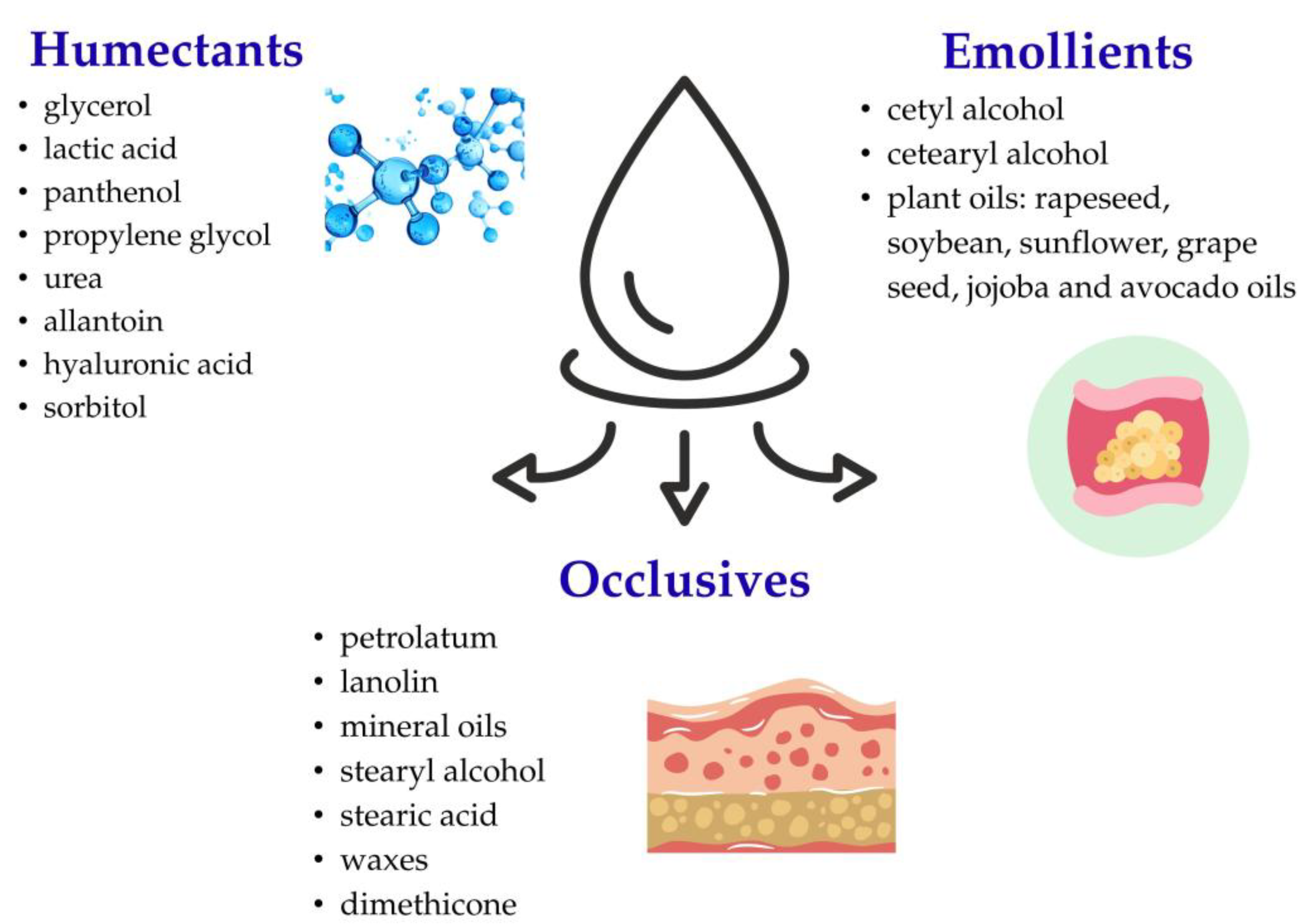
2]. The main function of moisturizers is to enhance the skin barrier and increase water content in the stratum corneum. Moisturizing ingredients are generally divided into three categories: humectants, occlusives and emollients (
Figure 1). Humectants are hydrophilic compounds containing functional groups capable of binding water molecules, which reduces water loss from the skin and prevents the preparation from drying out. Typical humectants include glycerol, lactic acid, panthenol, propylene glycol, urea, allantoin, hyaluronic acid and sorbitol [
3,
4,
5]. Occlusive agents, on the other hand, reduce transepidermal water loss (TEWL) by forming a hydrophobic layer on the skin’s surface. Typical occlusive agents include petrolatum, lanolin, mineral oils, stearyl alcohol, stearic acid, waxes, cholesterol, and silicone derivatives such as dimethicone. The third group is emollients, which are lipid-based substances often similar in composition to the intercellular lipids naturally found in the skin. Emollients improve skin softness and smoothness, providing a pleasant sensory sensation. Examples include cetyl alcohol, cetearyl alcohol, and plant-derived oils such as rapeseed, soybean, sunflower, grape seed, jojoba, and avocado oils. It has been demonstrated that combining humectants, occlusives, and emollients in a single dermatological formulation enhances the general moisturizing efficacy of the product [
6,
7]. The condition of the epidermal barrier and the skin’s moisture level can be assessed using various instrumental techniques. TEWL, measured using a Tewameter, is a widely accepted parameter for assessing the integrity of the skin barrier by quantitatively determining the amount of water diffusing from the skin through the stratum corneum into the surrounding environment. Skin hydration is typically assessed using a corneometer, which measures the electrical capacitance of the stratum corneum. Higher electrical conductivity values indicate greater water content in the stratum corneum and consequently a higher level of skin hydration [
8,
9].
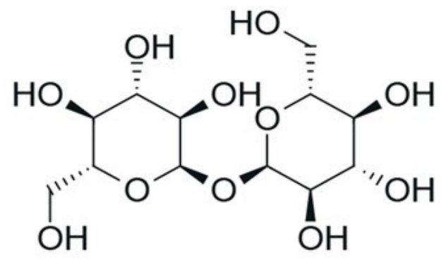
Trehalose is a natural non-reducing sugar. It consists of two glucose molecules joined by an α,α-1,1-glycosidic bond (
Figure 2). Trehalose exhibits hydrophilic properties and is stable at high temperatures and across a wide pH range (3.5–10). It is easily hydrolyzed to glucose, which is essential for many metabolic pathways, such as glycolysis. By forming hydrogen bonds between the hydroxyl groups of the sugar and the phosphate groups of the membrane phospholipids, trehalose enhances the integrity of cellular structures, particularly membranes and enzymatic proteins. It protects membranes from drying out or freezing, maintaining the fluidity and permeability of bilayer lipid structures. As a result, the cell membrane retains its functions and enables the cell to survive in unfavorable environmental conditions [
10,
11]. It is known as the “sugar of life” because it helps desert plants protect their internal structure during dry periods and enables them to regenerate as soon as the rains come. These mechanisms are also utilized by fungi, nematodes, bacteria, and insects, particularly in situations of increased nutrient demand, such as during spore incubation, toxic substance effects, or exposure to extremely low or high temperatures [
12,
13].
Trehalose, due to its properties and ability to stabilize proteins and lipids, is used in many industries, including food, cosmetics, and pharmaceutical fields. The use of trehalose in skin care products is associated with its properties and effects: it inhibits TEWL, strengthens the skin barrier, has nutritional and regenerating properties, and inhibits the breakdown of fatty acids on the skin to prevent unpleasant odors (in deodorants). As a raw material, it occurs as a white crystalline powder (pH 4.5–6.5) and is commonly used in moisturizing, anti-aging, and firming preparations (for the body, face, and hair), and in oral care products [
10,
14].
Among topical dosage forms, emulsions are considered one of the most widely used dermatological carriers, because they incorporate both hydrophilic and lipophilic ingredients into a single formula. Simple ointments or traditional moisturizers, often based on a single mechanism of action, such as occlusion or water binding, can provide only short-term relief from the symptoms of dry skin. In contrast, modern emulsion-based formulations combine moisturizing, emollient, and occlusive agents into a single delivery system, providing complementary mechanisms of action. Such formulations not only improve skin hydration but also support the repair of the skin barrier, enhance sensory properties, and increase patient acceptance. Therefore, the development of multifunctional emulsion systems represents a promising strategy for the treatment of xerosis and other skin barrier disorders [
1,
3,
15].
Although the moisturizing properties of trehalose have been described previously, the innovative aspect of our study and its main objective were the development of an optimized dermatological emulsion carrier by comparing various oils and emulsifiers, then characterizing the formulation in terms of its physicochemical, rheological, mechanical, and adhesive properties. Furthermore, the selected formulation was evaluated in vivo using TEWL and corneometry measurements after the addition of various concentrations of trehalose (1%, 2.5% and 5%), which enabled the selection of the most effective formulation.
2. Materials and Methods
2.1. Materials
Trehalose, avocado oil, jojoba oil, Olivem 1000 (cetearyl olivate and sorbitan olivate), cetyl alcohol, GSC (glyceryl stearate citrate) and GSB (gluconolactone and sodium benzoate) were purchased from Zrob Sobie Krem (Prochowice, Poland). Liquid paraffin was supplied by Starpharma (Warszawa, Poland) and Vitamin E by Hasco (Wrocław, Poland). Glycerol (propan-1,2,3-triol) was obtained from COEL (Kraków, Poland), xanthan gum, bovine-skin gelatin, and triethanolamine from Sigma-Aldrich (Steinheim, Germany). Purified water was prepared by reverse osmosis and deionization (apparatus type WCR-RO 10-DPS, Cobrabid—Aqua, Warszawa, Poland).
2.2. Preliminary Studies—Design, Preparation, and Characterization of Emulsion
2.2.1. Preparation of Emulsions
To develop a base formulation for trehalose, six placebo emulsions were prepared. They differed in the emulsifiers and oils used. Emulsions E1–E3 contained Olivem 1000 (cetearyl olivate and sorbitan olivate) as the emulsifier, while emulsions E4–E6 included a mixture of emulsifiers: GSC (glycerol ester of citric and stearic acids) and cetyl alcohol in a ratio of 2:3, which was selected in preliminary studies.
The emulsions were prepared using different oils—avocado oil (E3, E6), jojoba oil (E2, E5), or a combination of both oils (E1, E4). The composition of the emulsions is presented in
Table 1. All emulsions were prepared using the hot method by heating the aqueous and oil phases separately to 70 °C ± 2 °C, followed by mixing them using a mechanical stirrer. The emulsions were mixed for 30 min at 400 rpm.
The aqueous phase of emulsions consisted of water, glycerol, and a preservative—a mixture of sodium benzoate and gluconolactone (GSB). The oil phase included liquid paraffin, the appropriate oil (jojoba oil, avocado oil), and the emulsifier (GSC, cetyl alcohol, Olivem). After cooling, vitamin E (an antioxidant) and an appropriate amount of xanthan gum were added to achieve the desired consistency (E2, E4, E5, E6). Emulsions E1 and E2 exhibited suitable consistency and did not require the addition of xanthan gum.
2.2.2. Visual, Centrifuge Stability Test, and pH Evaluation
The emulsions were visually evaluated to determine their appearance, including color, consistency, and homogeneity. The pH assessment of emulsions was carried out using a pH meter Orion Star A211 (Thermo Fisher Scientific, Waltham, MA, USA).
The emulsions were also subjected to stability assessment by centrifugation at 4000 rpm for 10 min [
16], using centrifuge MPW-223E (MPW Med. Instruments, Warszawa, Poland). Then, the emulsions were visually observed for phase separation. Each measurement was performed 24 h after emulsion preparation.
2.2.3. Viscosity and Rheological Assessment
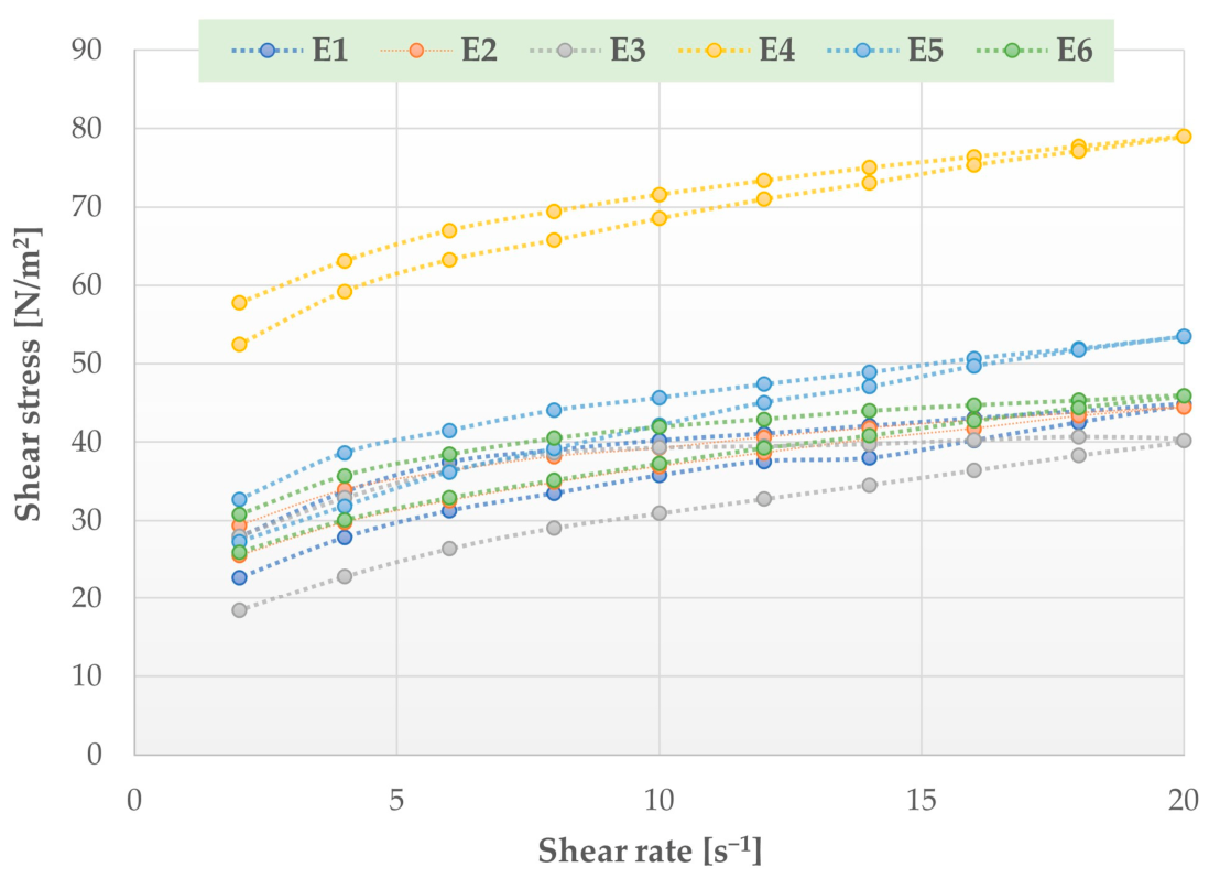
The rheological features of the emulsions were analyzed by the Brookfield DV—III Ultra viscosity meter (Ametek Brookfield, MA, USA). The apparent viscosity of samples (0.5 mL) was tested at 25 °C ± 1 °C. Rheological measurements were performed under increasing and then decreasing shear rates (from 2 s−1 to 20 s−1). The results are demonstrated as a plot of shear stress vs. shear rate (flow curves) and a plot of viscosity vs. shear rate (rheograms). Four repetitions of viscosity tests were carried out, and the average values with deviations were calculated.
2.2.4. Mechanical and Adhesion Analysis
The mechanical properties of the emulsion were measured using a TA.XT Plus texture analyzer (Stable Micro Systems, Godalming, UK). The measurement was performed at room temperature using a 35 mm diameter disk, which was immersed in the emulsion at a speed of 2 mm/s. Properties such as consistency, firmness, and cohesiveness were determined using the Texture Exponent 32 software and presented graphically.
Adhesive properties of the prepared emulsions were measured using a TA.XT Plus texture analyzer. A gelatin membrane was used in the test, and for this purpose, a 30% gelatin solution was prepared and poured into a Petri dish (0.3 g/cm3). In addition, measurements using porcine skin and hairless mouse skin were performed. The porcine and mouse skin samples were stored at −80 °C ± 1 °C (up to a maximum of 1 month). One day before the test, they were thawed, and prior to analysis, they were immersed in 0.9% NaCl for 30 min (25 °C). A 10 mm diameter gelatin disk or skin sample was attached to the head of the device using cyanoacrylate glue, and 0.5 g of the test emulsion was applied to the plate of the texture analyzer. The adhesion capacity of the preparations was measured at 32 °C ± 1 °C (water bath) under the following conditions: contact pressure of 0.5 N and contact time of 90 s. The adhesive properties were characterized based on the adhesion force (mN) and adhesion work (µJ). All measurements (mechanical and adhesion-related) were performed six times, and the mean values were then calculated.
Skin from hairless mice (Cby.Cg inbred strain—Foxn1nu/cmdb) was obtained from animals at the Center for Experimental Medicine (Medical University of Bialystok) that were intended for organ harvesting for use in in vitro research. Porcine skin (prepared from the front legs) was taken from animals weighing 200–250 kg (Bost butchery, Turosn Koscielna, Poland). The studies conducted on skin samples did not require approval from the Local Ethics Committee for Animal Experiments.
2.2.5. Spreadability of Emulsion
The spreadability coefficient of the preparations was determined using a modified method described by Rao et al. [
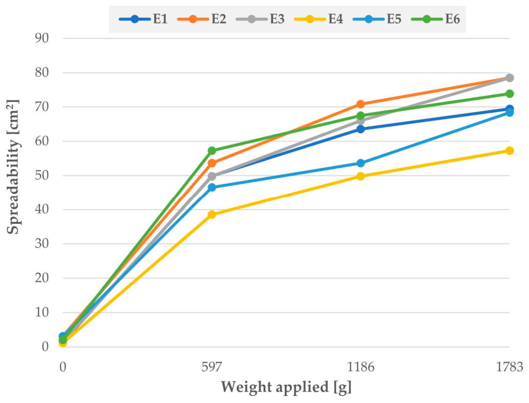
17], employing four glass slides; one was used as a platform for applying the emulsion, while the other three slides were used to weigh down the emulsion (each 597 g). A 1 g sample of the preparation was placed on a glass plate. After application, each plate was left for 2 min to remove air trapped between the plates and form a uniform layer of the preparation. After applying each weight, the surface area of the emulsion was calculated. The results are presented in a graph showing the relationship between surface area and applied weight.
2.3. Preparation and Evaluation of Emulsions with Trehalose
Based on preliminary studies of the placebo emulsion, the optimal base composition for trehalose was selected. The trehalose emulsions were prepared, as described in
Section 2.2.1. Trehalose was dissolved in the aqueous phase at concentrations of 1%, 2.5%, and 5%.
The pH of the trehalose emulsions was measured using the same method mentioned in
Section 2.2.2. To achieve a pH value for the emulsion close to the physiological pH of the skin, triethanolamine was added until the pH of the formulation reached approximately 5.5.
2.4. Measurement of Transepidermal Water Loss
TEWL test was performed using a Tewameter TM 300 (Courage-Khazaka Electronic GmbH, Cologne, Germany), a device designed to measure skin hydration and the condition of the skin barrier. The study was conducted to measure the level of TEWL from the studied skin area before and after the application of emulsions containing trehalose. TEWL was expressed in g/m2/h—water loss in grams per square meter per hour. The study included a group of 7 adults: healthy women aged 21–55 who had no contraindications to the measurements and showed no tendency to allergies. The first measurement was taken before applying the emulsion, further measurements at 1, 2, and 4 weeks after application, and two weeks after the end of application. Participants did not use any other moisturizing cosmetic products for at least 7 days prior to the study and during the trial. Each participant received two different emulsions containing trehalose and one placebo emulsion, in accordance with the predefined allocation scheme. Emulsions containing trehalose were applied to separate, designated areas on the inner side of the forearms, approximately 2–3 cm from the elbow fold. The placebo was applied to a separate area located below the site treated with trehalose on the left forearm. The preparations were applied twice a day, in the morning and in the evening, to separate areas to avoid interaction between them. TEWL measurement took place in a seated position after a 10 min period of rest and under constant environmental conditions (at room temperature—about 21 °C, at a humidity of about 50–60%). The study was evaluated by placing a probe (Tewameter TM Hex probe; Courage-Khazaka Electronic GmbH, Cologne, Germany) on the skin for 45 s. Each measurement was performed in triplicate.
The methods for TEWL and corneometer evaluation were approved by the Bioethics Committee at the Medical University of Bialystok (bioethical permission number APK.002.364.2024). The number of participants in the in vivo study (TEWL and hydration) was limited because these measurements were designed as preliminary studies.
2.5. Assessment of Skin Hydration
A corneometer CM 825 (Courage-Khazaka Electronic GmbH, Cologne, Germany) was used to measure skin hydration, measuring changes in the dielectric constant of the stratum corneum. The test was conducted in the same way as the test with the Tewameter (
Section 2.4); the only difference was the duration of a single measurement (1 s). Each measurement was performed in triplicate.
2.6. Statistical Analysis
Data were presented as mean ± standard deviation (SD). Statistical analysis was performed using Statistica 13.3 software (StatSoft, Krakow, Poland). Data obtained from viscosity, texture, and adhesion studies were evaluated using one-way ANOVA, followed by Tukey’s post hoc test. TEWL and skin hydration were measured repeatedly in the same volunteers during the study, and repeated-measures analysis of variance (repeated-measures ANOVA) was used to evaluate the effect of treatment over time. Differences were considered statistically significant at p < 0.05.
3. Results and Discussion
During the development of dermatological emulsions containing trehalose, liquid paraffin and the vegetable oils jojoba oil and avocado oil were used as the oily phase. Triglycerides and fatty acids are mainly responsible for the properties of oils. Avocado oil includes palmitic, palmitoleic and oleic acids with antimicrobial properties, and linoleic and α-linolenic acids with anti-inflammatory and regenerating properties. The content of these components in the oil can vary by about 5–10% due to the origin of the plant material and the production method. Avocado oil has an oleic acid to linoleic acid ratio of 2–3 to 1, which corresponds to a physiological ratio of 3:1 [
18,
19]. Jojoba oil consists of approximately 98% pure waxes (mainly wax esters, free fatty acids, alcohols, and hydrocarbons), as well as sterols and vitamins, so it is commonly known as liquid wax. Due to the presence of natural antioxidants (α, γ, and δ tocopherol), it shows high resistance to rancidity. Jojoba oil, thanks to its unique chemical composition, exhibits a wide range of biological activity. When used externally, it has antimicrobial, antioxidant, and anti-inflammatory properties. Due to its properties, it can be effective in the fight against acne or psoriasis. It softens the surface of the skin, shows good spreadability without feeling greasy, prevents excessive evaporation of moisture from the skin, and, thanks to its structural similarity to skin sebum, it smoothes the skin and inhibits excessive peeling [
20,
21]. Another oily ingredient used in the prepared emulsions was liquid paraffin. It is a purified mixture of liquid hydrocarbons obtained from the processing of crude oil. It creates an occlusion on the skin and prevents moisture loss. It has a softening and lubricating effect, but it can limit the absorption of substances. Studies confirm that the use of 10% liquid paraffin in combination with glycerol significantly reduces skin dryness and inhibits the associated itching [
17,
22].
All emulsions prepared with the use of the described emulsifiers were characterized by a uniform consistency and did not show signs of phase separation. The color of the emulsion depended on the type of oil used in the formulation; emulsions containing avocado oil (E1, E3, E4, E6) were light yellow or yellow. It was observed that emulsions containing Olivem 1000 (E1–E3) as an emulsifier had a higher consistency, and among them, only emulsion E2 required correction of the consistency by the addition of xanthan gum. In the case of emulsions with emulsifiers GSC and cetyl alcohol (E4–E6), each of the emulsions required the addition of xanthan gum, due to its rather thin consistency. Similar observations were obtained in the research conducted by Zuikina et al. [
23], for preparations using emulsifiers Olivem 1000 and Emulpharma 1000. It was shown that both types of emulsifiers made it possible to obtain stable emulsions with high viscosity, which provided beneficial performance properties.
It was shown that emulsions E1, E2 and E3 were stable and no phase separation was observed during the centrifuge test. For emulsions E4–E6, phase homogenization was less effective than in emulsions with the Olivem—it took longer to combine both phases. Finally, in the centrifuge test, homogeneous emulsions E5 and E6 were formed without phase separation; only in emulsion E4 was slight phase separation observed (a visible color gradient, which may indicate the separation of avocado oil from the other components).
The pH values of the emulsions were in the range of 4.07–4.54 (the lowest pH was in emulsion E1, the highest—E6), and they were adjusted to 5.5 by adding triethanolamine.
It should be noted that the stability assessment conducted in this study was preliminary and was limited to short-term visual observations, pH measurements, and centrifugation tests. Further studies are recommended to evaluate long-term and accelerated stability under controlled storage conditions.
3.1. Rheological Properties of Emulsions
The viscosity of the preparation affects the stability of the emulsion, and it should also be adapted to the purpose of the product and the type of packaging. High-viscosity preparations may spread less well on the skin and be more difficult to remove from the packaging, but they have better occlusive properties, desirable, for example, in regenerating preparations. When the viscosity is too low, the preparation is characterized by a lighter form, does not give a feeling of heaviness, and it can run off the surface of the skin during application [
16]. Statistically, the highest viscosity (
p < 0.05) was possessed by emulsion E4 (7157.1 ± 80.2 mPa·s), which is associated with the greatest concentration of xanthan gum (1%). The viscosity of the remaining emulsions was in the range of 3922–4565 mPa·s (
Table 2).
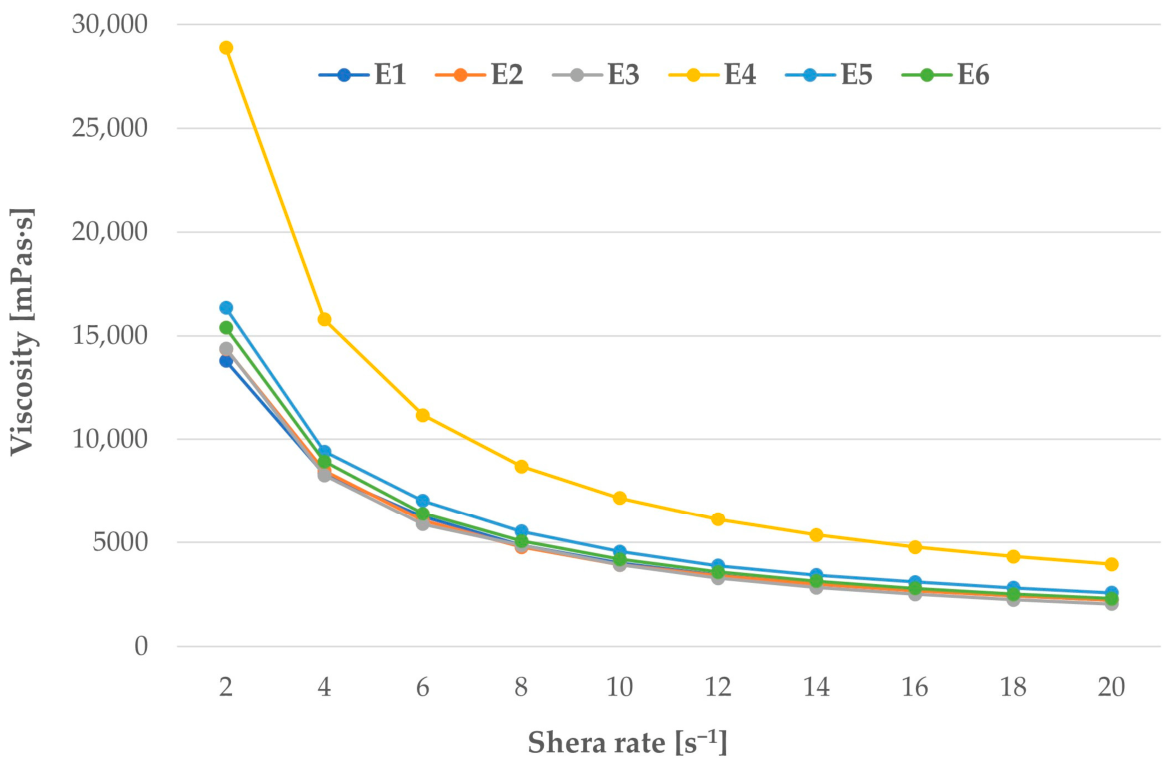
In order to further characterize the emulsions, the viscosity graphs were prepared, showing the dependence of viscosity on the shear rate (
Figure 3). The analysis of these curves enables the classification of the preparations as Newtonian or non-Newtonian bodies. The prepared emulsions showed a decrease in viscosity with increasing shear rate, indicating non-Newtonian shear-thinning behavior. Hysteresis loops (
Figure 4) present the phenomenon of thixotropy occurring in the prepared emulsions—the ability of the system to reproduce the consistency change as a result of shear stress. Topical pharmaceutical products often contain ingredients that affect the product’s thixotropy—a feature that improves the product’s application (ease of dosing and spreading), as well as its storage stability [
24,
25]. Emulsion E4 exhibited statistically significant differences (
p < 0.05) in rheological properties (flow and thixotropy features) compared to other emulsions.
The rheological data for all emulsions were fitted using the Power Law (Ostwald–de Waele) [
26,
27,
28]. The calculated flow index (
n) ranged from 0.136 to 0.207, indicating a shear-thinning system—viscosity decreases with increasing shear rate. Among the tested emulsions, preparation E4 exhibited the highest consistency coefficient (K = 52.43 Pa·s
n) and the lowest flow behavior index (
n = 0.136), suggesting the strongest internal structure and the greatest flow resistance. The determination coefficients (R
2 = 0.911–0.9996) confirmed a good fit of the experimental data to the Power Law model [
24].
3.2. Texture Evaluation of Emulsions
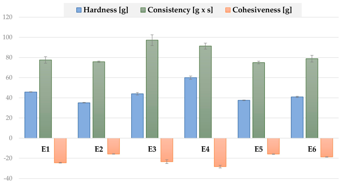
Emulsions intended for skin application should be easy to use and ensure adequate contact between the formulation and the skin surface. When designing an optimal formulation for topical use, it is important to maintain a balance between the adhesion of the emulsion and its consistency. Texture analysis provides valuable information regarding these properties. The prepared emulsions were subjected to texture evaluation 24–48 h following preparation, analyzing their mechanical and adhesion properties. Different parameters were measured: hardness (maximum positive force, in g), consistency (positive area below the curve, in g·s), and cohesiveness (minimum negative force, in g) (
Figure 5). The hardness of an emulsion expresses the applicability of the products to the skin and is defined as the force required to deform a sample, i.e., resistance of the emulsion to the applied pressure. Cohesiveness is the emulsion’s ability to return to its initial form after deformation, while consistency refers to the emulsion’s density and cohesiveness [
29,
30]. It was observed that emulsion E4 containing jojoba oil, avocado oil, GSC and cetyl alcohol was characterized by significantly (
p < 0.05) the highest hardness and consistency values and had the highest viscosity compared to other emulsions. The lowest values for all textural parameters were obtained for emulsions E2, E5 and E6.
Another parameter tested was the emulsions’ adhesive properties, i.e., the ability of the preparation to adhere to the model surface. To simulate in vivo conditions, porcine skin and mouse skin were used. However, the use of such materials may be associated with some limitations, such as biological variability, difficulties in standardization, and potential mechanical damage, which can affect the results. Therefore, an alternative model—a gelatin membrane—was additionally used. Gelatin is an animal protein and is frequently used as a layer in adhesion studies [
31,
32,
33]. The adhesive properties of the emulsions were determined by measuring two parameters: adhesion work (W
ad) and adhesion force (F
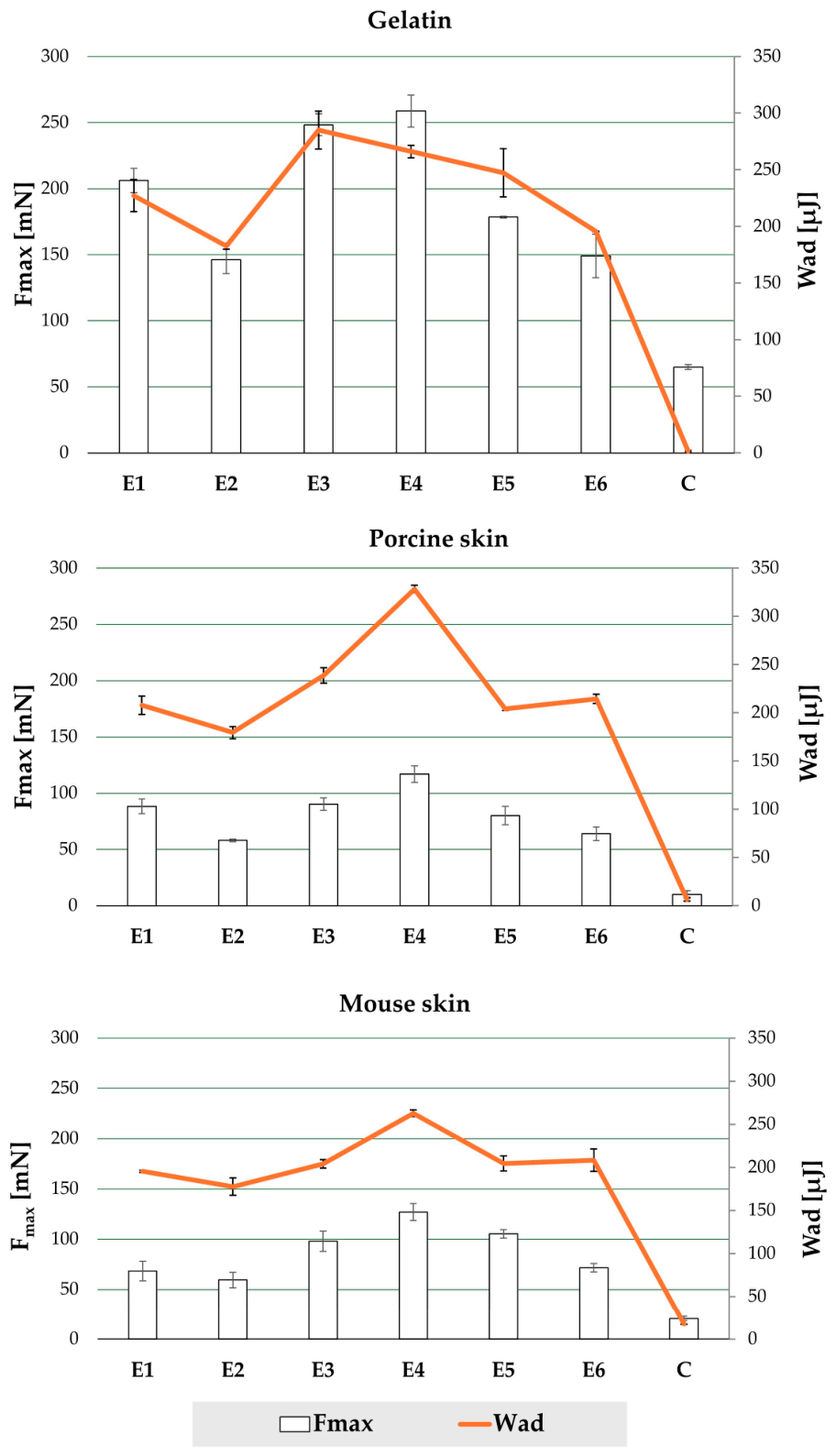
max). Adhesion work defines the amount of work required to separate the tested sample from the applied adhesive layer, while adhesion force describes the sample’s ability to bond with the tested surface. Emulsion E4 exhibited the highest values of adhesion force and work, as demonstrated on both gelatin film and porcine and mouse skin (
Figure 6). High parameter values were also obtained for emulsion E3. It was also observed that the adhesion values obtained using the gelatin membrane were significantly higher (
p < 0.05) than those measured on animal skins, which may be related to the hydrophilic and swelling properties of gelatin, stronger intermolecular interactions, and hydrogen bonding with the tested formulations [
31].
In conclusion, all tested emulsions exhibited adhesive properties. The values for Fmax and Wad were significantly higher (p < 0.05) compared to the control (cellulose paper).
Based on the spreadability results (
Figure 7), it was found that in all emulsions, spreadability increased as the applied mass of the glass plate increased, indicating good spreadability and ease of application. Among the tested formulations, E2 exhibited the highest spreadability at the maximum applied mass, suggesting the lowest flow resistance and best spreading efficiency. These values correlated with the lower viscosity values obtained in the viscosity test (
Section 3.1). E3 and E6 also exhibited high spreadability values, indicating favorable flow properties. In contrast, E4 exhibited the lowest spreadability, which may suggest higher viscosity and a higher consistency, corresponding to the results obtained in the rheological and mechanical evaluation.
In summary, based on the tests conducted on the placebo emulsions (without the trehalose), emulsions E3 and E4 exhibited the most favorable properties. Stability data were the crucial factor in the selection of the preferred emulsion. Emulsion E4 showed slight phase separation in the centrifuge test; therefore, emulsion E3—containing avocado oil and the emulsifier Olivem—was selected as the emulsion carrier for trehalose.
The prepared emulsion E3 required pH adjustment to values close to the physiological pH of the skin surface [
34]. For this purpose, triethanolamine was used to adjust the prepared emulsions to a pH of approximately 5.5. The emulsions were prepared in three concentrations: 1% (TE1), 2.5% (TE2.5), and 5% (TE5). Additionally, for reference purposes, placebo emulsions (PEs)—E3 emulsion without trehalose—were used in the TEWL and corneometer tests.
3.3. TEWL and Skin Hydration Measurement
TEWL measurement and corneometer (skin hydration) assessments were performed on the participants’ skin. The number of participants was limited because the in vivo assessment was designed as a preliminary study. During 1, 2, and 4 weeks of emulsion application, as well as 2 weeks after the end of their use, the hydration of the stratum corneum and TEWL were measured. TEWL determines the amount of water diffusing through the stratum corneum to the skin surface per unit of time. Under normal conditions, TEWL values for healthy skin range from 0 to 15 g/m
2/h [
35,
36]. The obtained TEWL might be affected by factors such as temperature, body site, sweating, or the emotional state of the patient, which is why the TEWL assessment was performed under similar conditions, in the same room, after a 10 min rest and after measuring skin moisture and temperature. On the other hand, skin hydration is measured by changes in the dielectric constant of the stratum corneum. The measurement results are presented in conventional units, ranging from 0 to 120. Values below 30 indicate very dry, dehydrated skin; a result of 30–40 indicates dry skin, and values above 40 indicate properly hydrated skin [
9,
37].
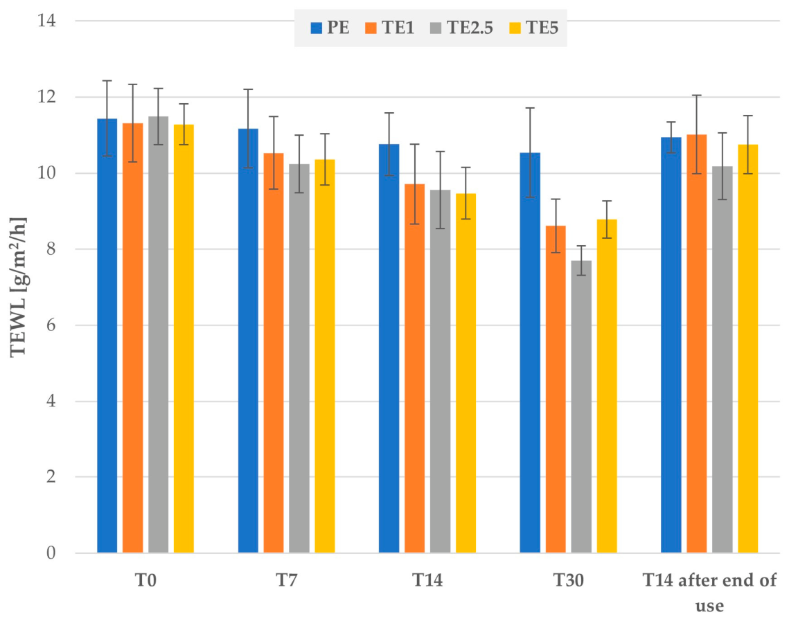
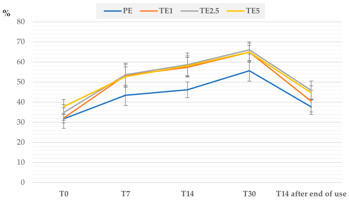
In all participants, a significant (
p < 0.05) decrease in TEWL and an increase in corneum hydration were observed during the 28-day application of the emulsion (
Figure 8 and
Figure 9). No erythema, irritation, itching, or other adverse skin reactions were observed during the in vivo test. The greatest reduction in TEWL (about 33%) was observed for the 2.5% emulsion (TE2.5). Emulsions with 1% (TE1) and 5% (TE5) decreased TEWL by approximately 24%, while the placebo emulsion (PE) decreased it by only 8%. The highest skin hydration after 4 weeks was noted for emulsion TE1 (102%), remaining formulations caused a hydration increase of 72–89%. It was observed that 14 days after finishing the application of the emulsion, TEWL increased and skin hydration decreased, indicating the need for regular use of the products to maintain the achieved effect.
It should be emphasized that the observed changes in TEWL and stratum corneum hydration cannot be attributed solely to trehalose. The emulsion base contained glycerol, avocado oil and liquid paraffin, which act as humectants, emollients and occlusives, and therefore might contribute to the overall moisturizing effect. Nevertheless, the greater changes observed in the trehalose-containing formulations compared to the placebo suggest that trehalose may provide additional hydration within the developed emulsion formulation.
In studies conducted by Maeda et al. [
14], TEWL and stratum corneum hydration were assessed following the application of a sodium trehalose sulfate solution at concentrations of 0.05%, 0.1%, and 0.2%. It was found that the water content in the stratum corneum was significantly higher than in the control areas, and the TEWL value decreased. Additionally, the amount of filaggrin and the mRNA levels of proteins involved in intercellular transport and lipid storage in the stratum corneum increased. Another study involving a group of 15 patients with very dry skin on the legs, conducted by Greco et al. [
12] confirmed the moisturizing effect of trehalose. Participants used a formulation containing trehalose, ceramides, lecithin, and cholesterol. After 4 weeks, a 54% reduction in TEWL and a 102% increase in hydration were observed. A significant improvement in skin parameters was demonstrated compared to both the placebo control and basal values.
4. Conclusions
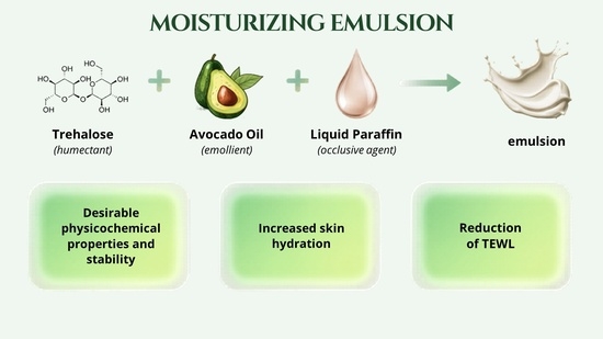
Among the tested formulations, emulsion E3, containing liquid paraffin and avocado oil, exhibited the most favorable physicochemical properties and stability. The developed emulsion demonstrated non-Newtonian shear-thinning and thixotropic behavior, which are considered advantageous for topical formulations.
In vivo evaluation—TEWL and skin hydration tests—showed that regular application of the trehalose-containing emulsions improved skin hydration and reduced TEWL. The formulation containing 2.5% trehalose showed the most beneficial effect in reducing TEWL, while emulsions containing 1% trehalose most significantly increased skin hydration. Therefore, no single concentration can be considered definitely optimal in terms of both parameters. However, the observed reduction in hydration and increase in TEWL after treatment discontinuation suggest that continuous application is necessary to maintain the beneficial effects.
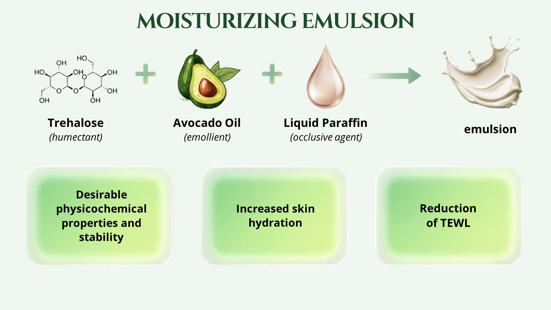
Moreover, the obtained results indicate that the final moisturizing effect of the emulsion was ensured by other excipients used in the designed formulations, by combining several agents: trehalose as a humectant, avocado oil as an emollient, and liquid paraffin as an occlusive agent. The developed emulsion might serve as a promising dermatological carrier for trehalose intended for dry skin care; however, further studies are required.