Article

A Triple-Helical Collagen Gel Modulates Electrophysiological Parameters in Isolated Rabbit Skin

by
Dominika Dąbrowska-Wisłocka
1,
Aleksandra Kalinoska
2,
Olga Zavyalova
1,
Beata Winiecka
2,
Karolina Pisanko
3,
Arkadiusz Jundziłł
3,
Karolina Szewczyk-Golec
4,* and
Iga Hołyńska-Iwan
2,*
1
Department of Chemical Technology and Pharmaceuticals, Faculty of Pharmacy, Collegium Medicum in Bydgoszcz, Nicolaus Copernicus University in Torun, 87-100 Torun, Poland
2
Department of Pathobiochemistry and Clinical Chemistry, Faculty of Pharmacy, Ludwik Rydygier Collegium Medicum in Bydgoszcz, Nicolaus Copernicus University in Torun, 85-084 Torun, Poland
3
Department of Plastic, Esthetic and Reconstructive Surgery, Faculty of Medicine, Ludwik Rydygier Collegium Medicum in Bydgoszcz, Nicolaus Copernicus University in Torun, 87-094 Torun, Poland
4
Department of Medical Biochemistry and Biology, Faculty of Medicine, Ludwik Rydygier Collegium Medicum in Bydgoszcz, Nicolaus Copernicus University in Torun, 87-092 Torun, Poland
*
Authors to whom correspondence should be addressed.
Cosmetics 2026, 13(4), 192; https://doi.org/10.3390/cosmetics13040192
Submission received: 26 May 2026 / Revised: 25 July 2026 / Accepted: 27 July 2026 / Published: 29 July 2026

Abstract

Collagen is widely used in the cosmetics industry as an active ingredient in skin-care formulations due to its biocompatibility, biodegradability, low antigenicity, and high biological activity. As a natural humectant, collagen binds water molecules within the skin, reduces transepidermal water loss, and helps maintain skin elasticity and hydration. Despite its broad application, the influence of collagen on epithelial ion transport remains insufficiently understood. This study evaluated the effect of a gel containing triple-helical collagen on sodium and chloride ion transport in the isolated rabbit skin. The collagen gel was applied to 25 skin specimens for 24 h and compared with 30 untreated control specimens. Electrophysiological analyses included measurements of transepithelial electrical potential (PD), electrical resistance (R), and potential changes during stimulation (PDmin and PDmax). Collagen gel significantly decreased R compared with control tissues, indicating altered tissue permeability. Collagen gel-treated tissues also exhibited a significantly more electropositive PDmin than controls, whereas PDmax values remained comparable between groups, suggesting that electrophysiological responsiveness was maintained under the experimental conditions. The observed electropositive shift in PDmin may reflect altered sodium ion transport. Whether these electrophysiological changes are associated with changes in tissue hydration requires direct investigation.

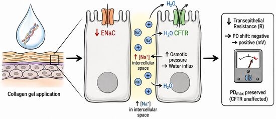
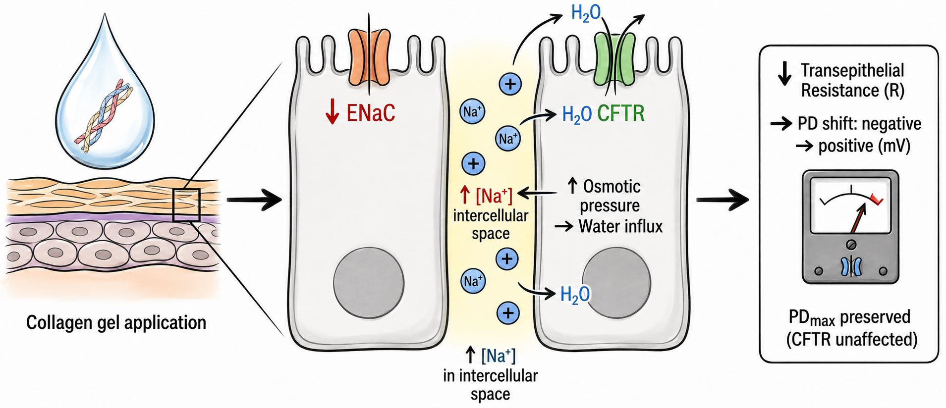
Graphical Abstract

1. Introduction

Collagen is a fibrillar protein and the most abundant structural protein in vertebrates [1,2,3,4,5,6]. It is most abundant in the extracellular matrix (EMC) [4,6]. It is a major component of the extracellular matrix (ECM) and is found predominantly in fibrous tissues such as the skin, ligaments, bones, blood vessels, tendons, muscles, cornea, and dentin [1,3,4,6,7,8]. Collagen provides structural support and preserves tissue integrity, contributing to tissue strength, stability, and elasticity [7,8,9,10].
Due to its biocompatibility, biodegradability, favorable mechanical properties, low antigenicity, and high biological activity [5,9,10], collagen has attracted considerable interest in tissue engineering [3,7,8], regenerative medicine [11,12], as well as in the medical [13], pharmaceutical [14], and cosmetic industries [15,16]. Common sources of collagen include animal-derived collagen (porcine, bovine, or fish), autologous collagen, and collagen derived from cultured human fibroblasts [13,17,18]. In biomedical and cosmetic applications, the most frequently used forms are hydrolyzed collagen (collagen peptides), native collagen (the undenatured form), and gelatin (the fully denatured form) [7,17].
Collagen is used in a wide range of formulations, including hydrogels [2,7,19], sponges [2,7], films [2,20], emulsions [7], nanoparticles [2], and nanofibers [2,7]. It is also incorporated into biomaterials designed to support drug delivery systems, often in combination with anti-inflammatory and antimicrobial agents [2,7,11]. In the cosmetics industry, collagen is primarily applied as an active ingredient in products intended for the care of the skin and its appendages. Importantly, due to its natural humectant properties, collagen can bind water molecules within the skin, thereby reducing transepidermal water loss (TEWL) and helping maintain skin elasticity and hydration [16,21,22].
Nearly half of the collagen in the human body is located in the skin, predominantly as type I and type III collagen [23,24,25]. High-molecular-weight collagen is responsible for maintaining the structural integrity of the skin by forming a dense network of fibers within the dermis [26]. In contrast, low-molecular-weight collagen contributes to increasing the density and diameter of collagen fibers [26,27]. With aging, progressive collagen degradation leads to reduced skin firmness, altered facial contours, and the formation of wrinkles [24,28,29,30,31,32,33,34]. Collagen, particularly low-molecular-weight collagen and collagen-derived peptides, may improve skin hydration, elasticity, and barrier function [35,36]. These effects have been attributed to enhanced water retention, improved extracellular matrix organization, stimulation of fibroblast activity, and modulation of skin barrier integrity. Collagen may influence the skin microenvironment by affecting extracellular hydration and tissue permeability. However, despite substantial evidence supporting the beneficial effects of collagen on skin hydration and barrier-related parameters, little is known about its direct influence on ion transport mechanisms and electrophysiological properties of skin tissue. Understanding these mechanisms may provide new insight into how collagen modulates skin hydration and microenvironment homeostasis.
Epithelial cells forming the epidermis exhibit a characteristic polarized structure, and transepithelial ion transport across these cells is essential for maintaining skin homeostasis and proper tissue function [37,38]. The electrostatic field generated at the skin surface by ion transport contributes to the skin’s proper appearance and reactivity [39]. Therefore, maintaining homeostasis in the transport of ions and water, both within epidermal cells and across individual skin layers, is crucial for preserving skin integrity [39,40]. Disturbances in water distribution are closely associated with alterations in ion transport, particularly involving sodium and chloride ions. Likewise, increased ion transport affects water movement within the tissue [37,41,42].
Numerous studies have demonstrated that the epithelial sodium channel (ENaC) is involved in regulating cellular hydration and maintaining the local microenvironment [38,40]. Moreover, altered sodium ion transport has been associated with changes in local intercellular osmolality [43], which have been implicated in immune activation and may contribute to allergies, skin hypersensitivity, and hypertrophic scarring [38,39,42]. ENaC channels have also been shown to actively participate in wound healing processes [44], keratinocyte differentiation [45], and normal epidermal growth [38,42,46,47]. Moreover, ENaC dysfunction may be associated with increased transport of oxidized low-density lipoproteins (ox-LDLs) and the initiation of an inflammatory response [48].
Chloride ion transport occurs primarily through the cystic fibrosis transmembrane conductance regulator (CFTR) channel, which is involved in regulating both the amount and composition of sweat secretion [47]. Similarly to ENaC, CFTR participates in the activation of immunocompetent cells [40]. Furthermore, abnormalities in chloride channel function may impair skin healing and regeneration, disrupt water transport, leading to cellular dehydration or overhydration, and disturb in the surrounding microenvironment [39,40,44,45,47].
The growing interest in collagen and its widespread use in cosmetology makes it important to determine the mechanisms through which this protein affects skin tissue. Since ion transport plays a key role in maintaining skin hydration and microenvironment homeostasis, investigating collagen-induced changes in these parameters may provide new mechanistic insight into its biological activity. We hypothesized that collagen gel might alter skin electrophysiological properties by modulating ion transport, with potential downstream effects on hydration and barrier function. In the present study, a fish-derived triple-helical collagen preparation was used. A ready-to-use cosmetic gel with a known, stable composition and uniformly distributed collagen fragments, which would yield reproducible, reliable data, was selected. Due to limited experimental data on the effect of collagen on ion transport in the skin, an experimental model was developed to evaluate the prolonged influence of collagen gel on the electrophysiological parameters of isolated rabbit skin fragments. To explore the possible involvement of sodium- and chloride-transport pathways in the observed electrophysiological responses, tissues were sequentially exposed to amiloride, an ENaC blocker, and bumetanide, an inhibitor of the sodium-potassium-chloride cotransporter (NKCC).

2. Materials and Methods

The experiment was conducted on 55 isolated skin fragments collected from five New Zealand White rabbits. The animals of both sexes were approximately 2–3 months old and weighed between 3.5 and 4.0 kg. The animals were handled in accordance with European Union regulations governing animal welfare. All animals were housed under identical standard conditions, with free access to food and water (ad libitum), in an environment with controlled humidity (55–60%) and a temperature of 22–23 °C, maintained on a 12-h day/night cycle. All animals were healthy and showed no signs of inflammation or skin lesions. Furthermore, they had not been subjected to any experimental procedures or interventions that could have altered their physiological status. The animals were euthanized by CO2 asphyxiation using a 60% concentration in air. This experiment did not involve live animals and, in accordance with European Union law, did not require approval from a bioethics committee. The following tissue samples were also collected from the sacrificed experimental animals for other studies: abdominal skin, large intestine, respiratory tract, tendons, liver, kidneys, bile, and blood.

2.1. Chemicals and Solutions

The following reagents and solutions were used in the experiment:
-
Ringer solution (RS): the isoosmotic solution that includes K+ 4.0 mM; Na+ 147.2 mM; Mg2+ 2.6 mM; Ca2+ 2.2 mM; Cl 160.8 mM (Avantor Performance Materials Poland S.A, Gliwice, Poland); 4-(2-hydroxyethyl)piperazine-1-ethanesulfonic acid 10.0 mM (Sigma-Aldrich, St. Louis, MO, USA). RS was used for incubation and mechanical stimulation.
-
Amiloride (Ami): 0.1 mM (3,5-diamino-6-chloro-2-carboxylic acid) 266.09 g/mol (Sigma-Aldrich, St. Louis, MO, USA) in RS. Ami was used as an inhibitor of the sodium ion transport pathway.
-
Bumetanide (Bume): 0.1 mM (3-butylamino-4-phenoxy-5-sulfamoylbenzoic acid) 364.42 g/mol (Sigma-Aldrich, St. Louis, MO, USA) in RS. Bume was used as an inhibitor of the chloride ion transport pathway.
-
Collagen—a gel, ready-to-use cosmetic product (Inventia Polish Technologies Sp. z o. o., Żuławka, Poland). Dry product contains 89.9% of protein substances, of which collagen proteins constitute 75%. The water content of the preparation is around 95%. The collagen proteins in the preparation form a triple helix consisting of the following subunits: alpha1 (120 kDa, 43%), alpha2 (130 kDa, 23%), and alpha3 (220 kDa, 34%). Additionally, the gel contains, in amounts below 0.20%, the following compounds: caprylyl glycol, elastin, and lactic acid; the pH of the cosmetic product is ~7.4.

2.2. Experimental Procedure

Tissue samples for testing were collected from the inner surface of the auricle. All samples were then rinsed with RS and randomly divided into two groups, which were incubated for 24 h in an incubator at 25 °C, 60% humidity, in the dark:
(1)
Control (n = 30): skin specimens incubated in RS.
(2)
Collagen (n = 25): skin specimens treated with collagen in the amount of gel 1 g/cm2 and incubated in RS.
Each rabbit contributed skin fragments to both the control and collagen-treated groups. The collagen treatment was applied independently to each skin fragment, which was used only once. Skin specimens were incubated at room temperature (25 °C) to maintain stable ex vivo conditions during prolonged exposure to the collagen preparation and to preserve functional ion transport. This temperature also reflects the practical conditions under which topical collagen gels are handled and initially applied before equilibrating with skin temperature.
After incubation, electrophysiological parameters of the skin were measured using a modified Ussing chamber under stationary conditions and during mechanical-chemical stimulation. Skin tissue fragments of 1 cm2 were placed horizontally in the Ussing chamber filled with RS. The design and operation of the modified Ussing chamber have been described previously [39]. Briefly, the modified Ussing chamber consists of two symmetrical chambers, enabling electrical isolation and incubation of horizontally arranged tissue samples in fluid. The chamber modification enables mechanical or mechanical-chemical stimulation using a nozzle connected to a peristaltic pump at a flow rate of 0.05 mL/s, thereby stimulating the superficial layer of the skin fragments. The stimulation nozzle was located 3 mm above the tissue. Below the nozzle level, on the other side of the chamber, vent holes allowed excess liquid to flow freely, eliminating the pressure difference. The experiment lasted 24 h of incubation in RS (Control) and/or collagen (Collagen), followed by 30 min of electrophysiological parameter measurements. The experiment involved measuring the following electrophysiological parameters:
-
PD—transepithelial electric potential [mV] measured continuously during 30 min of experiment.
-
PDmin—minimal transepithelial electric potential [mV] measured during 15-sec stimulation.
-
PDmax—maximal transepithelial electric potential [mV] measured during 15-sec stimulation.
-
R—transepithelial resistance [Ω·cm2], was determined by applying a constant current of 10 µA across the tissue sample using a pair of electrodes positioned on opposite sides of the sample. Then the voltage change was measured using the voltage-measuring electrode placed on the epidermal side of the tissue sample, corresponding to the surface exposed to the external environment, and the reference electrode positioned on the opposite (internal) side of the tissue. The value of R was calculated using Ohm’s law.
After stabilization of electrophysiological parameters for each skin fragment, three stimulations were performed sequentially using solutions containing RS, Ami, and Bume, respectively. The order of stimulation was fixed and was not randomized. The stimulation solutions were applied as 0.75-mL aliquots onto the tissue surface through the stimulation nozzle. Following application, the solution was dispersed within the 10-mL apical chamber and was continuously diluted and removed through the chamber outlet by the flowing RS. The subsequent stimulation was performed only after the electrophysiological parameters had returned to their pre-stimulation baseline values. Although the experimental protocol was designed to minimize carry-over and order effects, the potential influence of the fixed stimulation order cannot be completely excluded and should be acknowledged as a methodological limitation of the present study.

2.3. Data Analysis

Data were recorded in accordance with the experimental protocol for the EVC 4000 instrument (WPI, Worcester, MA, USA). The instrument was connected to an MP150 data acquisition system, which sequentially transferred the acquired data to AcqKnowledge 3.8.1 (Biopac Systems, Inc., Goleta, CA, USA) software for data collection and analysis. Statistical analysis was performed using Statistica 13.3 (StatSoft, Inc., Kraków, Poland). The distribution of the data was assessed using the Kolmogorov–Smirnov test with the Lilliefors correction, in accordance with recommendations for statistical analysis in the biological and medical sciences. Since most variables did not meet the assumption of normal distribution, non-parametric tests were applied. The Wilcoxon signed-rank test was used for paired comparisons within the same tissue samples under the same incubation conditions. The Mann–Whitney U test was used for unpaired comparisons between independent groups of skin tissue samples (control vs. collagen gel-treated samples). Statistical significance was set at p < 0.05. For comparisons performed using the Mann–Whitney U test, the effect size (r) was calculated from the standardized Z statistic using the formula r = | Z | / N , where N is the total number of observations. Effect sizes were interpreted as negligible (r < 0.10), small (0.10 ≤ r < 0.30), moderate (0.30 ≤ r < 0.50), and large (r ≥ 0.50).

3. Results

Incubation of tissues in collagen gel resulted in a significant reduction in R measured at the beginning of the experiment from 11,779 Ω·cm2 (median, R initial, control fragments) to 1675 Ω·cm2 (median, R initial, collagen gel). R values measured at the end of the experiment did not differ significantly between the examined tissues, both in the control and in the collagen gel-treated fragments (Table 1; Wilcoxon test). The collagen gel had a large effect on the tested skin fragments in terms of changes in R (r > 0.6, Table 1). The PD values measured at the beginning and end of the experiment for collagen gel-treated tissues were electropositive (PD initial 0.41 mV and PD final 0.45 mV, respectively), statistically significantly higher than for the control fragments (PD initial −0.22 mV and PD final −0.32 mV, respectively, Table 1, Mann–Whitney U test). No significant changes in PD were observed throughout the experiment in either the control or collagen gel-treated group. Collagen gel did not produce an effect on constant ion transport, as measured by PD changes.
The PDmax and PDmin values measured during stimulation differed significantly in both control tissues and tissues treated with collagen gel (Table 2; Wilcoxon test). The PDmax values observed in collagen gel-treated tissues were comparable to those recorded in the control samples, regardless of the stimulation solution applied. In contrast, the median PDmin value in specimens treated with collagen gel was 0 mV for all stimulation conditions.
Comparison of PDmax values between the two tissue groups revealed no statistically significant differences, regardless of the stimulation solution applied. However, analysis of PDmin demonstrated a significant increase from electronegative values to a median of 0 mV in collagen gel-treated specimens for RS and Bume stimulation. Nevertheless, following Ami stimulation, the shift toward a more electropositive potential persisted, although the difference did not reach statistical significance and the effect size was small (Table 3). For PDmax, the effect size was negligible following Ami and Bume stimulation and small following RS stimulation, whereas moderate effect sizes were observed for PDmin following RS and Bume stimulation (Table 3).

4. Discussion

Studies investigating the effects of externally applied collagen preparations on the skin are essential for understanding the mechanisms of action of cosmetic preparations within the unique skin microenvironment [16,18,19]. Experiments on cosmetic preparations conducted on living tissues help elucidate their mechanisms of action and provide a reliable model for reflecting potential changes in living organisms [2,13,26,31]. The proposed experimental model seems especially valuable because it uses full-thickness skin fragments that remain viable and fully responsive, with preserved neural connections and cells capable of rapidly responding to environmental and chemical stimuli [39,41,42,49].
In the present study, the application of collagen gel to skin tissue resulted in a significant reduction in R compared with the control specimens (median: 1675 Ω·cm2 vs. 11,779 Ω·cm2, respectively). However, statistical significance should be interpreted in light of the potential within-animal correlation of skin fragments. Despite the observed decrease in R, the maintained electrophysiological responses suggest that tissue function was preserved, although subtle structural alterations cannot be excluded, while ion permeability increased (large effect side, Table 1). The R values measured throughout the experiment remained stable and did not change significantly in either the control or collagen gel-treated preparations. Changes in electrical resistance have been described as useful for assessing tissue barrier integrity, including layer tightness, the absence of structural damage or microdeformations, as well as the effects of various substances on ion and water transport across the skin [37,50,51]. Although a single measurement of electrical resistance cannot directly demonstrate the absence of microdamage, the maintenance of stable electrophysiological responses throughout the experiment indicates that the tissue retained its functional ion-transport capacity. Active transepithelial ion transport requires metabolically active cells with preserved plasma membrane integrity and adequate adenosine 5’-triphosphate (ATP) production. Severely damaged or non-viable tissue would not be expected to generate a measurable transepithelial PD or reproducible changes in PD in response to sequential chemical stimulation. Therefore, our findings support the conclusion that the tissue remained functionally responsive under the experimental conditions, but they do not exclude subtle structural alterations or microdamage. Based on the observed R values, we presume that after collagen gel application, the tissue remained functionally responsive and retained active transepithelial ion transport under the experimental conditions. The maintained electrophysiological responsiveness suggests that the collagen gel did not markedly compromise the functional viability of the skin fragments [18,52]. Instead, the observed reduction in R may suggest increased skin permeability, potentially associated with enlargement of intercellular spaces resulting from water influx accompanying ion transport. Therefore, it is important to ensure that collagen preparations are applied with adequate hydration to avoid disrupting skin barrier function [53]. Overall, collagen gel treatment may contribute to enhanced hydration of the examined tissue fragments; however, this interpretation should be considered with caution [7,30,32]. Further studies combining electrophysiological, histological, and hydration measurements are required to clarify the underlying mechanisms.
An electropositive PD measured under stationary conditions was observed in collagen gel-treated skin fragments (median: 0.41 mV), indicating increased ion permeability. The stationary potential remained stable regardless of the type of stimulation applied or the tissue group analyzed. The results of the Wilcoxon test demonstrated that the potential measured under stationary conditions (i.e., in the absence of stimulation) differed significantly from the potential recorded during both mechanical and mechanical–chemical stimulation. Each type of stimulation induced reproducible, measurable changes in potential in all tested skin fragments (Table 2). Importantly, collagen gel application did not alter the responsiveness of skin cells to mechanical stimuli in the analyzed system. The ability of the tissue to perceive and respond to mechanical and mechanical–chemical stimulation remained comparable to that observed in the control group following collagen gel administration. The results confirm that collagen-containing preparations do not alter stimulus perception when applied to the skin [2,7,17,18,31].
In the study group, a significant increase in PDmin was observed compared with the control group (median: 0 mV vs. −0.5 mV, respectively), which may be consistent with partial inhibition of transepithelial sodium ion transport. However, this proposed mechanism was not directly examined in the present study. PDmin values are primarily dependent on intensive intracellular sodium transport and, to a lesser extent, on the inhibition of chloride ion transport. The observed increase in potential may therefore be associated with decreased sodium transport in keratinocytes [39]. Retention of sodium ions within the intercellular spaces may enhance local hydration, thereby explaining the observed reduction in R. Changes in tissue hydration following collagen application have been reported in studies involving both human participants [24,27,29] and cell culture models treated with collagen [31,32,52,54]. Importantly, the absence of a collagen gel-induced increase in sodium ion transport may be a favorable finding, as excessive sodium transport has been implicated in promoting inflammatory responses and the recruitment of immunocompetent cells [40,46]. However, because inflammatory markers were not evaluated in the present study, the proposed association between reduced sodium transport and anti-inflammatory effects remains speculative and requires further investigation.
PDmax values measured under stimulated conditions did not differ significantly between the analyzed groups. The PDmax parameter is primarily associated with intensive intracellular chloride transport and, to a lesser extent, with inhibition of sodium influx into cells. Therefore, the lack of significant changes in PDmax may suggest that collagen gel treatment did not affect chloride ion secretion from the cells. Analysis of changes in chloride ion transport is particularly important for initiating regenerative processes. Therefore, the mild regulatory effect on chloride ion secretion observed in the present study may be associated with the limited influence of collagen-containing preparations on the initiation of cellular regeneration processes [24,44,45], tissue reconstruction, and/or the maintenance of pro-regenerative properties and adequate hydration of intercellular spaces [34]. The lack of significant changes in PDmax following sequential application of Bume and Ami may help narrow the possible mechanisms underlying the observed electrophysiological changes. Since Bume and Ami affect distinct ion transport pathways, the absence of measurable differences after their application may suggest that collagen gel treatment did not substantially alter chloride ion transport under the experimental conditions used. However, it should be noted that PDmax is an indirect marker of chloride secretion, and the presented interpretation should be considered with caution.
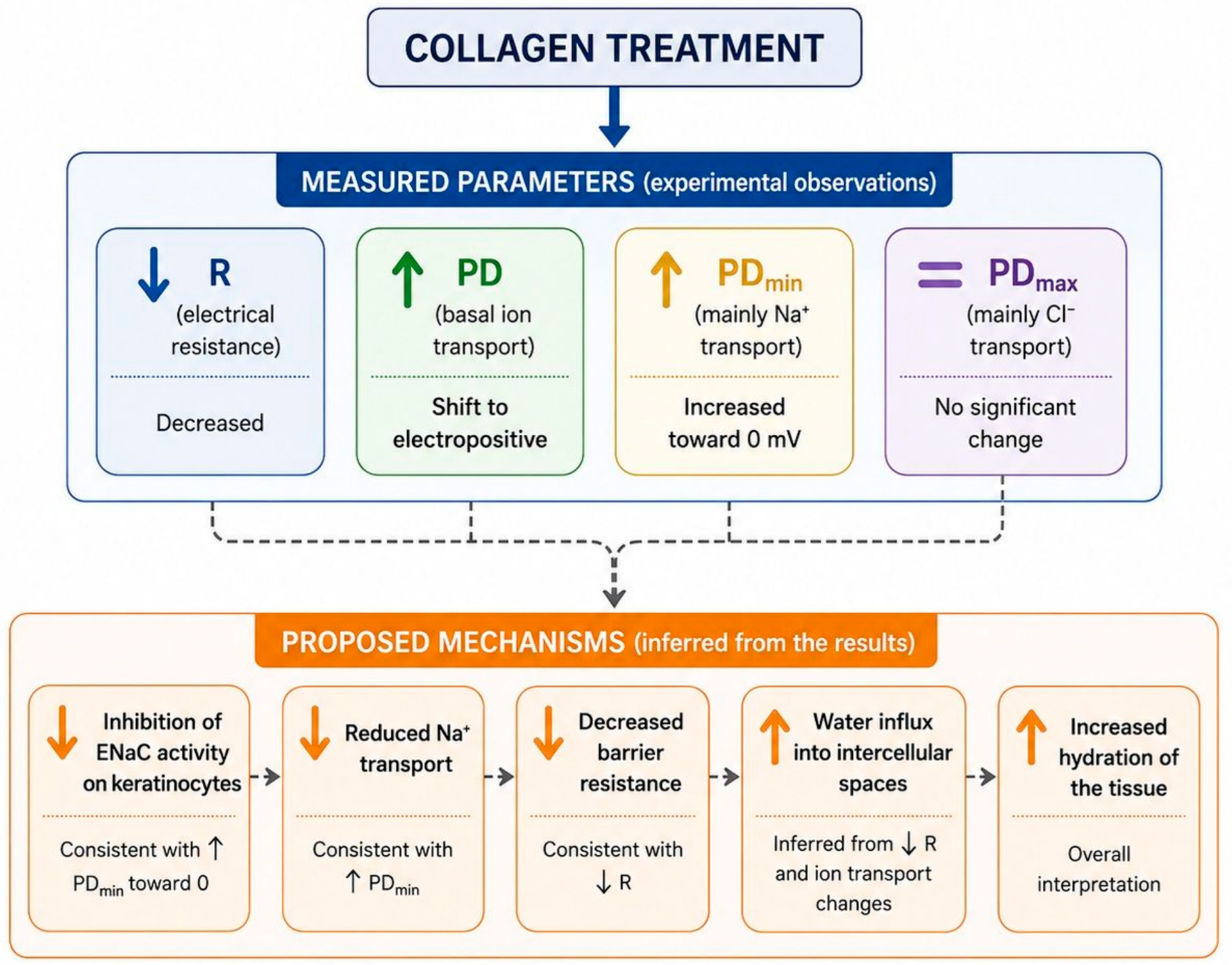
The electrophysiological effects of collagen gel application on the skin are summarized in Figure 1.
In collagen gel-treated preparations, PDmax values remained comparable to those measured in control tissues, regardless of the type of stimulation applied. In contrast, PDmin values increased toward electropositive values. This phenomenon may be explained, at least in part, by a putative inhibitory effect of collagen gel on sodium ion transport, which could be consistent with reports describing the low allergenic potential of collagen-containing preparations [1,7,18]. The application of the Ami solution elicited a response nonsignificantly modified to that observed in the control group, as also reported following the use of ointment with vitamin A [42]. Importantly, tissues treated with collagen gel retained the full ability to regulate the activation and inhibition of selected ion transport pathways, including those involving potassium ions. The collagen preparation had a mild effect and did not impair the physiological capacity of the skin to modulate ion transport pathways, which may be beneficial for users of collagen-based products. Furthermore, the applied collagen gel did not affect the ability of the tissue to respond to the stimulation used in the experiment.
The presented study has some limitations that should be explicated. One of the major limitations is the use of rabbit auricle skin as the experimental model. Although this model is well-suited for controlled electrophysiological studies, rabbit skin differs from human skin in structural organization, barrier properties, permeability, and potentially ion transport characteristics. Therefore, caution should be exercised when directly extrapolating these findings to human skin. Further studies using human skin models are needed to confirm the translational relevance of the observed effects. Another limitation is that multiple skin fragments originated from the same rabbits. Although fragments were randomly allocated to both treatment groups and treated independently ex vivo, potential within-animal correlations cannot be completely excluded because hierarchical statistical modeling could not be performed. While this allocation strategy reduced the potential influence of inter-animal variability on group comparisons, animal-level clustering may still have affected the estimated variability. Therefore, statistical significance should be interpreted with appropriate caution, and future studies with a larger number of animals should incorporate animal-level clustering into the statistical analysis. Additionally, a limitation of the present study is the lack of a vehicle-gel control. Therefore, the observed electrophysiological effects should be interpreted as those of the collagen preparation rather than of collagen alone, since the possible contributions of the gel matrix, hydration, occlusive properties, or minor formulation components cannot be completely excluded. Future studies incorporating a collagen-free vehicle with otherwise identical physicochemical properties will be required to determine the specific contribution of collagen.

5. Conclusions

In conclusion, the results of the present study indicate that the application of triple-helical collagen-containing gel preparation significantly affects the electrophysiological properties of skin tissue. Skin specimens treated with collagen gel remained responsive throughout the experiment, maintaining their ability to react to mechanical and mechanical–chemical stimulation.
Collagen gel treatment was associated with reduced transepithelial resistance and changes in ion transport-related parameters, suggesting altered tissue permeability and modulation of ion transport pathways. The observed changes in PDmin, together with the reduction in tissue resistance, may indicate that the mechanism of action of the collagen preparation may be associated with altered sodium transport in keratinocytes, potentially contributing to altered water distribution within intercellular spaces. However, the presented findings represent preliminary electrophysiological effects of the collagen gel and do not directly demonstrate ENaC inhibition, changes in skin hydration, or cosmetic efficacy of the preparation examined.
At the same time, collagen gel treatment did not significantly alter chloride ion transport-related parameters or the capacity of skin tissue to respond to stimulation, suggesting preserved electrophysiological responsiveness. These findings provide preliminary insight into the potential effects of collagen-based preparations on skin electrophysiology and support further studies investigating their potential role in modulating the skin microenvironment.

Author Contributions

D.D.-W. writing—original draft preparation, investigation, validation; A.K. writing—review and editing, investigation, conceptualization, data curation; O.Z. writing—review and editing, visualization; B.W. writing—review and editing, investigation; K.P. writing—review and editing, software; A.J. writing—review and editing, resources; K.S.-G. writing—review and editing, funding acquisition, project administration; I.H.-I. methodology, writing—review and editing, supervision, formal analysis. All authors have read and agreed to the published version of the manuscript.

Funding

This research received no external funding.

Institutional Review Board Statement

No experiments involving human participants were performed in the study. The presented experiment did not include living animals, and according to the Polish and European Union law, the bioethical committee agreement was not required. Animal care was in accordance with the guidelines and regulations stipulated by the Polish Animal Protection Act and the European Directive on the Protection of Animals Used for Scientific Purposes (2010/63/EU). All applicable institutional and national guidelines for the care and use of animals were followed (Consent for euthanizing animals no. 14/2016, date 20 October 2016, Nicolaus Copernicus University).

Informed Consent Statement

Not applicable.

Data Availability Statement

The data supporting the findings of this study are available from the corresponding author (I.H.-I.) upon reasonable request: igaholynska@cm.umk.pl.

Acknowledgments

ChatGPT was used to prepare figures according to the authors' instructions and to correct some English sentences. The authors reviewed and verified all generated content and take full responsibility for the final manuscript.

Conflicts of Interest

The authors declare no conflicts of interest.

Abbreviations

The following abbreviations are used in this manuscript:
AmiAmiloride 0.1 mM solution
BumeBumetanide 0.1 mM solution
CFTRCystic Fibrosis Transmembrane Regulator
ENaCEpithelial Sodium Channel
PDTransepithelial electric potential measured during stationary conditions (mV)
PDmaxMaximal transepithelial electric potential measured during 15-s stimulation (mV)
PDminMinimal transepithelial electric potential measured during 15-s stimulation (mV)
RResistance (Ω/cm2)
RSRinger solution

References

  1. Carvalho, A.M.; Marques, A.P.; Silva, T.H.; Reis, R.L. Evaluation of the Potential of Collagen from Codfish Skin as a Biomaterial for Biomedical Applications. Mar. Drugs 2018, 16, 495. [Google Scholar] [CrossRef] [Scilit] [PubMed]
  2. Gajbhiye, S.; Wairkar, S. Collagen fabricated delivery systems for wound healing: A new roadmap. Biomater. Adv. 2022, 142, 213152. [Google Scholar] [CrossRef] [Scilit] [PubMed]
  3. Jafari, H.; Lista, A.; Siekapen, M.M.; Ghaffari-Bohlouli, P.; Nie, L.; Alimoradi, H.; Shavandi, A. Fish Collagen: Extraction, Characterization, and Applications for Biomaterials Engineering. Polymers 2020, 12, 2230. [Google Scholar] [CrossRef] [Scilit] [PubMed]
  4. Mienaltowski, M.J.; Birk, D.E. Structure, physiology, and biochemistry of collagens. Adv. Exp. Med. Biol. 2014, 802, 5–29. [Google Scholar] [CrossRef] [Scilit] [PubMed]
  5. Rezvani Ghomi, E.; Nourbakhsh, N.; Akbari Kenari, M.; Zare, M.; Ramakrishna, S. Collagen-based biomaterials for biomedical applications. J. Biomed. Mater. Res. B Appl. Biomater. 2021, 109, 1986–1999. [Google Scholar] [CrossRef] [Scilit] [PubMed]
  6. Tanrikulu, I.C.; Westler, W.M.; Ellison, A.J.; Markley, J.L.; Raines, R.T. Templated Collagen “Double Helices” Maintain Their Structure. J. Am. Chem. Soc. 2020, 142, 1137–1141. [Google Scholar] [CrossRef] [Scilit] [PubMed]
  7. Sharma, S.; Rai, V.K.; Narang, R.K.; Markandeywar, T.S. Collagen-based formulations for wound healing: A literature review. Life Sci. 2022, 290, 120096. [Google Scholar] [CrossRef] [Scilit] [PubMed]
  8. Li, H.; Chen, R.; Jia, Z.; Wang, C.; Xu, Y.; Li, C.; Xia, H.; Meng, D. Porous fish collagen for cartilage tissue engineering. Am. J. Transl. Res. 2020, 12, 6107–6121. [Google Scholar] [PubMed]
  9. Meyer, M. Processing of collagen based biomaterials and the resulting materials properties. Biomed. Eng. OnLine 2019, 18, 24. [Google Scholar] [CrossRef] [Scilit] [PubMed]
  10. Liu, S.; Lau, C.S.; Liang, K.; Wen, F.; Teoh, S.H. Marine collagen scaffolds in tissue engineering. Curr. Opin. Biotechnol. 2022, 74, 92–103. [Google Scholar] [CrossRef] [Scilit] [PubMed]
  11. Lim, Y.S.; Ok, Y.J.; Hwang, S.Y.; Kwak, J.Y.; Yoon, S. Marine Collagen as A Promising Biomaterial for Biomedical Applications. Mar. Drugs 2019, 17, 467. [Google Scholar] [CrossRef] [Scilit] [PubMed]
  12. Peng, W.; Li, D.; Dai, K.; Wang, Y.; Song, P.; Li, H.; Tang, P.; Zhang, Z.; Li, Z.; Zhou, Y.; et al. Recent progress of collagen, chitosan, alginate and other hydrogels in skin repair and wound dressing applications. Int. J. Biol. Macromol. 2022, 208, 400–408. [Google Scholar] [CrossRef] [Scilit] [PubMed]
  13. Nilforoushzadeh, M.A.; Khodaverdi Darian, E.; Afzali, H.; Amirkhani, M.A.; Razzaghi, M.; Naser, R.; Amiri, A.B.; Alimohammadi, A.; Nikkhah, N.; Zare, S. Role of Cultured Skin Fibroblasts in Regenerative Dermatology. Aesthetic Plast. Surg. 2022, 46, 1463–1471. [Google Scholar] [CrossRef] [Scilit] [PubMed]
  14. León-López, A.; Morales-Peñaloza, A.; Martínez-Juárez, V.M.; Vargas-Torres, A.; Zeugolis, D.I.; Aguirre-Álvarez, G. Hydrolyzed Collagen-Sources and Applications. Molecules 2019, 24, 4031. [Google Scholar] [CrossRef] [Scilit] [PubMed]
  15. Venkatesan, J.; Anil, S.; Kim, S.K.; Shim, M.S. Marine Fish Proteins and Peptides for Cosmeceuticals: A Review. Mar. Drugs 2017, 15, 143. [Google Scholar] [CrossRef] [Scilit] [PubMed]
  16. Sionkowska, A.; Adamiak, K.; Musiał, K.; Gadomska, M. Collagen Based Materials in Cosmetic Applications: A Review. Materials 2020, 13, 4217. [Google Scholar] [CrossRef] [Scilit] [PubMed]
  17. Shenoy, M.; Abdul, N.S.; Qamar, Z.; Bahri, B.M.; Al Ghalayini, K.Z.; Kakti, A. Collagen Structure, Synthesis, and Its Applications: A Systematic Review. Cureus 2022, 14, e24856. [Google Scholar] [CrossRef] [Scilit] [PubMed]
  18. de Souza, A.; de Almeida Cruz, M.; de Araújo, T.A.; Parisi, J.R.; do Vale, G.C.; Dos Santos Jorge Sousa, K.; Ribeiro, D.A.; Granito, R.N.; Renno, A.C.M. Fish collagen for skin wound healing: A systematic review in experimental animal studies. Cell Tissue Res. 2022, 388, 489–502. [Google Scholar] [CrossRef] [Scilit] [PubMed]
  19. Pien, N.; Pezzoli, D.; Van Hoorick, J.; Copes, F.; Vansteenland, M.; Albu, M.; De Meulenaer, B.; Mantovani, D.; Van Vlierberghe, S.; Dubruel, P. Development of photo-crosslinkable collagen hydrogel building blocks for vascular tissue engineering applications: A superior alternative to methacrylated gelatin? Mater. Sci. Eng. C Mater. Biol. Appl. 2021, 130, 112460. [Google Scholar] [CrossRef] [Scilit] [PubMed]
  20. Yue, C.; Ding, C.; Du, X.; Wang, Y.; Su, J.; Cheng, B. Self-assembly of collagen fibrils on graphene oxide and their hybrid nanocomposite films. Int. J. Biol. Macromol. 2021, 193, 173–182. [Google Scholar] [CrossRef] [Scilit] [PubMed]
  21. Mejía-Calvo, I.; López-Juárez, L.E.; Vázquez-Leyva, S.; López-Morales, C.A.; Montoya-Escutia, D.; Merlos Rivera, P.G.; Herbert-Pucheta, J.E.; Zepeda-Vallejo, L.G.; Velasco-Velázquez, M.; Pavón, L.; et al. Quality attributes of partially hydrolyzed collagen in a liquid formulation used for skin care. J. Cosmet. Dermatol. 2021, 20, 150–158. [Google Scholar] [CrossRef] [Scilit] [PubMed]
  22. Aguirre-Cruz, G.; León-López, A.; Cruz-Gómez, V.; Jiménez-Alvarado, R.; Aguirre-Álvarez, G. Collagen Hydrolysates for Skin Protection: Oral Administration and Topical Formulation. Antioxidants 2020, 9, 181. [Google Scholar] [CrossRef] [Scilit] [PubMed]
  23. Wang, J.; Hu, J.; Yuan, X.; Li, Y.; Song, L.; Xu, F. Recombinant collagen hydrogels induced by disulfide bonds. J. Biomed. Mater. Res. Part A 2022, 110, 1774–1785. [Google Scholar] [CrossRef] [Scilit] [PubMed]
  24. Inoue, N.; Sugihara, F.; Wang, X. Ingestion of bioactive collagen hydrolysates enhance facial skin moisture and elasticity and reduce facial ageing signs in a randomised double-blind placebo-controlled clinical study. J. Sci. Food Agric. 2016, 96, 4077–4081. [Google Scholar] [CrossRef] [Scilit] [PubMed]
  25. Jia, Z.; Li, H.; Cao, R.; Xiao, K.; Lu, J.; Zhao, D.; Wang, Z.; Zhang, Y.; Chen, J.; Zhang, W.; et al. Electrospun nanofibrous membrane of fish collagen/polycaprolactone for cartilage regeneration. Am. J. Transl. Res. 2020, 12, 3754–3766. [Google Scholar] [CrossRef] [Scilit] [PubMed]
  26. Samiei, M.; Alipour, M.; Khezri, K.; Saadat, Y.R.; Forouhandeh, H.; Abdolahinia, E.D.; Vahed, S.Z.; Sharifi, S.; Dizaj, S.M. Application of Collagen and Mesenchymal Stem Cells in Regenerative Dentistry. Curr. Stem Cell Res. Ther. 2022, 17, 606–620. [Google Scholar] [CrossRef] [Scilit] [PubMed]
  27. Maia Campos, P.M.; Melo, M.O.; Siqueira César, F.C. Topical application and oral supplementation of peptides in the improvement of skin viscoelasticity and density. J. Cosmet. Dermatol. 2019, 8, 1693–1699. [Google Scholar] [CrossRef] [Scilit] [PubMed]
  28. Li, K.; Meng, F.; Li, Y.R.; Tian, Y.; Chen, H.; Jia, Q.; Cai, H.; Jiang, H.B. Application of Nonsurgical Modalities in Improving Facial Aging. Int. J. Dent. 2022, 2022, 8332631. [Google Scholar] [CrossRef] [Scilit] [PubMed]
  29. Rustad, A.M.; Nickles, M.A.; McKenney, J.E.; Bilimoria, S.N.; Lio, P.A. Myths and media in oral collagen supplementation for the skin, nails, and hair: A review. J. Cosmet. Dermatol. 2022, 21, 438–443. [Google Scholar] [CrossRef] [Scilit] [PubMed]
  30. Yazaki, M.; Yamada, M.; Goulas, S.; Teramoto, S.; Nakaya, M.-A.; Ohno, S.; Yamaguchi, K. Oral Ingestion of Collagen Hydrolysate Leads to the Transportation of Highly Concentrated Gly-Pro-Hyp and Its Hydrolyzed Form of Pro-Hyp into the Bloodstream and Skin. J. Agric. Food Chem. 2017, 65, 2315–2322. [Google Scholar] [CrossRef] [PubMed]
  31. Lin, P.; Hua, N.; Hsu, Y.; Kan, K. Oral Collagen Drink for Antiaging: Antioxidation, Facilitation of the Increase of Collagen Synthesis, and Improvement of Protein Folding and DNA Repair in Human Skin Fibroblasts. Oxid. Med. Cell. Longev. 2020, 2020, 8031795. [Google Scholar] [CrossRef] [Scilit] [PubMed]
  32. Lee, J.H.; Park, J.; Shin, D.W. The Molecular Mechanism of Polyphenols with Anti-Aging Activity in Aged Human Dermal Fibroblasts. Molecules 2022, 27, 4351. [Google Scholar] [CrossRef] [Scilit] [PubMed]
  33. de Bengy, A.F.; Lamartine, J.; Sigaudo-Roussel, D.; Fromy, B. Newborn and elderly skin: Two fragile skins at higher risk of pressure injury. Biol. Rev. Camb. Philos. Soc. 2022, 97, 874–895. [Google Scholar] [CrossRef] [Scilit] [PubMed]
  34. de Miranda, R.B.; Weimer, P.; Rossi, R.C. Effects of hydrolyzed collagen supplementation on skin aging: A systematic review and meta-analysis. Int. J. Dermatol. 2021, 60, 1449–1461. [Google Scholar] [CrossRef] [Scilit] [PubMed]
  35. Choi, E.; Joo, H.; Kim, M.; Kim, D.U.; Chung, H.C.; Kim, J.G. Low-Molecular-Weight Collagen Peptide Improves Skin Dehydration and Barrier Dysfunction in Human Dermal Fibrosis Cells and UVB-Exposed SKH-1 Hairless Mice. Int. J. Mol. Sci. 2025, 26, 6427. [Google Scholar] [CrossRef] [Scilit] [PubMed]
  36. Seong, S.H.; Lee, Y.I.; Lee, J.; Choi, S.; Kim, I.A.; Suk, J.; Jung, I.; Baeg, C.; Kim, J.; Oh, D.; et al. Low-molecular-weight collagen peptides supplement promotes a healthy skin: A randomized, double-blinded, placebo-controlled study. J. Cosmet. Dermatol. 2024, 23, 554–562. [Google Scholar] [CrossRef] [Scilit] [PubMed]
  37. Ferreira, D.M.; Silva, C.S.; Souza, M.N. Electrical impedance model for evaluation of skin irritation in rabbits and humans. Ski. Res. Technol. 2007, 13, 259–267. [Google Scholar] [CrossRef] [Scilit] [PubMed]
  38. Hanukoglu, I.; Boggula, V.R.; Vaknine, H.; Sharma, S.; Kleyman, T.; Hanukoglu, A. Expression of epithelial sodium channel (ENaC) and CFTR in the human epidermis and epidermal appendages. Histochem. Cell Biol. 2017, 147, 733–748. [Google Scholar] [CrossRef] [Scilit]
  39. Hołyńska-Iwan, I.; Szewczyk-Golec, K. Analysis of changes in sodium and chloride ion transport in the skin. Sci. Rep. 2020, 10, 18094. [Google Scholar] [CrossRef] [Scilit] [PubMed]
  40. Xu, W.; Hong, S.J.; Zeitchek, M.; Cooper, G.; Jia, S.; Xie, P.; Qureshi, H.A.; Zhong, A.; Porterfield, M.D.; Galiano, R.D.; et al. Hydration status regulates sodium flux and inflammatory pathways through epithelial sodium channel (ENaC) in the skin. J. Investig. Dermatol. 2015, 135, 796–806. [Google Scholar] [CrossRef] [Scilit]
  41. Zavyalova, O.; Dąbrowska-Wisłocka, D.; Misiura, K.; Hołyńska-Iwan, I. Chitosan-glycolic acid gel modification of chloride ion transport in mammalian skin: An in vitro study. Molecules 2023, 28, 6581. [Google Scholar] [CrossRef] [Scilit] [PubMed]
  42. Dłubała, K.; Wasiek, S.; Pilarska, P.; Szewczyk-Golec, K.; Mila-Kierzenkowska, C.; Łączkowski, K.; Sobiesiak, M.; Gackowski, M.; Tylkowski, B.; Hołyńska-Iwan, I. The influence of retinol ointment on rabbit skin (Oryctolagus cuniculus) ion transport—An in vitro study. Int. J. Mol. Sci. 2024, 25, 9670. [Google Scholar] [CrossRef] [Scilit] [PubMed]
  43. Sure, F.; Rapedius, M.; Diakov, A.; Bertog, M.; Obergrussberger, A.; Fertig, N.; Korbmacher, C.; Ilyaskin, A.V. Automated patch-clamp recordings for detecting activators and inhibitors of the epithelial sodium channel (ENaC). Pflugers Arch. 2025, 477, 857–872. [Google Scholar] [CrossRef] [Scilit]
  44. Chen, J.; Chen, Y.; Chen, Y.; Yang, Z.; You, B.; Ruan, Y.C.; Peng, Y. Epidermal CFTR Suppresses MAPK/NF-κB to Promote Cutaneous Wound Healing. Cell. Physiol. Biochem. 2016, 39, 2262–2274. [Google Scholar] [CrossRef] [Scilit] [PubMed]
  45. Wang, J.; Luo, J.; Huang, W.; Liu, C.; Zeng, D.; Liu, H.; Qu, X.; Liu, C.; Xiang, Y.; Qin, X. Increased intracellular Cl concentration by activating FAK promotes airway epithelial BEAS-2B cells proliferation and wound healing. Arch. Biochem. Biophys. 2020, 680, 108225. [Google Scholar] [CrossRef] [Scilit] [PubMed]
  46. Zhao, J.; Jia, S.; Xie, P.; Friedrich, E.; Galiano, R.D.; Qi, S.; Mao, R.; Mustoe, T.A.; Hong, S.J. Knockdown of sodium channel Nax reduces dermatitis symptoms in rabbit skin. Lab. Investig. 2020, 100, 751–761. [Google Scholar] [CrossRef] [Scilit] [PubMed]
  47. Kim, J.; Farahmand, M.; Dunn, C.; Milla, C.E.; Horii, R.I.; Thomas, E.A.; Moss, R.B.; Wine, J.J. Sweat rate analysis of ivacaftor potentiation of CFTR in non-CF adults. Sci. Rep. 2018, 8, 16233. [Google Scholar] [CrossRef] [Scilit] [PubMed]
  48. Xu, L.; Zhou, S.-L.; Bai, L.; Yu, C.-L.; Niu, N. Epithelial sodium channel in dendritic cells participates in high-fat-induced inflammatory responses. Mol. Biol. Rep. 2025, 52, 856. [Google Scholar] [CrossRef] [Scilit] [PubMed]
  49. Alsamad, F.; Stamatas, G. Directional assessment of the skin barrier function in vivo. Ski. Res. Technol. 2023, 29, e13346. [Google Scholar] [CrossRef] [Scilit] [PubMed]
  50. Abdayem, R.; Callejon, S.; Portes, P.; Kirilov, P.; Demame, F.; Pirot, F.; Jannin, V.; Haftek, M. Modulation of transepithelial electric resistance (TEER) in reconstructed human epidermis by excipients known to permeate intestinal tight junctions. Exp. Dermatol. 2015, 24, 686–691. [Google Scholar] [CrossRef] [Scilit] [PubMed]
  51. Barker, A.T.; Jaffe, L.F.; Vanable, J.W. The glabrous epidermis of cavies contains a powerfull baterry. Am. J. Physiol. 1982, 242, R358–R366. [Google Scholar] [CrossRef] [Scilit] [PubMed]
  52. Lin, X.; Chen, Y.; Jin, H.; Zhao, Q.; Liu, C.; Li, R.; Yu, F.; Chen, Y.; Huang, F.; Yang, Z.; et al. Collagen Extracted from Bigeye Tuna (Thunnus obesus) Skin by Isoelectric Precipitation: Physicochemical Properties, Proliferation, and Migration Activities. Mar. Drugs 2019, 17, 261. [Google Scholar] [CrossRef] [Scilit] [PubMed]
  53. Zoio, P.; Lopes-Ventura, S.; Marto, J.; Oliva, A. Open-Source Human Skin Model with an In Vivo-like Barrier for Drug Testing. Altex 2022, 39, 405–418. [Google Scholar] [CrossRef] [Scilit] [PubMed]
  54. Anderegg, U.; Halfter, N.; Schnabelrauch, M.; Hintze, V. Collagen/glycosaminoglycan-based matrices for controlling skin cell responses. Biol. Chem. 2021, 402, 1325–1335. [Google Scholar] [CrossRef] [Scilit] [PubMed]
Figure 1. Collagen gel-induced changes in electrophysiological parameters of epithelial tissue. Hypothetical mechanisms responsible for the observed changes are presented in parentheses. Solid lines and arrows (upper panel) indicate experimentally measured parameters. Dashed lines and arrows (lower panel) indicate inferred relationships not directly measured. Abbreviations: PD—transepithelial electrical potential, R—electrical resistance, PDmin—minimal transepithelial potential difference measured during a 15-sec stimulation of skin surface, PDmax—maximal transepithelial potential difference measured during a 15-sec stimulation of skin surface.
Cosmetics 13 00192 g001
Table 1. Values of transepithelial resistance (R) and transepithelial electric potential (PD) of the control skin specimens and samples exposed to collagen gel for 24 h.
Control (n = 30)Collagen Gel (n = 25)Mann–Whitney Test (p/r)
R [Ω·cm2]PD [mV]R [Ω·cm2]PD [mV]PD Control vs. Collagen GelR Control vs.
Collagen Gel
initialmedian11,779−0.2216750.410.00319/0.32479<0.001/0.623499
lower quartile5417−0.56705−0.55
upper quartile28,493041430.65
finalmedian12,907−0.3220010.450.01419/0.13959<0.001/0.618945
lower quartile6565−0.57707−0.56
upper quartile28,891−0.1243630.64
Wilcoxon test (p)PD initial vs. PD final 0.22047 0.07524
R initial vs. R final0.05875 0.45908
Abbreviations: Control—skin specimens incubated in Ringer solution (RS), Collagen gel—skin specimens treated with collagen gel 1 g/cm2 for 24 h and incubated in RS, p < 0.05, r—effect size according to Mann–Whitney results.
Table 2. Values of the minimal (PDmin) and maximal (PDmax) transepithelial electric potential measured during 15-sec stimulation of the control skin specimens and samples exposed to collagen gel for 24 h.
Control (n = 30)Collagen Gel (n = 25)
PDmax (mV)PDmin (mV)Wilcoxon Test PDmax vs. PDminPDmax (mV)PDmin (mV)Wilcoxon Test PDmax vs. PDmin
RSmedian0.88−0.5<0.0010.760<0.001
lower quartile0.21−1.070−0.79
upper quartile2.72−0.211.50.58
Bumemedian0.7−0.64<0.0010.730<0.001
lower quartile0.08−1.560.12−0.82
upper quartile2.2−0.391.190.52
Amimedian0.92−0.54<0.0010.790<0.001
lower quartile0.08−1.220.12−1.37
upper quartile1.82−0.281.680.34
Abbreviations: Control—skin specimens incubated in Ringer solution (RS), Collagen gel—skin specimens treated with Collagen gel 1 g/cm2 for 24 h and incubated in RS, Ami—amiloride (0.1 mmol/L) solution, Bume—bumetanide (0.1 mmol/L) solution, p < 0.05.
Table 3. Results of the Mann–Whitney U test of control skin specimens and samples exposed to collagen gel for 24 h.
Stimulation FluidPDminprPDmaxpr
RSControl vs.
Collagen gel
0.001270.33981Control vs.
Collagen gel
0.203810.283762
BumeControl vs.
Collagen gel
0.006200.38735Control vs.
Collagen gel
0.755120.042044
AmiControl vs.
Collagen gel
0.052050.26187Control vs.
Collagen gel
0.407310.002402
Abbreviations: Control—skin specimens incubated in Ringer solution (RS), Collagen gel—skin specimens treated with collagen gel 1 g/cm2 for 24 h and incubated in RS, Ami—amiloride (0.1 mmol/L) solution, Bume—bumetanide (0.1 mmol/L) solution, PDmin—minimal transepithelial potential difference measured during a 15-sec stimulation of skin surface (mV), PDmax—maximal transepithelial potential difference measured during a 15-sec stimulation of skin surface (mV), p < 0.05, r—effect size according to Mann–Whitney results.
Disclaimer/Publisher’s Note: The statements, opinions and data contained in all publications are solely those of the individual author(s) and contributor(s) and not of MDPI and/or the editor(s). MDPI and/or the editor(s) disclaim responsibility for any injury to people or property resulting from any ideas, methods, instructions or products referred to in the content.