Novel Vegan Exosome-like Biomimetic Vesicles for Skin and Hair Follicle Protection and Rejuvenation: Structural and Functional Characterization and Placebo-Controlled Clinical Efficacy Studies


Abstract
1. Introduction
2. Materials and Methods
2.1. Production Concept of the Vegan Biomimetic Exosomes
2.2. Physical and Structural Characterization
2.3. Lipidomic and Proteomic Analyses
2.4. Comparative Transcriptomic Assessment of Functional Biomimetism
2.5. Dermal Penetration Study Against Conventional Liposomes
2.6. Fibroblast Wound Healing Assay
2.7. Ex Vivo UV-A Photoaging Model and Vitamin C Comparison
2.8. Ex Vivo Human Hair Follicle Study
2.9. Placebo-Controlled Clinical Study
3. Results
3.1. In Vitro Results
3.1.1. Structural Biomimetism: Vesicle Architecture, Lipidomics, and Proteomics
3.1.2. Functional Biomimetism: Comparison with Human Fibroblast-Derived Exosomes in NHDFs
3.1.3. Penetration Study: Comparison with Conventional Liposomes
3.1.4. Wound Healing Activity in Fibroblasts
3.2. Ex Vivo Results
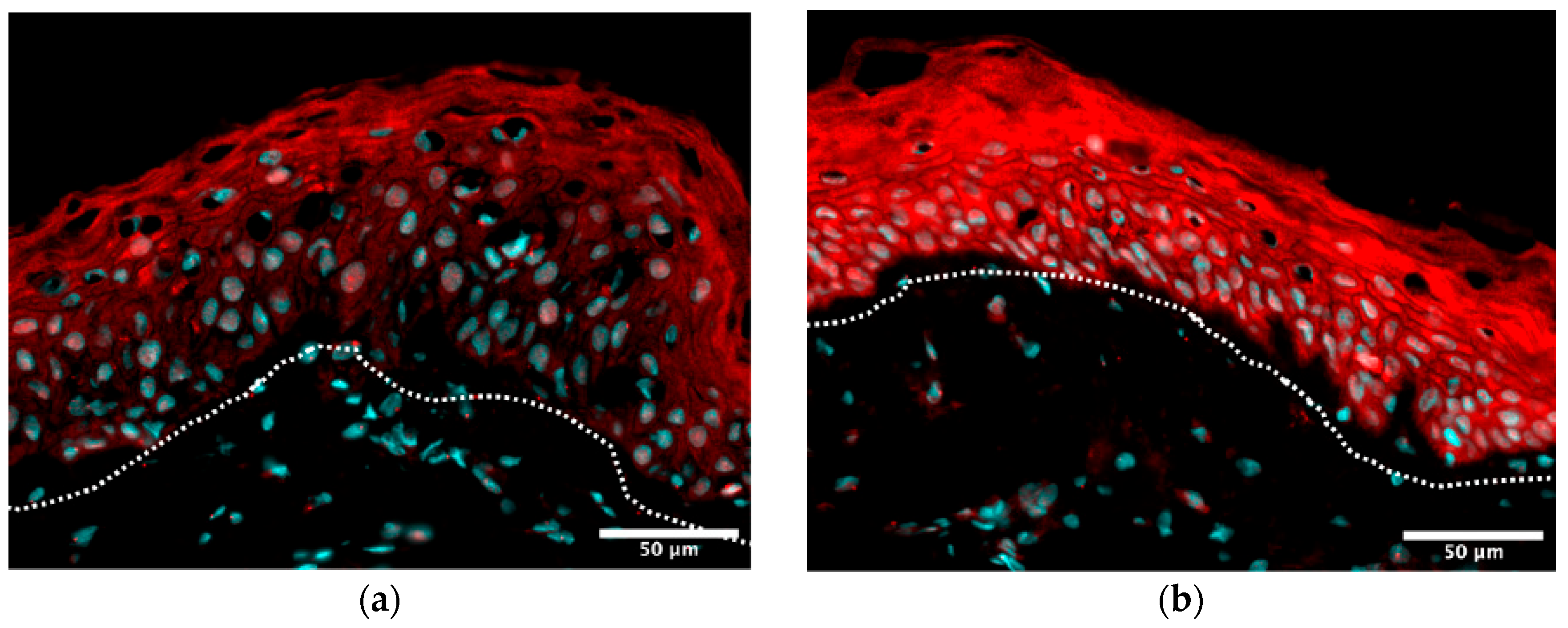
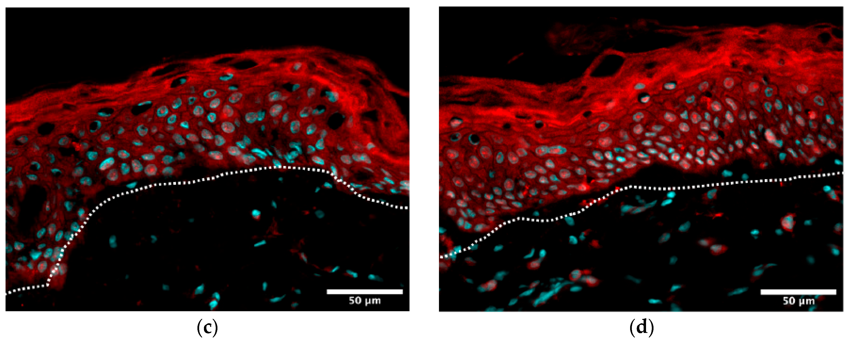
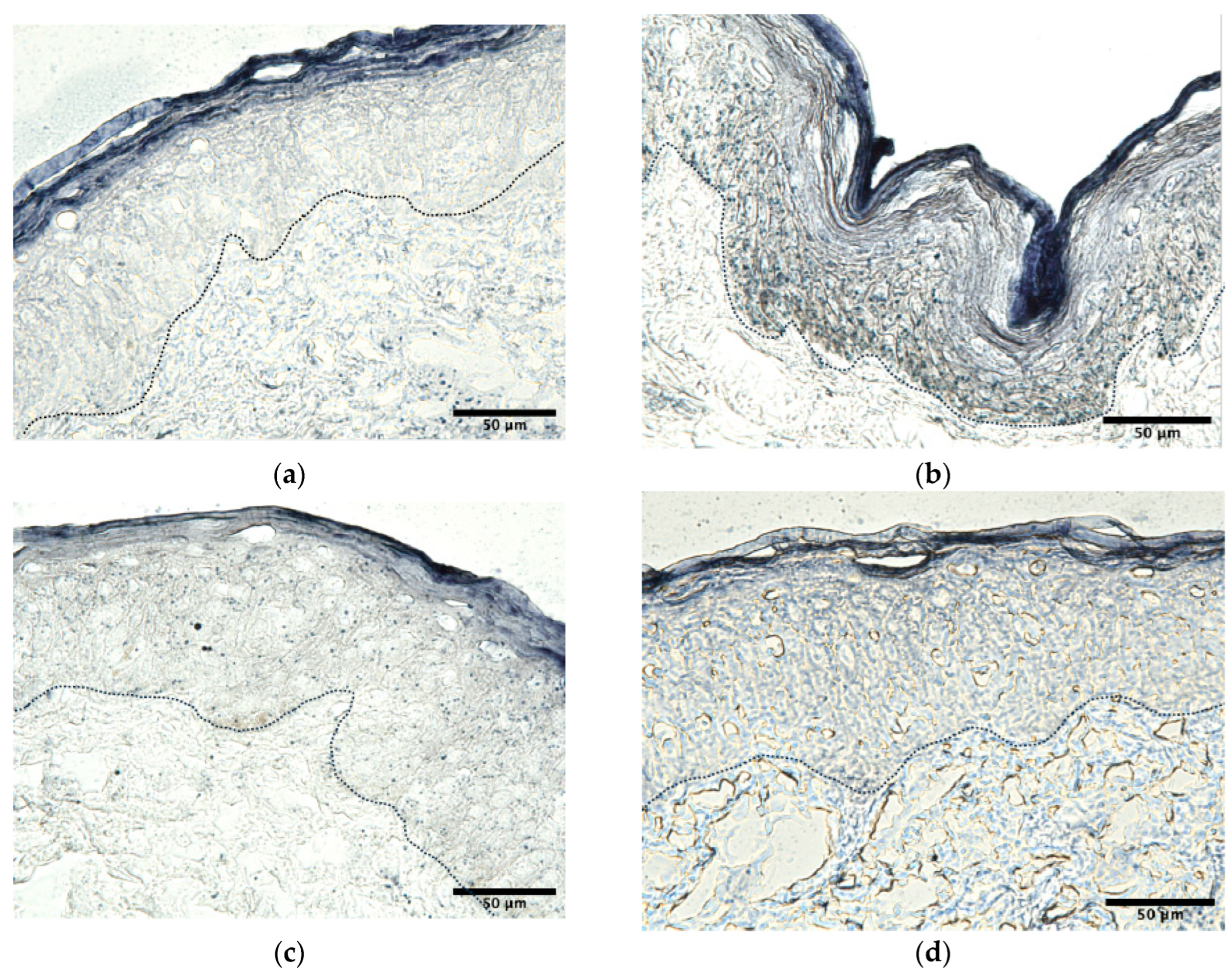
3.2.1. UV-A-Challenged Skin Explants: Antioxidant, Pigmentary, Matrix, and Barrier Protection
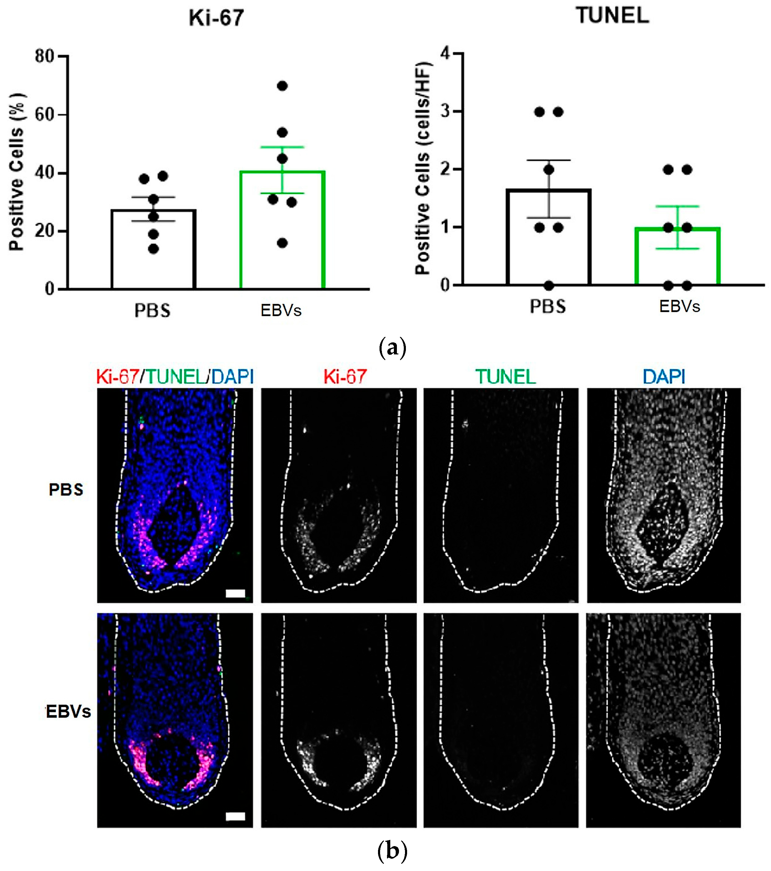
3.2.2. Ex Vivo Human Hair Follicles: Safety and Anagen-Supportive Activity
3.3. In Vivo Results
Placebo-Controlled 2% Cream Clinical Study
4. Discussion
5. Conclusions
6. Patents
- PCT Application: International application number PCT/EP2026/050007, filed on 2 January 2026.
- Chinese National Phase: Application number 2026100000733, filed on 5 January 2026.
Author Contributions
Funding
Institutional Review Board Statement
Informed Consent Statement
Data Availability Statement
Acknowledgments
Conflicts of Interest
References
- He, R.; Sun, M.; Liu, T.; Geng, L.; Xing, C.; Xu, P.; Peng, Y.; Fang, Y. Hallmarks of the Aging Skin Microenvironment: Components and Mechanisms. J. Cell. Physiol. 2025, 240, e70111. [Google Scholar] [CrossRef] [PubMed]
- Nan, L.; Guo, P.; Hui, W.; Xia, F.; Yi, C. Recent advances in dermal fibroblast senescence and skin aging: Unraveling mechanisms and pioneering therapeutic strategies. Front. Pharmacol. 2025, 16, 1592596. [Google Scholar] [CrossRef]
- López-Otín, C.; Blasco, M.A.; Partridge, L.; Serrano, M.; Kroemer, G. Hallmarks of aging: An expanding universe. Cell 2023, 186, 243–278. [Google Scholar] [CrossRef] [PubMed]
- Shin, S.H.; Lee, Y.H.; Rho, N.-K.; Park, K.Y. Skin aging from mechanisms to interventions: Focusing on dermal aging. Front. Physiol. 2023, 14, 1195272. [Google Scholar] [CrossRef] [PubMed]
- Bollag, W.B.; Aitkens, L.; White, J.; Hyndman, K.A. Aquaporin-3 in the epidermis: More than skin deep. Am. J. Physiol. Cell Physiol. 2020, 318, C1144–C1153. [Google Scholar] [CrossRef]
- Hara-Chikuma, M.; Verkman, A.S. Roles of aquaporin-3 in the epidermis. J. Investig. Dermatol. 2008, 128, 2145–2151. [Google Scholar] [CrossRef] [PubMed]
- Hajialiasgary Najafabadi, A.; Soheilifar, M.H.; Masoudi-Khoram, N. Exosomes in skin photoaging: Biological functions and therapeutic opportunity. Cell Commun. Signal. 2024, 22, 32. [Google Scholar] [CrossRef]
- Tienda-Vázquez, M.A.; Hanel, J.M.; Márquez-Arteaga, E.M.; Salgado-Álvarez, A.P.; Scheckhuber, C.Q.; Alanis-Gómez, J.R.; Espinoza-Silva, J.I.; Ramos-Kuri, M.; Hernández-Rosas, F.; Melchor-Martínez, E.M.; et al. Exosomes: A promising strategy for repair, regeneration and treatment of skin disorders. Cells 2023, 12, 1625. [Google Scholar] [CrossRef]
- Li, M.; Li, S.; Du, C.; Zhang, Y.; Li, Y.; Chu, L.; Han, X.; Galons, H.; Zhang, Y.; Sun, H.; et al. Exosomes from different cells: Characteristics, modifications, and therapeutic applications. Eur. J. Med. Chem. 2020, 207, 112784. [Google Scholar] [CrossRef]
- Sreeraj, H.; AnuKiruthika, R.; Tamilselvi, K.; Subha, D. Exosomes for skin treatment: Therapeutic and cosmetic application. Nano TransMed 2024, 3, 100048. [Google Scholar] [CrossRef]
- Kalluri, R.; LeBleu, V.S. The biology, function, and biomedical applications of exosomes. Science 2020, 367, eaau6977. [Google Scholar] [CrossRef]
- Welsh, J.A.; Goberdhan, D.C.I.; O’DRiscoll, L.; Buzas, E.I.; Blenkiron, C.; Bussolati, B.; Cai, H.; Di Vizio, D.; Driedonks, T.A.P.; Erdbrügger, U.; et al. Minimal information for studies of extracellular vesicles (MISEV2023): From basic to advanced approaches. J. Extracell. Vesicles 2024, 13, e12404, Erratum in J. Extracell. Vesicles 2024, 13, e12451. [Google Scholar] [CrossRef]
- Mathieu, M.; Martin-Jaular, L.; Lavieu, G.; Théry, C. Specificities of secretion and uptake of exosomes and other extracellular vesicles for cell-to-cell communication. Nat. Cell Biol. 2019, 21, 9–17. [Google Scholar] [CrossRef]
- van Niel, G.; D’Angelo, G.; Raposo, G. Shedding light on the cell biology of extracellular vesicles. Nat. Rev. Mol. Cell Biol. 2018, 19, 213–228. [Google Scholar] [CrossRef]
- Pegtel, D.M.; Gould, S.J. Exosomes. Annu. Rev. Biochem. 2019, 88, 487–514. [Google Scholar] [CrossRef] [PubMed]
- Yáñez-Mó, M.; Siljander, P.R.-M.; Andreu, Z.; Bedina Zavec, A.; Borràs, F.E.; Buzas, E.I.; Buzas, K.; Casal, E.; Cappello, F.; Carvalho, J.; et al. Biological properties of extracellular vesicles and their physiological functions. J. Extracell. Vesicles 2015, 4, 27066. [Google Scholar] [CrossRef]
- Bin Dayel, S.; Hussein, R.S. Exosomes in Dermatology: Emerging Roles in Skin Health and Disease. Pharmaceutics 2025, 17, 600. [Google Scholar] [CrossRef] [PubMed]
- Domaszewska-Szostek, A.; Krzyżanowska, M.; Polak, A.; Puzianowska-Kuźnicka, M. Effectiveness of extracellular vesicle application in skin aging treatment and regeneration: Do we have enough evidence from clinical trials? Int. J. Mol. Sci. 2025, 26, 2354. [Google Scholar] [CrossRef] [PubMed]
- Vyas, K.S.; Kaufman, J.; Munavalli, G.S.; Robertson, K.; Behfar, A.; Wyles, S.P. Exosomes: The latest in regenerative aesthetics. Regen. Med. 2023, 18, 181–194. [Google Scholar] [CrossRef]
- Mehta, R.C.; Smith, S.R.; Grove, G.L. Fibroblast-derived growth factors and exosomes as cosmeceuticals. Dermatol. Rev. 2023, 4, 250–259. [Google Scholar] [CrossRef]
- Syromiatnikova, V.; Prokopeva, A.; Gomzikova, M. Methods of the large-scale production of extracellular vesicles. Int. J. Mol. Sci. 2022, 23, 10522. [Google Scholar] [CrossRef]
- Ahn, S.H.; Ryu, S.W.; Choi, H.; You, S.; Park, J.; Choi, C. Manufacturing therapeutic exosomes: From bench to industry. Mol. Cells 2022, 45, 284–290. [Google Scholar] [CrossRef]
- Busatto, S.; Vilanilam, G.; Ticer, T.; Lin, W.-L.; Dickson, D.W.; Shapiro, S.; Bergese, P.; Wolfram, J. Tangential flow filtration for highly efficient concentration of extracellular vesicles from large volumes of fluid. Cells 2018, 7, 273. [Google Scholar] [CrossRef]
- Dilsiz, N. A comprehensive review on recent advances in exosome isolation and characterization: Toward clinical applications. Transl. Oncol. 2024, 50, 102121. [Google Scholar] [CrossRef]
- Jin, Z.; Na, J.; Lin, X.; Jiao, R.; Liu, X.; Huang, Y. Plant-derived exosome-like nanovesicles: A novel nanotool for disease therapy. Heliyon 2024, 10, e30630. [Google Scholar] [CrossRef]
- Cao, M.; Diao, N.; Cai, X.; Chen, X.; Xiao, Y.; Guo, C.; Chen, D.; Zhang, X. Plant exosome nanovesicles: Green delivery platforms. Mater. Horiz. 2023, 10, 3879–3894. [Google Scholar] [CrossRef]
- Kameli, N.; Dragojlovic-Kerkache, A.; Savelkoul, P.; Stassen, F.R. Plant-derived extracellular vesicles: Current findings, challenges, and future applications. Membranes 2021, 11, 411. [Google Scholar] [CrossRef] [PubMed]
- Mu, N.; Li, J.; Zeng, L.; You, J.; Li, R.; Qin, A.; Liu, X.; Yan, F.; Zhou, Z. Plant-derived exosome-like nanovesicles: Current progress and prospects. Int. J. Nanomed. 2023, 18, 4987–5009. [Google Scholar] [CrossRef] [PubMed]
- Bayat, F.; Afshar, A.; Baghban, N. Algal cells-derived extracellular vesicles: A review with special emphasis on their antimicrobial effects. Front. Microbiol. 2021, 12, 785716. [Google Scholar] [CrossRef] [PubMed]
- Picciotto, S.; Santonicola, P.; Paterna, A.; Rao, E.; Raccosta, S.; Romancino, D.P.; Noto, R.; Touzet, N.; Manno, M.; Di Schiavi, E.; et al. Extracellular vesicles from microalgae: Uptake studies in human cells and caenorhabditis elegans. Front. Bioeng. Biotechnol. 2022, 10, 830189. [Google Scholar] [CrossRef]
- Adamo, G.; Fierli, D.; Romancino, D.P.; Picciotto, S.; Barone, M.E.; Aranyos, A.; Božič, D.; Morsbach, S.; Raccosta, S.; Stanly, C.; et al. Nanoalgosomes: Introducing extracellular vesicles produced by microalgae. J. Extracell. Vesicles 2021, 10, e12081. [Google Scholar] [CrossRef] [PubMed]
- Shen, H.; Li, S.; Lin, L.; Wu, Q.; Dong, Z.; Xu, W. Advancements in plant-derived exosome-like vesicles: Versatile bioactive carriers for targeted drug delivery systems. J. Pharm. Anal. 2025, 15, 101300. [Google Scholar] [CrossRef] [PubMed]
- Choi, D.-S.; Kim, D.-K.; Kim, Y.-K.; Gho, Y.S. Proteomics of extracellular vesicles: Exosomes and ectosomes. Mass Spectrom. Rev. 2020, 39, 34–53. [Google Scholar] [CrossRef]
- Palomar-Alonso, N.; Lee, M.; Kim, M. Exosomes: Membrane-associated proteins, challenges and perspectives. Biochem. Biophys. Rep. 2023, 37, 101599. [Google Scholar] [CrossRef]
- Moll, P.; Ante, M.; Seitz, A.; Reda, T. QuantSeq 3′ mRNA sequencing for RNA quantification. Nat. Methods 2014, 11, i–iii. [Google Scholar] [CrossRef]
- Dobin, A.; Davis, C.A.; Schlesinger, F.; Drenkow, J.; Zaleski, C.; Jha, S.; Batut, P.; Chaisson, M.; Gingeras, T.R. STAR: Ultrafast universal RNA-seq aligner. Bioinformatics 2013, 29, 15–21. [Google Scholar] [CrossRef] [PubMed]
- Love, M.I.; Huber, W.; Anders, S. Moderated estimation of fold change and dispersion for RNA-seq data with DESeq2. Genome Biol. 2014, 15, 550. [Google Scholar] [CrossRef]
- Schneider, C.A.; Rasband, W.S.; Eliceiri, K.W. NIH Image to ImageJ: 25 years of image analysis. Nat. Methods 2012, 9, 671–675. [Google Scholar] [CrossRef]
- Purba, T.S.; Haslam, I.S.; Poblet, E.; Jiménez, F.; Gandarillas, A.; Izeta, A.; Paus, R. Human epithelial hair follicle stem cells and their progeny: Current state of knowledge, the widening gap in translational research and future challenges. Bioessays 2014, 36, 513–525. [Google Scholar] [CrossRef]
- Langan, E.A.; Philpott, M.P.; Kloepper, J.E.; Paus, R. Human hair follicle organ culture: Theory, application and perspectives. Exp. Dermatol. 2015, 24, 903–911. [Google Scholar] [CrossRef]
- Yang, C.C.; Cotsarelis, G. Review of hair follicle dermal cells. J. Dermatol. Sci. 2010, 57, 2–11. [Google Scholar] [CrossRef]
- Soma, T.; Tajima, M.; Kishimoto, J. Hair cycle-specific expression of versican in human hair follicles. J. Dermatol. Sci. 2005, 39, 147–154. [Google Scholar] [CrossRef]
- du Cros, D.L.; LeBaron, R.G.; Couchman, J.R. Association of versican with dermal matrices and its potential role in hair follicle development and cycling. J. Investig. Dermatol. 1995, 105, 426–431. [Google Scholar] [CrossRef]
- Vidal, V.P.; Chaboissier, M.-C.; Lützkendorf, S.; Cotsarelis, G.; Mill, P.; Hui, C.-C.; Ortonne, N.; Ortonne, J.-P.; Schedl, A. Sox9 is essential for outer root sheath differentiation and the formation of the hair stem cell compartment. Curr. Biol. 2005, 15, 1340–1351. [Google Scholar] [CrossRef]
- Qiu, W.; Lei, M.; Li, J.; Wang, N.; Lian, X. Activated hair follicle stem cells and Wnt/β-catenin signaling involve in pathnogenesis of sebaceous neoplasms. Int. J. Med. Sci. 2014, 11, 1022–1028. [Google Scholar] [CrossRef] [PubMed]
- Kishimoto, J.; Burgeson, R.E.; Morgan, B.A. Wnt signaling maintains the hair-inducing activity of the dermal papilla. Genes Dev. 2000, 14, 1181–1185. [Google Scholar] [CrossRef] [PubMed]
- Johnson, B.Z.; Stevenson, A.W.; Prêle, C.M.; Fear, M.W.; Wood, F.M. The role of IL-6 in skin fibrosis and cutaneous wound healing. Biomedicines 2020, 8, 101. [Google Scholar] [CrossRef]
- Jiang, W.G.; Sanders, A.J.; Ruge, F.; Harding, K.G. Influence of interleukin-8 (IL-8) and IL-8 receptors on the migration of human keratinocytes, the role of PLC-γ and potential clinical implications. Exp. Ther. Med. 2012, 3, 231–236. [Google Scholar] [CrossRef]
- Apte, A.; Liechty, K.W.; Zgheib, C. Immunomodulatory biomaterials on chemokine signaling in wound healing. Front. Pharmacol. 2023, 14, 1084948. [Google Scholar] [CrossRef] [PubMed]







| Attribute | Animal/Human Derived Exosomes | Natural Vegan EVs | Conventional Exosome-like Liposomes | EBVs |
|---|---|---|---|---|
| Ethical acceptability | Limited | High | High | High |
| Scalability | Low to moderate | Low | High | High |
| Batch consistency | Variable | Variable | High | High |
| True multi-component membrane complexity | High | Moderate to high | Low | High |
| Need for lecithin-driven artificial assembly | No | No | Yes | No |
| Communication- oriented biomimicry | High | Species specific | Low | High |
| Regulatory/cosmetic feasibility | Challenging | Moderate | High | High |
| Parameter | Result | Interpretation |
|---|---|---|
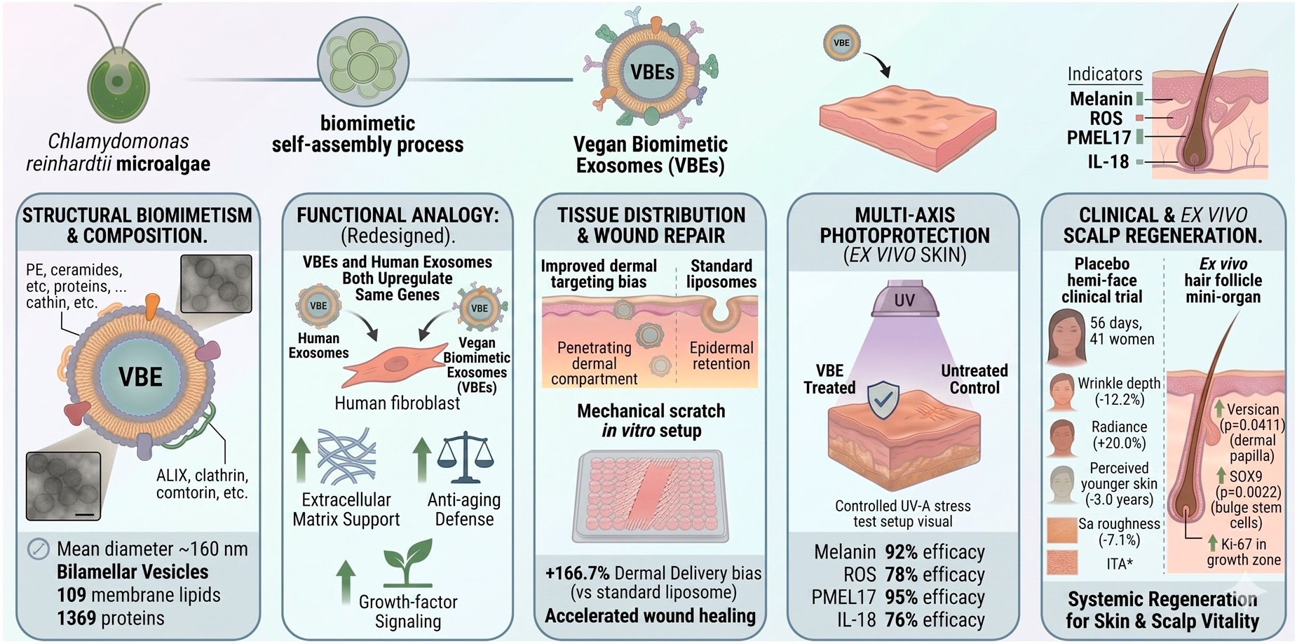
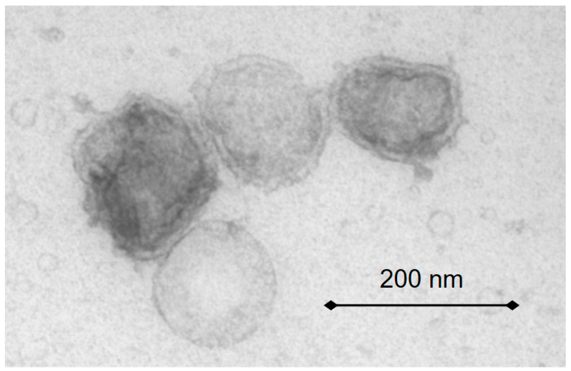
| Mean vesicle diameter | ~160 nm (final product) for C. reinhardtii-derived vesicles | Within exosome-relevant nanoscale range |
| Vesicle concentration | >10 billion vesicles/mL | Enables batch standardization and reproducible dosing |
| Morphology | Bilamellar vesicular architecture by TEM | Supports exosome-inspired membrane organization |
| Proteomic complexity | 1369 proteins; 1086 protein families | Far exceeds simplified carrier systems |
| Lipidomic complexity | 109 lipids across 23 classes | Supports membrane specialization and cell interaction |
| Representative membrane lipids | PE, PG, ceramides, CE, TG, N-acyl ethanolamines, and sphingolipids | Associated with curvature, rigidity, fusion, microdomains, and signaling |
| Representative proteins | FAS1, TM9SF, HSP60/70/90, clathrin, ALIX/BRO1, RAB1A/6/18, antioxidant enzymes, growth factor-like proteins, and DNA repair-related proteins | Align with exosomal adhesion, trafficking, stress adaptation, and biological cargo logic |
| Delivery System | Epidermal Fluorescence (a.u.) | Dermal Fluorescence (a.u.) | Dermis/Epidermis Ratio |
|---|---|---|---|
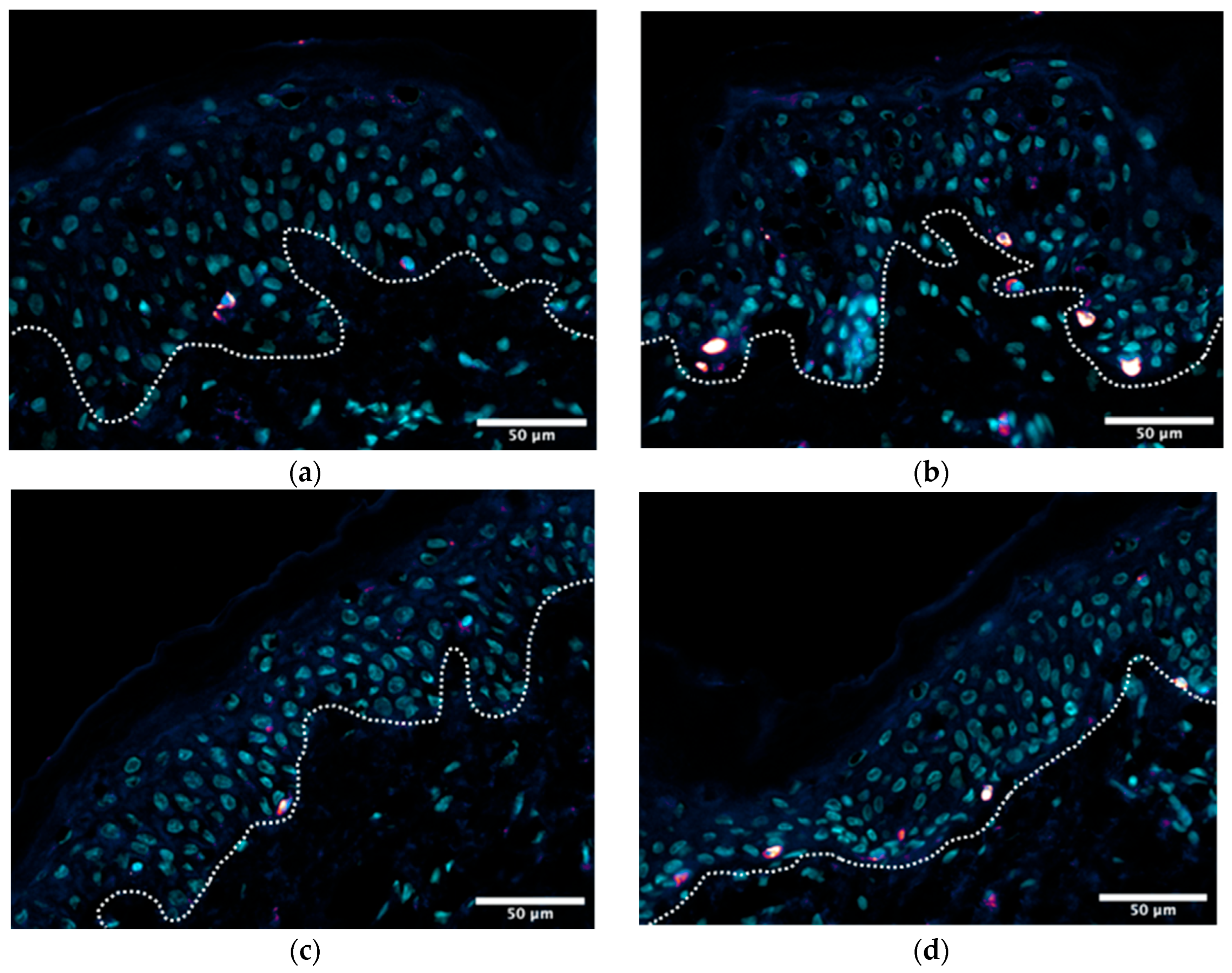
| EBVs | 7194.67 | 2320.67 | 0.32 |
| Conventional liposomes | 13,965.00 | 1656.33 | 0.12 |
| Time (h) | Control (% vs. T0) | EBVs 1.0% (% vs. T0) | p vs. Control (1.0%) |
|---|---|---|---|
| 2 | −3.7 | −12.8 | 0.034246 |
| 4 | −3.6 | −15.7 | 0.044286 |
| 6 | −7.5 | −18.4 | 0.211 |
| 8 | −17.4 | −30.4 | 0.0131 |
| Biomarker | Stress | 2% EBVs + Stress | 5% Vit C + Stress | 2.5% Vit C + 1% EBVs + Stress |
|---|---|---|---|---|
| Melanin | 298.0% | 116.7% (92% efficacy ***) | 113.5% (93% efficacy ***) | 71.7% (100% efficacy ***) |
| ROS | 196.5% | 121.1% (78% efficacy ***) | 119.1% (80% efficacy ***) | 104.2% (96% efficacy ***) |
| PMEL17 | 159.8% | 103.1% (95% efficacy ***) | 119.1% (68% efficacy ***) | 102.3% (96% efficacy ***) |
| IL-18 | 183.2% | 120.2% (76% efficacy ***) | 134.1% (59% efficacy ***) | 106.0% (93% efficacy ***) |
| Lipofuscin | 132.7% | 115.8% (52% efficacy ***) | 103.9% (88% efficacy ***) | 103.0% (91% efficacy ***) |
| Readout | Outcome with EBVs | Interpretation |
|---|---|---|
| LDH release | No overt increase over 7 days | No detectable cytotoxicity in cultured follicles |
| Amputated follicle elongation | Higher growth tendency vs. PBS on days 3–7 | Supports growth-promoting activity |
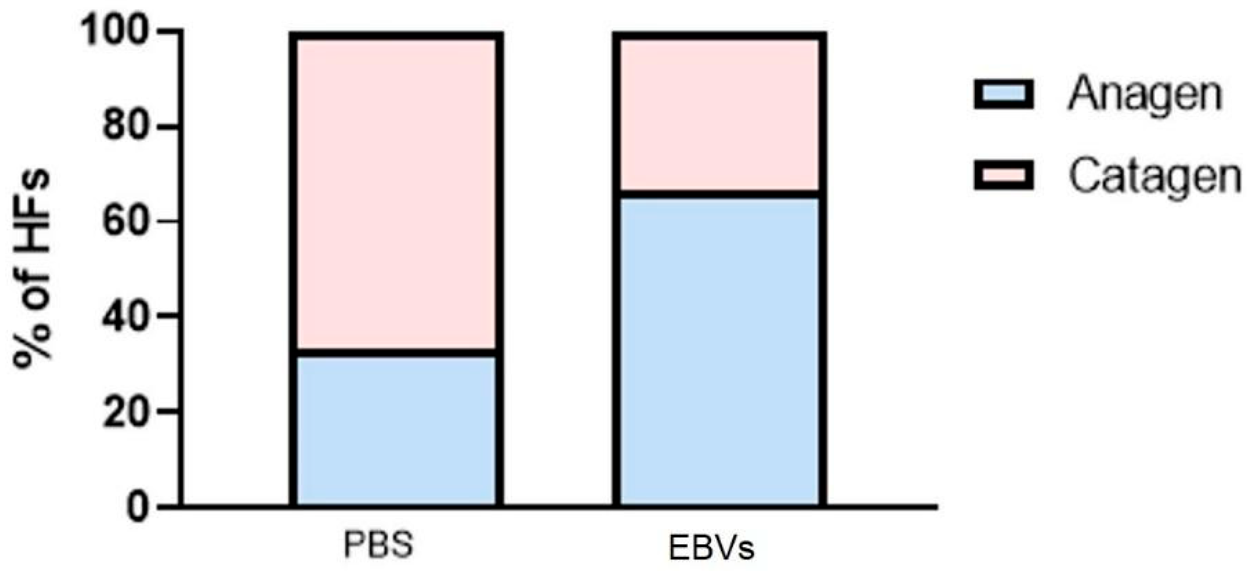
| Macroscopic hair cycle staging | More follicles remained in anagen at end of culture | Anagen-supportive effect |
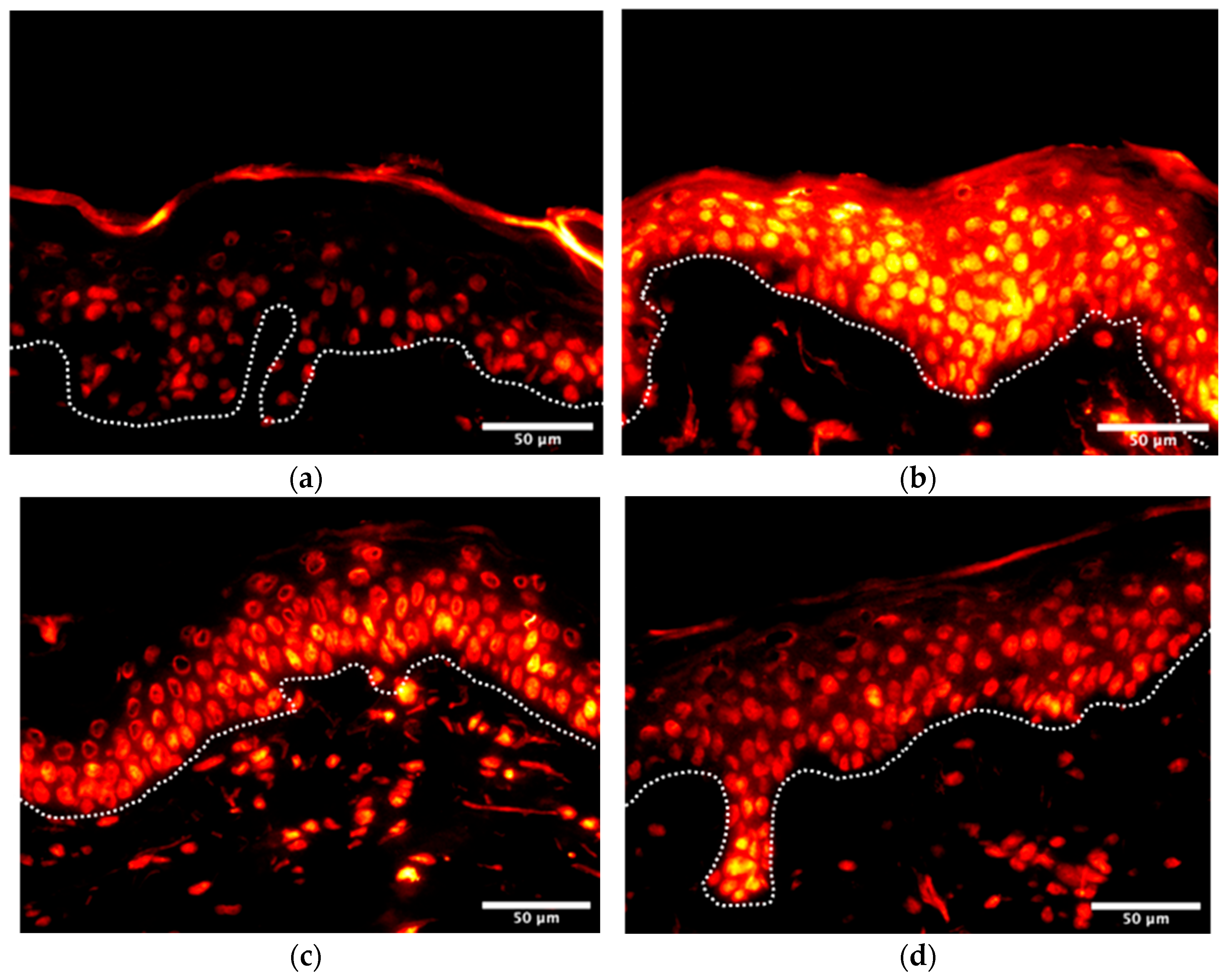
| Ki-67-positive germinative matrix cells | 41.0% vs. 27.7% in control | Higher proliferative activity in growth zone |
| TUNEL staining | Only a few positive cells in both groups | No evidence of damage-driven pseudo-effect |
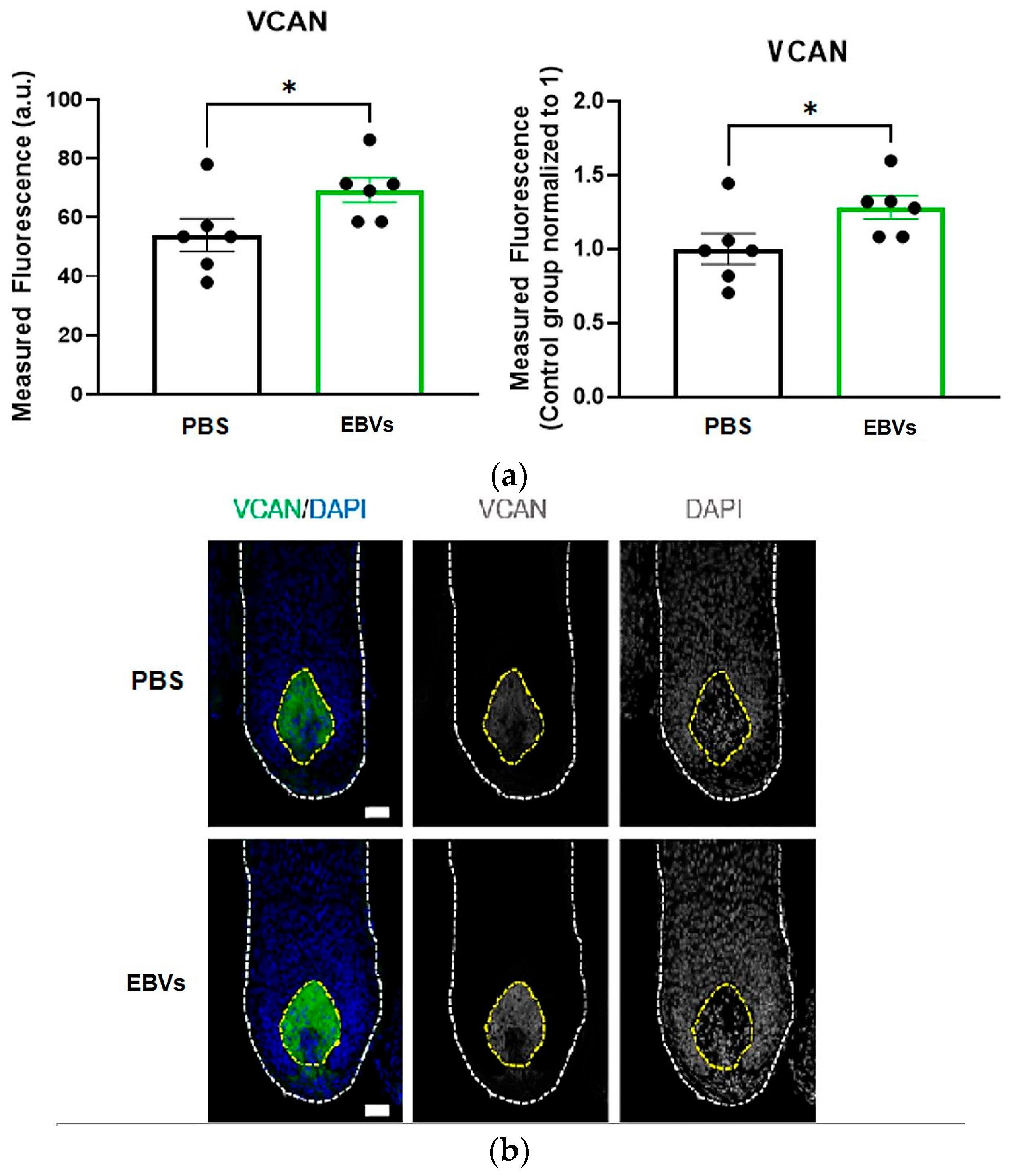
| Versican (VCAN) in dermal papilla | Higher in EBV group; p = 0.0411 | Supports inductive/anagen dermal papilla state |
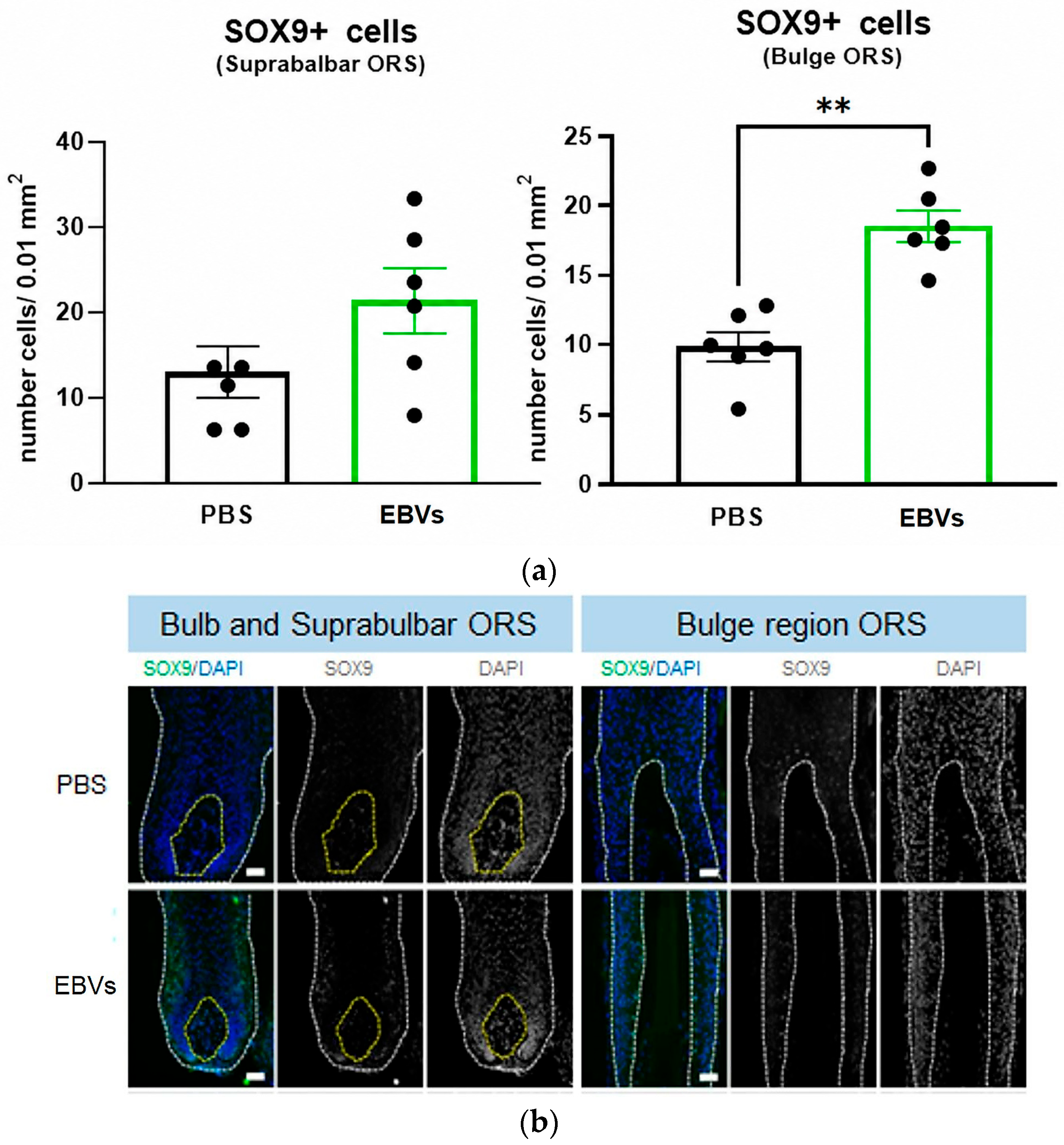
| SOX9-positive bulge ORS cells | Higher in EBV group; p = 0.0022 | Supports progenitor/stem-associated follicular compartment |
| Endpoint | Day 14 | Day 28 | Day 56 | Placebo Summary |
|---|---|---|---|---|
| Wrinkle depth | −9.0% *** | −11.5% *** | −12.2% *** | No meaningful reduction (−1.6% at day 56); active globally superior |
| Sa roughness/smoothness | −3.9% * | −4.6% * | −7.1% *** | Minimal change (−1.0% at day 56) |
| Perceived younger skin | −2.2 years | −2.8 years | −3.0 years | About −0.5 to −0.6 years (at day 56) |
| Overall skin ITA° | +4.5% ** | +8.0% *** | +10.3% *** | +2.6% only at day 56 |
| Dark spot ITA° | +8.7% *** | +18.9% *** | +22.8% *** | +4.3% at day 28; +6.9% at day 56 |
| Elasticity (R2) | +2.0% ** | +3.7% *** | +4.9% *** | +1.4% at day 56 |
| Firmness (R0) | −3.3% *** | −4.6% *** | −7.4% *** | −1.0%, −1.4%, −2.9% |
| Radiance (gloss) | +9.3% *** | +16.1% *** | +20.0% *** | +2.7%, +3.8%, +4.1% |
| Reshaping/skin sagging | n.d. | −0.160 mm *** | −0.336 mm *** | Smaller effect (+0.054 mm at day 56) |
| Clinical smoothness responders | n.s. | 58.5% | 70.7% | Significant but not clinically relevant (31.7% at day 56) |
Disclaimer/Publisher’s Note: The statements, opinions and data contained in all publications are solely those of the individual author(s) and contributor(s) and not of MDPI and/or the editor(s). MDPI and/or the editor(s) disclaim responsibility for any injury to people or property resulting from any ideas, methods, instructions or products referred to in the content. |
© 2026 by the authors. Licensee MDPI, Basel, Switzerland. This article is an open access article distributed under the terms and conditions of the Creative Commons Attribution (CC BY) license.
Share and Cite
García-Delgado, N.; Lapeyre, A.; Ayats, J. Novel Vegan Exosome-like Biomimetic Vesicles for Skin and Hair Follicle Protection and Rejuvenation: Structural and Functional Characterization and Placebo-Controlled Clinical Efficacy Studies. Cosmetics 2026, 13, 120. https://doi.org/10.3390/cosmetics13030120
García-Delgado N, Lapeyre A, Ayats J. Novel Vegan Exosome-like Biomimetic Vesicles for Skin and Hair Follicle Protection and Rejuvenation: Structural and Functional Characterization and Placebo-Controlled Clinical Efficacy Studies. Cosmetics. 2026; 13(3):120. https://doi.org/10.3390/cosmetics13030120
Chicago/Turabian StyleGarcía-Delgado, Noemí, Alexandre Lapeyre, and Jordi Ayats. 2026. "Novel Vegan Exosome-like Biomimetic Vesicles for Skin and Hair Follicle Protection and Rejuvenation: Structural and Functional Characterization and Placebo-Controlled Clinical Efficacy Studies" Cosmetics 13, no. 3: 120. https://doi.org/10.3390/cosmetics13030120
APA StyleGarcía-Delgado, N., Lapeyre, A., & Ayats, J. (2026). Novel Vegan Exosome-like Biomimetic Vesicles for Skin and Hair Follicle Protection and Rejuvenation: Structural and Functional Characterization and Placebo-Controlled Clinical Efficacy Studies. Cosmetics, 13(3), 120. https://doi.org/10.3390/cosmetics13030120