Abstract
Photocatalytic technologies represent an innovative method to reduce microbial load on surfaces, even considering recent public health emergencies involving coronaviruses and other microorganisms, whose presence has been detected on surfaces. In this review paper, the antimicrobial efficacy of various photocatalysts applied by different coating methods on different surfaces has been compared and critically discussed. Publications reviewing the use of photocatalytic coatings on surfaces for antimicrobial effectiveness have been examined. Clear search parameters were employed to analyze the PubMed, Scopus, and WOS databases, resulting in 45 papers published between 2006 to 2023 that met the inclusion criteria. The paper assessed various types of photocatalytic coatings that targeted different microbial objectives. Based on the pooled data analysis, the TiO2 coating exhibited a substantial effect in decreasing bacteria strains, both Gram-positive and -negative (99.4%). Although the diversity of these technologies poses significant obstacles to obtaining a comprehensive final assessment of their effectiveness and feasibility for surface application, subgroup analysis indicated significant variations in the removal efficiency of Gram-positive strains based on different surface types (p = 0.005) and time of exposure (p = 0.05). Photocatalytic coatings provide a promising approach to combating the spread of microorganisms on surfaces. Further “in-field” investigations are necessary in the foreseeable future to explore and optimize this novel and exciting health technology.
1. Introduction
Ensuring human health is the primary challenge of the twenty-first century. The COVID-19 pandemic has brought to light how health threats can spread rapidly on a global scale. One of the ways infectious diseases can spread is through “indirect contact” or “fomite” exposure. Contaminated surfaces have the potential to transfer pathogens to the mucous membranes of individuals, thereby making them more vulnerable to infections [1]. Indirect contact can play an important role in the spread of respiratory diseases [2,3,4]. More resistant pathogens have a higher likelihood of spreading through the air or staying on surfaces until they meet susceptible individuals [5]. The risk of transmission through indirect contact depends on various factors, including the surface type [6,7].
Traditional disinfection methods typically involve using chemicals, ultraviolet radiation (UV), or other physical treatments to reduce the microbial load and lower the infective dose. Nevertheless, some bacteria can form biofilms and become resistant to these disinfection methods, making it more challenging to eliminate them [8,9]. Biofilms are present in healthcare facilities and are not easily eliminated by disinfectants. Indeed, biofilms are a breeding ground for pathogens, including multi-drug resistant organisms, and they are linked to healthcare-associated infections (HAIs) [10]. Biofilms can form themselves to various surfaces such as metals, plastics, or tissues. Their growth on medical devices and implants, such as heart valves, pacemakers, vascular grafts, catheters, prosthetic joints, intrauterine devices, sutures, and contact lenses, is a significant concern because it can lead to infections. There are several types of biofilms in healthcare settings, including hydrated biofilms and dry surface biofilms, and these cannot be treated in the same way. The inability to find an adequate technique significantly increases the disease burden on patients and healthcare systems. Thus, it is essential to advance innovative methods to combat the expansion of biofilms [11].
Several technologies, such as hydrogen peroxide steam, UV light, and heavy metal-coated surfaces (copper and silver), have been proven to be effective for disinfecting environmental surfaces. Researchers have been investigating designing surfaces with bactericidal or bacteriostatic activities for several years [12]. Various strategies have been used to combat pathogens, including surface coatings with antibiotics, biocides, metals, enzymes, and organic compounds [13]. The use of photocatalysts to coat surfaces, bestowing antimicrobial properties, is becoming increasingly useful. During the photocatalysis process, the interaction of light with semiconductors results in the formation of highly reactive oxygen species (ROS) such as hydrogen peroxide, singlet oxygen, superoxide radical anions, and hydroxyl radicals [14,15,16,17]. During this process, ROS act as antimicrobial agents, causing serious damage to nucleic acids, lipids, and proteins and inhibiting or exterminating microorganisms and pathogens. The field of nanobiotechnology has advanced significantly in recent years, allowing for the synthesis of nanomaterials with specific shapes and sizes. This has greatly improved the effectiveness of antimicrobial materials. Nanoparticles are particularly effective for antibacterial activity due to their unique chemical and physical properties, large surface areas, high heat stability and resistance, and broad-spectrum antibacterial activities [14,15]. Current research is concentrating on developing nanostructured surfaces for disinfection using photocatalytic materials and visible light. Recently, new strategies were proposed to overcome the limits of photocatalysts, such as the need to use high-energy UV light, looking toward using visible light-driven photocatalysts [18,19,20]. The ideal material or coating should be activated under artificial light conditions, especially considering the application in a hospital setting [18,19,20,21,22].
Based on the typologies of the coating process, the antimicrobial surfaces can be classified as passive, reducing the adhesion of microorganisms, or active, killing microorganisms upon contact. Passive or active surfaces can have several proprieties such as super-wettability, super-hydrophobicity, superoleophobicity, and omniphobicity [21]. Several technologies have been achieved to immobilize photocatalysts onto surfaces [22]. The synthesis of nanostructured materials can be realized by approaches such as sol-gel routes, hydrothermal and solvothermal methods, vapor- or plasma-assisted methods, or deposition of pre-synthesized nanostructured materials exploiting a wet-chemical process such as impregnation, dip, or spin coating. Each synthesis process can have advantages and disadvantages, and recent reviews have underlined, through a descriptive approach, the several applications to contrast microbial loads and future thoughts in hospital settings through descriptive approaches [23,24,25,26].
The purpose of the present systematic review and meta-analysis was to explore the antimicrobial effectiveness of several photocatalytic coatings on different surfaces, analyzing the data coming from the available literature on this topic through a quantitative approach and showing perspectives for the future.
2. Materials and Methods
2.1. Study Design and Strategy of Search
The Preferred Reporting Items for Systematic Review and Meta-Analysis (PRISMA) guidelines were used to identify eligible articles to explore the antimicrobial effectiveness of photocatalytic coatings on surfaces [27]. The search strategy has been registered in PROSPERO (reference number CRD42023449501).
Relevant literature on this theme was collected through a systematic search of three electronic databases (PubMed, Scopus and Web of Science) that were interrogated using the following terms: (“(antimicrobial or antibacterial)” AND “surfaces” AND “photocatalysis” AND “coating”). A search was conducted on three databases using different search criteria such as title, abstract, MeSH terms, and keywords. The period considered for the article collection was extensive to obtain a total overview of the topic (from June 2000 to 31 July 2023). The reference lists of each article were also checked to find additional relevant citations.
2.2. Inclusion and Exclusion Criteria
This review only considered studies that were based on the English language; analytic study designs; and “in vivo”, “in vitro”, and “in field” studies. Studies such as clinical trials, reviews, meta-analyses, case studies, case reports, proceedings, qualitative studies, editorials, commentary studies, studies without a control group, studies with incomplete designs (such as ecological studies), and any other types of study were excluded from the database. Extracted data from the three databases, such as titles and abstracts, were transferred to the site Covidence—Better systematic review management [28] for the relevance assessment process. The process of selecting studies involved a several-step exclusion process, involving four reviewers who independently investigated the titles and abstracts of the studies. During this multi-step exclusion process, reviewer consensus was obtained. Titles and abstracts acquired from the three databases were transferred to the reference site Covidence—Better systematic review management for the relevance assessment process. The next step was screening by title and abstracts the potentially eligible studies, following the inclusion criteria stated above; the screening was conducted by 4 authors (F.V., F.U., V.V., and G.L.) independently. Then, full texts were read independently by the 4 authors (F.V., F.U., V.V., and G.L.) with a later discussion about their inclusion in the review. Disagreements were achieved by consensus among the authors. We included articles from the inception to July 2023. The review process is represented in Figure 1 (PRISMA flow diagram of the systematic review process).
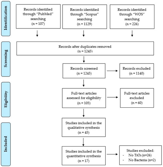
Figure 1.
PRISMA flow diagram of the systematic review process.
2.3. Data Synthesis
We used Comprehensive Meta-Analysis (CMA) software v.4 (Biostat Inc., Englewood, NJ, USA) to combine data. Our goal was to compare the effectiveness of a functionalized surface coated with TiO2 against different bacterial strains (negative and positive Gram strains). To do this, we collected information on the rate of bacterial reduction, the wavelength of the light source, the time of exposure, the type of surfaces, and the method of coating. We calculated the eradication rates in both the case and control groups, as well as any side effects, and reported them as an event rate. The 95% confidence interval (95% CI) was also calculated. Hedges’ g standardized mean difference statistic was used to calculate fixed and random effects model estimates. To evaluate statistically significant heterogeneity, we used the I2 (percentage of variation reflecting true heterogeneity), τ2 (random-effects between study variance), and p-value from Cochran’s Q test. When there was good homogeneity amongst the studies included (I2 < 50%, p > 0.1), we employed the fixed effects model. Conversely, the random effects model was used in cases where the studies included shown significant heterogeneity (I2 ≥ 50%, p ≤ 0.1). To perform a sensitivity analysis, the effects model was altered, or individual studies were excluded. Funnel plots were also utilized to explore potential publication bias. Meta-regression and subgroup analyses were performed to explore the sources of heterogeneity expected [29,30,31]. For meta-regression analysis, the wavelength, the time of exposition, the type of surfaces, and the method of coating of the studies were considered.
3. Results and Discussion
3.1. Articles Selection
There has been a growing interest in using photocatalytic coatings to eliminate microorganisms, and this is well-reflected in scientific literature. Research related to the combination of “antimicrobial or antibacterial” properties, “surfaces”, “photocatalysis”, and “coatings” has increased exponentially in recent decades, as can be seen in Figure 2A. Additionally, bibliometric analysis of the literature shows that a significant number of researchers are actively studying this subject in more countries around the world (Figure 2B).
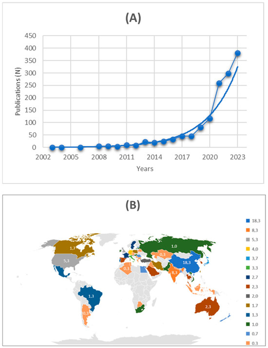
Figure 2.
Graphics reporting the bibliometric analysis of literature. (A) The trend in the number of publications per year in the total of 1462 records found (date of search: from the inception to databases and July 2023) using the following combinations of topic keywords: “antimicrobial or antibacterial” properties, “surfaces”, “photocatalysis”, and “coatings”. (B) The percentage of distribution of research in the countries of the world (using Bing Technologies and sources of data: Australian Bureau of Statistics, GeoNames, Geospatial Data Edit, Microsoft, Naviinfo, Open Places, OpenStreetMap, TomTom, Wikipedia, and Zenrin).
It is important to note that regions with lower research activity on this issue overlap with those that should prioritize antimicrobial resistance surveillance, such as Sub-Saharan Africa [32].
A total of 1462 records were found, and, after screening, 1245 were included, and 105 were assessed for eligibility. In total, 5 papers were excluded because they did not include any control group, 24 articles because they considered textile surfaces, 5 articles did not use any light source, and 26 articles because they were not pertinent. Finally, 45 articles met the inclusion criteria and were included in the qualitative synthesis [33,34,35,36,37,38,39,40,41,42,43,44,45,46,47,48,49,50,51,52,53,54,55,56,57,58,59,60,61,62,63,64,65,66,67,68,69,70,71,72,73,74,75,76,77]. For each article, the following data were reported: author, year, country, type of surface, type of photocatalyst, dose of photocatalyst, type of coating method, details of coating method, and main results (Table 1); author, year, country, microbial target, initial CFU (Colony Forming Units), microbial reduction, light source, wavelength of light, distance of light source from surface, characteristics of light source, time of light exposition, and test for evaluation of antimicrobial activity (Table 2).

Table 1.
A summary of the key findings and main features of the studies that were included in the systematic review.
Table 2.
Details of the microbial activity of the papers considered in the review.
3.2. Characteristics of the Selected Studies
The 45 articles included in the systematic review were published between 2006 [33] and 2023 [34,35,36,37], showing a positive growth trend: one article was published in 2006 [33], two in 2007 [38,39], three in 2008 [40,41,42], two in 2009 [43,44], one in 2010 [45], two in 2011 [46,47], one in 2012 [48], three in 2013 [49,50,51], four in 2014 [52,53,54,55], two in 2016 [56,57], five in 2017 [58,59,60,61,62], three in 2018 [63,64,65], four in 2019 [66,67,68,69], three in 2020 [70,71,72], five in 2022 [73,74,75,76,77], and four in 2023 [34,35,36,37]. Research related to the antimicrobial properties of photocatalytic processes has exponentially increased over the last few decades. Identifying alternative technologies to traditional methods is a necessary challenge for human health and ecosystem protection. Furthermore, the awareness of co-infection about COVID-19 has compelled researchers to explore potential solutions. This section discusses methods that have been used to prevent the growth of bacteria and fungi or as antiviral agents, such as carbon-based nanomaterials [78,79,80,81]. It is worth noting that photocatalysis has proven to be highly effective in inactivating various microorganisms, even resulting in their complete decomposition [82,83,84]. The main application of photocatalysis is in the preparation of self-cleaning surfaces. Several in vitro studies have shown its potential effectiveness as a semiconductor active in matrices such as water, air, and surfaces against various microorganisms. Although it has been tested in hospital settings, further research is needed to determine its effectiveness in real-world scenarios [85,86,87,88,89]. Moreover, there has been a recent increase in interest in using photocatalysis for indoor air purification and water treatment but also for assembly of masks and clothes and medical purposes such as wound healing [90,91,92].
Research in this field shows a global dimension and the studies were achieved in different countries: seven trials were performed in China [41,51,55,56,72,74,77], five in Spain [35,59,64,69,73], four in UK [38,39,43,67], three in USA [60,62,65], two in France [54,66], two in Taiwan [33,50], two in Turkey [44,47], two in Thailand [40,49], two in Argentina [52,71], one in Ireland [57], one in Singapore [75], one in Slovenia [70], one in Vietnam [48], one in Bulgaria [37], one in Israel [36], one in Germany [45], one in Latvia [76], one in Sweden [34], one in Brazil [61], one in Italy [50], one in Hungary [53], one in New Zealand [68], one in Poland [46], one in Japan [42], and one in Bangladesh [63]. All the studies considered a particular type of surface, a microbial target and a photocatalyst. Furthermore, they all presented microbial reduction data and a test for the evaluation of antibacterial properties.
For each study included in the systematic review, several factors that affected the effectiveness of disinfection were extracted, such as type of surface and photocatalyst, dosage, the process of deposition techniques, type of microorganisms, light source, and time of exposure. This operational parameter could affect the efficiency of the photocatalytic disinfection process. As shown in Table 1, 28 articles considered glass surface [33,34,35,36,37,39,43,44,45,47,49,51,52,53,54,55,57,58,59,63,64,65,66,67,69,71,73,76], 4 ceramics [46,48,50,60], 3 steel [33,38,68], 3 polyvinyl chloride (PVC) [41,56,77], 2 polyurethane [61,74], 1 polyurea [75], 1 polystyrene [70], 1 polypropylene [40], 1 titanium alloy rods (Ti-6Al-4V) [72], 1 polyethylene [62], 1 polydimethylsiloxane [61], and 1 silicone [42].
Regarding photocatalysts, 19 articles considered TiO2 [33,38,40,41,45,46,50,51,54,59,60,61,62,64,66,67,68,71,73], 8 Ag with TiO2 [34,35,36,39,42,47,53,65], 2 ZnO [49,69], 1 SiO2/TiO2 [48], 1 ZnO/Ag2O [58], 1 I-TiO2 [56], 1 Photocatalytic conductor polymer (PTET-T-COOH) [74], 1 N-doped TiO2 [43], 1 Fe-doped TiO2–MWCNT (multiwalled carbon nanotubes) [63], 1 F, Cu-doped TiO2 [57], 1 La-, Ce-, Pr-, and Gd (RE-dopants)-doped nano-ZnO [75], 1 Cu-TiO2 [70], 1 Ag-SiO2/TiO2 [52], 1 TiO2-Sn4+ [44], 1 PtSe2 [37], 1 Ag/AgCl/α-Fe2O3 [76], 1 Fe-doped TiO2 with chitosan [55], 1 Ag-decorated β-Bi2O3/Bi2O2.7 [77], and 1 MoO3-SiO2-Ag2O [72]. Among several materials tested, TiO2 is the most suitable for use in photocatalytic processes compared to ZnO, CeO2, SnO2, ZrO2, CdS, and others. TiO2, also known as white pigment, is commonly used as an additive to building and coating materials due to its high photocatalytic activity, physical and chemical stability in the dark, non-toxicity, lack of corrosion, and low cost [24].
The photocatalyst’s effectiveness depends on the rate of ROS production at the semiconductor surface, which is influenced by various factors. These factors include material morphologies, element doping, oxidant addition, high surface area, and high light intensity [93]. The surface morphology of the photocatalyst has a direct impact on the adsorption of contaminants, which is crucial for photo-mineralization. The structure and features of the substrate significantly impact the effectiveness of disinfection. A greater pore structure and rougher surface can enhance the loading capacity of photocatalytic materials, as well as the adsorption capacity and contact area of the photocatalyst [94]. In addition, the loose texture structure and light scattering performance have significantly increased the specific surface area and light absorption capacity [95]. Moreover, the other aspects affected the disinfection efficiency, such as the wettability of surfaces depending on the super-hydrophilic, super-hydrophobic, and super-amphiphilic properties of the materials [96].
The dosage of photocatalysts is a crucial factor in defining the efficiency of disinfection or microbial load inactivation. However, the dose of photocatalyst used was always not mentioned in all the works, and the quantity is very different in several experiment designs: 6 mL of Ti[O(CH2)3CH3]4; 0.2 g AgNO3 [47]; 1 mg cm−2 [50]; 0.5 ± 0.05 mg [67], 2.6 mL of titanium (IV) bis(acetylacetonate) diisopropoxide (75 wt % in isopropanol) [35]; 200 mL TiO2 sol, 5 mL HI [56]; 14 mg [74]; 79.87 g/mol [51]; 2 mL [59], 16.5 mL [64]; 400 mg of the H2Ti3O7 nanotube; 100 mL of 0.5 mM solution of Cu2+ [70]; 17.02 g Titanium n-butoxide; 0.8510 g silver nitrate [39]; 8.4 g (w/w = 10); 18.8 g (w/w = 20); 32.2 g (w/w = 30); 50.1 g (w/w = 40); 75.2 g (w/w = 50) [44]; 0.6 Mg/cm2 [53]; 13.9 g/L [54]; 0.200 g of AgNO3; 0.200 mL of CH2Cl2; 0.400 g α-Fe2O [76]; 0.05 g [55]; and 0.0625 Mg/cm2 [62]. There are various deposition techniques available to obtain nanostructured materials, including conventional and established methods, as well as emergent and alternative approaches. These methods involve coating directly on the surface or deposition of pre-synthesized nanostructured materials. The type of coating was not indicated in the works of Álvarez et al., 2022 [73], and Li et al., 2022 [75]. In the other works, the type of coating was: (i) method based on coating directly on the surface as a physical vapor deposition, including sputtering deposition [33,35,37,41,46,58,66,72] and spray coating [35,36,46,53,62,64,69]; (ii) deposition of pre-synthesized nanostructured materials, including dip coating [33,36,39,41,42,45,48,51,52,56,57,65,67,77], spin coating [34,44,47,71,76], drop coating [54,55,63,74], atmospheric pressure chemical vapor deposition (APCVD) [38,43,46], chemical process [50], manual coating [40], flame-assisted CVD (FACVD) [38], smearing [59,69], impregnation [59], pulsed-pressure metalorganic chemical vapor deposition (pp-MOCVD) [68], atomic layer deposition (ALD) [61], and multiple coating [60].
As shown in Table 2, 27 articles considered Escherichia coli as a microbial target [33,34,35,36,37,38,39,40,41,42,43,48,49,50,51,52,54,55,56,58,62,63,65,68,72,75,77], 16 Staphylococcus aureus [34,36,39,42,46,50,53,57,58,59,60,64,67,69,72,76], 4 Pseudomonas aeruginosa [34,42,71,75], 3 Candida albicans [55,61,72], 2 Pseudomonas putida [50,59], 2 Aspergillus niger [45,55], 2 Listeria monocytogenes [52,66], 2 Staphylococcus epidermidis [47,74], 1 Legionella pneumophila [70], 1 MS2 virus [36], 1 Kocuria rhizophila [45], 1 Bacillus atrophaeus [45], 1 Bacillus cereus [39], 1 Salmonella typhimurium [72], 1 Bacillus anthracis [52], 1 Clostridium perfringens [52], 1 Listeria innocua [50], and 1 Human coronavirus 229E [73]. Bacteria can be classified in “Gram-positive” and “Gram-negative”, based on the color they take on in Gram staining. This method uses crystal violet dye, which is retained by the thick peptidoglycan cell wall present in Gram-positive bacteria (20 to 80 nm, compared to 2–3 nm in Gram-negative bacteria). Therefore, this reaction gives these microorganisms a blue color [78]. Specifically, considering Gram strains, 21 articles of this systematic review considered Gram-positive bacteria (S. aureus, L. monocytogenes, S. epidermidis, K. rhizophila, B. atrophaeus, B. cereus, and B. anthracis), and 29 articles considered Gram-negative bacteria (E. coli, P. aeruginosa, P. putida, L. pneumophila, S. typhimurium, C. perfringens, and L. innocua). Also, two articles considered viruses (Human coronavirus 229E and MS2 virus) and three articles fungi (C. albicans and A. niger). The physiology and microbial structure determine the photocatalytic inactivation efficiency. The structure of microorganisms is a crucial factor in determining their resistance to photocatalytic disinfection. Microorganisms have varying levels of resistance to photocatalytic disinfection. The order of disinfection susceptibility is as follows: molds, yeasts, Gram-positive bacteria, Gram-negative bacteria, and viruses [97]. Thus, in the case of viruses, relatively few outer structures are present, offering less resistance to inactivation by photocatalysis; for bacteria, Gram-positive bacteria have higher peptidoglycan content than Gram-negative bacteria and, for this reason, are more resistant [98,99,100,101,102,103,104]. The most widely used test for evaluation of antimicrobial activity was plate counting [33,34,35,36,38,39,40,41,43,45,46,48,49,50,51,53,54,55,56,57,58,60,61,62,63,66,67,68,69,70,71,72,74,76,77]; the other tests were: LIVE/DEAD Biofilm Viability Kit [37,52,59,64,65,69,75]; disk diffusion assay [47]; UV-induced bactericidal test [47]; qualitative Ag ion release in bacteria inoculated agar media [47]; surface topographical examination by laserscan profilometry [47]; endpoint titration method [73]; membrane integrity evaluation [71]; and cell attachment method [42].
Thirty-two articles considered UV as light source [33,35,36,37,38,39,40,41,42,43,44,45,46,47,49,50,51,52,54,59,60,61,62,64,65,66,67,68,69,70,71,75], 14 visible light [33,34,41,48,53,55,56,57,58,60,63,72,74,76,77], and one study used D65 (radiation that emulates daylight) [73]. The wavelength of the light varied from 254 [43,46,62] to 750 nm [73], and the distance of light source from the surface varied from 57 mm [46] to 30 cm [58].
Finally, the time of light exposition varied from 120 s [46] to 24 h [43,48,57,67,70]. In particular, nine studies considered 1 h of exposition [34,42,44,49,50,51,61,63,72]; seven studies 3 h [38,40,47,50,58,62,71]; five studies 2 h [52,53,55,59,69]; five studies 6 h [37,39,47,54,74]; five studies 24 h [43,48,57,67,70]; four studies 30 min [34,50,56,76]; four studies 90 min [34,36,41,42]; three studies 4 h [35,45,68]; three studies 20 min [42,50,66]; two studies 45 min [52,65]; one study 120 s [46]; one study 15 min [34]; one study 25 min [75]; one study 234 min [73]; one study 5 h [33]; one study 12 h [47]; and one study 18 h [64].
A meta-analysis was conducted based on 17 studies selected from 45 included in the systematic review (Table 3). In particular, 26 articles were excluded from the meta-analysis because they did not consider TiO2 and 2 articles because they did not consider bacteria. For the meta-analysis, the bacteria considered in the included articles were grouped into Gram-positive (L. monocytogenes; S. aureus; L. innocua; and K. rhizophila) and Gram-negative (E. coli; P. putida; and P. aeruginosa). The surface types of the articles included in the meta-analysis were grouped into four groups (glass, ceramics, plastic, and steel); therefore, polypropylene [40], PVC [41], and polyethylene [62] were included in the “plastic” group. Coating types were also grouped: FACVD, APCVD [38], and ppMOCVD [68] were included in the “chemical process” group; smearing and impregnation [59] and sputtering deposition, APCVD, and spray [46] were included in the “multiple coating” group. Light sources were grouped according to nanometers into “UVA”, “UVB”, “UVC”, “Visible”, and “UV all” when not better specified. Finally, exposition time was also grouped into four groups: “2–30 min”; “31–90 min”; “91–180 min”; and “>180 min”.
Table 3.
Characteristics of the studies included in the meta-analysis.
3.3. Antimicrobial Efficacy of Coatings
After analyzing the results of the included studies, we have concluded that nanoparticle coating led to an increase in antimicrobial effectiveness. (Table 2). In the literature, different studies have assessed the variations in antimicrobial activity bases modified with TiO2 nanoparticles. By analysis of pooled data, the TiO2 coating had a strong explanatory force for the reduction of both Gram-positive and -negative bacteria strains (99.4%, Figure 3), and the subgroup analysis showed variations in removal efficiency for different surfaces (p = 0.005) and the time of exposure (p = 0.05) for Gram-positive strains. In particular, the glass surface was found to be the best in terms of antimicrobial efficacy, and the best time of light exposition was the one longer than 180 min (p = 0.001). Surface glass is mainly used in healthcare as bioactive glass [105]. In medicine and dentistry, it has several clinical applications involving hard tissue regeneration. In dentistry, photocatalysis has various applications, including dental restorative materials, mineralizing agents, coatings for dental implants, pulp capping, root canal treatment, and air abrasion. In medicine, photocatalysis has a wide range of applications, from orthopedics to soft tissue restoration [105]. Photocatalysis can also be used to inactivate harmful microbes, making it useful in various settings, such as medical, laboratory, industrial, and wastewater treatment [106,107]. UV light irradiation in the presence of a photocatalyst can be used to sterilize medical devices and body implants, such as dental implants. Photocatalyst coatings are commonly employed for this purpose [108]. TiO2 films on chromium steel and titanium substrates allow for disinfection of implants that may be at risk of infection by bacteria such as S. aureus [108]. Furthermore, dressings used in medical treatment can be coated with polymer–metal nanocomposites to make them microbe-free. Photocatalysis is a great method also for preventing the spread of biological contaminants through the air, with implications for anthrax and other infectious contaminants [109].
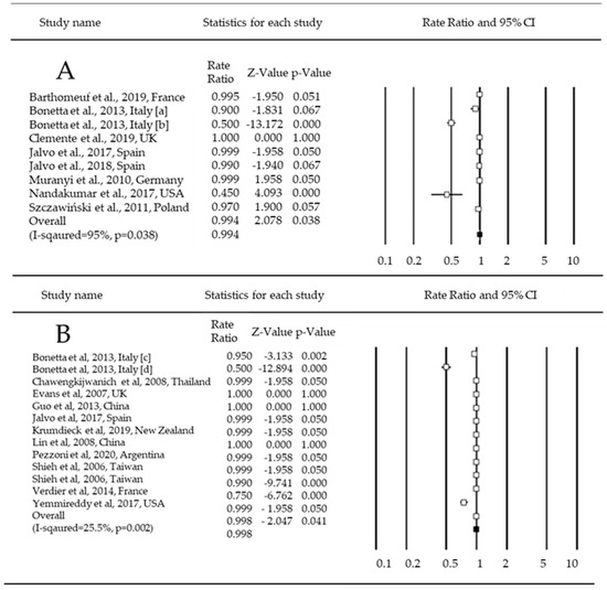
Figure 3.
Forest plot. The bacterial reduction rate of coated-TiO2, as indicated by the mean effect size and 95% confidence interval. (A) = Gram-positive; (B) = Gram-negative [33,38,40,41,45,46,50,51,54,59,60,62,64,66,67,68,71].
The meta-analysis showed that Gram-positive bacteria were more reduced than Gram-negative ones. This result is promising because Gram-positive bacteria are among the most widespread resistant pathogens, posing significant clinical challenges due to their immense genetic ability to acquire and develop resistance to antimicrobials. Gram-positive bacteria can generate spores that can survive in the environment [70,71,78,79]. These spores are one of the most resistant forms of life known to date and can tolerate various stresses, such as heat, chemicals, and harsh physical conditions [72,80]. The included meta-analysis articles are plotted in the forest plot below, and the bacteria strains are divided based on their classification (A = Gram-positive, and B = Gram-negative). The mechanism of TiO2 toxicity towards microorganisms depends on the rupture of the cytoplasmic membrane and subsequent leakage of intracellular components [110,111]. Thus, hydroxyl radicals produced on the coating attack the cytoplasmic membrane, and the different morphologies of the outer layers of different Gram-positive and -negative bacteria hinder hydroxyl radical attack in different ways. The Gram-positive bacterium S. aureus, for example, has little protection from radicals, having only a periplasmic space and a peptidoglycan layer that, although thick, is composed of a rather open polymeric network of polysaccharide chains of N-acetylmuramic acid and N-acetylglucosamine with peptide bridges. The Gram-negative bacterium E. coli, on the other hand, has a layer of peptidoglycan in addition to an outer membrane composed of lipids, lipopolysaccharides, and proteins. This logic would explain the greater antimicrobial activity of Ag-TiO2 against Gram-positive bacteria. Cell death occurs when the membrane is disrupted because there are no other barriers, such as TiO2 [110,112].
The Q value for the influencing factors was very high (Q = 202.8, p = 0.001), showing that the type of the surface and process of coating can modulate bacterial removal efficiency and are influenced by each other. The study suggests that the interactions between different factors affecting bacterial removal in coatings should be further explored. Among the surface types, dip coating is the most effective (R = 0.0005, and p = 0.0001) in reducing bacteria, especially Gram-positive. Indeed, the dip coating method is one of the most widely used to deposit TiO2 NPs. Among the principal proprieties, there are simplicity, reliability, reproducibility, and cost-effectiveness [113]. Dip coating has several advantages, such as being suitable to cover surfaces with different geometries, enabling coating of both sides of a substrate at once, and deposition is suitable for application in largescale processes [114]. Furthermore, dip coating methods can be used to coat a wide range of substrates, including metallic, ceramic, and polymeric surfaces, among others [115]. Similarly, spray coating is a commonly used deposition technique for applying TiO2 NPs-based coatings on large surfaces due to its mild operating conditions and cost-effectiveness [113,114,115]. The superior wear resistance of TiO2 nanoparticle coating may contribute to its antimicrobial effect [81,82]. Furthermore, even if surface defects appear after a certain number of times, the antifouling and antimicrobial properties can be maintained as long as the surface is surrounded by a significant coating of TiO2 nanoparticles [83]. The future of this research field is focused on developing innovative photocatalytic and photo-electrocatalytic surfaces for microbial inactivation. These solutions should address gaps such as low utilization efficiency of sunlight [18,19,20] and the need for nanostructured photoanodes that can provide better electron transport and oxygen vacancy materials [116,117]. Additionally, synergic connection with other processes such as fuel cells or ozonation can improve disinfection performance [22].
3.4. Limitations of the Study
This systematic review and meta-analysis have limitations. Firstly, there is heterogeneity among the selected studies, with some not presenting an initial bacterial load, some failing to clarify the dose of photocatalyst used, and others lacking details on the wavelength, distance, and characteristics of the light source employed. Significant variations in the coating method exist, thereby restricting comparability and potentially undermining the consistency of the findings. This systematic review and meta-analysis mark the first endeavor to establish the antimicrobial efficacy of various photocatalysts adhered to distinct surfaces employing diverse coating techniques [23,24,25,26]. This initiates new possibilities for forthcoming research that can identify the most effective coating method for disinfecting pathogenic microorganisms that pose a danger to human health.
4. Conclusions
Microbial-based diseases and their spread remain a significant burden on the healthcare systems and economies of countries worldwide. Moreover, over the decades, microorganisms generated resistance against existing drugs due to misuse or overuse. To address these life-threatening problems, new alternatives have been sought. Antimicrobial photocatalyst-based materials have emerged as a tool to fight against pathogens, as highlighted in this review. These materials have been used as surface coatings to destroy SARS-CoV-2. Antibacterial activity due to photocatalysis works by disrupting the cell envelope of bacteria. This means that the likelihood of pathogens developing resistance against photocatalysts is low or null, unlike conventional antibiotics that target specific areas. The goal is to develop antimicrobial coatings that are safe and can be used as an alternative to current antibiotics or disinfectants. This systematic review and meta-analysis work lays a promising foundation for this. Indeed, it was found that coating surfaces with photocatalysts has excellent disinfectant properties regardless of the type of coating, and it is effective on various microorganisms, even very resistant ones. The best surface is the glass, and the dip coating seems to be the better technology for the deposition of TiO2. Moving forward, it is essential to include multi-drug-resistant and clinically isolated pathogens in research and development efforts. Moreover, proposals for developing novel materials that combine electrospinning and advanced oxidation technologies can be made. This can include synthetic strategies that take advantage of the unique properties of polymers and overcome the limits of current photocatalysts.
Author Contributions
Conceptualization, F.U. and F.V.; methodology, F.U., F.V., G.L. and V.V.; software, F.U., F.V. and V.V.; formal analysis, F.U., F.V., G.L. and V.V.; investigation, F.U., F.V., G.L. and V.V.; data curation, F.U., F.V. and V.V.; writing—original draft preparation, F.U. and F.V.; writing—review and editing, F.U., F.V. and G.L.; supervision F.V. and V.R.S. All authors have read and agreed to the published version of the manuscript.
Funding
This study was partially funded by the MIUR-Fund-PON R&I 2014–2020 React-EU and IUSM Projects (CUP H83C23000160001; Prot. 1007-2023).
Institutional Review Board Statement
Not applicable.
Informed Consent Statement
Not applicable.
Data Availability Statement
Not applicable.
Acknowledgments
The authors thank Manuela Camerino and Tiziana Zilli for library assistance.
Conflicts of Interest
The authors declare no conflicts of interest.
References
- Wilson, A.M.; Weir, M.H.; King, M.F.; Jones, R.M. Comparing approaches for modelling indirect contact transmission of infectious diseases. J. R. Soc. Interface 2021, 18, 20210281. [Google Scholar] [CrossRef]
- Atkinson, M.P.; Wein, L.M. Quantifying the routes of transmission for pandemic influenza. Bull. Math. Biol. 2008, 70, 820–867. [Google Scholar] [CrossRef]
- Reed, S.E. An investigation of the possible transmission of rhinovirus colds through indirect contact. J. Hyg. 1975, 75, 249–258. [Google Scholar] [CrossRef] [PubMed]
- Mubareka, S.; Lowen, A.C.; Steel, J.; Coates, A.L.; Carcia-Sastre, A.; Palese, P. Transmission of influenza virus via aerosols and fomites in the guinea pig model. J. Infect. Dis. 2009, 199, 858–865. [Google Scholar] [CrossRef] [PubMed]
- Walther, B.A.; Ewald, P.W. Pathogen survival in the external environment and the evolution of virulence. Biol. Rev. 2004, 79, 849–869. [Google Scholar] [CrossRef]
- Sze-To, G.N.; Yang, Y.; Kwan, J.K.; Yu, S.C.; Chao, C.Y. Effects of surface material, ventilation, and human behavior on indirect contact transmission risk of respiratory infection. Risk. Anal. 2014, 34, 818–830. [Google Scholar] [CrossRef]
- Tuson, H.H.; Weibel, D.B. Bacteria–surface interactions. Soft. Matter. 2013, 9, 4368. [Google Scholar] [CrossRef] [PubMed]
- Dancer, S.J. Controlling hospital-acquired infection: Focus on the role of the environment and new technologies for decontamination. Clin. Microbiol. Rev. 2014, 27, 665–690. [Google Scholar] [CrossRef]
- Kumar, A.; Alam, A.; Rani, M.; Ehtesham, N.Z.; Hasnain, S.E. Biofilms: Survival and defense strategy for pathogens. Int. J. Med. Microbiol. 2017, 307, 481–489. [Google Scholar] [CrossRef]
- Maillard, J.Y.; Centeleghe, I. How biofilm changes our understanding of cleaning and disinfection. Antimicrob. Resist. Infect. Control. 2023, 12, 95. [Google Scholar] [CrossRef]
- Krukiewicz, K.; Kazek-Kęsik, A.; Brzychczy-Włoch, M.; Łos, M.J.; Ateba, C.N.; Mehrbod, P.; Ghavami, S.; Shyntum, D.Y. Recent Advances in the Control of Clinically Important Biofilms. Int. J. Mol. Sci. 2022, 23, 9526. [Google Scholar] [CrossRef] [PubMed]
- Protano, C.; Cammalleri, V.; Romano Spica, V.; Valeriani, F.; Vitali, M. Hospital environment as a reservoir for cross transmission: Cleaning and disinfection procedures. Ann. Ig. 2019, 31, 436–448. [Google Scholar]
- Cloutier, M.; Mantovani, D.; Rosei, F. Antibacterial Coatings: Challenges, Perspectives, and Opportunities. Trends Biotechnol. 2015, 33, 637–652. [Google Scholar] [CrossRef] [PubMed]
- Banu, R.; Salvi, N.; Gupta, S.; Ameta, C.; Ameta, R.; Punjabi, P.B. A facile synthesis of GO/CuO nanocomposite with enhancing photocatalytic activity for the degradation of azure-B dye and its antimicrobial behavior. Arab. J. Sci. Eng. 2021, 47, 365–378. [Google Scholar] [CrossRef]
- Kong, X.; Liu, X.; Zheng, Y.; Chu, P.K.; Zhang, Y.; Wu, S. Graphitic carbon nitride-based materials for photocatalytic antibacterial application. Mater. Sci. Eng. R. 2021, 145, 100610. [Google Scholar] [CrossRef]
- Atacan, K.; Güy, N.; Özacar, M. Recent advances in photocatalytic coatings for antimicrobial surfaces. Curr. Opin. Chem. Eng. 2022, 36, 100777. [Google Scholar] [CrossRef]
- Ganguly, P.; Byrne, C.; Breen, A.; Pillai, S.C. Antimicrobial activity of photocatalysts: Fundamentals, mechanisms, kinetics and recent advances. Appl. Catal. B 2018, 225, 51–75. [Google Scholar] [CrossRef]
- Shahsavandi, F.; Amirjani, A.; Hosseini, H.R.M. Plasmon-enhanced photocatalytic activity in the visible range using AgNPs/polydopamine/graphitic carbon nitride nanocomposite. Appl. Surf. Sci. 2022, 585, 152728. [Google Scholar] [CrossRef]
- Amirjani, A.; Amlashi, N.B.; Ahmadiani, Z.R. Plasmon-Enhanced Photocatalysis Based on Plasmonic Nanoparticles for Energy and Environmental Solutions: A Review. ACS Appl. Nano Mater. 2023, 6, 9085–9123. [Google Scholar] [CrossRef]
- Liu, L.; Shen, Z.; Wang, C. Highly efficient visible-light-driven photocatalytic disinfection of flowing bioaerosol using mono/multilayer MXene based catalyst. Chem. Eng. J. 2023, 457, 141327. [Google Scholar] [CrossRef]
- Zhang, P.; Lin, L.; Zang, D.; Guo, X.; Liu, M. Designing bioinspired anti-biofouling surfaces based on a superwettability strategy. Small 2017, 13, 1503334. [Google Scholar] [CrossRef] [PubMed]
- Valenzuela, L.; Faraldos, M.; Bahamonde, A.; Rosal, R. Critical review on the use of photocatalysis and photoelectrocatalysis to create antimicrobial surfaces. Curr. Opin. Chem. Eng. 2021, 34, 100762. [Google Scholar] [CrossRef]
- Schutte-Smith, M.; Erasmus, E.; Mogale, R.; Marogoa, N.; Jayiya, A.; Visser, H.G. Using visible light to activate antiviral and antimicrobial properties of TiO2 nanoparticles in paints and coatings: Focus on new developments for frequent-touch surfaces in hospitals. J. Coat. Technol. Res. 2023, 20, 789–817. [Google Scholar] [CrossRef]
- Rabajczyk, A.; Zielecka, M.; Klapsa, W.; Dziechciarz, A. Self-Cleaning Coatings and Surfaces of Modern Building Materials for the Removal of Some Air Pollutants. Materials 2021, 14, 2161. [Google Scholar] [CrossRef] [PubMed]
- Margarucci, L.M.; Romano Spica, V.; Protano, C.; Gianfranceschi, G.; Giuliano, M.; Di Onofrio, V.; Mucci, N.; Valeriani, F.; Vitali, M.; Romano, F. Potential antimicrobial effects of photocatalytic nanothecnologies in hospital settings. Ann. Ig. 2019, 31, 461–473. [Google Scholar] [PubMed]
- Byrne, J.A.; Dunlop, P.S.; Hamilton, J.W.; Fernández-Ibáñez, P.; Polo-López, I.; Sharma, P.K.; Vennard, A.S. A review of heterogeneous photocatalysis for water and surface disinfection. Molecules 2015, 20, 5574–5615. [Google Scholar] [CrossRef] [PubMed]
- Page, M.J.; McKenzie, J.E. The PRISMA 2020 statement: An updated guideline for reporting systematic reviews. BMJ 2021, 372, 71. [Google Scholar] [CrossRef]
- Covidence-Better Systematic Review Management. Available online: https://www.covidence.org/ (accessed on 31 July 2023).
- Bender, R.; Friede, T.; Koch, A.; Kuss, O.; Schlattmann, P.; Schwarzer, G.; Skipka, G. Methods for evidence synthesis in the case of very few studies. Res. Synth. Methods 2018, 9, 382–392. [Google Scholar] [CrossRef] [PubMed]
- Santabárbara, J.; Olaya, B.; Bueno-Notivol, J.; Pérez-Moreno, M.; Gracia-García, P.; Ozamiz-Etxebarria, N.; Idoiaga-Mondragon, N. Prevalence of depression among medical students during the COVID-19 pandemic. A systematic review and meta-analysis. Rev. Med. Chil 2021, 149, 1579–1588. [Google Scholar] [CrossRef] [PubMed]
- Santabárbara, J.; Ozamiz-Etxebarria, N.; Idoiaga, N.; Olaya, B.; Bueno-Novitol, J. Meta-Analysis of Prevalence of Depression in Dental Students during COVID-19 Pandemic. Medicina 2021, 57, 1278. [Google Scholar] [CrossRef]
- Oldenkamp, R.; Schultsz, C.; Mancini, E.; Cappuccio, A. Filling the gaps in the global prevalence map of clinical antimicrobial resistance. Proc. Natl. Acad. Sci. USA 2021, 118, e2013515118. [Google Scholar] [CrossRef]
- Shieh, K.J.; Li, M.; Lee, Y.H.; Sheu, S.D.; Liu, Y.T.; Wang, Y.C. Antibacterial performance of photocatalyst thin film fabricated by defection effect in visible light. Nanomedicine 2006, 2, 121–126. [Google Scholar] [CrossRef] [PubMed]
- Bletsa, E.; Merkl, P.; Thersleff, T.; Normark, S.; Henriques-Normark, B.; Sotiriou, G.A. Highly durable photocatalytic titanium suboxide–polymer nanocomposite films with visible light-triggered antibiofilm activity. Chem. Eng. J. 2023, 454, 139971. [Google Scholar] [CrossRef]
- Cuadra, J.G.; Molina-Prados, S.; Mínguez-Vega, G. Multifunctional silver-coated transparent TiO2 thin films for photocatalytic and antimicrobial applications. Appl. Surf. Sci. 2023, 617, 156519. [Google Scholar] [CrossRef]
- Fu, H.; Yaniv, V.; Betzalel, Y.; Mamane, H.; Gray, K.A. Creating anti-viral high-touch surfaces using photocatalytic transparent films. Chemosphere 2023, 323, 138280. [Google Scholar] [CrossRef]
- Todorova, N.; Minev, N.; Marinova, V.; Buchkov, K.; Videva, V.; Todorov, R.; Rafailov, P.; Strijkova, V.; Psycharis, V.; Giannakopoulou, T.; et al. Two-dimensional PtSe2 coatings with antibacterial activity. Appl. Surf. Sci. 2023, 611, 155534. [Google Scholar] [CrossRef]
- Evans, P.; Sheel, D.W. Photoactive and antibacterial TiO2 thin films on stainless steel. Surf. Coat. Technol. 2007, 201, 9319–9324. [Google Scholar] [CrossRef]
- Page, K.; Palgrave, R.G.; Parkin, I.P.; Wilson, M.; Savin, S.L.; Chadwick, A.V. Titania and silver–titania composite films on glass—Potent antimicrobial coatings. J. Mater. Chem. 2007, 17, 95–104. [Google Scholar] [CrossRef]
- Chawengkijwanich, C.; Hayata, Y. Development of TiO2 powder-coated food packaging film and its ability to inactivate Escherichia coli in vitro and in actual tests. Int. J. Food. Microbiol. 2008, 123, 288–292. [Google Scholar] [CrossRef]
- Lin, H.; Xu, Z.; Wang, X.; Long, J.; Su, W.; Fu, X.; Lin, Q. Photocatalytic and antibacterial properties of medical-grade PVC material coated with TiO2 film. J. Biomed. Mater. Res. B Appl. Biomater. 2008, 87, 425–431. [Google Scholar] [CrossRef]
- Yao, Y.; Ohko, Y.; Sekiguchi, Y.; Fujishima, A.; Kubota, Y. Self-sterilization using silicone catheters coated with Ag and TiO2 nanocomposite thin film. J. Biomed. Mater. Res. B Appl. Biomater. 2008, 85, 453–460. [Google Scholar] [CrossRef] [PubMed]
- Dunnill, C.W.H.; Aiken, Z.A.; Pratten, J. Enhanced photocatalytic activity under visible light in N-doped TiO2 thin films produced by APCVD preparations using t-butylamine as a nitrogen source and their potential for antibacterial films. J. Photochem. Photobiol. A Chem. 2009, 207, 244–253. [Google Scholar] [CrossRef]
- Sayilkan, F.; Asiltürk, M.; Kiraz, N.; Burunkaya, E.; Arpaç, E.; Sayilkan, H. Photocatalytic antibacterial performance of Sn4+-doped TiO2 thin films on glass substrate. J. Hazard Mater. 2009, 162, 1309–1316. [Google Scholar] [CrossRef] [PubMed]
- Muranyi, P.; Schraml, C.; Wunderlich, J. Antimicrobial efficiency of titanium dioxide-coated surfaces. J. Appl. Microbiol. 2010, 108, 1966–1973. [Google Scholar] [CrossRef] [PubMed]
- Szczawiński, J.; Tomaszewski, H.; Jackowska-Tracz, A.; Szczawińska, M.E. Survival of Staphylococcus aureus exposed to UV radiation on the surface of ceramic tiles coated with TiO2. Pol. J. Vet. Sci. 2011, 14, 41–46. [Google Scholar] [CrossRef]
- Akgun, B.A.; Wren, A.W.; Durucan, C.; Towler, M.R.; Mellott, N.P. Sol–gel derived silver-incorporated titania thin films on glass: Bactericidal and photocatalytic activity. J. Sol.-Gel. Sci. Technol. 2011, 59, 228–238. [Google Scholar] [CrossRef]
- Chien, D.M.; Dung, D.T.M.; Dam, L.D. Preparation of nitrogen co-doped SiO2/TiO2 thin films on ceramic with enhanced photocatalytic activity under visible-light irradiation. J. Exp. Nanosci. 2012, 7, 254–262. [Google Scholar] [CrossRef]
- Thongsuriwong, K.; Amornpitoksuk, P.; Suwanboon, S. Structure, morphology, photocatalytic and antibacterial activities of ZnO thin films prepared by sol–gel dip-coating method. Adv. Powder Technol. 2013, 24, 275–280. [Google Scholar] [CrossRef]
- Bonetta, S.; Bonetta, S.; Motta, F.; Strini, A.; Carraro, E. Photocatalytic bacterial inactivation by TiO2-coated surfaces. AMB Express 2013, 3, 59. [Google Scholar] [CrossRef]
- Guo, M.Z.; Ling, T.C.; Poon, C.S. Nano-TiO2-based architectural mortar for NO removal and bacteria inactivation: Influence of coating and weathering conditions. Cem. Concr. Compos. 2013, 36, 101–108. [Google Scholar] [CrossRef]
- Roldán, M.V.; de Oña, P.; Castro, Y.; Durán, A.; Faccendini, P.; Lagier, C.; Grau, R.; Pellegri, N.S. Photocatalytic and biocidal activities of novel coating systems of mesoporous and dense TiO2-anatase containing silver nanoparticles. Mater. Sci. Eng. C 2014, 43, 630–640. [Google Scholar] [CrossRef] [PubMed]
- Tallósy, S.P.; Janovák, L.; Ménesi, J. LED-light Activated Antibacterial Surfaces Using Silver-modified TiO2 Embedded in Polymer Matrix. J. Adv. Oxid. Technol. 2014, 17, 9–16. [Google Scholar] [CrossRef]
- Verdier, T.; Coutand, M.; Bertron, A.; Roques, C. Antibacterial Activity of TiO2 Photocatalyst Alone or in Coatings on E. coli: The Influence of Methodological Aspects. Coatings 2014, 4, 670–686. [Google Scholar] [CrossRef]
- Xiao, G.; Zhang, X.; Zhao, Y.; Su, H.; Tan, T. The behavior of active bactericidal and antifungal coating under visible light irradiation. Appl. Surf. Sci. 2014, 292, 756–763. [Google Scholar] [CrossRef]
- Deng, W.; Ning, S.; Lin, Q.; Zhang, H.; Zhou, T.; Lin, H.; Long, J.; Lin, Q.; Wang, X. I-TiO2/PVC film with highly photocatalytic antibacterial activity under visible light. Colloids Surf. B Biointerfaces 2016, 144, 196–202. [Google Scholar] [CrossRef]
- Leyland, N.S.; Podporska-Carroll, J.; Browne, J.; Hinder, S.J.; Quilty, B.; Pillai, S.C. Highly Efficient F, Cu doped TiO2 anti-bacterial visible light active photocatalytic coatings to combat hospital-acquired infections. Sci. Rep. 2016, 6, 24770. [Google Scholar] [CrossRef]
- Chuang, K.T.; Abdullah, H.; Leu, S.J.; Cheng, K.B.; Kuo, D.H.; Chen, H.C.; Chien, J.H.; Hu, W.T. Metal oxide composite thin films made by magnetron sputtering for bactericidal application. J. Photochem. Photobiol. A Chem. 2017, 337, 151–164. [Google Scholar] [CrossRef]
- Jalvo, B.; Faraldos, M.; Bahamonde, A.; Rosal, R. Antimicrobial and antibiofilm efficacy of self-cleaning surfaces functionalized by TiO2 photocatalytic nanoparticles against Staphylococcus aureus and Pseudomonas putida. J. Hazard. Mater. 2017, 340, 160–170. [Google Scholar] [CrossRef]
- Nandakumar, V.; Han, Z.; Fritz, Z.; Krishna, V.; Koopman, B.; Moudgil, B. Visible Light Photocatalytic Bacterial Inactivation on Titanium Dioxide Coatings. KONA Powder Part. J. 2017, 34, 234–240. [Google Scholar] [CrossRef]
- Pessoa, R.S.; dos Santos, V.P.; Cardoso, S.B.; Doria, A.C.; Figueira, F.R.; Rodrigues, B.V.; Testoni, G.E.; Fraga, M.A.; Marciano, F.R.; Lobo, A.O.; et al. TiO2 coatings via atomic layer deposition on polyurethane and polydimethylsiloxane substrates: Properties and effects on C. albicans growth and inactivation process. Appl. Surf. Sci. 2017, 422, 73–84. [Google Scholar] [CrossRef]
- Yemmireddy, V.K.; Hung, Y.C. Photocatalytic TiO2 coating of plastic cutting board to prevent microbial cross-contamination. Food. Control 2017, 77, 88–95. [Google Scholar] [CrossRef]
- Hossain, M.A.; Elias, M.; Sarker, D.R.; Diba, Z.R.; Mithun, J.M.; Azad, M.A.; Siddiquey, I.A.; Rahman, M.M.; Uddin, J.; Uddin, M.N. Synthesis of Fe- or Ag-doped TiO2–MWCNT nanocomposite thin films and their visible-light-induced catalysis of dye degradation and antibacterial activity. Res. Chem. Intermed. 2018, 44, 2667–2683. [Google Scholar] [CrossRef]
- Jalvo, B.; Faraldos, M.; Bahamonde, A.; Rosal, R. Antibacterial surfaces prepared by electrospray coating of photocatalytic nanoparticles. Chem. Eng. J. 2018, 334, 1108–1118. [Google Scholar] [CrossRef]
- Won, Y.; Schwartzenberg, K.; Gray, K.A. TiO2-based transparent coatings create self-cleaning surfaces. Chemosphere 2018, 208, 899–906. [Google Scholar] [CrossRef] [PubMed]
- Barthomeuf, M.; Castel, X.; Le Gendre, L.; Louis, J.; Denis, M.; Pissavin, C. Effect of Titanium Dioxide Film Thickness on Photocatalytic and Bactericidal Activities Against Listeria monocytogenes. Photochem. Photobiol. 2019, 95, 1035–1044. [Google Scholar] [CrossRef]
- Clemente, A.; Ramsden, J.J.; Wright, A.; Iza, F.; Morrissey, J.A.; Li Puma, G.; Malik, D.J. Staphylococcus aureus resists UVA at low irradiance but succumbs in the presence of TiO2 photocatalytic coatings. J. Photochem. Photobiol. B 2019, 193, 131–139. [Google Scholar] [CrossRef]
- Krumdieck, S.P.; Boichot, R.; Gorthy, R.; Land, J.G.; Lay, S.; Gardecka, A.J.; Polson, M.I.; Wasa, A.; Aitken, J.E.; Heinemann, J.A.; et al. Nanostructured TiO2 anatase-rutile-carbon solid coating with visible light antimicrobial activity. Sci. Rep. 2019, 9, 1883. [Google Scholar] [CrossRef]
- Valenzuela, L.; Iglesias, A.; Faraldos, M.; Bahamonde, A.; Rosal, R. Antimicrobial surfaces with self-cleaning properties functionalized by photocatalytic ZnO electrosprayed coatings. J. Hazard. Mater. 2019, 369, 665–673. [Google Scholar] [CrossRef]
- Oder, M.; Koklič, T.; Umek, P.; Podlipec, R.; Štrancar, J.; Dobeic, M. Photocatalytic biocidal effect of copper doped TiO2 nanotube coated surfaces under laminar flow, illuminated with UVA light on Legionella pneumophila. PLoS ONE 2020, 15, e0227574. [Google Scholar] [CrossRef]
- Pezzoni, M.; Catalano, P.N.; Delgado, D.C.; Pizarro, R.A.; Bellino, M.G.; Costa, C.S. Antibiofilm effect of mesoporous titania coatings on Pseudomonas aeruginosa biofilms. J. Photochem. Photobiol. B Biol. 2020, 203, 111762. [Google Scholar] [CrossRef]
- Zhao, Y.; Xu, J.; Li, Z.; Fu, T.; Jiang, S. In vitro antibacterial properties of MoO3/SiO2/Ag2O nanocomposite coating prepared by double cathode glow discharge technique. Surf. Coat. Technol. 2020, 397, 125992. [Google Scholar] [CrossRef]
- Álvarez, Á.L.; Dalton, K.P.; Nicieza, I.; Abade Dos Santos, F.A.; de la Peña, P.; Domínguez, P.; Martin-Alonso, J.M.; Parra, F. Virucidal Properties of Photocatalytic Coating on Glass against a Model Human Coronavirus. Microbiol. Spectr. 2022, 10, e0026922. [Google Scholar] [CrossRef] [PubMed]
- Du, J.; Li, Z.; Guo, H.; Zhu, E.; Liu, C.; Ren, B.; Che, G. The facile preparation and antibacterial performance of a conductive polymer-PU coating under visible light. Prog. Org. Coat. 2022, 165, 106755. [Google Scholar] [CrossRef]
- Li, Y.; Liu, Y.; Yao, B.; Narasimalu, S.; Dong, Z. Rapid preparation and antimicrobial activity of polyurea coatings with RE-Doped nano-ZnO. Microb. Biotechnol. 2022, 15, 548–560. [Google Scholar] [CrossRef] [PubMed]
- Vihodceva, S.; Šutka, A.; Otsus, M.; Vija, H.; Grase, L.; Kahru, A.; Kasemets, K. Visible-Light Active Flexible and Durable Photocatalytic Antibacterial Ethylene-co-vinyl Acetate—Ag/AgCl/α-Fe2O3 Composite Coating. Nanomaterials 2022, 12, 1984. [Google Scholar] [CrossRef] [PubMed]
- Xu, X.; Wang, Y.; Zhang, D.; Wang, J.; Yang, Z. In situ growth of photocatalytic Ag-decorated β-Bi2O3/Bi2O2.7 heterostructure film on PVC polymer matrices with self-cleaning and antibacterial properties. Chem. Eng. J. 2022, 429, 131058. [Google Scholar] [CrossRef]
- Serrano-Aroca, Á.; Takayama, K.; Tuñón-Molina, A.; Seyran, M.; Hassan, S.S.; Pal Choudhury, P.; Uversky, V.N.; Lundstrom, K.; Adadi, P.; Palù, G.; et al. Carbon-based nanomaterials: Promising antiviral agents to combat COVID-19 in the microbial-resistant era. ACS Nano 2021, 15, 8069–8086. [Google Scholar] [CrossRef]
- Chong, Y.; Ge, C.; Fang, G.; Wu, R.; Zhang, H.; Chai, Z.; Chen, C.; Yin, J.-J. Light-enhanced antibacterial activity of graphene oxide, mainly via accelerated electron transfer. Environ. Sci. Technol. 2017, 51, 10154–10161. [Google Scholar] [CrossRef]
- Elias, L.; Taengua, R.; Frígols, B.; Salesa, B.; Serrano-Aroca, Á. Carbon nanomaterials and LED irradiation as antibacterial strategies against gram-positive multidrug-resistant pathogens. Int. J. Mol. Sci. 2019, 20, 3603. [Google Scholar] [CrossRef]
- Endo-Kimura, M.; Kowalska, E. Plasmonic photocatalysts for microbiological applications. Catalysts 2020, 10, 824. [Google Scholar] [CrossRef]
- Endo, M.; Wei, Z.S.; Wang, K.L.; Karabiyik, B.; Yoshiiri, K.; Rokicka, P.; Ohtani, B.; Markowska-Szczupak, A.; Kowalska, E. Noble metalmodified titania with visible-light activity for the decomposition of microorganisms. Beilstein J. Nanotechnol. 2018, 9, 829–841. [Google Scholar] [CrossRef] [PubMed]
- Kowalska, E.; Wei, Z.; Karabiyik, B.; Herissan, A.; Janczarek, M.; Endo, M.; Markowska-Szczupak, A.; Remita, H.; Ohtani, B. Silvermodified titania with enhanced photocatalytic and antimicrobial properties under UV and visible light irradiation. Catal. Today 2015, 252, 136–142. [Google Scholar] [CrossRef]
- Janczarek, M.; Endo-Kimura, M.; Wei, Z.; Bielan, Z.; Mogan, T.R.; Khedr, T.M.; Wang, K.; Markowska-Szczupak, A.; Kowalska, E. Novel structures and applications of graphene-based semiconductor photocatalysts: Faceted particles, photonic crystals, antimicrobial and magnetic properties. Appl. Sci. 2021, 11, 1982. [Google Scholar] [CrossRef]
- Herrmann, J.M. Heterogeneous photocatalysis: State of the art and present applications. Top. Catal. 2005, 34, 49–65. [Google Scholar] [CrossRef]
- Kong, D.B.; Ma, C.C.; Wang, W.; Liu, C.; Tian, Y.; Wang, T.; Zhao, Z.P.; Zhang, C.Y.; Feng, H.M.; Chen, S.G. Two birds with one stone: Interfacial controls and pH response for long-term and high efficiency Cu2O antibacterial materials. Chem. Eng. J. 2022, 427, 131734. [Google Scholar] [CrossRef]
- Salvadores, F.; Reli, M.; Alfano, O.M.; Kocí, K.; Ballari, M.D. Efficiencies evaluation of photocatalytic paints under indoor and outdoor air conditions. Front. Chem. 2020, 8, 551710. [Google Scholar] [CrossRef]
- Maulidiyah, M.; Susilowati, P.E.; Mudhafar, N.K.; Salim, L.A.; Wibowo, D.; Muzakkar, M.Z.; Irwan, I.; Arham, Z.; Nurdin, M. Photo-inactivation Staphylococcus aureus by using formulation of Mn-N-TiO2 composite coated wall paint. Biointerface Res. Appl. Chem. 2022, 12, 1628–1637. [Google Scholar]
- Abdulagatov, I.M.; Ragimov, R.M.; Khamidov, M.A.; Maksumova, A.M.; Abdullaeva, N.M. ALD coated polypropylene hernia meshes for prevention of mesh-related post-surgery complications: An experimental study in animals. Biomed. Mater. 2022, 17, 1. [Google Scholar] [CrossRef]
- Ma, J.Z.; Liu, C.Y.; Yan, K. CQDs-MoS2 QDs loaded on Dendritic fibrous Nanosilica/Hydrophobic waterborne polyurethane acrylate for antibacterial coatings. Chem. Eng. J. 2022, 429, 132170. [Google Scholar] [CrossRef]
- Tuñón-Molina, A.; Takayama, K.; Redwan, E.M.; Uversky, V.N.; Andrés, J.; Serrano-Aroca, Á. Protective face masks: Current status and future trends. ACS Appl. Mater. Interfaces 2021, 13, 56725–56751. [Google Scholar] [CrossRef]
- Lu, Y.; Guan, S.; Hao, L.; Yoshida, H.; Nakada, S.; Takisawa, T.; Itoi, T. Inactivation of SARS-CoV-2 and photocatalytic degradation by TiO2 photocatalyst coatings. Sci. Rep. 2022, 12, 16038. [Google Scholar] [CrossRef] [PubMed]
- Margarucci, L.M.; Gianfranceschi, G.; Romano Spica, V.; D’Ermo, G.; Refi, C.; Podico, M.; Vitali, M.; Romano, F.; Valeriani, F. Photocatalytic Treatments for Personal Protective Equipment: Experimental Microbiological Investigations and Perspectives for the Enhancement of Antimicrobial Activity by Micrometric TiO2. Int. J. Environ. Res. Public Health 2021, 18, 8662. [Google Scholar] [CrossRef] [PubMed]
- Guillard, C.; Lachheb, H.; Houas, A.; Ksibi, M.; Elaloui, E.; Herrmann, J.-M. Influence of chemical structure of dyes, of pH and of inorganic salts on their photocatalytic degradation by TiO2 comparison of the efficiency of powder and supported TiO2. J. Photochem. Photobiol. A Chem. 2003, 158, 27–36. [Google Scholar] [CrossRef]
- Yang, Y.; Yan, Z.; Yang, S.; Tang, Z.; Li, W.; Yang, B.; Su, W.; Ji, T. Effect of substrate roughness on NOx removal of poly heptazine imides coated cement pastes exposed to washing and weathering. J. Clean. Prod. 2022, 377, 134397. [Google Scholar] [CrossRef]
- Zhang, J.; Tan, H.; Deng, X. NOx removal ability of photocatalytic cement-based materials with porous structure. J. Clean. Prod. 2022, 377, 134396. [Google Scholar] [CrossRef]
- Li, F.; Liu, G.; Liu, F.; Yang, S. A review of self-cleaning photocatalytic surface: Effect of surface characteristics on photocatalytic activity for NO. Environ. Pollut. 2023, 327, 121580. [Google Scholar] [CrossRef]
- Ahlawat, K.; Jangra, R.; Ish, A.; Dixit, A.; Fulwani, D.; Jain, N.; Prakash, R. Analysis of a UV photocatalytic oxidation-based disinfection system for hydroxyl radicals, negative air ions generation and their impact on inactivation of pathogenic micro-organisms. Rev. Sci. Instrum. 2023, 94, 104103. [Google Scholar] [CrossRef]
- Sizar, O.; Leslie, S.W.; Unakal, C.G. Gram-Positive Bacteria. In StatPearls [Internet]; StatPearls Publishing: Treasure Island, FL, USA, 2023; Available online: https://www.ncbi.nlm.nih.gov/books/NBK470553/ (accessed on 31 December 2023).
- Karaman, R.; Jubeh, B.; Breijyeh, Z. Resistance of Gram-Positive Bacteria to Current Antibacterial Agents and Overcoming Approaches. Molecules 2020, 25, 2888. [Google Scholar]
- Wohlgemuth, S.; Kämpfer, P. BACTERIA|Bacterial Endospores. In Encyclopedia of Food Microbiology, 2nd ed.; Batt, C.A., Tortorello, M.L., Eds.; Academic Press: Cambridge, MA, USA, 2014; pp. 160–168. [Google Scholar]
- Sawada, T.; Yoshino, F.; Kimoto, K.; Takahashi, Y.; Shibata, T.; Hamada, N.; Sawada, T.; Toyoda, M.; Lee, M.C. ESR detection of ROS Generated by TiO2 coated with fluoridated apatite. J. Dent. Res. 2010, 89, 848–853. [Google Scholar] [CrossRef]
- Kado, D.; Sakurai, K.; Sugiyama, T.; Ueda, T. Evaluation of cleanability of a titanium dioxide (TiO2)-coated acrylic resin denture base. Prosthodont. Res. Pract. 2005, 4, 69–76. [Google Scholar] [CrossRef]
- Alrahlah, A.; Fouad, H.; Hashem, M.; Niazy, A.A.; AlBadah, A. Titanium oxide (TiO2)/polymethylmethacrylate (PMMA) denture base nanocomposites: Mechanical, viscoelastic and antibacterial behavior. Materials 2018, 11, 1096. [Google Scholar] [CrossRef] [PubMed]
- Skallevold, H.E.; Rokaya, D.; Khurshid, Z.; Zafar, M.S. Bioactive Glass Applications in Dentistry. Int. J. Mol. Sci. 2019, 20, 5960. [Google Scholar] [CrossRef] [PubMed]
- Khan, K.A.; Ghatak, H.R.; Ahuja, S.M. Photocatalytic technology: A review of environmental protection and renewable energy application for sustainable development. Environ. Technol. Innov. 2020, 19, 100893. [Google Scholar]
- Ray, S.K.; Hur, J. A critical review on modulation of NiMoO4-based materials for photocatalytic applications. J. Environ. Manag. 2021, 278, 111562. [Google Scholar] [CrossRef]
- Kumaravel, V.; Nair, K.M.; Mathew, S.; Bartlett, J.; Kennedy, J.E.; Manning, H.G.; Whelan, B.J.; Leyland, N.S.; Pillai, S.C. Antimicrobial TiO2 nanocomposite coatings for surfaces, dental and orthopaedic implants. Chem. Eng. J. 2021, 416, 129071. [Google Scholar] [CrossRef] [PubMed]
- Rahimi, M.; Noruzi, E.; Sheyksaran, E.; Ebadi, B.; Kariminezhad, Z.; Molaparast, M.; Mehrabani, M.G.; Mehramouz, B.; Yousefi, M.; Ahmadi, R.; et al. Carbohydrate polymer-based silver nanocomposites: Recent progress in the antimicrobial wound dressings. Carbohydr. Polym. 2020, 231, 115696. [Google Scholar] [CrossRef]
- Huang, Z.; Maness, P.C.; Blake, D.M.; Wolfrum, E.J.; Smolinski, S.L.; Jacoby, W.A. Bactericidal mode of titanium dioxide photocatalysis. J. Photochem. Photobiol. A 2000, 130, 163. [Google Scholar] [CrossRef]
- Lu, Z.X.; Zhou, L.; Zhang, Z.L.; Shi, W.L.; Xie, Z.X.; Xie, H.Y.; Pang, D.W.; Shen, P. Cell Damage Induced by Photocatalysis of TiO2 Thin Films. Langmuir 2003, 19, 8765. [Google Scholar] [CrossRef]
- Sunada, K.; Watanabe, T.; Hashimoto, K. Studies on photokilling of bacteria on TiO2 thin film. J. Photochem. Photobiol. A 2003, 156, 227. [Google Scholar] [CrossRef]
- Dell’Edera, M.; Lo Porto, C.; De Pasquale, I.; Petronella, F.; Curri, M.L.; Agostiano, A.; Comparelli, R. Photocatalytic TiO2-based coatings for environmental applications. Catal. Today 2021, 380, 62–83. [Google Scholar] [CrossRef]
- Nabi, I.; Bacha, A.U.; Li, K.; Cheng, H.; Wang, T.; Liu, Y.; Ajmal, S.; Yang, Y.; Feng, Y.; Zhang, L. Complete Photocatalytic Mineralization of Microplastic on TiO2 Nanoparticle Film. IScience 2020, 23, 101326. [Google Scholar] [CrossRef] [PubMed]
- Kim, J.Y.; Youn, D.H. Nanomaterials for Advanced Photocatalytic Plastic Conversion. Molecules 2023, 28, 6502. [Google Scholar] [CrossRef] [PubMed]
- Kouchehbaghi, N.H.; Sohrabi, M.; Razbin, M.; Daryakenari, A.A.; Abbasi, M.; Bahrami, S.A. Soft computing procedure to optimize the electrospinning parameters of polyacrylonitrile nanofibrous air filter. J. Text. Inst. 2023. [Google Scholar] [CrossRef]
- Lv, H.; Liu, Y.; Bai, Y.; Shi, H.; Zhou, W.; Chen, Y.; Liu, Y.; Yu, D.-G. Recent Combinations of Electrospinning with Photocatalytic Technology for Treating Polluted Water. Catalysts 2023, 13, 758. [Google Scholar] [CrossRef]
Disclaimer/Publisher’s Note: The statements, opinions and data contained in all publications are solely those of the individual author(s) and contributor(s) and not of MDPI and/or the editor(s). MDPI and/or the editor(s) disclaim responsibility for any injury to people or property resulting from any ideas, methods, instructions or products referred to in the content. |
© 2024 by the authors. Licensee MDPI, Basel, Switzerland. This article is an open access article distributed under the terms and conditions of the Creative Commons Attribution (CC BY) license (https://creativecommons.org/licenses/by/4.0/).