Review

Multifunctional Coatings on Implant Materials—A Systematic Review of the Current Scenario

by
Vinita Vishwakarma
1,*,
Gobi Saravanan Kaliaraj
1 and
Kamalan Kirubaharan Amirtharaj Mosas
2
1
Centre for Nanoscience and Nanotechnology, Sathyabama Institute of Science and Technology, Chennai 600119, India
2
Coating Department, FunGlass—Centre for Functional and Surface Functionalized Glass, Alexander Dubcek University of Trencin, 91150 Trencin, Slovakia
*
Author to whom correspondence should be addressed.
Coatings 2023, 13(1), 69; https://doi.org/10.3390/coatings13010069
Submission received: 30 November 2022 / Revised: 24 December 2022 / Accepted: 26 December 2022 / Published: 30 December 2022

Abstract

:
The challenges and demands of implant materials are changing as a result of the substantial expansion in the global population. Suitable implants are required for aged people, physical injuries, patients who need revised surgeries, contaminated implants, and accident victims. Hence, the requirement for implants is drastically increasing day by day. Metals, ceramics, and polymers are used as implant materials by biomedical industries for long-term suffering patients. Stainless steel, titanium and its alloys, aluminum alloys, cobalt, zirconium, etc. (metals), hydroxyapatite (ceramic), polyurethane, polyethylene, polyimide, etc. (polymers), are some of the examples that fulfill the implant requirements. There are many other obstructions, such as adhesion, inflammation, and bacterial attack, which minimize the implant’s performance and its activity. However, coatings on ideal implant materials are significant to avoid its failure and to enhance its durability and longevity. Advanced techniques, such as physical and chemical methods, are suitable coating approaches to promote the surface of implants with respect to mechanical, biological, and other multifunctional activities. This review paper focuses on and investigates several strategies for bioactive implants’ coatings, analysis, and emerging applications for biomedical industries.

1. Introduction

Biomedical implants are a boon for the medical industry, but the selection of implant materials and extending their lives is a challenging task to meet its ideal requirements. Implants for cardiovascular, breast, dental, facial, ophthalmic, etc., have a big market worldwide due to growing chronic diseases. The global implant market is expected to be worth 86.3 billion USD in 2020, rising to 145.6 billion USD by 2030 [1]. Implants are prepared from bone, tissues, skin, metals, ceramics, and polymers based on the user’s demand [2]. They are prone to the biological environment, which creates many shortcomings, such as adhesion, inflammation, bacterial attack, corrosion susceptibility, changes in the surface chemistry, etc. Microbial invasion and its infection of implants are complex processes that affect the surface properties of materials and surrounding environments [3]. Implant materials had to be compatible with the host tissue in terms of material, structure, and surface bonding. Implant microroughness reduces performance and long-term survival activity [4]. Surface properties of implants, such as wettability and surface energy, influence the host’s response to biointerfaces [5]. However, coatings of ideal implant materials are significant to maintain the superior mechanical properties, corrosion resistance, antimicrobial properties, and biocompatibility of the implants and to avoid its early failure. Commonly used implant materials are metals (stainless steel, titanium and its alloys, aluminum alloys, cobalt, zirconium), ceramics (hydroxyapatite), and polymers (polyurethane, polyethylene, polyimide, etc.).
It was found that metal implants leach metal ions, which are toxic for the body and hence fail to support the patient for a long time [6]. There are many other obstructions that reduce the implant’s performance and activity, such as poor osseointegration, inflammation, bacterial infection, age factor of the patient, systemic disorders, size and shape of the implant, etc. The surface of implant materials has different properties and has unusual cell interactions. The invasion of microorganisms on implants occurs frequently during surgery, preventing tissue integration on the surface [7]. Stainless steel, commercial pure titanium (Cp-Ti), Ti alloys, aluminum (Al) alloys, cobalt alloys, gold alloys, zirconium (Zr), etc., are reported as the best implant materials for different parts of the body because they exhibit superior biocompatibility and less foreign body interaction compared to other conventional materials [8]. Some implants, such as Ti, have good biocompatibility properties, but their poor wear resistance leads to allergic reactions, which may cause pain in the joint implant area, weakness, and thereby bacterial infections and implant loosening after a few years of implantation [9].
Hence, biocompatibility and bioactivity are essential requirements for an artificial implant to reduce allergic reactions (joint pain and inflammations) and to also exhibit chemical bonding to living tissues and the formation of a bone-like apatite layer on its surface. Microbial invasion by implant materials on surrounding tissues must also be avoided. Bone regeneration at the surface of implants is subjected to surface properties and biological components, such as proteins, ions, and cells [10]. During implantations, several reactions are involved between the implant and tissue, such as exchange of ions, protein adsorption, clotting of blood, and attachment of cells [11]. It is a difficult challenge to have native tissue osseointegrate with implant materials. Suitable bioactive coatings, such as osteoconductive coatings, biocompatible coatings, hard coatings, antimicrobial coatings, coatings for sustainable antibiotic release, antimicrobial paints in clinical environments, and corrosion resistant coatings, are promising techniques to achieve the ideal surface properties in the implant materials with respect to mechanical, biological, and other multifunctional properties. The current review paper focuses on and investigates several implant coating techniques, which are mentioned and discussed in detail. Figure 1 shows the types of implants, types of coatings, and their applications in the biomedical industry.

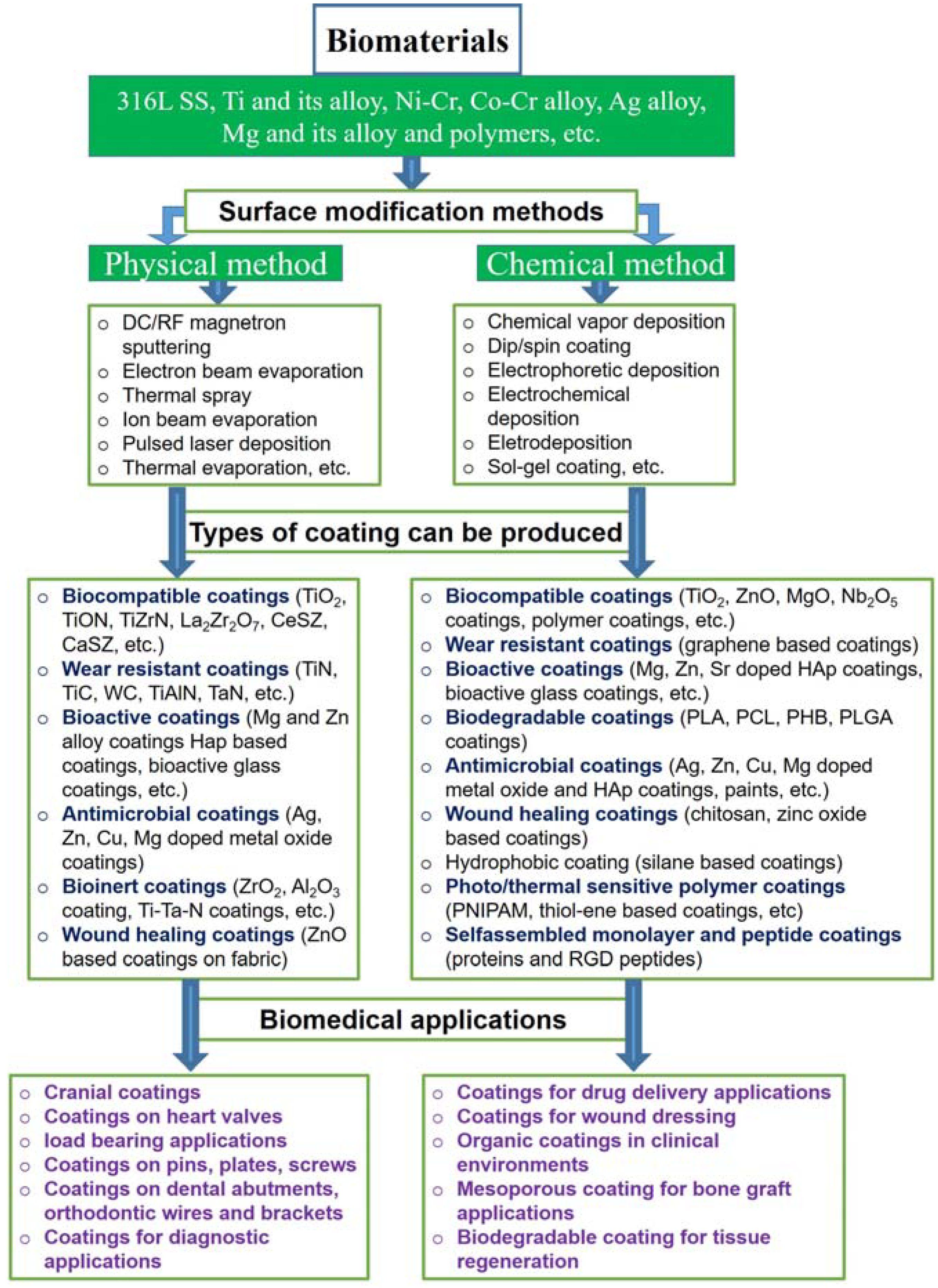
2. Types of Implant Coatings

2.1. Biocompatible Coatings

The orthopedic implant material is constantly in contact with the biological organism. The organism/implant response is significantly influenced by the biomaterial’s surface. The implant surface exposed to a corrosive environment could release ions into the body, which could lead to unfavorable reactions, such as pain and inflammation. It might also be subject to wear circumstances, as in joints, which would release wear particles into the surrounding tissue and possibly cause the implant to loosen. Surface modifications of the implants are required to improve their properties in order to avoid premature intervention and increase their shelf life.
The growth of bone tissue and its stability on the implant surface are tedious tasks. Osteoconductive coatings or creating a macro level rough surface on implant surface is a significant approach to develop bone tissue on implants surface apart from their mechanical properties, compositions, purity of chemicals and crystallinity nature. Osteoconduction is a bone grafting process for implant materials. Hydroxyapatite, as an osteoconductive material, is a combination of calcium and phosphate (10/6) and is suitable for bone regeneration. This material is widely used for bone grafting, which has wide application in hip arthroplasty. However, this chemical is also clinically proven compared to tricalcium phosphate and calcium phosphates similar to calcium fluoride phosphate, magnesium whitlockites, or phosphate mineral brushite [12]. Calcium orthophosphates are used as osteoconductive coating materials and also used as scaffolds or cementing substances [13]. Chitosan and gelatin composites have good interactions as scaffold materials on implant coating because of their mechanical strength, non-toxicity, biocompatibility, biodegradability, and low immunogenicity [14].

2.1.1. Calcium Based Apatite Coatings

The surfaces of implant materials, such as metals, ceramics, and polymers, are non-calcified. Apatite is a chemical component of calcium and phosphate, which demonstrate superior biocompatibility and osteoconductivity [15]. The blast apatite coating on implants responds well to bone. Surface modification of titanium was accomplished using hydrothermal hot pressing at 300 °C with hydroxyapatite ceramic coatings to accelerate apatite densification [16].

2.1.2. Bone Morphogenetic Protein Coatings (BMP)

BMP is a natural protein that promotes the healing of bone tissues and cartilage automatically in our body. In terms of bone regeneration capacity, implant materials have limited potential. Bone augmentation on implants is essential to recover the damaged sites. To increase the function of BMP, the surface topography of Ti implant was acid etched and sandblasted, which significantly enhanced bone contact [17]. Hydroxyapatite (HAp) based coatings improved the bioactivity of the implant by osseointegration properties. A porous hydroxyapatite coating on implant surfaces could trigger bone morphogenetic protein (BMP) expression and enhance mineralization.

2.1.3. RGD Peptide Based Coatings

Arginyl-glycyl-aspartic acid (RGD) peptide, which is a sequence of three amino-acids, supports cell adhesion on implants by integrin receptors [18] and transfers the signals through cytoplasmatic signaling pathways to the nucleus. Rejection of dental implants is 10% due to lack of osseointegration in the early stages due to lack of response from the biological host, such as extracellular matrix (ECM) protein absorption, surface interactions, tissue growth, etc. [19]. Rapid bone tissue formation makes the dental implant’s surface active, which is possible by conjugating the surface with bioactive peptides found in ECM proteins [20,21].

2.1.4. Mg Based Coating

Magnesium (Mg) is a trace element that is essential for bone metabolism in the human body. It has several applications in dentistry and orthopedics, as these coatings have been shown to enhance adhesion as well as the osseointegration process [22]. The effect of magnesium addition with zirconia-calcium phosphate coatings was studied by Pardun et al. [23]. These coatings were made by varying the amounts of magnesium fluoride or magnesium oxide added to yttria stabilized zirconia (YSZ) and hydroxyapatite (HAp). The magnesium content influenced coating surface morphology, mechanical strength, and calcium dissolution [24]. Furthermore, in vitro findings obtained with human osteoblasts show that the presence of Mg2+ ions improves biological performance. The Mg-containing coatings outperformed the pure YSZ-HAp coatings in terms of cell proliferation and differentiation. These findings show that adding magnesium to zirconia-calcium phosphate coatings increases their bioactivity potential, making them an excellent candidate for coatings on bone implants.
Bandyopadhyay et al. [25] examined the effect of MgO and ZnO dopants on the physical, mechanical, and biological properties of tricalcium phosphate (TCP) ceramics. The study of cell-material interactions and in vitro strength degradation were investigated over time and received special attention. TCP-MgO-ZnO proved good biocompatibility with osteoblastic precursor cells. These findings indicate that the addition of MgO to TCP ceramics promotes better cell spreading and attachment than TCP alone. On titanium (Ti) substrates, Mg and Mg-HAp coatings were deposited using RF/DC magnetron sputtering by Park et al. [26]. Mg and Mg-HAp coatings promoted the differentiation of MC3T3-E1 osteoblastic cells and speed up osseointegration. Mg-HAp and Ti-Mg coated Ti substrates increased osteocalcin (OCN) mRNA expression by 1.5 and 1.4-fold, respectively.
The antibacterial effect of Mg-based coatings was investigated by two in vitro studies, one of which examined bovine-derived HAp (BHAp) with MgO or MgF2, and the other looked at Zn-Mg co-implanted titanium surfaces. Fluorination treatment is primarily used to produce MgF2 coatings. The process of immersing Mg alloy in a specific concentration of hydrofluoric acid solution to develop a protective layer of MgF2 on the surface is referred to as fluorination treatment [27]. According to Mihailes Cu et al. [28], BHAp/MgO and BHAp/MgF2 coatings showed four-fold higher inhibition activity against Enterococcus sp., Candida albicans and Micrococcus sp., strains. The in vitro studies prove that Mg-based coatings enhance cellular behavior in terms of proliferation and morphology, while also improving the osteogenic markers expression and having significant antimicrobial activity. In contrast to surfaces that had only Mg incorporated, Yu et al. [29] demonstrated that Zn-Mg implanted Ti surfaces had the strongest antibacterial activity against bacteria, such as Streptococcus, Fusobacterium nucleatum, and Porphyromonasgingivalis mutans. The AZ31B alloy screw was successfully coated with MgF2 by Sun et al. [30] and investigated the impact of the coating on the corrosion rate and osteogenic activity of the implant in the body. In comparison to uncoated AZ31B screws, MgF2 coated screws had a good corrosion resistance and a slower release rate of Mg2+ ions at the beginning of implantation. Microscopic anatomy of biological tissues study showed that Mg substrate coated with MgF2 showed less inflammation reaction and good osteogenic activity. In vivo studies of fluorine coated Mg-alloy screws at different durations up to three months show less inflammation reaction and well osteogenic activity in the rabbit model (Figure 2). This suggests that fluorine modified Mg-alloy shows better osteogenic activity with fewer inflammatory effects.
Mihailescu et al. [28] used a pulsed laser deposition technique to produce (MgF2, MgO) doped BHAp thin films deposited on Ti substrates. The deposited thin films were subjected to compositional, structural, morphological, and biological studies. All doped BHAp films show adequate bonding strength, and these results support their potential use in biomedical applications. The surface modified implants with doped BHAp thin films resulted in good biocompatibility, according to the findings of in vitro cell culture studies compared to undoped BHAp coatings, the doped BHAp (BHAp:MgO > BHAp:MgF2) had greater anti-biofilm activity. These findings suggest that BHAp coatings doped with MgO or MgF2 are suitable for dental applications, as they not only ensure outstanding adhesion with the surface of implant and improve cytocompatibility, but also effectively prevent microbial colonization.
Zhao et al. [31] investigated the corrosion resistance of MgF2 deposited Mg alloy by comparing the MgF coating with the natural collagen film. According to the corrosion study, MgF2 significantly reduced the degradation rate compared to collagen coating. Drynda et al. [32] investigated a selection of fluoride-coated binary Mg-Ca alloys. MgF2 coating was found to reduce the corrosion rate of all binary Mg-Ca alloys. Furthermore, after implantation in a living body, there were no adverse effects, such as inflammation and hyperplasia. Jo et al. [33] successfully coated pure Mg with MgF2 and HAp layers using aerosol deposition techniques. The coating had a strong bond to the substrate and was uniformly dense. The MgF2/HAp composite layers in the simulated body fluid (SBF) solution slowed the corrosion rate at which uncoated Mg is corroded. In vitro studies revealed that the layer significantly increased cell proliferation. Additionally, the composite layers demonstrated good biocompatibility and acceptable corrosion resistance.

2.1.5. ZnO Based Coatings

Due to its high biocompatibility with human organs, absence of toxicity, high photocatalytic performance to eliminate a variety of infectious pathogens, good stability, and affordability, zinc oxide (ZnO) is one of the best materials [34]. Nanoparticles of ZnO are biocompatible and have improved antibacterial performance with improved corrosion resistance towards a variety of living microorganisms. When bacteria adhered to the surface of the implant, the ZnO coating induced the formation of a bacterial biofilm, but degenerated during prolonged exposure, typically after 24 h.
Numerous applications that have been considered for the prevention of bacterial biofilms, such as food packaging and in the field of antibacterial applications, may benefit from the hydrophobic phenomenon of ZnO nanostructures on metal surfaces. Combining ZnO to create nanocomposites with other antibacterial active materials is recommended as a solution to the ZnO insufficient bacterial inhibition properties. ZnO nanoparticles can be utilized to coat implants to provide antibacterial activity and corrosion prevention. For instance, at pH level of 13.2, the PEO/ZnO coating on AZ31B alloy implant demonstrated reduction activity toward E. coli and S. aureus colonies [35]. Upon illuminating with UV radiation to excite more electron-hole pairs and radical species, the antibacterial activity can be further improved. In contrast to the sample without UV irradiation, which only had bacterial inhibition activities of 67.70% against E. coli and 82.47% against S. aureus, the UV-irradiated sample had bacterial inhibition activities of 98.95 % against E. coli and 94.5% against S. aureus. Figure 3 shows the bacterial growth inhibition rate against S. aureus and E. coli with an increase in the concentration of nanoparticles from 1 to 4 g/L (Z1 to Z4) and exposure time (2 h, 4 h and 6 h) [35].
Due to the low shear resistance of the oxide layer, Ti implants have poor tribological properties in biomedical applications. The application of ZnO coating through sol-gel method improved their tribological properties and protected from wear [36]. Nanocomposite coatings of Ag2O/ZnO/NiO thin films can improve corrosion resistance. Varshney et al. [37] stated that the mechanical behavior of HAp/MgO/ZnO bioceramics had improved. By improving the crystallinity and physical characteristics of HAp, the composite increases its mechanical properties. Additionally, a high degree of chemical stability can be attained, which will be beneficial for use in bone regeneration.

2.1.6. TiO2 Based Coatings

Titanium dioxide (TiO2) is one of the commercially available antimicrobial coatings due to its stability, reactivity, reusability, durability, and low-cost. Due to its biocompatibility, mechanical strength, and strong resistance to corrosion, TiO2 has numerous applications in the biomedical field [38]. Hou et al. [39] successfully deposited a TiO2 thin film with a 400 nm thickness onto Mg-Zn alloy surface. According to their findings, the corrosion rate of Mg-Zn alloy was significantly reduced by the application of TiO2 coating, which was found to be in a dense and amorphous state. Cell experiments further demonstrated the prepared coating’s ability to improve endothelial cell viability and adhesion, significantly reduce hemolysis and platelet adhesion, and exhibit high biocompatibility. Peron et al. [40] used sputtering and atomic layer deposition (ALD) to deposit TiO2 thin layer with a thickness of 40 nm on AZ31 Mg alloy substrate and compared corrosion resistance. The potentiodynamic polarization (PDP) study and hydrogen evolution experiment revealed that both sputtered and ALD coated TiO2 could significantly increase the corrosion resistance and hydrogen evolution, but ALD coating was more noticeable, particularly in the case of the porous structure.
Park et al. [41] showed that by adjusting the nucleation growth time during the deposition process, the antibacterial activity against S. aureus could be improved. TiO2 films nucleated for 2 h and 4 h demonstrated high cell viability more than 95% in the cytotoxicity test using human dermal fibroblast (HDF) cells (Figure 4), whereas the TiO2 films nucleated for 6 h had a slight cytotoxicity with cell viability of less than 80%. The production of reactive oxygen species explains TiO2 antibacterial effect.
Recently, TiO2 nanotubes were coated with antimicrobial (Ag) and bioactive calcium and phosphorus using micro-arc oxidation process [42]. By using the plate count method, the antimicrobial capability of Ca-P-Ag/TiO2 coating was examined against S. aureus. The porous features in the coating enhance adhesion as well as growth of osteoblasts. By using RF magnetron sputtering and varying bias voltages, the corrosion resistance of TiO2 coated on 316 L stainless steel (SS) substrates was investigated, as was the corrosion behavior in a 3.5% NaCl solution after deposition using potentiodynamic polarization (PDP), and it was found that TiO2 displayed better corrosion potential (Ecorr) than uncoated substrates [42].

2.1.7. Carbon Based Coatings

DLC coatings have gotten a lot of attention in the biomedical field, owing to their good biocompatibility, non-toxicity, non-carcinogenicity, and blood compatibility, all of which are important for DLC applications [43]. DLCs have also been taken into consideration for biomedical pressure sensors because of their high piezo resistivity [44]. DLC coatings have been applied to artificial heart valves, medical wires, joint prostheses, and vascular stents [45]. Aluminium and its alloys are suitable for use in biomedicine when coated with DLCs. Artificial joints in orthopedic applications have to maintain patients wide ranges of motion, therefore, wear and friction are inevitable. The DLC coated metallic implants are suitable for hip and knee joint applications by providing a low coefficient of friction and corrosion resistance in human body conditions during service.
Due to their biocompatibility and particularly their antimicrobial property, DLC coatings have a potential use in surgical instruments because it is known that there is a negative association between surface energy and bacterial adhesion [46]. The sp2 C fraction, dopants, and hydrogen content are efficient ways to modify surface energy [47]. The amount of Staphylococci bacterial adhesion on the DLC coated biomaterial surface is significantly less than that of uncoated biomaterials. By controlling hydrogen content and dopant, DLC bacterial adhesion can be significantly reduced. The presence of Ag can break down DNA, rupture the cell wall and cell membrane and it is suggested to use it with DLC to make it more effective in antibacterial activity [48]. Schwarz et al. [49] prepared Ag-DLC film through an ion induced polymer densification technique and investigated the antibacterial properties. The number of bacteria that stick to Ag-DLCs surface is less than that of a pure DLC because of the release of silver ions (Figure 5). By making the DLC surface smoother, the amount of bacterial adhesion can be decreased [50]. There are not many studies on DLC’s ability to inhibit biofilm formation. The DLC surface is very vulnerable to biofilm formation against C. albicans [51], P. aeruginosa and E. coli [52]. A few metal doped DLCs are also effective at combating biofilm formation [53].

2.1.8. TiN and CrN Based Coatings

To enhance the surface characteristics and metallic implant materials biological performance, nitride-based coatings have been applied [54]. Titanium nitride (TiN) is a popular coating for orthopedic implant materials because of its biocompatible, low coefficient of wear and friction, high hardness, and corrosion resistance [55]. With the application of a TiN ceramic coating, the release of metallic ions from the implant surface to the organism is decreased, which inhibits bacterial growth. In addition to acting as a physical barrier between the substrate and environment, ceramic coatings have improved corrosion resistance [56]. There is a decrease in the release of wear particles due to the improved wear resistance of the coating property. The material fatigue strength is another enhanced quality. With respect to hemolysis percentage that was almost zero, TiN has encouraging blood compatibility properties [41,57]. As a result, TiN-coatings are used in cardiology for producing pacemaker leads and ventricular assist devices for patients suffering from heart failure [58]. TiN-coated electrodes are being researched in neurology for the creation of chronically implanted devices for treating spinal cord injury [59]. Subramanian et al. [60] investigated the significance of surface modification by applying coatings based on nitrides, oxynitrides, and ternary nitrides, such as TiN, TiAlN, and TiON, onto medical grade stainless steels and investigated their electrochemical behavior using SBF solution as an electrolyte. According to the findings, the ternary nitride coating TiAlN outperformed TiON, TiN and Ti metallic substrates in terms of corrosion resistance.
CrN coatings have been proven to increase wear resistance and hardness when compared to CoCr alloys, as well as reduce metal ion release from the substrate to the surrounding body. CrN was found to have enhanced corrosion resistance when compared to TiN, TiAlN, and DLC in a study [61]. Another study comparing CrN, TiN, and CrCN coatings found that CrN and CrCN coatings had a 36-fold lower wear rate than uncoated implants. The additional benefit of CrN coatings is the potential to develop a CrN layer through plasma nitriding. CrN is occasionally used as an interlayer between a substrate and a top ceramic coating due to its potential for strong adhesion and to minimize interfacial stress [62]. Additionally, there was a significant reduction in ion release. Another way to develop a CrN coating is through utilizing reactive plasma, nitrogen can also be incorporated into the surface of CoCr to produce a surface rich in CrN. Liu et al. [63] investigated and discovered that plasma nitriding and plasma carbonitriding of CoCr alloy increased its wear resistance and hardness than untreated CoCr alloy substrate. Additionally, corrosion resistance was improved for both treatments, with the carbonitrided surface outperforming than nitrided surface. Overall, TiN and CrN-based coatings may be appropriate for joint implants due to their low wear rates and potential for good adhesion.

2.2. Polymer Based Antimicrobial Coatings

Microbial associated implant infection is a difficult task in the implant industry, causing implant loosening, chronic pain, bone loss in the affected site, and other complications. Following implantation, adhered microbes on the implant surface migrate rapidly to the surrounding site and colonize. After a microbial infection, the traditional method of treating it with systemic antibiotic use is ineffective in reducing the rate of infection. As a result, surface modification of materials with antimicrobial properties is an effective method of reducing microbial invasion in health care environments. Antimicrobial agents are substances that have inherent antimicrobial properties that prevent microbe growth. Low molecular weight antimicrobial agents, in particular, are used in the food preservation industry, antimicrobial drugs, and water/soil sterilization [64]. However, a wide range of applications are limited by short-term antimicrobial performance with low sensitivity and environmental toxicity [65]. Polymer-based antimicrobial agents have received a lot of attention because of their (a) broad spectrum ability against human pathogens, (b) long term performance, (c) stability in harsh environments, (d) compatibility with the environment, (e) ease of synthesis, and (f) cost-effectiveness [66].

2.2.1. Polymers

Polymers have attracted much attention in a variety of applications due to their ease of fabrication, low cost, and simple surface modification [67,68]. Polymers have been used for a variety of applications ranging from drug delivery, tissue engineering, antimicrobial coatings, bioinert coatings, wound healing, and bone graft applications. Polymers, including polyquaternary ammonium salts (PQAS), polyethyleneimine (PEI), chitosan, poly cationic hydrogels exhibit intrinsic antimicrobial activity for biomedical applications. Biodegradable polymers, such as PLLA, PLGA, PGA, PEE, PDLLA and PBT, are increasingly used to fabricate 3D porous scaffolds for peripheral nerve growth and creating artificial blood vessels [69]. Fibrous scaffold polymer coatings, such as PLGA, PEVA, PLLA-CL, silk fibroin and gelatin, are used as porous scaffolds for musculoskeletal engineering construct, including ligament, bone, cartilage, vascular and nerve tissue engineering applications [70,71]. In addition, polymers such as PLGA, collagen and PLA polymers are effectively used in cartilage tissue regeneration applications [72].

2.2.2. Coatings for Sustainable Antibiotic Release

In the medical industry, prosthetic joint infections cause antibiotic resistant biofilm, which often creates antibiofouling or antimicrobial surfaces for the prosthetic treatment. Several antibiotics are conventionally available to treat infections, such as cellulitis, arthritis, endocarditis, pneumonia and biliary tract infections. Usage of standard protocols, such as systemic delivery of antibiotics for both prevention and treatment for suffering liver and renal complications, needs to look for it. However, poor penetration of antibiotic into affected sites which requires hospitalized monitoring [73]. Particularly, more efficient way of antibiotic release is highly desirable; as a result, variety of in situ drug releasing surfaces is under consideration to develop drug eluting surfaces. The main advantage of in situ drug release is to deliver the therapeutic dosage at or near target sites to maximize the drug efficiency as well as avoiding side effects. The sustainable drug release comprises several ways, including (a) the entrapment of biocidal agents within the device (b) adsorption of antibiotic in porous structure (c) loading antibiotic in the internal cavities and (d) deposition of drug/antimicrobial agents onto the surface [74].
Chitosan (CH) is an abundant natural polymer that can be used in both tissue and dental applications due to its outstanding biodegradability, biocompatibility, and non-toxicity nature [75]. Doxorubicin-CH was formed by electrophoretic deposition and implanted near the tumour site to arrest cancer metastasis. Results stated that the doxorubicin-CH coated Ti6Al4V implant effectively inhibit G-292 cancer cell growth as that of doxorubicin powder inhibition effect. Similarly, drug release kinetics of doxorubicin are decreased due to the addition of hydroxyapatite to the coating (doxorubicin-CH) formulation [76]. Ag nanoparticles/gentamycin loaded into silk fibroin coated on Ti implants with CH as a barrier was prepared and investigated CH loaded silver nanoparticles exhibited a prolonged drug release profile even in the acidic environment [77]. Deposition of the paclitaxel drug, which is used for lung, ovarian, and breast cancer, onto Ti6Al4V alloy by electrochemical deposition and during treatment, HY926 cells exposed to the paclitaxel drug showed a significantly lower survival rate. It is also noticed that the electrodeposition did not affect the physicochemical properties of the drug [78].
In addition, HAp coated samples could absorb more drug than uncoated HA samples. From their investigation, they stated that electrochemical deposition did not alter the medical function of paclitaxel drug as well as porous HAp coating enhanced paclitaxel drug loading efficiency. Antibacterial efficiency was checked by the electrodeposition method using S. aureus for vancomycin-CH/hydroxyapatite coating [79]. After a few hours of immersion treatment, 80% of vancomycin was released in phosphate buffered saline condition (pH 7.4) which significantly arrested the growth of S. aureus. Zone of inhibition (ZOI) assays revealed that vancomycin drug exhibited the antibacterial zone of 30 mm for more than a month. In addition, vancomycin-CH/hydroxyapatite coating significantly enhanced the attachment, proliferation and differentiation behavior of osteoblasts, such as G292 cells [79]. Curcumin/CH loaded coating onto the layer of bioactive glasses, polyether ether ketone (PEEK) was then deposited onto a medical grade 316 L SS substrate. The drug deposited stainless steel samples were performed for antibacterial activity against E.coli and S.carnosus. Further, multilayer coating triggered an apatite-like layer on the implant surface [80]. HA/ciplastin coating was prepared onto a magnesium substrate using electrochemical deposition for anti-tumor applications, whereas a slow and steady increase in the drug profile indicated that cisplatin exhibited sustainable release and was better employed for anti-tumour treatment in pH 7.4 buffer solutions [81].

2.2.3. Corrosion Resistant Nature of Coatings

Post implantation, deterioration of the surface of implanted materials is an avoidable situation, wherein, the metal ions will leach from the surface of the implant materials to the surrounding tissues. Numerous ions, such as Na+, K+, Cl, SO4, etc., present in the blood plasma frequently interact with the implant surface. It is well known that every material exhibits their unique potential (voltage), in which, it evolves corrosion protection nature. Once the body current exceeds the limit, then there is a possibility of breaking down of passive layer on the implants, thereby, pitting corrosion occurs. Pitting is the condition of forming a microhole on the surface of the implant, from which metal ions are drastically released and deposited in the surrounding tissues. The deposition of metal ions tends to cause an immunogenic response, and subsequent immune cells will be recruited at the site of the inflammation. The above situation causes severe health issues, such as pain, bacterial infection, morbidity, and mortality.
Hence, there is a need to modify the surface of the implant, through which corrosion protection can be improved. At the same time, surface modification should not affect its mechanical behavior. Different types of materials, including ceramics, metal nitrides, biopolymers, and metal oxide coatings, have been used to enhance the implant materials’ performance. Biocompatible ceramic coatings improve surface hardness, mineralization, and corrosion resistance. Especially, zirconia (ZrO2) exhibited toughness and corrosion protection nature against body fluids, such as artificial blood plasma and artificial saliva solution [82]. Further, ZrO2 and its allotropes (tetragonal zirconia) performed with superior wear and scratch resistance properties, which is the idealistic approach for implant applications. Titanium and zirconium carbide (TiC and ZrC) deposition onto medical implants using physical vapor deposition (PVD) have been considered for total knee and hip replacement prosthesis. Though carbide and carbonitride coatings failed to express microbicidal behavior, it exhibits superior wear resistance properties for improving durability of the implants [83].
Hydroxyapatite coating by electrochemical deposition significantly improved bioactivity and corrosion resistance [84]. In addition, HAp admixed with metal oxide nanocomposites are effectively elevate the mechanical and corrosion protection nature. Consumption of antibiotics in the post implant surgery is prerequisite procedure to avoid unnecessary microbial infection as well as inflammatory conditions. However, prolonged antibiotic consumption may lead to organ damage, which further leads to morbidity and mortality. Fabrication of antimicrobial agent containing ceramic nanocomposite coatings show positive response even to the multi drug resistant bacterial strains, from which antibiotic consumption and its subsequent side effects can be minimized [85,86]. Table 1 shows the multifunctional materials and their mechanical, antimicrobial, and biological behavior for biomedical applications.

3. Commercially Available Multifunctional Coatings

In the implant industry, numerous implants have been used for a variety of biomedical applications for the past 50 years. However, very few materials and compounds have been commercially available in the market. Among them, Sree Chitra Tirunal Institute for Medical Sciences and Technology (SIRTRA), India, developed a TiN coated Co-based alloy and produced a low cost cardiac valve [92]. Marcin Kozakiewicz et al. fabricated custom-made zirconium oxide implants for reconstruction of cranial bones [93]. The company named “Copper Development Association Inc. has developed a multifunctional corrosion and wear resistant TiN coating. In addition, clinical professors, Cassagnol and Saad fabricated sirolimus drug eluting polymer coated metallic stents to prevent restenosis, preventing excess growth of neointima. The developed drug eluted stents were approved by the United States for the patients undergoing percutaneous coronary intervention (PCI) [94]. Houssam Sahwil reported that DDS labs produced FDA approved, metal free and high flexural strength, white color, low elastic modulus of zirconia dental implant to improve durability [95]. Gradinaru et al. developed a hydroxyapatite based ocular implant especially for eviscerated patients [96]. In addition, various metal oxide nanocomposites and polymers are in the queue for commercialization in the near future.

4. Conclusions and Future Directions

Implant and bone interface properties are significant because of fast osseointegration. In order to improve the strength as well as the quality of bone-implant contact, multifunctional coatings are applied based on the application of implants. Ideal implants have superior mechanical and corrosion resistance properties, but to achieve the superior surface properties, osteoconductive based coatings, biocompatible coatings, and polymer based antimicrobial coatings are effective to enhance the implant’s life. Surface modification of materials is a prerequisite process for improving the performance of bulk materials, including metals and metal alloys, polymers, ceramics, etc. In the biomedical industry, surface modification is mainly involved to enhance the bioactivity, biocompatibility, and mechanical stability of the materials. Different types of materials have been involved for the above three major applications in the human body. For cranial applications, zirconia and its allotropes and alumina coatings were used on the Ti implant surface. Different types of coatings are used for oral applications, such as TiN/TiO2, ZrN and TiON on orthodontic wire, for improving corrosion resistance and avoiding bacterial invasion in artificial saliva solutions. Hydroxyapatite coatings on dental abutments are performed effectively to improve osseointegration properties as well as reduce the risk of bacterial infection. Drug eluting polymer coating also used effectively in dental implants, heart stent and orthopedic implants to release antibiotic drug, morphogenetic protein delivery for improving durability of the implants [97]. Wear resistance coatings have also taken considerable attention to withstand the implant material from wear and deterioration. TiC, ZrC, AlCrN, TiZrAlN and its multilayer coatings have been used to modify the surface of total hip and knee replacements and ankle implants. In addition, HAp and metal oxide nanocomposites coatings are employed to reduce dissolution behavior of HAp and improve its performance. At the same time, silver, zinc, and copper containing ceramic coatings drastically reduced bacterial invasion as well as their biocompatibility. The above antimicrobial agent containing surface modification system could help to avoid prolonged consumption and its subsequent impacts.
Last but not least, various types of surface modification techniques are used, such as physical vapor deposition, chemical vapor deposition, electrochemical deposition, spin coating, dip coating, sol-gel coating, electrodeposition, and electroless deposition. From our literature survey, we summarize and suggest that the physical vapor deposition method is an effective tool to modify the surface mainly due to its intrinsic characteristics, including better adhesion strength, high crystalline fill without defect on the coating surface, which causes better performance of the surface coated implant even in an aggressive body fluid environment. This paper has critically reviewed different types of bioactive coatings for multifunctional properties of implants materials that are effective for the patient.

Author Contributions

Conceptualization, V.V. and G.S.K.; methodology, V.V.; validation, V.V. and G.S.K.; writing—original draft preparation, V.V., G.S.K. and K.K.A.M.; writing—review and editing, K.K.A.M. and G.S.K.; supervision, V.V. All authors have read and agreed to the published version of the manuscript.

Funding

This research received no external funding.

Institutional Review Board Statement

Not applicable.

Informed Consent Statement

Not applicable.

Data Availability Statement

Not applicable.

Acknowledgments

Authors thank the management, Sathyabama Institute of Science and Technology, Chennai for their support.

Conflicts of Interest

The authors declare no conflict of interest.

References

  1. Medical Implants Market Size to Surpass US$ 145.6 Bn by 2030. Available online: https://www.precedenceresearch.com/medical-implants-market (accessed on 24 December 2022).
  2. Saini, M.; Singh, Y.; Arora, P.; Arora, V.; Jain, K. Implant Biomaterials: A Comprehensive Review. World J. Clin. Cases 2015, 3, 52. [Google Scholar] [CrossRef]
  3. Ribeiro, M.; Monteiro, F.J.; Ferraz, M.P. Infection of Orthopedic Implants with Emphasis on Bacterial Adhesion Process and Techniques Used in Studying Bacterial-Material Interactions. Biomatter 2012, 2, 176–194. [Google Scholar] [CrossRef] [PubMed] [Green Version]
  4. Kligman, S.; Ren, Z.; Chung, C.H.; Perillo, M.A.; Chang, Y.C.; Koo, H.; Zheng, Z.; Li, C. The Impact of Dental Implant Surface Modifications on Osseointegration and Biofilm Formation. J. Clin. Med. 2021, 10, 1641. [Google Scholar] [CrossRef] [PubMed]
  5. Rupp, F.; Gittens, R.A.; Scheideler, L.; Marmur, A.; Boyan, B.D.; Schwartz, Z.; Geis-Gerstorfer, J. A Review on the Wettability of Dental Implant Surfaces I: Theoretical and Experimental Aspects. Acta Biomater. 2014, 10, 2894–2906. [Google Scholar] [CrossRef] [PubMed] [Green Version]
  6. Sansone, V.; Pagani, D.; Melato, M. The Effects on Bone Cells of Metal Ions Released from Orthopaedic Implants. A Review. Clin. Cases Miner. Bone Metab. 2013, 10, 34–40. [Google Scholar] [CrossRef]
  7. Subbiahdoss, G.; Kuijer, R.; Grijpma, D.W.; van der Mei, H.C.; Busscher, H.J. Microbial Biofilm Growth vs. Tissue Integration: “The Race for the Surface” Experimentally Studied. Acta Biomater. 2009, 5, 1399–1404. [Google Scholar] [CrossRef]
  8. Eisenbarth, E.; Velten, D.; Müller, M.; Thull, R.; Breme, J. Biocompatibility of β-Stabilizing Elements of Titanium Alloys. Biomaterials 2004, 25, 5705–5713. [Google Scholar] [CrossRef]
  9. Gautam, S.; Bhatnagar, D.; Bansal, D.; Batra, H.; Goyal, N. Recent Advancements in Nanomaterials for Biomedical Implants. Biomed. Eng. Adv. 2022, 3, 100029. [Google Scholar] [CrossRef]
  10. Han, C.M.; Jin, G.Z.; Kim, H.W. Recent Update on Implant Surface Tailoring to Improve Bone Regenerative Capacity. Tissue Eng. Regen. Med. 2014, 11, 266–273. [Google Scholar] [CrossRef]
  11. Zakaria, O.; Madi, M.; Kasugai, S. A Novel Osteogenesis Technique: The Expansible Guided Bone Regeneration. J. Tissue Eng. 2012, 3, 2041731412441194. [Google Scholar] [CrossRef]
  12. Geesink, R.G.T. Osteoconductive coatings for total joint arthroplasty. In Clinical Orthopaedics and Related Research; Lippincott Williams and Wilkins: Amsterdam, The Netherlands, 2002; Volume 395, pp. 53–65. [Google Scholar]
  13. Zhou, H.; Yang, L.; Gbureck, U.; Bhaduri, S.B.; Sikder, P. Monetite, an Important Calcium Phosphate Compound—Its Synthesis, Properties and Applications in Orthopedics. Acta Biomater. 2021, 127, 41–55. [Google Scholar] [CrossRef]
  14. Manzoor, K.; Ahmad, S.; Soundarajan, A.; Ikram, S.; Ahmed, S. Chitosan based nanomaterials for biomedical applications. In Handbook of Nanomaterials for Industrial Applications; Elsevier: London, UK, 2018; pp. 543–562. ISBN 9780128133514. [Google Scholar]
  15. Mutsuzaki, H.; Yokoyama, Y.; Ito, A.; Oyane, A. Formation of Apatite Coatings on an Artificial Ligament Using a Plasma- and Precursor-Assisted Biomimetic Process. Int. J. Mol. Sci. 2013, 14, 19155–19168. [Google Scholar] [CrossRef] [Green Version]
  16. Onoki, T. Porous apatite coating on various titanium metallic materials via low temperature processing. In Biomaterials Science and Engineering; InTech: Philadelphia, PA, USA, 2011. [Google Scholar]
  17. Yeo, I.-S. Reality of Dental Implant Surface Modification: A Short Literature Review. Open Biomed. Eng. J. 2014, 8, 114–119. [Google Scholar] [CrossRef]
  18. Matsuura, T.; Hosokawa, R.; Okamoto, K.; Kimoto, T.; Akagawa, Y. Diverse Mechanisms of Osteoblast Spreading on Hydroxyapatite and Titanium. Biomaterials 2000, 21, 1121–1127. [Google Scholar] [CrossRef]
  19. Buser, D.; Broggini, N.; Wieland, M.; Schenk, R.K.; Denzer, A.J.; Cochran, D.L.; Hoffmann, B.; Lussi, A.; Steinemann, S.G. Enhanced Bone Apposition to a Chemically Modified SLA Titanium Surface. J. Dent. Res. 2004, 83, 529–533. [Google Scholar] [CrossRef]
  20. Raczkowska, J.; Ohar, M.; Stetsyshyn, Y.; Zemła, J.; Awsiuk, K.; Rysz, J.; Fornal, K.; Bernasik, A.; Ohar, H.; Fedorova, S.; et al. Temperature-Responsive Peptide-Mimetic Coating Based on Poly(N-Methacryloyl-l-Leucine): Properties, Protein Adsorption and Cell Growth. Colloids Surf. B Biointerfaces 2014, 118, 270–279. [Google Scholar] [CrossRef]
  21. Rezania, A.; Healy, K.E. Biomimetic Peptide Surfaces That Regulate Adhesion, Spreading, Cytoskeletal Organization, and Mineralization of the Matrix Deposited by Osteoblast-like Cells. Biotechnol. Prog. 1999, 15, 19–32. [Google Scholar] [CrossRef]
  22. Almehmadi, A.H. Effect of Magnesium-Based Coatings on Titanium or Zirconia Substrates on Bone Regeneration and Implant Osseointegration—A Systematic Review. Front. Mater. 2021, 8, 482. [Google Scholar] [CrossRef]
  23. Pardun, K.; Treccani, L.; Volkmann, E.; Streckbein, P.; Heiss, C.; Gerlach, J.W.; Maendl, S.; Rezwan, K. Magnesium-Containing Mixed Coatings on Zirconia for Dental Implants: Mechanical Characterization and in Vitro Behavior. J. Biomater. Appl. 2015, 30, 104–118. [Google Scholar] [CrossRef] [Green Version]
  24. Tong, P.; Sheng, Y.; Hou, R.; Iqbal, M.; Chen, L.; Li, J. Recent Progress on Coatings of Biomedical Magnesium Alloy. Smart Mater. Med. 2022, 3, 104–116. [Google Scholar] [CrossRef]
  25. Bandyopadhyay, A.; Bernard, S.; Xue, W.; Bose, S. Calcium Phosphate-Based Resorbable Ceramics: Influence of MgO, ZnO, and SiO2 Dopants. J. Am. Ceram. Soc. 2006, 89, 2675–2688. [Google Scholar] [CrossRef]
  26. Park, K.D.; Lee, B.A.; Piao, X.H.; Lee, K.K.; Park, S.W.; Oh, H.K.; Kim, Y.J.; Park, H.J. Effect of Magnesium and Calcium Phosphate Coatings on Osteoblastic Responses to the Titanium Surface. J. Adv. Prosthodont. 2013, 5, 402–408. [Google Scholar] [CrossRef] [PubMed] [Green Version]
  27. Lin, Z.; Zhao, Y.; Zhang, Z.; Xi, Y.; Yeung, K. Antibacterial Properties, Hemolysis and Biocompatibility of Biodegradable Medical Magnesium Alloys. Xiyou Jinshu Cailiao Yu Gongcheng/Rare Met. Mater. Eng. 2018, 47, 403–408. [Google Scholar]
  28. Mihailescu, N.; Stan, G.E.; Duta, L.; Chifiriuc, M.C.; Bleotu, C.; Sopronyi, M.; Luculescu, C.; Oktar, F.N.; Mihailescu, I.N. Structural, Compositional, Mechanical Characterization and Biological Assessment of Bovine-Derived Hydroxyapatite Coatings Reinforced with MgF2 or MgO for Implants Functionalization. Mater. Sci. Eng. C 2016, 59, 863–874. [Google Scholar] [CrossRef] [PubMed]
  29. Yu, Y.; Jin, G.; Xue, Y.; Wang, D.; Liu, X.; Sun, J. Multifunctions of Dual Zn/Mg Ion Co-Implanted Titanium on Osteogenesis, Angiogenesis and Bacteria Inhibition for Dental Implants. Acta Biomater. 2017, 49, 590–603. [Google Scholar] [CrossRef] [Green Version]
  30. Sun, W.; Zhang, G.; Tan, L.; Yang, K.; Ai, H. The Fluoride Coated AZ31B Magnesium Alloy Improves Corrosion Resistance and Stimulates Bone Formation in Rabbit Model. Mater. Sci. Eng. C 2016, 63, 506–511. [Google Scholar] [CrossRef]
  31. Zhao, N.; Workman, B.; Zhu, D. Endothelialization of Novel Magnesium-Rare Earth Alloys with Fluoride and Collagen Coating. Int. J. Mol. Sci. 2014, 15, 5263–5276. [Google Scholar] [CrossRef] [Green Version]
  32. Drynda, A.; Seibt, J.; Hassel, T.; Bach, F.W.; Peuster, M. Biocompatibility of Fluoride-Coated Magnesium-Calcium Alloys with Optimized Degradation Kinetics in a Subcutaneous Mouse Model. J. Biomed. Mater. Res. A 2013, 101A, 33–43. [Google Scholar] [CrossRef]
  33. Jo, J.H.; Kang, B.G.; Shin, K.S.; Kim, H.E.; Hahn, B.D.; Park, D.S.; Koh, Y.H. Hydroxyapatite Coating on Magnesium with MgF2 Interlayer for Enhanced Corrosion Resistance and Biocompatibility. J. Mater. Sci. Mater. Med. 2011, 22, 2437–2447. [Google Scholar] [CrossRef]
  34. Puspasari, V.; Ridhova, A.; Hermawan, A.; Amal, M.I.; Khan, M.M. ZnO-Based Antimicrobial Coatings for Biomedical Applications. Bioprocess Biosyst. Eng. 2022, 45, 1421–1445. [Google Scholar] [CrossRef]
  35. Seyfi, M.; Fattah-alhosseini, A.; Pajohi-Alamoti, M.; Nikoomanzari, E. Effect of ZnO Nanoparticles Addition to PEO Coatings on AZ31B Mg Alloy: Antibacterial Effect and Corrosion Behavior of Coatings in Ringer’s Physiological Solution. J. Asian Ceram. Soc. 2021, 9, 1114–1127. [Google Scholar] [CrossRef]
  36. Çomakli, O.; Yazici, M.; Yetim, T.; Yetim, F.; Celik, A. Tribological and Electrochemical Behavior of Ag2O/ZnO/NiO Nanocomposite Coating on Commercial Pure Titanium for Biomedical Applications. Ind. Lubr. Tribol. 2019, 71, 1166–1176. [Google Scholar] [CrossRef]
  37. Varshney, S.; Nigam, A.; Singh, A.; Samanta, S.K.; Mishra, N.; Tewari, R.P. Antibacterial, Structural, and Mechanical Properties of MgO/ZnO Nanocomposites and Its HA-Based Bio-Ceramics; Synthesized via Physio-Chemical Route for Biomedical Applications. Mater. Technol. 2022, 37, 2503–2516. [Google Scholar] [CrossRef]
  38. Hou, S. Solvothermal Fabrication of TiO2 Nanosheet Films on Degradable Mg–Zn Alloys. Surf. Eng. 2016, 32, 745–749. [Google Scholar] [CrossRef]
  39. Hou, S.; Yu, W.; Yang, Z.; Li, Y.; Yang, L.; Lang, S. Properties of Titanium Oxide Coating on MgZn Alloy by Magnetron Sputtering for Stent Application. Coatings 2020, 10, 999. [Google Scholar] [CrossRef]
  40. Peron, M.; bin Afif, A.; Dadlani, A.; Berto, F.; Torgersen, J. Comparing Physiologically Relevant Corrosion Performances of Mg AZ31 Alloy Protected by ALD and Sputter Coated TiO2. Surf. Coat Technol. 2020, 395, 125922. [Google Scholar] [CrossRef]
  41. Park, S.; Park, J.; Heo, J.; Hong, B.Y.; Hong, J. Growth Behaviors and Biocidal Properties of Titanium Dioxide Films Depending on Nucleation Duration in Liquid Phase Deposition. Appl. Surf. Sci. 2017, 425, 547–552. [Google Scholar] [CrossRef]
  42. Yu, S.; Guo, D.; Han, J.; Sun, L.; Zhu, H.; Yu, Z.; Dargusch, M.; Wang, G. Enhancing Antibacterial Performance and Biocompatibility of Pure Titanium by a Two-Step Electrochemical Surface Coating. ACS Appl. Mater. Interfaces 2020, 12, 44433–44446. [Google Scholar] [CrossRef]
  43. Peng, J.; Yang, M.; Bi, J.; Sheng, R.; Li, L. Hydrogen Existence State of a Hydrogenated Amorphous Carbon Coating and Its Thermal Stability. Diam. Relat. Mater. 2019, 99, 107535. [Google Scholar] [CrossRef]
  44. Petersen, M.; Bandorf, R.; Bräuer, G.; Klages, C.P. Diamond-like Carbon Films as Piezoresistors in Highly Sensitive Force Sensors. Diam. Relat. Mater. 2012, 26, 50–54. [Google Scholar] [CrossRef]
  45. Roy, R.K.; Lee, K.-R. Biomedical Applications of Diamond-like Carbon Coatings: A Review. J. Biomed. Mater. Res. B Appl. Biomater. 2007, 83B, 72–84. [Google Scholar] [CrossRef]
  46. Schwarz, F.P.; Hauser-Gerspach, I.; Waltimo, T.; Stritzker, B. Antibacterial Properties of Silver Containing Diamond like Carbon Coatings Produced by Ion Induced Polymer Densification. Surf. Coat. Technol. 2011, 205, 4850–4854. [Google Scholar] [CrossRef]
  47. Liu, C.; Zhao, Q. The CQ Ratio of Surface Energy Components Influences Adhesion and Removal of Fouling Bacteria. Biofouling 2011, 27, 275–285. [Google Scholar] [CrossRef]
  48. Wei, C.; Peng, K.S.; Hung, M.S. The Effect of Hydrogen and Acetylene Mixing Ratios on the Surface, Mechanical and Biocompatible Properties of Diamond-like Carbon Films. Diam. Relat. Mater. 2016, 63, 108–114. [Google Scholar] [CrossRef]
  49. Tang, S.; Zheng, J. Antibacterial Activity of Silver Nanoparticles: Structural Effects. Adv. Healthc. Mater. 2018, 7, 1701503. [Google Scholar] [CrossRef]
  50. Peng, Y.; Peng, J.; Wang, Z.; Xiao, Y.; Qiu, X. Diamond-like Carbon Coatings in the Biomedical Field: Properties, Applications and Future Development. Coatings 2022, 12, 1088. [Google Scholar] [CrossRef]
  51. Santos, T.B.; Vieira, A.A.; Paula, L.O.; Santos, E.D.; Radi, P.A.; Khouri, S.; Maciel, H.S.; Pessoa, R.S.; Vieira, L. Flexible Camphor Diamond-like Carbon Coating on Polyurethane to Prevent Candida Albicans Biofilm Growth. J. Mech. Behav. Biomed. Mater. 2017, 68, 239–246. [Google Scholar] [CrossRef]
  52. Watari, S.; Wada, K.; Araki, M.; Sadahira, T.; Ousaka, D.; Oozawa, S.; Nakatani, T.; Imai, Y.; Kato, J.; Kariyama, R.; et al. Intraluminal Diamond-like Carbon Coating with Anti-adhesion and Anti-biofilm Effects for Uropathogens: A Novel Technology Applicable to Urinary Catheters. Int. J. Urol. 2021, 28, 1282–1289. [Google Scholar] [CrossRef]
  53. Cazalini, E.M.; Miyakawa, W.; Teodoro, G.R.; Sobrinho, A.S.S.; Matieli, J.E.; Massi, M.; Koga-Ito, C.Y. Antimicrobial and Anti-Biofilm Properties of Polypropylene Meshes Coated with Metal-Containing DLC Thin Films. J. Mater. Sci. Mater. Med. 2017, 28, 1–10. [Google Scholar] [CrossRef] [Green Version]
  54. Kaliaraj, G.S.; Siva, T.; Ramadoss, A. Surface Functionalized Bioceramics Coated on Metallic Implants for Biomedical and Anticorrosion Performance—A Review. J. Mater. Chem. B 2021, 9, 9433–9460. [Google Scholar] [CrossRef]
  55. Gobbi, S.J. Orthopedic Implants: Coating with TiN. Biomed. J. Sci. Tech. Res. 2019, 16, 11740–11742. [Google Scholar] [CrossRef]
  56. van Hove, R.P.; Sierevelt, I.N.; van Royen, B.J.; Nolte, P.A. Titanium-Nitride Coating of Orthopaedic Implants: A Review of the Literature. Biomed. Res. Int. 2015, 2015, 485975. [Google Scholar] [CrossRef] [PubMed] [Green Version]
  57. Dion, I.; Baquey, C.; Candelon, B.; Monties, J.R. Hemocompatibility of Titanium Nitride. Int. J. Artif. Organs 1992, 15, 617–621. [Google Scholar] [CrossRef] [PubMed]
  58. Sin, D.C.; Kei, H.L.; Miao, X. Surface Coatings for Ventricular Assist Devices. In Coatings for Biomedical Applications; Elsevier: Amsterdam, The Netherlands, 2012; pp. 264–283. [Google Scholar]
  59. Cogan, S.F. Neural Stimulation and Recording Electrodes. Annu. Rev. Biomed. Eng. 2008, 10, 275–309. [Google Scholar] [CrossRef] [Green Version]
  60. Subramanian, B.; Muraleedharan, C.V.; Ananthakumar, R.; Jayachandran, M. A Comparative Study of Titanium Nitride (TiN), Titanium Oxy Nitride (TiON) and Titanium Aluminum Nitride (TiAlN), as Surface Coatings for Bio Implants. Surf. Coat. Technol. 2011, 205, 5014–5020. [Google Scholar] [CrossRef]
  61. Wang, Q.; Zhou, F.; Wang, C.; Yuen, M.F.; Wang, M.; Qian, T.; Matsumoto, M.; Yan, J. Comparison of Tribological and Electrochemical Properties of TiN, CrN, TiAlN and a-C:H Coatings in Simulated Body Fluid. Mater. Chem. Phys. 2015, 158, 74–81. [Google Scholar] [CrossRef]
  62. Skjöldebrand, C.; Tipper, J.L.; Hatto, P.; Bryant, M.; Hall, R.M.; Persson, C. Current Status and Future Potential of Wear-Resistant Coatings and Articulating Surfaces for Hip and Knee Implants. Mater. Today Bio 2022, 15, 100270. [Google Scholar] [CrossRef]
  63. Liu, R.; Li, X.; Hu, X.; Dong, H. Surface Modification of a Medical Grade Co-Cr-Mo Alloy by Low-Temperature Plasma Surface Alloying with Nitrogen and Carbon. Surf. Coat. Technol. 2013, 232, 906–911. [Google Scholar] [CrossRef] [Green Version]
  64. Kenawy, E.-R.; Abdel-Hay, F.I.; El-Shanshoury, A.E.-R.R.; El-Newehy, M.H. Biologically Active Polymers. V. Synthesis and Antimicrobial Activity of Modified Poly(Glycidyl Methacrylate-Co-2-Hydroxyethyl Methacrylate) Derivatives with Quaternary Ammonium and Phosphonium Salts. J. Polym. Sci. A Polym. Chem. 2002, 40, 2384–2393. [Google Scholar] [CrossRef]
  65. Sauvet, G.; Dupond, S.; Kazmierski, K.; Chojnowski, J. Biocidal Polymers Active by Contact. V. Synthesis of Polysiloxanes with Biocidal Activity. J. Appl. Polym. Sci. 2000, 75, 1005–1012. [Google Scholar] [CrossRef]
  66. Kenawy, E.R.; Worley, S.D.; Broughton, R. The Chemistry and Applications of Antimicrobial Polymers: A State-of-the-Art Review. Biomacromolecules 2007, 8, 1359–1384. [Google Scholar] [CrossRef]
  67. Kumar, B.; Mathur, A.; Pathak, R.; Sardana, K.; Gautam, H.K.; Kumar, P. Evaluation of Antimicrobial Efficacy of Quaternized Poly[Bis(2-Chloroethyl)Ether-Alt-1,3-Bis[3-(Dimethylamino)Propyl]Urea] against Targeted Pathogenic and Multi-Drug-Resistant Bacteria. J. Bioact. Compat. Polym. 2016, 31, 467–480. [Google Scholar] [CrossRef]
  68. Belbekhouche, S.; Bousserrhine, N.; Alphonse, V.; Carbonnier, B. From Beta-Cyclodextrin Polyelectrolyte to Layer-by-Layer Self-Assembly Microcapsules: From Inhibition of Bacterial Growth to Bactericidal Effect. Food Hydrocoll. 2019, 95, 219–227. [Google Scholar] [CrossRef]
  69. Stetsyshyn, Y.; Raczkowska, J.; Harhay, K.; Gajos, K.; Melnyk, Y.; Dąbczyński, P.; Shevtsova, T.; Budkowski, A. Temperature-Responsive and Multi-Responsive Grafted Polymer Brushes with Transitions Based on Critical Solution Temperature: Synthesis, Properties, and Applications. Colloid Polym. Sci. 2021, 299, 363–383. [Google Scholar] [CrossRef]
  70. Gunatillake, P.; Mayadunne, R.; Adhikari, R. Recent Developments in Biodegradable Synthetic Polymers. Biotechnol. Annu. Rev. 2006, 12, 301–347. [Google Scholar]
  71. Vasita, R.; Katti, D.S. Nanofibers and Their Applications in Tissue Engineering. Int. J. Nanomed. 2006, 1, 15–30. [Google Scholar] [CrossRef]
  72. Tachibana, A.; Furuta, Y.; Takeshima, H.; Tanabe, T.; Yamauchi, K. Fabrication of Wool Keratin Sponge Scaffolds for Long-Term Cell Cultivation. J. Biotechnol. 2002, 93, 165–170. [Google Scholar] [CrossRef]
  73. Ruszczak, Z. Collagen as a Carrier for On-Site Delivery of Antibacterial Drugs. Adv. Drug Deliv. Rev. 2003, 55, 1679–1698. [Google Scholar] [CrossRef]
  74. Webb, J.C.J.; Spencer, R.F. The Role of Polymethylmethacrylate Bone Cement in Modern Orthopaedic Surgery. J. Bone Jt. Surg. Ser. B 2007, 89, 851–857. [Google Scholar] [CrossRef]
  75. Lyndon, J.A.; Boyd, B.J.; Birbilis, N. Metallic Implant Drug/Device Combinations for Controlled Drug Release in Orthopaedic Applications. J. Control. Release 2014, 179, 63–75. [Google Scholar] [CrossRef]
  76. Giacometti, A.; Cirioni, O.; Ghiselli, R.; Orlando, F.; Mocchegiani, F.; Silvestri, C.; Licci, A.; de Fusco, M.; Provinciali, M.; Saba, V.; et al. Comparative Efficacies of Quinupristin-Dalfopristin, Linezolid, Vancomycin, and Ciprofloxacin in Treatment, Using the Antibiotic-Lock Technique, of Experimental Catheter-Related Infection Due to Staphylococcus Aureus. Antimicrob Agents Chemother 2005, 49, 4042–4045. [Google Scholar] [CrossRef] [PubMed] [Green Version]
  77. Zhou, W.; Li, Y.; Yan, J.; Xiong, P.; Li, Q.; Cheng, Y.; Zheng, Y. Construction of Self-Defensive Antibacterial and Osteogenic AgNPs/Gentamicin Coatings with Chitosan as Nanovalves for Controlled Release. Sci. Rep. 2018, 8, 13432. [Google Scholar] [CrossRef] [PubMed]
  78. Lai, Y.L.; Lai, S.-B.; Yen, S.K. Paclitaxel/Hydroxyapatite Composite Coatings on Titanium Alloy for Biomedical Applications. Mater. Sci. Eng. C 2017, 79, 622–628. [Google Scholar] [CrossRef] [PubMed]
  79. Yang, C.C.; Lin, C.C.; Liao, J.W.; Yen, S.K. Vancomycin-Chitosan Composite Deposited on Post Porous Hydroxyapatite Coated Ti6Al4V Implant for Drug Controlled Release. Mater. Sci. Eng. C 2013, 33, 2203–2212. [Google Scholar] [CrossRef] [PubMed]
  80. Virk, R.S.; Rehman, M.A.U.; Munawar, M.A.; Schubert, D.W.; Goldmann, W.H.; Dusza, J.; Boccaccini, A.R. Curcumin-Containing Orthopedic Implant Coatings Deposited on Poly-Ether-Ether-Ketone/Bioactive Glass/Hexagonal Boron Nitride Layers by Electrophoretic Deposition. Coatings 2019, 9, 572. [Google Scholar] [CrossRef] [Green Version]
  81. Lai, Y.-L.; Lin, C.-C.; Hsu, S.-R.; Yen, S.-K. Electrochemical Deposition of Cisplatin on Pure Magnesium. J. Electrochem. Soc. 2018, 165, D196–D205. [Google Scholar] [CrossRef]
  82. Kaliaraj, G.S.; Muthaiah, B.; Alagarsamy, K.; Vishwakarma, V.; Kirubaharan, A.M.K. Role of Bovine Serum Albumin in the Degradation of Zirconia and Its Allotropes Coated 316L SS for Potential Bioimplants. Mater. Chem. Phys. 2021, 258, 123859. [Google Scholar] [CrossRef]
  83. Kumar, D.D.; Kaliaraj, G.S. Multifunctional Zirconium Nitride/Copper Multilayer Coatings on Medical Grade 316L SS and Titanium Substrates for Biomedical Applications. J. Mech. Behav. Biomed. Mater. 2018, 77, 106–115. [Google Scholar] [CrossRef]
  84. Yugeswaran, S.; Yoganand, C.P.; Kobayashi, A.; Paraskevopoulos, K.M.; Subramanian, B. Mechanical Properties, Electrochemical Corrosion and in-Vitro Bioactivity of Yttria Stabilized Zirconia Reinforced Hydroxyapatite Coatings Prepared by Gas Tunnel Type Plasma Spraying. J. Mech. Behav. Biomed. Mater. 2012, 9, 22–33. [Google Scholar] [CrossRef]
  85. Kaliaraj, G.S.; Bavanilathamuthiah, M.; Kirubaharan, K.; Ramachandran, D.; Dharini, T.; Viswanathan, K.; Vishwakarma, V. Bio-Inspired YSZ Coated Titanium by EB-PVD for Biomedical Applications. Surf. Coat. Technol. 2016, 307, 227–235. [Google Scholar] [CrossRef]
  86. Lishchynskyi, O.; Stetsyshyn, Y.; Raczkowska, J.; Awsiuk, K.; Orzechowska, B.; Abalymov, A.; Skirtach, A.G.; Bernasik, A.; Nastyshyn, S.; Budkowski, A. Fabrication and Impact of Fouling-Reducing Temperature-Responsive Poegma Coatings with Embedded CaCo3 Nanoparticles on Different Cell Lines. Materials 2021, 14, 1417. [Google Scholar] [CrossRef]
  87. Kaliaraj, G.S.; Thukkaram, S.; Alagarsamy, K.; Kirubaharan, A.M.K.; Paul, L.K.; Abraham, L.; Vishwakarma, V.; Sagadevan, S. Silver-Calcia Stabilized Zirconia Nanocomposite Coated Medical Grade Stainless Steel as Potential Bioimplants. Surf. Interfaces 2021, 24, 101086. [Google Scholar] [CrossRef]
  88. Kumar, D.D.; Kaliaraj, G.S.; Kirubaharan, A.M.K.; Alagarsamy, K.; Vishwakarma, V.; Baskaran, R. Biocorrosion and Biological Properties of Sputtered Ceramic Carbide Coatings for Biomedical Applications. Surf. Coat. Technol. 2019, 374, 569–578. [Google Scholar] [CrossRef]
  89. Dhinasekaran, D.; Kaliaraj, G.S.; Jagannathan, M.; Rajendran, A.R.; Prakasarao, A.; Ganesan, S.; Subramanian, B. Pulsed Laser Deposition of Nanostructured Bioactive Glass and Hydroxyapatite Coatings: Microstructural and Electrochemical Characterization. Mater. Sci. Eng. C 2021, 130, 112459. [Google Scholar] [CrossRef]
  90. Alagarsamy, K.; Vishwakarma, V.; Kaliaraj, G.S.; Vasantha, N.C.; Samuel, S.J.R. Biological Adhesion and Electrochemical Behavior of Ag-ZrO2 Bioceramic Coatings for Biomedical Applications. J. Adhes. Sci. Technol. 2020, 34, 349–368. [Google Scholar] [CrossRef]
  91. Walczak, M.; Pasierbiewicz, K.; Szala, M. Adhesion and Mechanical Properties of TiAlN and Altin Magnetron Sputtered Coatings Deposited on the DMSL Titanium Alloy Substrate. Acta Phys. Pol. A 2019, 136, 294–298. [Google Scholar] [CrossRef]
  92. Sankarkumar, R.; Bhuvaneshwar, G.S.; Magotra, R.; Muralidharan, S.; Rajan, R.S.; Saha, D.; Subba Rao, K.S.V.K.; Valiathan, M.S.; Radhakrishna, S.; Ramani, A.V. Chitra Heart Valve: Results of a Multicenter Clinical Study. J. Heart Valve Dis. 2001, 10, 619–627. [Google Scholar]
  93. Kozakiewicz, M.; Gmyrek, T.; Zajdel, R.; Konieczny, B. Custom-Made Zirconium Dioxide Implants for Craniofacial Bone Reconstruction. Materials 2021, 14, 840. [Google Scholar] [CrossRef]
  94. Cassagnol, M. Maha Saad US Pharmacist; Jobson Medical Information LLC: New York, NY, USA, 2010; p. 37. [Google Scholar]
  95. Zirconia Dental Implants—Pros and Cons. Available online: https://blog.ddslab.com/zirconia-dental-implants-pros-and-cons (accessed on 24 December 2022).
  96. Gradinaru, S.; Popescu, V.; Leasu, C.; Pricopie, S.; Yasin, S.; Ciuluvica, R.; Ungureanu, E. Hydroxyapatite Ocular Implant and Non-Integrated Implants in Eviscerated Patients. J. Med. Life 2015, 8, 90–93. [Google Scholar]
  97. Nastyshyn, S.; Stetsyshyn, Y.; Raczkowska, J.; Nastishin, Y.; Melnyk, Y.; Panchenko, Y.; Budkowski, A. Temperature-Responsive Polymer Brush Coatings for Advanced Biomedical Applications. Polymers 2022, 14, 4245. [Google Scholar] [CrossRef]
Figure 1. Schematic view of types of biomaterials, choices of coatings and its biomedical applications.
Coatings 13 00069 g001
Figure 2. Hematoxylin and eosin (HE) stained sections imaging around the implants made of (A) uncoated AZ31 Mg alloy screw, (T) Ti alloy screw, and (F) AZ31 Mg alloy coated with fluorine, with different intervals. Reprinted with permission from Ref. [30]. Copyright 2016, Elsevier.
Coatings 13 00069 g002
Figure 3. Bacterial growth inhibition rate against (a) E. coli and (b) S. aureus for the time exposed to 2 h, 4 h, and 6 h, respectively [35].
Coatings 13 00069 g003
Figure 4. Cytotoxicity study of TiO2 films with respect to nucleation time. Reprinted with permission from Ref. [41]. Copyright 2017, Elsevier.
Coatings 13 00069 g004
Figure 5. Dual fluorescent staining analysis for vital (Green) and dead (Red) bacterial cells on (a) uncoated and (b) Ag-DLC coated Ti6Al4V substrates. Reprinted/adapted with permission from Ref. [49]. Copyright 2011, Elsevier.
Coatings 13 00069 g005
Table 1. Multifunctional coatings and its mechanical, antibacterial and biological properties.
CoatingDeposition TechniqueMechanical PropertiesAntimicrobial PropertiesOsteogenesis FunctionCorrosion Behavior AnalysisRef.
Ag-CaSZ coatingE-beam evaporationAg-CaSZ and CaSZ coating improved wear resistant
property
Ag-CaSZ improved antibacterial property against P.aeruginosaCaSZ and Ag-CaSZ improved osteoblast cell proliferationImproved corrosion protection[87]
TiC and ZrC coatingDC sputtering techniqueTiC significantly improved
mechanical properties
Failed to prove antibacterial activity against P.aeruginosaImproved cell adhesion and proliferationImproved corrosion protection property[88]
HAp and bioactive glass coatingPulsed laser deposition technique-Bioactive glass improved significant antibacterial propertyBioactive glass and HAp coating enhanced osteoblast cell proliferationBioactive glass and HAp coating improved corrosion protection nature in simulated body fluid (SBF) condition[89]
Ag-ZrO2 bioceramic coatingDC sputtering techniqueAg-ZrO2 coating improved adhesion strength amidst coating and steel surfaceAg-ZrO2 significantly improved antibacterial property against E.coli and S.speciesHigh concentration of Ag-ZrO2 diminished cellular adhesion and growthAll the coated samples improved corrosion protection property[90]
TiAlN and AlTiN coatingMagnetron sputteringTiAlN and AlTiN coating
exhibited higher surface hardness and scratch resistant property on Ti alloy
substrate
---[91]
Disclaimer/Publisher’s Note: The statements, opinions and data contained in all publications are solely those of the individual author(s) and contributor(s) and not of MDPI and/or the editor(s). MDPI and/or the editor(s) disclaim responsibility for any injury to people or property resulting from any ideas, methods, instructions or products referred to in the content.

