You are currently viewing a new version of our website. To view the old version click .
Antibiotics
  • Article
  • Open Access

8 September 2025

Comparative Analysis of Phenotypic and Genotypic Antibiotic Susceptibility of Pasteurella multocida Isolated from Various Host Species in France and Hungary

,
,
,
and
1
HUN-REN Veterinary Medical Research Institute, 1143 Budapest, Hungary
2
National Laboratory of Infectious Animal Diseases, Antimicrobial Resistance, Veterinary Public Health and Food Chain Safety, University of Veterinary Medicine, 1078 Budapest, Hungary
3
Ceva Biovac, 49070 Beaucouzé, France
*
Author to whom correspondence should be addressed.
This article belongs to the Section Antibiotics in Animal Health

Abstract

Background/Objectives: Pasteurella multocida is responsible for a wide variety of animal diseases worldwide, causing major economic losses. These infections are usually treated with antibiotics; however, the emergence of multidrug-resistant (MDR) strains is increasingly hindering. Understanding antibiotic resistance in P. multocida is important for effective treatment strategies and public health, as it impacts both animal and human welfare. Methods: In this study, the antibiotic susceptibility of 80 P. multocida isolates was evaluated by phenotypic (disk diffusion and broth microdilution) and genotypic analysis via whole-genome sequencing, with particular attention to the occurrence of MDR strains. The strains were tested against antibiotics from nine antimicrobial classes (penicillins, cephalosporins, aminoglycosides, tetracyclines, macrolides, fluoroquinolones, lincosamides, phenicols, and sulfonamides). Antimicrobial resistance gene (ARG) sequences and single-nucleotide polymorphisms (SNPs) were evaluated in paired reads using the Bacterial and Viral Bioinformatics Resource Center (BV-BRC) and the Comprehensive Antibiotic Resistance Database (CARD) via Resistance Gene Identifier (RGI), respectively. Results: Phenotypic results indicated that cephalosporins and phenicols were the most effective drugs against P. multocida; however, the majority of strains were also susceptible to fluoroquinolones and tetracyclines. In contrast, high resistance rates were observed to sulfamethoxazole and clindamycin. The most prevalent resistance genes were strA, sul2, and tetH, while none of the strains harbored the bla-TEM or erm (42) genes. Conclusions: Of the two phenotypic methods, MIC values showed a stronger positive correlation with genotypic results, making it a more suitable method for determining antibiotic susceptibility. The phenotypic results for phenicols, tetracyclines, and fluoroquinolones showed a strong correlation with the detected resistance genes. In contrast, resistance to sulfamethoxazole, β-lactams, and macrolides remained genetically unexplained, suggesting the existence of additional resistance mechanisms to be explored.

1. Introduction

P. multocida is a significant veterinary pathogen with zoonotic potential, posing a risk to humans through animal contact. It is the etiological agent of fowl cholera [], atrophic rhinitis in pigs [], hemorrhagic septicemia in cattle and buffaloes [], and snuffles in rabbits []. As an opportunistic pathogen, it is a major contributor to the development of the respiratory disease complex in cattle and swine []. Humans might be infected through scratches and bites from cats and dogs [].
The spread of antimicrobial resistance is one of the most threatening global challenges of our time, and has now become one of the top 10 global health risks []. It is currently estimated that nearly 700,000 people die every year due to antimicrobial resistance. This could rise to 10 million by 2050, according to predictions []. The majority of antibiotics used to treat animal diseases are also important in human medicine. Antibiotics are essential in combating P. multocida infections, but the rise of multidrug-resistant (MDR) strains underscores the urgency of antimicrobial stewardship under the One Health framework []. The P. multocida strain is classified as MDR if it is resistant to at least one agent representing three or more antibiotic classes []. It is essential to establish a susceptibility profile before therapy in order to achieve successful treatment. There are various methods to determine the antibiotic susceptibility of strains and to detect MDR strains. Phenotypic susceptibility testing, including disk diffusion and broth microdilution method, remains the gold standard for therapy []. Genotypic antibiotic susceptibility testing mainly involves the identification of resistance genes (ARGs) using different polymerase chain reaction (PCR)-based methods []. In recent years, whole-genome sequencing has become increasingly prevalent, enabling a more efficient identification of ARGs and point mutations [,].
Considering the results of previous publications, the main hypotheses of this research were the following: (a) a correlation can be observed between phenotypic and genotypic resistance for each antibiotic class, (b) the broth dilution microdilution method is more reliable for phenotypic susceptibility testing than the disc diffusion method, and (c) most P. multocida strains are susceptible to cephalosporins, fluoroquinolones, and phenicols but resistant to sulfonamides and clindamycin. To verify the aforementioned hypotheses, the aim of this study was to determine the phenotypic and genotypic antibiotic susceptibility profiles of P. multocida strains, to compare the efficacy of these methods, and identify the most effective drugs for treatment.

2. Results

The antibiotic susceptibility profiles of the tested strains obtained by disk diffusion are summarized in Figure 1. All strains were resistant to clindamycin, and with a high resistance rate (75.0%) to sulfamethoxazole. The strains were highly susceptible (>90%) to cephalosporins, tetracyclines, and phenicols. On the other hand, there was a decrease in the susceptibility of strains to penicillins (sensitivity (S): 67.5–83.8%), fluoroquinolones (S: 76.3–83.5%), and gentamicin (56.8%). A high percentage of strains (73.8%) showed intermediate susceptibility to erythromycin.
Figure 1. Antibiotic susceptibility rates of P. multocida isolates from avian and mammalian hosts, determined by disk diffusion (16 antibiotics). Abbreviations: PEN: penicillin; AMX: amoxicillin; AMP: ampicillin; FUR: ceftiofur; CFX: cephalexin; GEN: gentamicin; TTC: tetracycline; DOX: doxycycline; ERY: erythromycin; CD: clindamycin; FFC: florfenicol; CHL: chloramphenicol; SMX: sulfamethoxazole; CPR: ciprofloxacin; ENO: enrofloxacin; UB: flumequine; S: susceptible; I: intermediate; R: resistant.
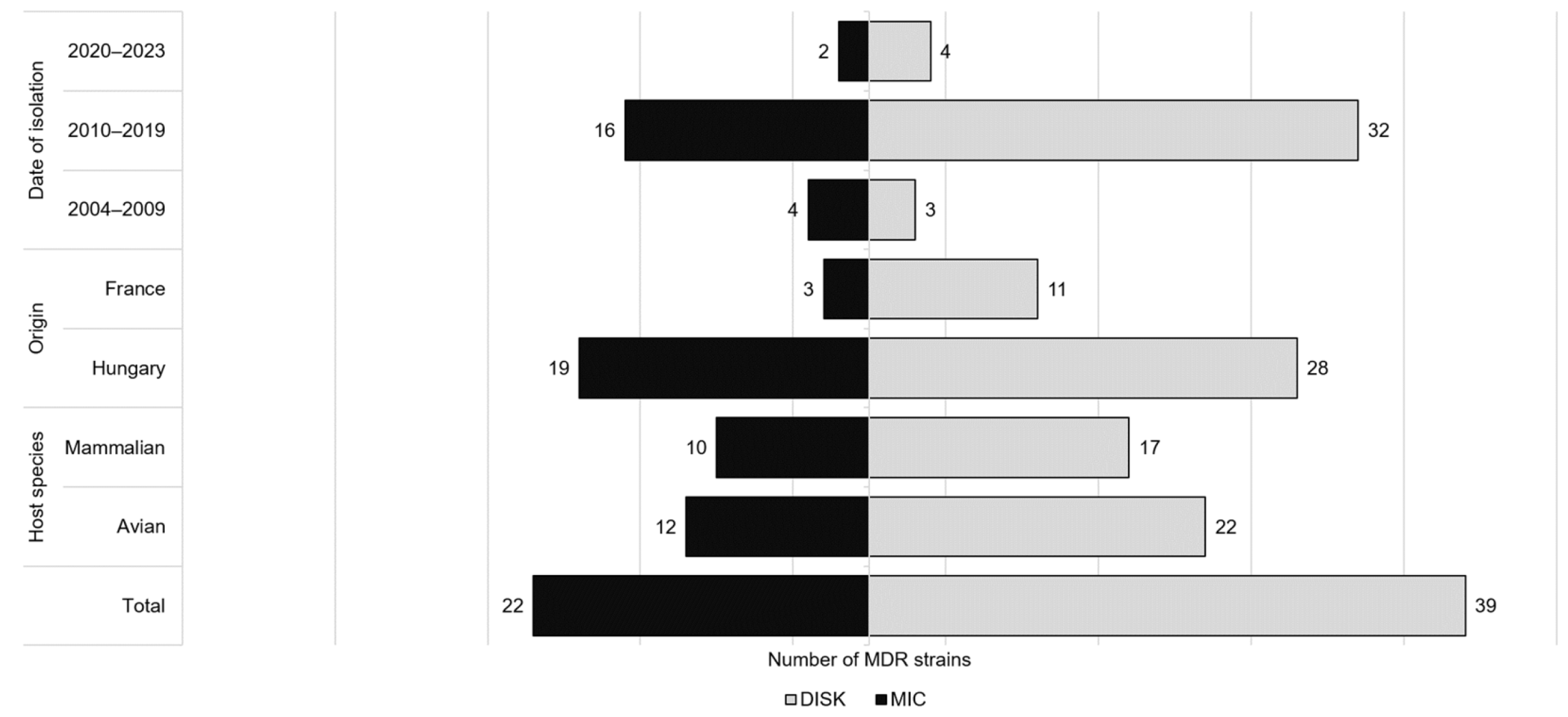
A total of 39 MDR strains were identified, most of which showed resistance to three (n = 18), four (n = 15), and in some cases to five, six, seven, and eight antibiotic classes (Table 1). Among MDR strains, the most frequent resistance combinations were penicillins–lincosamides–sulfonamides (n = 8) and penicillins–aminoglycosides–lincosamides–sulfonamides (n = 6). Overall, these strains were most resistant to clindamycin (100%), sulfamethoxazole (95%), and ampicillin (59%). MDR strains were isolated from both avian (n = 22) and mammalian (n = 17) hosts (Figure 2).
Table 1. Antibiotic susceptibility profiles and host species of multidrug-resistant strains identified by disk diffusion. Strains in bold were also found to be multidrug-resistant by the broth microdilution method. Abbreviations: PN: penicillins; AG: aminoglycosides; TET: tetracyclines; MAC: macrolides; LA: lincosamides; SA: sulfonamides; FC: phenicols; FQ: fluoroquinolones; CEP: cephalosporins; n: number of bacterial strains.
Figure 2. Distribution of MDR P. multocida isolates by host species, geographic origin, and year of isolation, based on disk diffusion and broth microdilution results.
The susceptibility data based on Minimal Inhibitory Concentration (MIC) values obtained by the broth microdilution method are summarized in Figure 3. The strains showed high susceptibility (>90%) to ceftiofur, tetracyclines, florfenicol and enrofloxacin. For these antibiotics, the calculated MIC50 and MIC90 values (Table 2) were both in the susceptible range. There was reduced susceptibility of strains to antibiotics of the penicillin class (S: 47.5–72.5%) and to tilmicosin (88.8%). For antibiotics of the penicillin class, the MIC90 values were always in the more resistant category. Complete resistance to clindamycin and high resistance rate (70.0%) to sulfamethoxazole were observed. The majority of strains (68.8%) had moderate susceptibility to erythromycin. The epidemiological breakpoint (ECOFF) defined by the European Committee on Antimicrobial Susceptibility Testing (EUCAST) that distinguishes wild-type and non-wild-type strains is presented in the last column of Table 2. These values were always higher than the calculated MIC50 values but lower than the MIC90 values, except for tetracyclines, tilmicosin, and florfenicol. The prevalence of non-wild-type strains was less than 10% for tetracycline, doxycycline, erythromycin, tilmicosin, and florfenicol, but higher than 50% for amoxicillin and ceftiofur. For ampicillin, enrofloxacin, and flumequine, this prevalence ranged from 17% to 33%. The susceptibility profiles of strains isolated from avian and mammalian host species are illustrated in Figure 3. A total of 22 MDR strains were identified which showed resistance to 3 (n = 10), 4 (n = 6), and 5 (n = 6) antibiotic classes. The most frequent resistance combination was penicillins-lincosamides-sulfonamides (n = 4) (Table 3). Overall, these strains were mostly resistant to clindamycin (100%), sulfamethoxazole (100%) and erythromycin (54.5%). Nearly equal proportions of MDR strains were detected from avian (54.5%) and mammalian (45.5%) hosts (Figure 2).
Figure 3. Antibiotic susceptibility rates of P. multocida isolates from avian and mammalian hosts, determined by broth microdilution (12 antibiotics). Abbreviations: PEN: penicillin; AMX: amoxicillin; AMP: ampicillin; FUR: ceftiofur; TTC: tetracycline; DOX: doxycycline; ERY: erythromycin; TIL: tilmicosin; CD: clindamycin; FFC: florfenicol; SMX: sulfamethoxazole; ENO: enrofloxacin; S: susceptible; I: intermediate; R: resistant.
Table 2. Distribution of MIC values per antibiotic, including MIC50 and MIC90, calculated resistance rates, and epidemiological cut-off value (ECOFF) values. Bold vertical lines indicate breakpoints for resistance. Abbreviations: S: susceptible; I: intermediate; R: resistant; PEN: penicillin; AMX: amoxicillin; AMP: ampicillin; FUR: ceftiofur; TTC: tetracycline; DOX: doxycycline; ERY: erythromycin; TIL: tilmicosin; CD: clindamycin; FFC: florfenicol; ENO: enrofloxacin; SMX: sulfamethoxazole; UB: flumequine; APR: apramycin.
Table 3. Antibiotic susceptibility profiles and host species of multidrug-resistant strains identified by the broth microdilution method. The strains in bold were also found to be multidrug-resistant by disk diffusion. Abbreviations: PN: penicillins; TET: tetracyclines; MAC: macrolides; LA: lincosamides; SA: sulfonamides; FC: phenicols; FQ: fluoroquinolones; CEP: cephalosporins; n: number of bacterial strains.
Whole-genome sequencing of 80 P. multocida isolates resulted in 1,844,984–36,016,201 reads, with average genome coverage ranging from 119× to 2295×. Metadata of the sequenced isolates, including host species and geographic origin, are detailed in Supplementary Table S1. The detection frequencies of the ARGs tested is summarized in Supplementary Table S2. None of the strains harbored the bla-TEM or erm (42) resistance genes. The most prevalent resistance genes were strA (n = 7), sul2, and tetH (n = 6). A total of 19 strains had amino acid modifications in the gyrA gene. The most frequently detected amino acid substitution was Ser83Ile, affecting six strains. A total of seven strains had Ser83Arg change (five strains with AGC → AGA, and two strains with AGC → AGG single-nucleotide polymorphism (SNP)), five strains contained Asp87Asn, and one had Asp87Gly substitution. The gyrB gene of the tested strains did not have relevant point mutations. In the parC gene sequence, substitution at Glu84Lys and three strains contained an amino acid change at Ser80Leu. The most extensively resistant strain was a Hungarian bovine isolate (ID: 4221), which harbored 11 resistance genes (tetH, sul2, msrE, mphE, strA, aphA1, strB, bla-ROB-1, bla-OXA-2, aadB, and aadA25). However, no amino acid changes in the sequence of gyrA, gyrB, and parC genes responsible for fluoroquinolone resistance were identified in this strain.
A mutation within 16S rRNA conferring resistance to spectinomycin was detected in all isolates using Bacterial and Viral Bioinformatics Resource Center (BV-BRC). An SNP in 23S rRNA conferring resistance to clindamycin was found only in three isolates where the query and template sequence pairwise identity was ≥90%. No SNP was found in four strains. In the rest of the isolates, the presence of this SNP was also observed, but the pairwise sequence identity between the query sequence and the reference sequence was less than 90% (query coverage: 74.04–89.26%; reference coverage: 84.0–90.76%).
According to the correlation analyses, disk diffusion results showed a strong positive correlation between antibiotics belonging to the same classes, such as tetracyclines (r = 0.9), cephalosporins (r = 0.89), and a positive correlation in the case of fluoroquinolones (r = 0.32–0.63) and penicillins (r = 0.35–0.55). In addition, correlations were observed between antibiotics belonging to the class of penicillins and gentamicin (r = 0.23–0.44), erythromycin, and flumequine (r = 0.3) (Figure 4).
Figure 4. Pearson correlation heatmap based on disk diffusion results for P. multocida isolates. The phi values (r) range from −1 to +1, where +1 indicates a perfect positive correlation (resistance to both drugs tends to co-occur), 0 indicates no linear relationship, and −1 indicates a perfect negative correlation (resistance to one drug is associated with susceptibility to the other). Correlations were calculated using resistance data (1 = resistant; 0.5 = intermediate; 0 = susceptible) based on Clinical and Laboratory Standards Institute (CLSI) breakpoints.
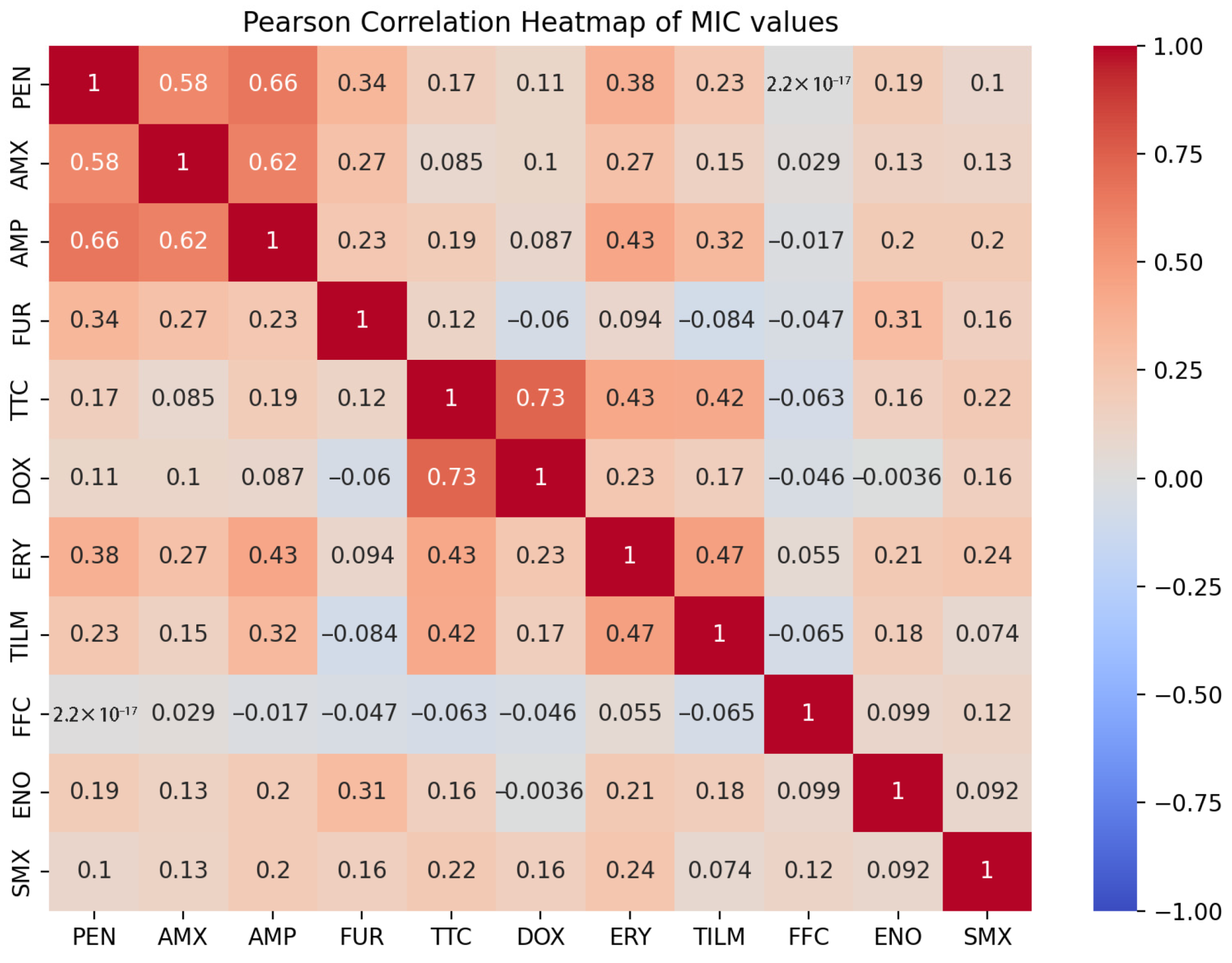
The same correlations were also detected when analyzing MIC values. In addition, a correlation was noted within the class of macrolides (r = 0.47), as well as between macrolides and tetracycline (r = 0.43) (Figure 5).
Figure 5. Pearson correlation heatmap based on MIC values from broth microdilution assays for P. multocida isolates. The phi values (r) range from −1 to +1, where +1 indicates a perfect positive correlation (resistance to both drugs tends to co-occur), 0 indicates no linear relationship, and −1 indicates a perfect negative correlation (resistance to one drug is associated with susceptibility to the other). Correlations were calculated using resistance data (1 = resistant; 0.5 = intermediate; 0 = susceptible) based on CLSI breakpoints.
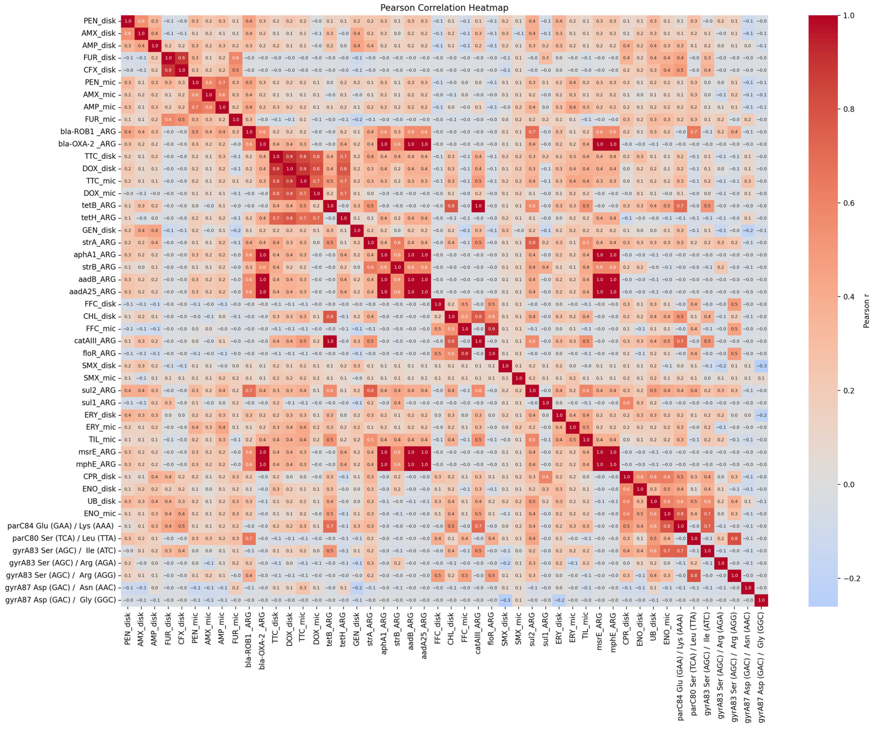
For fluoroquinolones, a correlation was observed between phenotypically confirmed resistance and the SNPs in parC (Glu84Lys, r = 0.41–0.77) and in gyrA (Ser83Ile, r = 0.36–0.74). The strongest correlations were found between enrofloxacin MIC values and the two aforementioned SNPs (r = 0.74–0.77). The other SNPs showed no significant correlation with the phenotypic results. A negligible correlation was observed between phenotypic resistance to gentamicin and the resistance genes, whereas a strong correlation was noted between the occurrence of ARGs responsible for aminoglycoside resistance (r = 0.36–1.0). A very low correlation was detected between the phenotypic results of sulfamethoxazole, penicillins, cephalosporins, macrolides, and the ARGs responsible for resistance. In the case of tetracyclines, strong positive correlations were observed between the results of phenotypic methods and the tetH (r = 0.7–0.8) and a positive correlation in the case of tetB (r = 0.2–0.5). Similarly, the phenotypic results for florfenicol were highly correlated with the floR gene (r = 0.5–0.9), and the disk diffusion results for chloramphenicol with the catAIII gene (r = 0.8) (Figure 6).
Figure 6. Integrated correlation heatmap showing relationships among phenotypic susceptibility, MIC values, and resistance gene/SNP presence. The phi values (r) range from −1 to +1, where +1 indicates a perfect positive correlation, 0 indicates no linear relationship, and −1 indicates a perfect negative correlation (resistance to one drug is associated with susceptibility to the other). Correlations were calculated using resistance data (1 = resistant or contain ARG/SNP; 0.5 = intermediate; 0 = susceptible) based on CLSI breakpoints.

3. Discussion

In this study, the antibiotic susceptibility profiles of 80 P. multocida strains were determined using phenotypic and genotypic methods. To date, phenotypic tests, primarily the disk diffusion assay, have been used to establish diagnostic and therapeutic protocols []. However, several studies have reported that disk diffusion is less reliable due to its low reproducibility and sensitivity, and MIC determination is preferred [,]. Comparing the results of the two phenotypic methods, the only significant difference was for antibiotics belonging to the penicillin class. This may be because the disk diffusion breakpoints used in this study were calculated values [], which mainly referred to P. multocida strains of porcine origin, and the lack of an intermediate category made the comparison with the MIC values very limited. Between the two phenotypic tests, MIC values exhibited stronger positive correlations with genotypic findings for macrolides, enrofloxacin, and phenicols. So, in these cases, the working hypothesis was confirmed. In contrast, disk diffusion values showed higher correlation coefficients for tetracycline-class antibiotics. Both phenotypic assays showed that the majority of MDR strains were isolated during the 2010s, mainly from avian hosts in Hungary.
Disk diffusion results showed that avian strains were more susceptible to cephalosporins, whereas mammalian strains were more susceptible to fluoroquinolones, sulfamethoxazole, tetracyclines, and erythromycin. In terms of MIC values, the Hungarian strains were more susceptible to sulfamethoxazole, amoxicillin, and tetracyclines than the French isolates. Avian strains showed lower MIC values for enrofloxacin, tilmicosin, ampicillin, and penicillin than mammalian strains. These differences are probably due to different treatment protocols and antibiotics used in each country and species.
Based on the phenotypic methods, the majority of the strains were highly susceptible to phenicols. Although no resistance was detected by disk diffusion, the broth microdilution method indicated two resistant strains. These strains were of avian origin and harbored the floR resistance gene, confirming the results obtained from the MIC values. Resistance to chloramphenicol was only tested by disk diffusion, which identified three resistant strains (two mammalian, one avian). Two of the strains carried the catAIII gene responsible for chloramphenicol resistance, while one strain carried the floR gene. The phenotypic results of florfenicol highly correlated with the floR gene (r = 0.5–0.9), and the disk diffusion results of chloramphenicol with the catAIII gene (r = 0.8). All resistant strains originated from Hungary. The high susceptibility of P. multocida strains from Hungary to phenicols has been reported previously. Sellyei et al. observed high susceptibility to florfenicol and chloramphenicol in strains of avian and porcine origin []. More recently, Kerek et al. have shown similarly high susceptibility to florfenicol in poultry strains []. Similar susceptibility profiles have been observed globally [,,], although recently the number of resistant strains has been increasing [,,].
The tested strains were highly susceptible to cephalosporins. Phenotypic susceptibility to cephalexin was tested by disk diffusion only, and no resistant strains were observed. Only one strain showed reduced susceptibility (ID: 4149), this bovine strain was also resistant to ceftiofur. By disk diffusion, only one strain was resistant to ceftiofur, while three resistant strains were detected by MIC testing. All these strains were originated from Hungary. Our findings are consistent with the observations previously reported in the scientific literature that cephalosporin antibiotics are highly effective for the treatment of pasteurellosis [,,,,,].
Within the tetracycline class, tetracycline and doxycycline were tested, and both phenotypic methods showed the latter to be more effective against the strains. Resistance to tetracycline was found in almost equal proportions by the two methods, whereas for doxycycline, the degree of resistance was higher by disk diffusion. Of the resistant or intermediate strains detected by phenotypic methods, almost all harbored tetH or tetB resistance genes. A strong positive correlation was observed between the results of phenotypic methods and the tetH (r = 0.7–0.8), and a positive correlation with tetB (r = 0.2–0.5). Overall, these results indicated that the Hungarian and French strains are essentially susceptible to tetracyclines. Resistant strains were more frequently identified from avian sources. Some authors have found similarly high levels of susceptibility [,]; however, the number of reports of resistance to these drugs has been increasing [,,,,].
In general, P. multocida strains show high susceptibility to ciprofloxacin and enrofloxacin [,,,,,], but the number of resistant bacterial strains is increasing year by year [,]. In this study, depending on the phenotypic method, 83.8–90.0% of strains were susceptible to these antibiotics. Resistance is mainly due to mutations affecting the gyrA, gyrB, and parC genes. Based on literature data, the most significant amino acid changes are Ser83Ile, Asp87Gly, and Asp87Asn (gyrA); Glu84Lys and Ser80Leu (parC); and Pro415Thr and Asp426Asn (gyrB) [,,,,]. No significant mutations were detected in the gyrB gene among the sequenced isolates. However, correlations were observed between phenotypically confirmed resistance and the SNPs parC Glu84Lys (r = 0.41–0.77) and gyrA Ser83Ile (r = 0.36–0.74). The strongest correlations were detected between the MIC values of enrofloxacin and the two aforementioned SNPs (r = 0.74–0.77). No correlation was found between other observed amino acid changes and phenotypic resistance.
Eighty-eight percent of the strains tested were susceptible to tilmicosin, and this sensitivity is globally widespread [,,]. Moderate susceptibility to erythromycin is quite common in P. multocida strains [,,], which is supported by the present observations. The erm (42) gene responsible for erythromycin resistance was not detected, and only a small proportion of genes responsible for other macrolide resistance were found, although a positive correlation (r = 0.4) between phenotypic and genotypic resistance was observed for tilmicosin regarding msrE and mphE. In addition to resistance genes, P. multocida can develop macrolide resistance through efflux mechanisms, target modification and drug inactivation, all of which can occur independently of resistance genes [].
Based on MIC values, a total of eight strains were resistant to all three tested penicillin antibiotics. These were all Hungarian strains, the majority (75%) of which were isolated from mammals. Disk diffusion showed resistance against all penicillins in nine strains. These strains, except for one isolate, were Hungarian, mostly found in mammals. Overall, compared to the MIC values (S: 47.5–72.5%), the strains showed higher susceptibility (S: 67.5–83.8%) to these drugs using the disk diffusion method. Again, this is probably because disk diffusion breakpoints were calculated values and the lack of an intermediate category made it very difficult to compare the results with the MIC values. In a number of cases, isolates were found to be resistant to a particular penicillin antibiotic, but the genetic background for this phenotypic resistance could not be confirmed. Sahay et al. observed a similar phenomenon: by disk diffusion, 46% of the strains tested showed resistance to penicillin, but none of the isolates carried either the bla-OXA-2 or bla-ROB-1 gene []. In this study, the bla-ROB-1 resistance gene was only present in strains that were found to be resistant to all antibiotics in the penicillins class by both methods. Furthermore, the analysis of these antibiotics shows that P. multocida strains had a diverse susceptibility profile. Tang et al. reported 100% resistance to amoxicillin in porcine strains in China []. On the other hand, Vilaró et al. reported high susceptibility (96.2%) to amoxicillin in strains form swine in Spain []. Timsit et al. reported 100% penicillin susceptibility using a microdilution method, while others described 95% sensitivity [,]. Sabsabi et al. assessed the antibiotic susceptibility in Malaysian poultry strains using disk diffusion and found 14% resistance to both penicillin and amoxicillin []. Schönecker et al. observed 27% resistance to penicillin, while Xiao et al. described 28% resistance to amoxicillin [,].
Gentamicin is recognized worldwide as an effective drug against P. multocida [,,,,]. The genotypic resistance was mainly due to genes strA (aminoglycoside-3-phosphotransferase), strB (aminoglycoside-6-phosphotransferase), aadA1 (aminoglycoside-3-adenyltransferase), aadA14 (adenyltransferase), aphA1 [aph (3’)-Ia] (aminoglycoside-3-phosphotransferase), aphA3 [aph (3’)-III], and aadB [ant (2”)-Ia], which may also be plasmid encoded [,,]. However, gentamicin, representing aminoglycosides in this study and tested only by disk diffusion, showed 16% resistance and a relatively high rate of reduced sensitivity (27%). On the other hand, the aforementioned genes were detected only in the minority of strains. Although aminoglycoside resistance in P. multocida is generally associated with resistance genes, other mechanisms may also contribute to reduced drug sensitivity including ribosomal mutations or modifications, biofilm formation, and variations in virulence factors and capsular serotypes [].
Nearly 70% of the strains were resistant to sulfamethoxazole by both phenotypic methods. French and avian strains were more resistant than Hungarian and mammalian isolates. Despite the high phenotypic resistance rate, only eight strains possessed either the sul1 or sul2 gene responsible for sulfonamide resistance. The observed phenotypic resistance rate is a widely described phenomenon [,,,]. Several publications have reported the frequent occurrence of the sul1 and sul2 genes in P. multocida isolates [], typically located on various plasmids [,]. In addition to resistance genes, P. multocida can also develop sulfonamide resistance through various mechanisms, including altered membrane permeability, efflux pumps, and changes in target enzyme. These mechanisms either prevent sulfonamides from reaching their target or reduce the effectiveness of the drug []. Although further analyses, such as transcriptome sequencing or enzyme structure studies, were not carried out, our results indicate that these mechanisms may be rather prevalent among P. multocida strains than the carriage of sul1 and sul2 genes.
Absolute resistance to clindamycin was observed with both phenotypic methods. Clindamycin targets the 50S ribosomal subunit, and methylation of the adenine residues in the 23S rRNA prevents antibiotic binding to its site []. The point mutation in the 23S rRNA gene responsible for clindamycin resistance was identified in all but four strains using BV-BRC. High rates of resistance to clindamycin in P. multocida strains have been reported in several previous publications [,,,]; therefore, it is not recommended for the treatment of pasteurellosis [].

4. Materials and Methods

4.1. Bacterial Strains

A total of 80 P. multocida isolates were examined, the majority of which originated from Hungary (n = 64) and a smaller proportion from France (n = 16). The strains were isolated between 2004 and 2023 from various mammalian (n = 38) and avian (n = 42) species, which were distributed among the host species as follows: cattle (n = 25), goose (n = 18), turkey (n = 10), duck (n = 10), albatross (n = 3), sheep (n = 3), goat (n = 3), swine (n = 3), dog (n = 1), human (n = 1), rabbit (n = 1), and fallow deer (n = 1). Samples were cultured on Columbia agar plates (Columbia agar, LAB M, Bury, UK) containing 5% sheep blood and incubated at 37 °C for 24 h under aerobic conditions. The identity of the strains was confirmed by PCR targeting the kmt gene, as described by Townsend et al. (1998) [].

4.2. Antibiotic Susceptibility Testing

In vitro phenotypic susceptibility of P. multocida isolates was evaluated by broth microdilution method and disk diffusion against nine antibiotic classes (penicillins, cephalosporins, aminoglycosides, tetracyclines, lincosamides, macrolides, phenicols, sulfonamides, and fluoroquinolones) according to the Clinical and Laboratory Standards Institute (CLSI) guidelines []. The selection of these antibiotic classes was based on their common use in veterinary medicine and the need to assess resistance patterns relevant to clinical practice.
For disk diffusion, 16 antimicrobial agents (Biolab Zrt., Budapest, Hungary) were tested: penicillin G (10 U), amoxicillin (10 µg), ampicillin (10 µg), ceftiofur (30 µg), cephalexin (30 µg), gentamicin (10 µg), tetracycline (30 µg), doxycycline (30 µg), clindamycin (2 µg), erythromycin (15 µg), florfenicol (30 µg), chloramphenicol (30 µg), sulfamethoxazole (300 U), enrofloxacin (5 µg), ciprofloxacin (5 µg), and flumequine (30 µg). Results were analyzed 24 h after inoculation. Quality control was ensured using Escherichia coli ATCC 25922 and Staphylococcus aureus ATCC 25923, which served as reference strains to validate the accuracy of the antibiotic susceptibility testing.
In the broth microdilution, 14 antimicrobial agents (Sigma-Aldrich, St. Louis, MO, USA) were examined: penicillin (0.015 to 16 µg/mL), amoxicillin (0.06 to 64 µg/mL), ampicillin (0.06 to 64 µg/mL), ceftiofur (0.015 to 16 µg/mL), apramycin (0.06 to 64 µg/mL), tetracycline (0.03 to 32 µg/mL), doxycycline (0.06 to 64 µg/mL), clindamycin (0.125 to 128 µg/mL), erythromycin (0.06 to 64 µg/mL), tilmicosin (0.25 to 256 µg/mL), florfenicol (0.06 to 64 µg/mL), sulfamethoxazole (0.5 to 512 µg/mL), enrofloxacin (0.015 to 16 µg/mL), and flumequine (0.06 to 64 µg/mL). For quality control, Escherichia coli ATCC 25922 and Staphylococcus aureus ATCC 29213 were used. After incubation at 37 °C for 24 h, the turbidity (quantified) of the suspensions was measured at 450 nm using a microplate reader. The MIC was defined as the lowest concentration that completely inhibited the bacterial growth. MIC50 (inhibiting the growth of 50% of tested isolates) and MIC90 (inhibiting the growth of 90% of tested isolates) values were also calculated.
The obtained values were classified into three categories (susceptible, intermediate, or resistant) according to CLSI recommendations [,]. For doxycycline, the range set for tetracycline was used. For antibiotics of the penicillin class, no CLSI limit was available in the case of disk diffusion. For these antibiotics, the limits calculated by Kumakawa et al. (2025) were used []. A strain was considered MDR if it was resistant to at least one agent representing three or more antibiotic classes []. MIC values were also evaluated based on EUCAST breakpoints (https://mic.eucast.org/search/, accessed on 17 August 2025).

4.3. Whole Genome Sequencing and Bioinformatical Analyses

A loopful of bacteria was suspended in 2 mL of Brain Heart Infusion Broth (BHI, Merck, Darmstadt, Germany) and incubated at 37 °C for 24 h under aerobic conditions. Genomic DNA was extracted using the Quick-DNA Fungal/Bacterial Miniprep Kit (Zymo Research Corporation, Irvine, CA, USA) according to the manufacturer’s instructions. The concentration of the obtained DNA was checked using a Nanodrop spectrophotometer (Thermo Fisher Scientific, Wilmington, DE, USA). Whole-genome sequencing of the strains was accomplished by SeqOmics Biotechnology Ltd. (Mórahalom, Hungary) on the Illumina MiSeq platform (San Diego, CA, USA). Sequences read quality was assessed using FastQC (v0.12.1). Reads were trimmed with BBDuk in Geneious Prime v. 2025.0.3 []. De novo genome assembly was conducted with SPAdes v. 4.0.0 integrated in Geneious Prime. Genome annotation was performed using Geneious Prime and the HN07 reference genome (NZ_CP007040). To identify SNPs in the gyrA, gyrB, and parC genes, the sequences were aligned to the corresponding loci of the HN07 reference strain. ARGs sequences or SNPs were evaluated in paired reads using BV-BRC (https://www.bv-brc.org/) and the Comprehensive Antibiotic Resistance Database (CARD) by Resistance Gene Identifier (RGI) (https://card.mcmaster.ca/analyze/rgi, accessed on 6 May 2025), with 90% identity cut-off.

4.4. Statistical Analyses

Correlation analyses were performed in R 4.2.1 to investigate the relationships between the resistance observed by the two phenotypic methods and the presence of ARGs and SNPs. Pearson’s correlation matrix was used to quantify the relationships. Heatmaps were generated using Julius.AI (https://julius.ai/, accessed on 27 June 2025).

5. Conclusions

In conclusion, phenicols and cephalosporins were the most effective drugs against P. multocida, though most strains are highly susceptible to fluoroquinolones and tetracyclines as well. However, the declining susceptibility rate of fluoroquinolones is a cause for concern. Enrofloxacin is a critically important antibiotic and has a key role in human medicine. The background of decreasing susceptibility is probably due to overuse or inappropriate application; therefore, it is not recommended as first-line treatment for P. multocida infections in poultry considering One Health perspective. Elevated resistance rates were observed for clindamycin and sulfamethoxazole, although genotypic resistance was not always confirmed for the latter. The resistances to β-lactams and macrolides observed by phenotypic testing remains genetically unexplained and further studies, such as transcriptomic or proteomic analyses, are needed to investigate differential gene expression or protein modifications associated with resistance. Of the two phenotypic methods, MIC values showed a stronger positive correlation with genotypic results, making it a more suitable method for determining antibiotic susceptibility. This method is particularly recommended for testing antibiotics of the penicillin class for P. multocida. The significant economic impact of P. multocida infections on animal husbandry underscores the importance of continuous monitoring of antimicrobial resistance to guide treatment protocols and maintain efficacy.

Supplementary Materials

The following supporting information can be downloaded at: https://www.mdpi.com/article/10.3390/antibiotics14090906/s1, Table S1: Metadata of P. multocida isolates (n = 80) including origin, host species, year of isolation, and BioSample accession numbers; Table S2: Phenotypic and genotypic antibiotic susceptibility profiles of P. multocida isolates across B-lactams; Table S3: Phenotypic and genotypic antibiotic susceptibility profiles of P. multocida isolates across tetracyclines; Table S4: Phenotypic and genotypic antibiotic susceptibility profiles of P. multocida isolates across aminoglycosides; Table S5: Phenotypic and genotypic antibiotic susceptibility profiles of P. multocida isolates across phenicols; Table S6: Phenotypic and genotypic antibiotic susceptibility profiles of P. multocida isolates across sulfonamids; Table S7: Phenotypic and genotypic antibiotic susceptibility profiles of P. multocida isolates across macrolides; Table S8: Phenotypic and genotypic antibiotic susceptibility profiles of P. multocida isolates across fluoroquinolones; Table S9: Phenotypic and genotypic antibiotic susceptibility profiles of P. multocida isolates across lincosamids.

Author Contributions

Conceptualization, K.P. and T.M.; Sample resources, T.M. and H.G.; formal analysis, K.P., E.W., M.D., and T.M.; investigation, K.P.; data curation, K.P.; writing—original draft preparation, K.P.; writing—review and editing, E.W., M.D., and T.M.; visualization, K.P. and M.D. All authors have read and agreed to the published version of the manuscript.

Funding

This research has been implemented with the support provided by the Ministry of Innovation and Technology of Hungary (legal successor: Ministry of Culture and Innovation of Hungary) from the National Research, Development, and Innovation Fund, financed under the TKP2021-EGA-01 and 2024-2.1.1-EKÖP (project no. 2024-2.1.1-EKÖP-2024-00018, granted for Krisztina Pintér) funding scheme of the National Research, Development, and Innovation Office. Additional support was provided by the National Laboratory for Infectious Animal Diseases, Antimicrobial Resistance, Veterinary Public Health, and Food Chain Safety, RRF-2.3.1-21-2022-00001.

Institutional Review Board Statement

Not applicable.

Data Availability Statement

Raw sequencing data for the P. multocida isolates have been deposited to the NCBI Sequence Read Archive under BioProject ID PRJNA1270947 and PRJNA1288180. (Will be available as soon as the necessary publications are accepted, or by 31 January 2026 at the latest.) Accession numbers are listed in Supplementary Table S1.

Acknowledgments

During the preparation of this study, the authors used Julius.AI for data visualization purposes. The authors have reviewed and edited the output and take full responsibility for the content of this publication.

Conflicts of Interest

Author Hubert Gantelet was employed by the company Ceva Biovac. The remaining authors declare that the research was conducted in the absence of any commercial or financial relationships that could be construed as a potential conflict of interest.

Abbreviations

The following abbreviations are used in this manuscript:
ARGantimicrobial resistance gene
BHIBrain Heart Infusion
BV-BRC-Bacterial and Viral Bioinformatics Resource Center
CARDComprehensive Antibiotic Resistance Database
CLSIClinical and Laboratory Standards Institute
ECOFFepidemiological cut-off value
EUCASTEuropean Committee on Antimicrobial Susceptibility Testing
MDRmultidrug-resistant
MICminimal inhibitory concentration
PCRpolymerase chain reaction
RGIResistance Gene Identifier
SNPsingle-nucleotide polymorphism

References

  1. Rhoades, K.R.; Rimler, R.B. Fowl Cholera. In Pasteurella and Pasteurellosis; Adam, C., Rutter, J.M., Eds.; Academic Press Limited: London, UK, 1989; pp. 95–113. ISBN 978-0-12-044274-4. [Google Scholar]
  2. Magyar, T.; Lax, A.J. Atrophic Rhinitis. In Polymicrobial Diseases; ASM Press: Washington, DC, USA, 2002. [Google Scholar]
  3. De Alwis, M.C.L. Haemorrhagic Septicaemia; ACIAR Monograph Series; Australian Centre for International Agricultural Research: Canberra, Australia, 1999; ISBN 978-1-86320-269-5. [Google Scholar]
  4. Wilson, B.A.; Ho, M. Pasteurella multocida: From Zoonosis to Cellular Microbiology. Clin. Microbiol. Rev. 2013, 26, 631–655. [Google Scholar] [CrossRef]
  5. Freshwater, A. Why Your Housecat’s Trite Little Bite Could Cause You Quite a Fright: A Study of Domestic Felines on the Occurrence and Antibiotic Susceptibility of Pasteurella multocida. Zoonoses Public Health 2008, 55, 507–513. [Google Scholar] [CrossRef]
  6. Bhargav, A.; Gupta, S.; Seth, S.; James, S.; Fatima, F.; Chaurasia, P.; Ramachandran, S. Knowledgebase of Potential Multifaceted Solutions to Antimicrobial Resistance. Comput. Biol. Chem. 2022, 101, 107772. [Google Scholar] [CrossRef] [PubMed]
  7. Sy, C.L.; Chen, P.-Y.; Cheng, C.-W.; Huang, L.-J.; Wang, C.-H.; Chang, T.-H.; Chang, Y.-C.; Chang, C.-J.; Hii, I.-M.; Hsu, Y.-L.; et al. Recommendations and Guidelines for the Treatment of Infections Due to Multidrug Resistant Organisms. J. Microbiol. Immunol. Infect. 2022, 55, 359–386. [Google Scholar] [CrossRef] [PubMed]
  8. Mackenzie, J.S.; Jeggo, M. The One Health Approach—Why Is It So Important? Trop. Med. Infect. Dis. 2019, 4, 88. [Google Scholar] [CrossRef] [PubMed]
  9. Magiorakos, A.-P.; Srinivasan, A.; Carey, R.B.; Carmeli, Y.; Falagas, M.E.; Giske, C.G.; Harbarth, S.; Hindler, J.F.; Kahlmeter, G.; Olsson-Liljequist, B.; et al. Multidrug-Resistant, Extensively Drug-Resistant and Pandrug-Resistant Bacteria: An International Expert Proposal for Interim Standard Definitions for Acquired Resistance. Clin. Microbiol. Infect. 2012, 18, 268–281. [Google Scholar] [CrossRef]
  10. Rubin, J.E. Antimicrobial Susceptibility Testing Methods and Interpretation of Results. In Antimicrobial Therapy in Veterinary Medicine; John Wiley & Sons, Ltd.: West Sussex, UK, 2013; pp. 11–20. ISBN 978-1-118-67501-4. [Google Scholar]
  11. Enne, V.I.; Bennett, P.M. Methods to Determine Antibiotic Resistance Gene Silencing. In Antibiotic Resistance Protocols, 2nd ed.; Methods in Molecular Biology; Gillespie, S.H., McHugh, T.D., Eds.; Humana Press: Totowa, NJ, USA, 2010; pp. 29–44. ISBN 978-1-60327-279-7. [Google Scholar]
  12. Zankari, E.; Hasman, H.; Kaas, R.S.; Seyfarth, A.M.; Agersø, Y.; Lund, O.; Larsen, M.V.; Aarestrup, F.M. Genotyping Using Whole-Genome Sequencing Is a Realistic Alternative to Surveillance Based on Phenotypic Antimicrobial Susceptibility Testing. J. Antimicrob. Chemother. 2013, 68, 771–777. [Google Scholar] [CrossRef]
  13. Oniciuc, E.A.; Likotrafiti, E.; Alvarez-Molina, A.; Prieto, M.; Santos, J.A.; Alvarez-Ordóñez, A. The Present and Future of Whole Genome Sequencing (WGS) and Whole Metagenome Sequencing (WMS) for Surveillance of Antimicrobial Resistant Microorganisms and Antimicrobial Resistance Genes across the Food Chain. Genes 2018, 9, 268. [Google Scholar] [CrossRef]
  14. Martineau, F.; Picard, F.J.; Lansac, N.; Ménard, C.; Roy, P.H.; Ouellette, M.; Bergeron, M.G. Correlation between the Resistance Genotype Determined by Multiplex PCR Assays and the Antibiotic Susceptibility Patterns of Staphylococcus aureus and Staphylococcus epidermidis. Antimicrob. Agents Chemother. 2000, 44, 231–238. [Google Scholar] [CrossRef]
  15. Kerek, Á.; Szabó, Á.; Jerzsele, Á. Antimicrobial Susceptibility Profiles of Pasteurella multocida Isolates from Clinical Cases of Waterfowl in Hungary between 2022 and 2023. Vet. Sci. 2024, 11, 194. [Google Scholar] [CrossRef]
  16. Kumakawa, M.; Akama, R.; Hosoi, Y.; Hiraoka, Y.; Harada, S.; Matsuda, M.; Kawanishi, M.; Sekiguchi, H. Establishing Tentative Cut-off Values for Disk Diffusion Method for Pasteurella multocida and Mannheimia haemolytica from Livestock Animals in Japan. J. Vet. Med. Sci. 2025, 87, 349–355. [Google Scholar] [CrossRef]
  17. Sellyei, B.; Varga, Z.; Szentesi-Samu, K.; Kaszanyitzky, É.; Magyar, T. Antimicrobial Susceptibility of Pasteurella multocida Isolated from Swine and Poultry. Acta Vet. Hung. 2009, 57, 357–367. [Google Scholar] [CrossRef]
  18. Cid, D.; Fernández-Garayzábal, J.F.; Pinto, C.; Domínguez, L.; Vela, A.I. Antimicrobial Susceptibility of Pasteurella multocida Isolated from Sheep and Pigs in Spain—Short Communication. Acta Vet. Hung. 2019, 67, 489–498. [Google Scholar] [CrossRef] [PubMed]
  19. Petrocchi-Rilo, M.; Gutiérrez-Martín, C.-B.; Pérez-Fernández, E.; Vilaró, A.; Fraile, L.; Martínez-Martínez, S. Antimicrobial Resistance Genes in Porcine Pasteurella multocida Are Not Associated with Its Antimicrobial Susceptibility Pattern. Antibiotics 2020, 9, 614. [Google Scholar] [CrossRef] [PubMed]
  20. Schönecker, L.; Schnyder, P.; Schüpbach-Regula, G.; Meylan, M.; Overesch, G. Prevalence and Antimicrobial Resistance of Opportunistic Pathogens Associated with Bovine Respiratory Disease Isolated from Nasopharyngeal Swabs of Veal Calves in Switzerland. Prev. Vet. Med. 2020, 185, 105182. [Google Scholar] [CrossRef] [PubMed]
  21. Zhu, D.; Yuan, D.; Wang, M.; Jia, R.; Chen, S.; Liu, M.; Zhao, X.; Yang, Q.; Wu, Y.; Zhang, S.; et al. Emergence of a Multidrug-Resistant Hypervirulent Pasteurella multocida ST342 Strain with a floR-Carrying Plasmid. J. Glob. Antimicrob. Resist. 2020, 20, 348–350. [Google Scholar] [CrossRef]
  22. Jeong, J.; Kang, M.S.; Jeong, O.M.; Lee, H.J.; Lee, J.Y.; Kwon, Y.K.; Park, J.W.; Kim, J.H. Investigation of Genetic Diversity of Pasteurella multocida Isolated from Diseased Poultry in Korea. Braz. J. Poult. Sci. 2021, 23, 1–10. [Google Scholar] [CrossRef]
  23. Sabsabi, M.A.; Zakaria, Z.; Abu, J.; Faiz, N.M. Molecular Characterisation and Antibiotic Sensitivity Profile of Pasteurella multocida Isolated from Poultry Farms in Malaysia. Austral J. Vet. Sci. 2021, 53, 121–126. [Google Scholar] [CrossRef]
  24. Vilaró, A.; Novell, E.; Enrique-Tarancón, V.; Balielles, J.; Vilalta, C.; Martinez, S.; Fraile Sauce, L.J. Antimicrobial Susceptibility Pattern of Porcine Respiratory Bacteria in Spain. Antibiotics 2020, 9, 402. [Google Scholar] [CrossRef]
  25. Oh, Y.-H.; Moon, D.-C.; Lee, Y.J.; Hyun, B.-H.; Lim, S.-K. Antimicrobial Resistance of Pasteurella multocida Strains Isolated from Pigs between 2010 and 2016. Vet. Rec. Open 2018, 5, e000293. [Google Scholar] [CrossRef]
  26. Jones, K.H.; Thornton, J.K.; Zhang, Y.; Mauel, M.J. A 5-Year Retrospective Report of Gallibacterium anatis and Pasteurella multocida Isolates from Chickens in Mississippi. Poult. Sci. 2013, 92, 3166–3171. [Google Scholar] [CrossRef]
  27. Khamesipour, F.; Momtaz, H.; Azhdary Mamoreh, M. Occurrence of Virulence Factors and Antimicrobial Resistance in Pasteurella multocida Strains Isolated from Slaughter Cattle in Iran. Front. Microbiol. 2014, 5, 536. [Google Scholar] [CrossRef]
  28. Tang, X.; Zhao, Z.; Hu, J.; Wu, B.; Cai, X.; He, Q.; Chen, H. Isolation, Antimicrobial Resistance, and Virulence Genes of Pasteurella multocida Strains from Swine in China. J. Clin. Microbiol. 2009, 47, 951–958. [Google Scholar] [CrossRef]
  29. Xiao, J.; Li, Y.; Hu, Z.; Zhang, Y.; Chang, Y.-F.; Zhou, Q.; Yan, Z.; Zhang, X.; Chen, L.; Li, W.; et al. Characterization of Pasteurella multocida Isolated from Ducks in China from 2017 to 2019. Microb. Pathog. 2021, 160, 105196. [Google Scholar] [CrossRef] [PubMed]
  30. Vila, J.; Ruiz, J.; Goñi, P.; De Anta, M.T. Detection of Mutations in parC in Quinolone-Resistant Clinical Isolates of Escherichia coli. Antimicrob. Agents Chemother. 1996, 40, 491–493. [Google Scholar] [CrossRef] [PubMed]
  31. Li, X.-S.; Qi, Y.; Xue, J.-Z.; Xu, G.-Y.; Xu, Y.-X.; Li, X.-Y.; Muhammad, I.; Kong, L.-C.; Ma, H.-X. Transcriptomic Changes and satP Gene Function Analysis in Pasteurella multocida with Different Levels of Resistance to Enrofloxacin. Vet. Sci. 2023, 10, 257. [Google Scholar] [CrossRef] [PubMed]
  32. Kong, L.-C.; Gao, D.; Gao, Y.-H.; Liu, S.-M.; Ma, H.-X. Fluoroquinolone Resistance Mechanism of Clinical Isolates and Selected Mutants of Pasteurella multocida from Bovine Respiratory Disease in China. J. Vet. Med. Sci. 2014, 76, 1655–1657. [Google Scholar] [CrossRef]
  33. Cárdenas, M.; Barbé, J.; Llagostera, M.; Miró, E.; Navarro, F.; Mirelis, B.; Prats, G.; Badiola, I. Quinolone Resistance-Determining Regions of gyrA and parC in Pasteurella multocida Strains with Different Levels of Nalidixic Acid Resistance. Antimicrob. Agents Chemother. 2001, 45, 990–991. [Google Scholar] [CrossRef]
  34. Dasgupta, N.; Paul, D.; Chanda, D.D.; Chetri, S.; Chakravarty, A.; Bhattacharjee, A. Observation of a New Pattern of Mutations in gyrA and parC within Escherichia coli Exhibiting Fluroquinolone Resistance. Indian J. Med. Microbiol. 2018, 36, 131–135. [Google Scholar] [CrossRef]
  35. Dayao, D.A.E.; Gibson, J.S.; Blackall, P.J.; Turni, C. Antimicrobial Resistance in Bacteria Associated with Porcine Respiratory Disease in Australia. Vet. Microbiol. 2014, 171, 232–235. [Google Scholar] [CrossRef]
  36. Nor Amdan, N.A.; Shahrulzamri, N.A.; Hashim, R.; Mohamad Jamil, N. Understanding the Evolution of Macrolides Resistance: A Mini Review. J. Glob. Antimicrob. Resist. 2024, 38, 368–375. [Google Scholar] [CrossRef]
  37. Sahay, S.; Natesan, K.; Prajapati, A.; Kalleshmurthy, T.; Shome, B.R.; Rahman, H.; Shome, R. Prevalence and Antibiotic Susceptibility of Mannheimia haemolytica and Pasteurella multocida Isolated from Ovine Respiratory Infection: A Study from Karnataka, Southern India. Vet. World 2020, 13, 1947–1954. [Google Scholar] [CrossRef]
  38. Timsit, E.; Hallewell, J.; Booker, C.; Tison, N.; Amat, S.; Alexander, T.W. Prevalence and Antimicrobial Susceptibility of Mannheimia haemolytica, Pasteurella multocida, and Histophilus somni Isolated from the Lower Respiratory Tract of Healthy Feedlot Cattle and Those Diagnosed with Bovine Respiratory Disease. Vet. Microbiol. 2017, 208, 118–125. [Google Scholar] [CrossRef] [PubMed]
  39. Kehrenberg, C.; Tham, N.T.T.; Schwarz, S. New Plasmid-Borne Antibiotic Resistance Gene Cluster in Pasteurella multocida. Antimicrob. Agents Chemother. 2003, 47, 2978–2980. [Google Scholar] [CrossRef] [PubMed]
  40. Michael, G.B.; Kadlec, K.; Sweeney, M.T.; Brzuszkiewicz, E.; Liesegang, H.; Daniel, R.; Murray, R.W.; Watts, J.L.; Schwarz, S. ICEPmu1, an Integrative Conjugative Element (ICE) of Pasteurella multocida: Structure and Transfer. J. Antimicrob. Chemother. 2012, 67, 91–100. [Google Scholar] [CrossRef]
  41. Giguère, S. Antimicrobial Drug Action and Interaction. In Antimicrobial Therapy in Veterinary Medicine; John Wiley & Sons, Ltd.: West Sussex, UK, 2013; pp. 1–10. ISBN 978-1-118-67501-4. [Google Scholar]
  42. Garneau-Tsodikova, S.; Labby, K.J. Mechanisms of Resistance to Aminoglycoside Antibiotics: Overview and Perspectives. Medchemcomm 2016, 7, 11–27. [Google Scholar] [CrossRef]
  43. Bedier, E.T.; Labib, S.R.; Ahmed, A.M. Characterization of Antimicrobial Resistance Genes of Pasteurella multocida Isolated from Diseased Chickens in Egypt. J. Hell. Vet. Med. Soc. 2022, 73, 4165–4172. [Google Scholar] [CrossRef]
  44. San Millan, A.; Escudero, J.A.; Gutierrez, B.; Hidalgo, L.; Garcia, N.; Llagostera, M.; Dominguez, L.; Gonzalez-Zorn, B. Multiresistance in Pasteurella multocida Is Mediated by Coexistence of Small Plasmids. Antimicrob. Agents Chemother. 2009, 53, 3399–3404. [Google Scholar] [CrossRef]
  45. Eliopoulos, G.M.; Huovinen, P. Resistance to Trimethoprim-Sulfamethoxazole. Clin. Infect. Dis. 2001, 32, 1608–1614. [Google Scholar] [CrossRef]
  46. Kulczycka-Mierzejewska, K.; Sadlej, J.; Trylska, J. Molecular Dynamics Simulations Suggest Why the A2058G Mutation in 23S RNA Results in Bacterial Resistance against Clindamycin. J. Mol. Model. 2018, 24, 191. [Google Scholar] [CrossRef]
  47. Cross, S.L.; Gelfand, M. Pasteurella multocida Infection Treatment & Management: Approach Considerations, Medical Care, Surgical Care. In Pasteurella multocida Infection; Medscape: New York, NY, USA, 2025. [Google Scholar]
  48. Townsend, K.M.; Frost, A.J.; Lee, C.W.; Papadimitriou, J.M.; Dawkins, H.J. Development of PCR Assays for Species- and Type-Specific Identification of Pasteurella multocida Isolates. J. Clin. Microbiol. 1998, 36, 1096–1100. [Google Scholar] [CrossRef]
  49. Winkler, M.A.; Cockerill, F.R.; Craig, W.A.; Dudley, M.N.; Eliopoulos, G.M.; Hecht, D.W.; Hindler, J.A.; Lowery, D.E.; Sheehan, D.J.; Tenover, F.C.; et al. Performance Standards for Antimicrobial Disk Susceptibility Tests; Approved Standard: M2-A9, 9th ed.; Clinical and Laboratory Standards Institute: Wayne, PA, USA, 2006; ISBN 1-56238-586-0. [Google Scholar]
  50. Shryock, T.R.; Apley, M.; Jones, R.N.; Lein, D.H.; Thornsberry, C.; Walker, R.D.; Watts, J.L.; White, D.G.; Wu, C.C. Performance Standards for Antimicrobial Disk and Dilution Susceptibility Tests for Bacteria Isolated from Animals: Approved Standard, 2nd ed.; NCCLS Document; NCCLS: Wayne, PA, USA, 2002; ISBN 978-1-56238-461-6. [Google Scholar]
  51. Papich, M.G.; Simjee, S.; Apley, M.; Frana, T.S.; Knapp, C.C.; Lubbers, B.V.; Rose, M.; Schwarz, S.; Silley, P.; Sweeney, M.T.; et al. Performance Standards for Antimicrobial Disk Susceptibility Tests for Bacteria Isolated from Animals: CLSI Supplement VET01S; Replaces VET01-S2, 3rd ed.; Clinical and Laboratory Standards Institute: Wayne, PA, USA, 2015; ISBN 978-1-56238-908-6. [Google Scholar]
  52. Kearse, M.; Moir, R.; Wilson, A.; Stones-Havas, S.; Cheung, M.; Sturrock, S.; Buxton, S.; Cooper, A.; Markowitz, S.; Duran, C.; et al. Geneious Basic: An Integrated and Extendable Desktop Software Platform for the Organization and Analysis of Sequence Data. Bioinformatics 2012, 28, 1647–1649. [Google Scholar] [CrossRef]
Disclaimer/Publisher’s Note: The statements, opinions and data contained in all publications are solely those of the individual author(s) and contributor(s) and not of MDPI and/or the editor(s). MDPI and/or the editor(s) disclaim responsibility for any injury to people or property resulting from any ideas, methods, instructions or products referred to in the content.

Article Metrics

Citations

Article Access Statistics

Multiple requests from the same IP address are counted as one view.