Abstract
A major problem of our time is the ever-increasing resistance to antimicrobial agents in bacterial populations. One of the most effective ways to prevent these problems is to target antibacterial therapies for specific diseases. In this study, we investigated the in vitro effectiveness of florfenicol against S. suis, which can cause severe arthritis and septicemia in swine herds. The pharmacokinetic and pharmacodynamic properties of florfenicol in porcine plasma and synovial fluid were determined. After a single intramuscular administration of florfenicol at 30 mg/kgbw, the AUC0–∞ was 164.45 ± 34.18 µg/mL × h and the maximum plasma concentration was 8.15 ± 3.11 µg/mL, which was reached in 1.40 ± 0.66 h, whereas, in the synovial fluid, these values were 64.57 ± 30.37 µg/mL × h, 4.51 ± 1.16 µg/mL and 1.75 ± 1.16 h, respectively. Based on the MIC values of the 73 S. suis isolates tested, the MIC50 and MIC90 values were 2 µg/mL and 8 µg/mL, respectively. We successfully implemented a killing–time curve in pig synovial fluid as a matrix. Based on our findings, the PK/PD breakpoints of the bacteriostatic (E = 0), bactericidal (E = −3) and eradication (E = −4) effects of florfenicol were determined and MIC thresholds were calculated, which are the guiding indicators for the treatment of these diseases. The AUC24h/MIC values for bacteriostatic, bactericidal and eradication effects were 22.22 h, 76.88 h and 141.74 h, respectively, in synovial fluid, and 22.42 h, 86.49 h and 161.76 h, respectively, in plasma. The critical MIC values of florfenicol against S. suis regarding bacteriostatic, bactericidal and eradication effects in pig synovial fluid were 2.91 ± 1.37 µg/mL, 0.84 ± 0.39 µg/mL and 0.46 ± 0.21 µg/mL, respectively. These values provide a basis for further studies on the use of florfenicol. Furthermore, our research highlights the importance of investigating the pharmacokinetic properties of antibacterial agents at the site of infection and the pharmacodynamic properties of these agents against different bacteria in different media.
1. Introduction
Antimicrobial resistance (AMR) is one of the leading health issues of our time, in human and veterinary medicine alike. This is underlined by the increasingly stringent regulation of the use and consumption of antibacterial agents [1]. In addition to the time-consuming and costly development of new antibacterial agents, the other solution is the repositioning of already authorized agents for new, different indications. In this case, a good method may be to use pharmacokinetic/pharmacodynamic (PK/PD) analysis [2]. Florfenicol is an example of why the accurate dose and time interval of the treatment for different indications should be determined, as it has been shown to promote the selection of florfenicol-resistant Escherichia coli strains in the microbiota despite high concentrations in the pig gastrointestinal tract [3].
Florfenicol, a member of the phenicol group, is a broad-spectrum antibacterial agent with a bacteriostatic mode of action, which is achieved by binding to the 50S subunit of the ribosome via inhibition of the enzyme peptidyl transferase [4,5,6,7,8]. It is widely used in the pig industry to treat respiratory diseases caused by Pasteurella multocida, Actinobacillus pleuropneumoniae, Mycoplasma hyopneumoniae, M. hyorhinis, Glässer-disease caused by Glaesserella parasuis and septicemia, polyserositis, meningitis and arthritis caused by Streptococcus suis. Furthermore, it is also used to treat bovine, sheep, goats, poultry and fish [5,9,10,11,12,13,14,15,16,17,18,19,20,21,22,23]. Although most products containing florfenicol are not authorized for diseases caused by S. suis in the European Union, studies to date have demonstrated its efficacy in septicemia caused by S. suis [23,24,25].
In order to provide data for the use of florfenicol in additional diseases caused by S. suis, the pharmacokinetic (PK) properties of florfenicol at the site of infection need to be investigated [26,27]. The pharmacokinetics of florfenicol in pigs have already been investigated in plasma and the lung. The pharmacokinetic properties of florfenicol vary widely between individual pigs [28]. Overall, it has excellent absorption and distribution in the pig body system, with a very low binding to plasma protein [24,28,29,30,31,32]. In a previous study, we investigated the pharmacokinetics of florfenicol in pig synovial fluid at a dose of 15 mg/kgbw following a single intramuscular administration. Based on this, it was concluded that florfenicol can only be used to treat arthritis caused by S. suis in pig if the minimum inhibitory concentration (MIC) of S. suis is less than or equal to 1.42 µg/mL [23]. In addition to the pharmacokinetic parameters, it is very important to continuously monitor the sensitivity of S. suis strains against florfenicol [26] because most studies show that the susceptibility of S. suis to florfenicol is not always as clear, as indicated by the MIC50 and MIC90 values of 2 µg/mL or lower in almost all countries, which is the breakpoint set by Clinical and Laboratory Standards Institute [15,16,22,23,24,25].
Similar studies needed to use PK/PD analysis to determine the dose and duration of florfenicol treatments and the time interval between two drug administrations as accurately as possible.
In a previous study, we investigated the PK of florefnicol in pig plasma and synovial fluid at a dose of 15 mg/kgbw following a single intramuscular administration, in which the results were inconclusive and we could not conclude that florfenicol can be recommended for the treatment of pig arthritis caused by S. suis in the approved dosage regimen. The aim of the present study was to determine the plasma and synovial PK of florfenicol at a dose of 30 mg/kgbw following a single intramuscular injection; thus, at a dose higher than the approved dosage regimen. In addition, the in vitro efficacy of florfenicol against S. suis bacteria isolated from clinical lesions in Hungary was investigated using PK/PD analysis. Moreover, our aim was to test the efficacy of florfenicol against S. suis strains in an in vitro experiment, characterized, in this case, by synovial fluid. For this purpose, we prepared killing curves of an S. suis isolate (SS96) in pig synovial fluid, plasma and cation-adjusted Mueller–Hinton broth (CA-MHB).
2. Results
2.1. Pharmacokinetics of Florfenicol
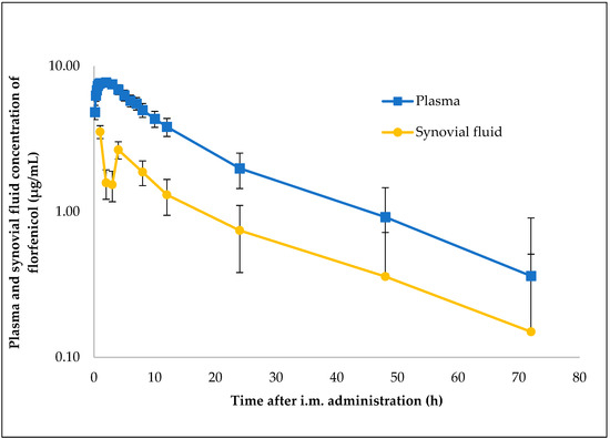
Pharmacokinetic parameters were computed via non-compartmental analysis from plasma and synovial fluid concentration data for 8 pigs. Florfenicol was administered intramuscularly at a single injection of 30 mg/kgbw. Table 1 presents mean PK parameters and standard deviation. The semi-logarithmic plasma and synovial fluid concentration–time curves of florfenicol after single i.m. administration of 30 mg/kgbw are illustrated in Figure 1. The mean Cmax of 8.15 ± 3.11 µg/mL in plasma was achieved with a Tmax of 1.40 ± 0.66 h. Florfenicol reached peak concentration in the synovial fluid more slowly after i.m. administration, and the Cmax 4.51 ± 1.16 µg/mL was achieved in 1.75 ± 1.16 h. The mean plasma and synovial fluid AUC24h following i.m. administration of florfenicol were 102.91 ± 19.90 µg/mL × h and 41.90 ± 16.93 µg/mL × h, respectively.

Table 1.
Plasma and synovial fluid PK parameters (mean ± SD) of florfenicol (Nuflor) in pigs following intramuscular administration of 30 mg/kgbw (n = 8).

Figure 1.
Semi-logarithmic plot illustrating the concentration–time curve of florfenicol in plasma and synovial fluid samples of pigs after a single i.m. administration of 30 mg/kgbw (n = 8).
2.2. MIC of Florfenicol against S. suis
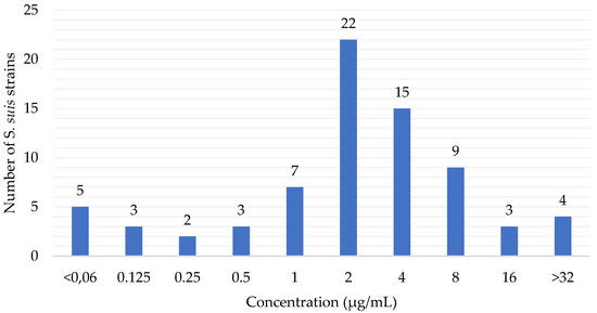
The florfenicol susceptibility of 73 S. suis isolates from pigs is summarized in Figure 2. The MIC50 and MIC90 were determined from the MIC values of 73 S. suis clinical isolates against florfenicol. According to the EUCAST [33] (The European Committee on Antimicrobial Susceptibility Testing) ECOFF (epidemiological cut-off) value (≤4 µg/L), 78.08% (57) of the S. suis isolates were wild-type, whereas 21.92% (16) were considered as non-wild-type to florfenicol. The CLSI breakpoints, however, indicate that 57.53% (42) of S. suis isolates are susceptible, 20.55% (15) are intermediate and 21.92% (16) are resistant to florfenicol.

Figure 2.
Minimum inhibitory concentration (MIC) distribution of florfenicol against S. suis in Hungary between 2018 and 2022 (n = 73).
2.3. PK and PD of Florfenicol after i.m. Administration of 30 mg/kgbw to Eight Healthy Pigs
The AUC/MIC50 and AUC/MIC90 values were calculated based on the AUC24h values determined in the plasma (102.91 ± 19.90 µg/mL × h) and synovial fluid (41.90 ± 16.93 µg/mL × h) of eight pigs for 24 h and the MIC50 and MIC90 values (2 µg/mL, 8 µg/mL) of 73 S. suis isolates. The AUC/MIC50 and AUC/MIC90 values in pig plasma were 51.45 ± 9.95 h and 12.86 ± 2.49 h, respectively. The AUC/MIC50 and AUC/MIC90 values in pig synovial fluid were 20.95 ± 8.47 h and 5.24 ± 2.12 h, respectively.
2.4. In Vitro Killing–Time Curves of Florfenicol against S. suis 96 Strain in Three Different Media (Pig Synovial Fluid, Pig Plasma, CA-MHB)
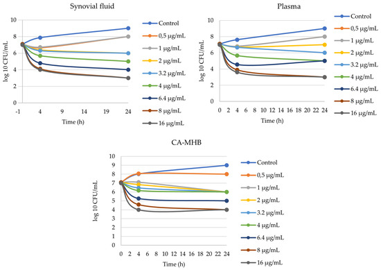
The MIC values of the investigated SS96 S. suis strain against florfenicol were 2 µg/mL, 2 µg/mL and 2 µg/mL in pig synovial fluid, pig plasma and CA-MHB, respectively. There was no difference between MIC values in different media. In all three media, a 3 log10 bacterial count decrease was achieved after 4 h at concentrations of 8 µg/mL and 16 µg/mL of florfenicol. The bacterial count reduction was 1 log10 or more when the concentration of florfenicol was greater than 2 µg/mL. The in vitro killing–time curves of florfenicol against SS96 S. suis strains in three different media are shown in Figure 3.

Figure 3.
In vitro killing–time curves of florfenicol against S. suis 96 strain in three different media (synovial fluid, plasma, CA-MHB).
2.5. PK/PD Integration
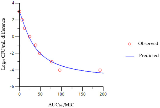
The relationship between synovial fluid AUC24h/MIC and the reduction in bacterial counts is shown in Figure 4. The AUC24h/MIC values required to result in a bacteriostatic effect were 22.22 h, 22.42 h and 14.21 h for florfenicol in synovial fluid, plasma and CA-MHB, respectively, as shown in Table 2. The corresponding values for bactericidal (E = −3) activity were 76.88 h, 86.49 h and 163.16 h, respectively. AUC24h/MIC values for bacterial eradication (E = −4) were higher in synovial fluid and plasma at 141.74 h and 161.76 h, respectively, whereas, in CA-MHB, florfenicol did not reach this level even at the highest concentration (16 µg/mL).

Figure 4.
Sigmoidal Emax correlation between SS96 bacterial count (CFU/mL) and in vitro AUC24h/MIC of florfenicol, illustrating the values required for bacteriostatic, bactericidal and eradication effects in synovial fluid in pig.

Table 2.
PK/PD breakpoints determined from the sigmoidal Emax inhibition equation in porcine synovial Fluid, plasma and CA-MHB.
2.6. Critical MIC Values of Florfenicol against S. suis Bacteriostatic, Bactericidal and Eradication Effects
Dividing the AUC24hss values calculated for eight pigs by the AUC24h/MIC breakpoints ratios gives the concentrations as MIC values, which can result in bacteriostatic, bactericidal and eradication effects in synovial fluid and plasma. Numerically, they were 2.91 ± 1.37, 0.84 ± 0.39 and 0.46 ± 0.21 for S. suis in synovial fluid and 7.34 ± 1.52, 1.90 ± 0.40 and 1.02 ± 0.21 in plasma, respectively (Table 3).

Table 3.
Critical MIC values of florfenicol against S. suis regarding bacteriostatic, bactericidal and eradication effects in pig synovial fluid and plasma.
3. Discussion
Florfenicol is characterized by a high lipid solubility and low protein binding, the latter being less than 15% in pig plasma, which results in a high Vd value for florfenicol [34,35]. The pharmacokinetics of florfenicol have been extensively studied in pigs; however, apart from our previous study, the concentration of florfenicol in synovial fluid has not yet been determined [23]. Our results regarding plasma florfenicol data are similar to previous studies. The maximum plasma concentration of florfenicol administered intramuscularly at a dose of 15 mg/kgbw was 3.04 ± 1.82 µg/mL, reached in 1.94 ± 0.87 h, which is almost identical to the results of our previous study, where Cmax and Tmax were 3.58 ± 1.51 µg/mL and 1.64 ± 1.74 h, respectively, in porcine plasma, whereas, in synovial fluid, they were 2.73 ± 1.2 µg/mL and 3.4 ± 1.67 h, respectively [23,32]. Following the intramuscular administration of florfenicol at a dose of 20 mg/kgbw, the maximum plasma concentration was 7.3 ± 6.0 µg/mL, which was reached in 2.3 ± 1.2 h [31]. In the present study, florfenicol was administered intramuscularly at a dose of 30 mg/kgbw, after which the Cmax and Tmax were 8.15 ± 3.11 µg/mL, which was reached in 1.40 ± 0.66 h. Here, the Cmax is higher than in a similar study, where the Cmax after application of the same dose was 4.44 ± 1.02 µg/mL [24]. All of this suggests that florfenicol is rapidly absorbed from the site of administration and rapidly distributed throughout the pig body system, including the synovial fluid. It can be clearly seen that higher values are also obtained in the synovial fluid following the administration of higher doses. Following administration at a dose of 30 mg/kgbw, the Cmax, AUC0–∞ and Tmax were 4.51 ± 1.16 µg/mL, 64.57 µg/mL × h and 1.75 ± 1.16 h, respectively. Figure 1 shows that lower concentrations are measured in the synovial fluid after the intramuscular administration of florfenicol compared to the first and fourth hours, which can be explained by the flip-flop kinetics of florfenicol, as the vehicle delays the absorption of the drug after intramuscular and subcutaneous administration.
Regarding the area under the concentration–time curve, florfenicol reaches higher values in plasma than in the synovial fluid, as already described in our previous publication; however, here, it is also valid at a higher dose. Since it has been shown in horses that, in acute arthritis, greater drug concentrations are achieved in synovial fluid than in healthy joints, we can assume that this is also the case in the synovial fluid of pigs [36].
In the present study, MIC values of 73 S. suis isolates were determined and the MIC50 and MIC90 were calculated. Among these values, the MIC50 is the same as the MIC50 value determined in a previous study, whereas the MIC90-values are closer to the results of studies conducted in other countries based on the results of our present study [16,37,38]. Although, in an Italian study, only 3% of 78 S. suis strains were resistant based on the CLSI breakpoint, in our study, 16% of 73 S. suis strains were resistant to florfenicol [39]. The susceptibility of S. suis to antibacterial agents is influenced by many factors, so continuous monitoring is recommended, even on a farm-by-farm basis, which is mandatory under current legislation, as antibacterial therapy can be used based on prior antibiotic susceptibility testing.
In the present study, we were the first to grow S. suis bacteria in pig synovial fluid and to implement an in vitro killing–time curve, the results of which will help to refine treatment protocols for arthritis caused by S. suis strains with florfenicol. The matrix effect, supported by several studies, could not be demonstrated in this study, as the MIC value of SS96 (2 µg/mL) was the same in all three media (synovial fluid, plasma, CA-MHB). The difference between the three media was observed in the failure to achieve the 4 log10 bacterial count reduction in the CA-MHB, even with the highest concentration of florfenicol (16 µg/mL). As no killing–time curve has been performed in synovial fluid before, our data could not be compared with other studies. Our data in plasma and in CA-MHB differ from the results of a previous study in which the AUC24h/MIC values for the bacteriostatic (E = 0), bactericidal (E = −3) and eradication (E = −4) effects of florfenicol determined ex vivo were 37.89 ± 4. 25 h, 44.02 ± 4.85 h and 46.42 ± 6.45 h in pig serum [24]. We obtained lower values for the bacteriostatic effect in all three media, whereas higher values were obtained for the bactericidal and eradication effects. The reasons for the difference may be that the two studies did not use the same S. suis strains, or that Lei at al. [24] used tryptic soy broth whereas we used CA-MHB. The differences in plasma could be due to the presence of antibodies. The importance of these studies is to characterize the behavior of the bacteria in the medium, with which in vitro models can be built at the site of infection. Modeling the site of infection will provide a basis for refining the use of antibiotics and thus increasing the effectiveness of treatments [40].
Based on the results of our present study, a bacteriostatic effect of florfenicol in swine arthritis caused by S. suis can be achieved below MIC values of 2 µg/mL, whereas, for septicemia, an MIC value of 7 µg/mL is recommended as a threshold value. A bactericidal effect can be expected if the MIC value for S. suis strains in arthritis is ≤0.8 µg/mL or in septicemia is ≤1.9 µg/mL in plasma. The critical MIC values for the eradication effect in arthritis and septicemia are ≤0.46 µg/mL and ≤1 µg/mL, respectively. It is important to note that these thresholds do not apply after intramuscular administration at the authorized dose of 15 mg/kgbw, but after a single intramuscular administration at the dose of 30 mg/kgbw used in our study, in which case, as we are deviating from the instructions for use, it is now the responsibility of the veterinarian to determine the withdrawal period, which should be taken into account for all therapies. On the basis of the known MIC values, our previous studies and other studies [23,24], we believe that florfenicol has a place in the treatment of swine arthritis caused by S. suis, but it would be worthwhile to perform further pharmacokinetic studies in an infection model and to confirm the results of the studies performed so far with clinical trials.
4. Materials and Methods
4.1. Experimental Animals and Design
In this study, we used 8 male pigs (Danish Landrace × Danish Yorkshire × Danish Duroc) with an average body weight (BW) of 28.93 ± 3.64 kg. In pig herds, clinical cases are most common between 4–8 weeks of age [41]. In our study, piglets were selected at 11 weeks of age, as this is also the age at which S. suis arthritis is most likely to occur, as maternal antibodies are certainly depleted. The animals were purchased from a local commercial pig farm in Hungary. The animals had not received any antimicrobial treatment prior to the experiment and were vaccinated against porcine circovirus 2 at 4 weeks of age. They were kept at 22–23 °C with adequate ventilation conditions, the relative humidity was 70% and the number of hours of light and darkness was 12 h each. Standard commercial feed and drinking water were provided ad libitum without medication prior to the experiment and no medication other than florfenicol was given to the pigs during the experiment. The pigs arrived at the experimental place a week earlier and did not show any clinical signs during this time, so it can be said that the investigation was carried out on clinically health animals. The study was authorized by the Local Animal Welfare Committee of the University of Veterinary Medicine, Budapest, and by the Government Office of Pest County, Food Chain Safety, Plant Protection and Soil Conservation Directorate, Budapest, Hungary (admission No. PE/EA/00367-6/2022).
Florfenicol (Nuflor injection A.U.V., Intervet International B.V., Boxmeer, Netherlands) was administered intramuscularly at a dose rate of 30 mg/kgbw. The drug was administered to the left neck muscles of the pigs after blind samples were taken. Subsequently, blood samples were then taken at the following times: 10, 20, 30, 40, 50, 60 min, 2, 3, 4, 5, 6, 7, 8, 10, 12, 24, 48 and 72 h, while synovial fluid samples were taken at the following time points: 1, 2, 3, 4, 8, 12, 24, 48 and 72 h. Blood samples were collected from the cranial vena cava of the animals using a 21 G × 2” needle and lithium heparin blood tube, and the blood samples were centrifuged at 1482× g for 10 min after sampling. For synovial fluid sampling, joint puncture was performed in the carpal and tarsal joints in continuous rotation using a 22 G × 1 ½” needle and 1 mL syringe. Samples were stored in low binding tubes at −80 °C until analysis.
4.2. Tandem Mass Spectrometry Analysis
Florfenicol was quantitated on the basis of the method published earlier [23]. Briefly: a Sciex 6500QTrap tandem mass spectrometer (Sciex, Framingham, MS, USA) was used in multiple reaction monitoring (MRM) mode, where the quantifier and qualifier transitions were 358.2/241 and 358.2/170, respectively. The mass spectrometer was operated in electrospray ionization with spray voltage of 5000 V. An Agilent 1100 HPLC system was coupled to the MS. A Kinetex XB C18 (50_2.1 mm, 2.6_m) column (Phenomenex) was applied for the separation by using water and acetonitrile, both containing 0.1% formic acid, in gradient mode. Five-point calibration model was applied. Analyst 1.6.3 software was used for data processing and controlling the measurements.
4.3. Pharmacokinetic Analysis
A non-compartmental pharmacokinetic analysis was used to determine the pharmacokinetic parameters of florfenicol in plasma and synovial fluid. The maximum drug concentration (Cmax) and the time of onset of maximum drug concentration (Tmax) were computed. The area under the 24 h concentration–time curve (AUC24h) and the area under the infinity extrapolated curve (AUC0–∞) were determined using a linear trapezoidal method. The half-life (T½), total body clearance (Cl/F) and mean residence time (MRT∞) were computed. For florfenicol, binding to plasma protein in pig was below 5% as it is negligible, so binding to plasma proteins was not considered [34,35,42]. Pharmacokinetic calculations and statistical analysis were performed using the Phoenix WinNonLin 8.3 software (Certara, Princeton, NJ, USA).
4.4. Minimum Inhibitory Concentration
The antibiotic susceptibility to florfenicol of 73 S. suis isolates from clinical samples of pig origin in Hungary were determined by broth microdilution method according to the CLSI (Clinical and Laboratory Standards Institute) description [43]. The isolation of S. suis was performed in 2022. The isolates were collected from Hungarian pig farms, each isolated from clinical lesions of dissected pigs. In each case, the samples were taken from untreated pigs.
The broth microdilution method was performed using CA-MHB (Mueller–Hinton Broth 2, Merck KGaA, Darmstadt, Germany). The isolates were stored at −80 °C and incubated in the presence of 5.0% CO2 at 37 °C for 24 h as recommended before broth microdilution method. After incubation, for the determination of the germ count, the bacterial suspensions were centrifuged at 3000× g for 10 min, washed in sterile physiological saline (Salsol solution infusion, TEVA Gyógyszergyár Zrt., Debrecen, Hungary), centrifuged again at 3000× g for 10 min and finally resuspended in physiological saline. The optical density of the suspensions at 600 nm was set to 0.1 (OD600 = 0.1), with the appropriate amount of physiological saline, which corresponded to 108 colony forming units (CFU)/mL bacterial density and a standard of 0.5 on the MacFarland scale. A suspension of 5 × 10⁵ CFU/mL was prepared with a 200-fold dilution. The germ count of the suspensions was tested with inoculation to blood agar plates and counting the number of CFUs. The sensitivity of S. suis isolates to florfenicol was tested by the broth microdilution method in the range of 32 µg/mL to 0.0625 µg/mL. Each isolate was included in the investigation with positive and negative control wells. This was followed by incubation period, and then the MIC values were read. This was the lowest concentration where no bacterial growth was detected. MIC50 and MIC90 values were computed as the MIC that inhibited the growth of 50% and 90%, respectively, of the isolates in different clusters. Determination of MIC and calculation of MIC50 and MIC90 were performed according to CLSI.
4.5. PK and PD of Florfenicol
PK/PD indices, calculated individual 8 pigs’ PK values of florfenicol after i.m. administration of 30 mg/kgbw and MIC50- and MIC90-values of 73 S. suis isolates were: AUC24h/MIC50, AUC24h/MIC90. Among the PK/PD relationships, AUC/MIC was selected as it better describes the clinical efficacy of long-acting formulations [44,45]. Since, in veterinary medicine, the AUC/MIC is not always projected to 24 h, we consider that, in this case, it is appropriate to use the unit h to denote this value [2].
4.6. In Vitro Model of Pig Synovial Fluid
To investigate the efficacy of florfenicol, an in vitro pig synovial fluid model was established and implemented as follows. Synovial fluid samples for bacterial growth were collected within 24 h prior to experiments from clinically healthy and untreated pigs. During the sampling, the skin surface of the joints was properly prepared, i.e., disinfected after shaving the hair, and then sampled for synovial fluid as described above. The syringes were then transported cooled at 4 °C and the needles were removed from syringes under laboratory conditions under laminar box to avoid contamination of the synovial fluid. Subsequently, 10 µL of each sample was inoculated on blood agar plate (Bak-Teszt Kft., Budapest, Hungary) and incubated at 37 °C for 18–24 h to check the sterility of the synovial fluids. Under the laminar box, the samples were individually placed in sterile tubes and stored at 4 °C until the start of the experiment. In the case where the synoval fluid was frozen (−20 °C), we observed a strong gelation, rendering the synovial fluids unsuitable for further investigation.
Before starting the experiment, we checked the blood agar results and included only those synovial fluid samples that were sterile; then these samples were in the ratio of 9:1 with sterile physiological saline, with the exception of the control (0 µg/mL florfenicol) sample, in order to inject the appropriate amount of florfenicol to form a two-fold dilution (0.125, 0.25, 0.5, 1.0, 2.0, 4.0, 8.0, 16.0 µg/mL) and to facilitate handling of the synovial fluid. These solutions were used to set up the killing–time curve.
4.7. Killing–Time Curve In Vitro
In order to generate the killing–time curves, we used an isolate of S. suis SS96, which was isolated from pig arthritis. Its MIC was determined in both pig serum and pig synovial fluid. The growth of the SS96 isolate was tested in florfenicol-free CA-MHB, pig serum and pig synovial fluid, which were the controls in the study, and the efficacy of florfenicol was also tested in the same media at the following concentrations: 0.125, 0.25, 0.5, 1.0, 2.0, 4.0, 8.0 and 16.0 µg/mL. Prior to the test, SS96 isolate was incubated in CA-MHB for 18 h in ambient air at 37 °C to achieve the appropriate initial germ count. This was determined as described above and the starting germ count was adjusted to 6.5 × 10⁴ CFU/mL in CA-MHB, pig serum and pig synovial fluid media. Subsequently, the different media were incubated in ambient air at 37 °C for 24 h at different concentrations of florfenicol. Following incubation, ten-fold dilution series were prepared and inoculated onto blood agar and, after incubation, in ambient air at 37 °C for 24 h with 5% CO2, the 24-h germ count was determined.
4.8. PK/PD Integration and Breakpoints
The sigmoidal Emax equation was used to model AUC24h/MIC data from killing–time curves using the non-linear regression program WinNonLin to predict plots of log10 change in CFU/mL versus AUC24h/MIC. PK/PD breakpoints were determined for three levels of growth inhibition after 24 h incubation: E = 0, bacteriostatic, which is a 0 log10 reduction in CFU/mL; E = −3, bactericidal, 3 log10 reduction in CFU/mL; and E = −4, 4 log10 reduction in bacterial count [2].
Dividing the AUC24hss values calculated for 8 pigs in vivo by the AUC24h/MIC breakpoints ratios gives the concentrations as critical MIC values, which can result in bacteriostatic, bactericidal and 4 log10 number reductions in synovial fluid and plasma in vitro.
Hill equation. E = log10-based change in live cell count, E0 = initial log10-based bacterial live cell count, Emax = maximum (response) kill capacity, C(t) = AUC24h/MIC, EC50 = in vitro concentration of florfenicol capable of half the maximum kill capacity Gamma= Hill coefficient (slope of the curve).
Author Contributions
Conceptualization, Z.S., P.M., R.S. and Á.J.; methodology, Z.S., Á.K., P.S., E.A. and I.B.; software, Z.S. and P.S.; validation, Z.S., P.S., Á.K. and E.A.; formal analysis, Z.S., P.M. and R.S.; investigation, Z.S., P.M., R.S., Á.K., P.M., P.S. and E.A.; resources, Z.S., P.M. and R.S.; data curation, Z.S.; writing—original draft preparation, Z.S., P.M., R.S. and P.S.; writing—review and editing, Z.S., Á.J. and I.B.; visualization, Z.S.; supervision, Á.J.; project administration, Z.S., P.M. and R.S.; funding acquisition, Á.J. All authors have read and agreed to the published version of the manuscript.
Funding
Project no. RRF-2.3.1-21-2022-00001 has been implemented with the support provided by the Recovery and Resilience Facility (RRF), financed under the National Recovery Fund budget estimate, RRF-2.3.1-21 funding scheme.
Institutional Review Board Statement
The study was authorized by the Local Animal Welfare Committee of the University of Veterinary Medicine, Budapest, and by the Government Office of Pest County, Food Chain Safety, Plant Protection and Soil Conservation Directorate, Budapest, Hungary (admission No. PE/EA/00367-6/2022).
Informed Consent Statement
Not applicable.
Data Availability Statement
The data presented in this study are available on request from the corresponding author.
Acknowledgments
The authors would like to give special thanks to Éva Borbás, Kata Balogh and Gergely Nagy for their professional assistance.
Conflicts of Interest
The authors declare no conflict of interest.
References
- Dewulf, J.; Joosten, P.; Chantziaras, I.; Bernaerdt, E.; Vanderhaeghen, W.; Postma, M.; Maes, D. Antibiotic Use in European Pig Production: Less Is More. Antibiotics 2022, 11, 1493. [Google Scholar] [CrossRef] [PubMed]
- Toutain, P.-L.; Pelligand, L.; Lees, P.; Bousquet-Mélou, A.; Ferran, A.A.; Turnidge, J.D. The Pharmacokinetic/Pharmacodynamic Paradigm for Antimicrobial Drugs in Veterinary Medicine: Recent Advances and Critical Appraisal. J. Vet. Pharmacol. Ther. 2021, 44, 172–200. [Google Scholar] [CrossRef] [PubMed]
- De Smet, J.; Boyen, F.; Croubels, S.; Rasschaert, G.; Haesebrouck, F.; De Backer, P.; Devreese, M. Similar Gastro-Intestinal Exposure to Florfenicol After Oral or Intramuscular Administration in Pigs, Leading to Resistance Selection in Commensal Escherichia Coli. Front. Pharmacol. 2018, 9, 1265. [Google Scholar] [PubMed]
- Syriopoulou, V.P.; Harding, A.L.; Goldmann, D.A.; Smith, A.L. In Vitro Antibacterial Activity of Fluorinated Analogs of Chloramphenicol and Thiamphenicol. Antimicrob. Agents Chemother. 1981, 19, 294–297. [Google Scholar] [CrossRef]
- Ueda, Y.; Ohtsuki, S.; Narukawa, N. Efficacy of Florfenicol on Experimental Actinobacillus Pleuropneumonia in Pigs. J. Vet. Med. Sci. 1995, 57, 261–265. [Google Scholar] [CrossRef] [PubMed]
- Marshall, S.A.; Jones, R.N.; Wanger, A.; Washington, J.A.; Doern, G.V.; Leber, A.L.; Haugen, T.H. Proposed MIC Quality Control Guidelines for National Committee for Clinical Laboratory Standards Susceptibility Tests Using Seven Veterinary Antimicrobial Agents: Ceftiofur, Enrofloxacin, Florfenicol, Penicillin G-Novobiocin, Pirlimycin, Premafloxacin, and Spectinomycin. J. Clin. Microbiol. 1996, 34, 2027–2029. [Google Scholar] [CrossRef]
- Ho, S.-P.; Hsu, T.-Y.; Chen, M.-H.; Wang, W.-S. Antibacterial Effect of Chloramphenicol, Thiamphenicol and Florfenicol against Aquatic Animal Bacteria. J. Vet. Med. Sci. 2000, 62, 479–485. [Google Scholar] [CrossRef]
- Hu, D.; Han, Z.; Li, C.; Lv, L.; Cheng, Z.; Liu, S. Florfenicol Induces More Severe Hemotoxicity and Immunotoxicity than Equal Doses of Chloramphenicol and Thiamphenicol in Kunming Mice. Immunopharmacol. Immunotoxicol. 2016, 38, 472–485. [Google Scholar] [CrossRef]
- Liu, J.-Z.; Fung, K.-F.; Chen, Z.-L.; Zeng, Z.-L.; Zhang, J. Tissue Pharmacokinetics of Florfenicol in Pigs Experimentally Infected with Actinobacillus Pleuropneumoniae. Eur. J. Drug Metab. Pharmacokinet. 2002, 27, 265–271. [Google Scholar] [CrossRef]
- Ali, B.H.; Al-Qarawi, A.A.; Hashaad, M. Comparative Plasma Pharmacokinetics and Tolerance of Florfenicol Following Intramuscular and Intravenous Administration to Camels, Sheep and Goats. Vet. Res. Commun. 2003, 27, 475–483. [Google Scholar] [CrossRef]
- Berge, A.C.B.; Sischo, W.M.; Craigmill, A.L. Antimicrobial Susceptibility Patterns of Respiratory Tract Pathogens from Sheep and Goats. J. Am. Vet. Med. Assoc. 2006, 229, 1279–1281. [Google Scholar] [CrossRef] [PubMed]
- Kim, Y.; Jung, J.; Kim, M.; Park, J.; Boxall, A.B.A.; Choi, K. Prioritizing Veterinary Pharmaceuticals for Aquatic Environment in Korea. Environ. Toxicol. Pharmacol. 2008, 26, 167–176. [Google Scholar] [CrossRef] [PubMed]
- Wasyl, D.; Hoszowski, A.; Szulowski, K.; Zając, M. Antimicrobial Resistance in Commensal Escherichia Coli Isolated from Animals at Slaughter. Front. Microbiol. 2013, 4, 221. [Google Scholar] [CrossRef] [PubMed]
- Liu, J.; Fung, K.-F.; Chen, Z.; Zeng, Z.; Zhang, J. Pharmacokinetics of Florfenicol in Healthy Pigs and in Pigs Experimentally Infected with Actinobacillus Pleuropneumoniae. Antimicrob. Agents Chemother. 2003, 47, 820–823. [Google Scholar] [CrossRef]
- Priebe, S.; Schwarz, S. In Vitro Activities of Florfenicol against Bovine and Porcine Respiratory Tract Pathogens. Antimicrob. Agents Chemother. 2003, 47, 2703–2705. [Google Scholar] [CrossRef]
- El Garch, F.; de Jong, A.; Simjee, S.; Moyaert, H.; Klein, U.; Ludwig, C.; Marion, H.; Haag-Diergarten, S.; Richard-Mazet, A.; Thomas, V.; et al. Monitoring of Antimicrobial Susceptibility of Respiratory Tract Pathogens Isolated from Diseased Cattle and Pigs across Europe, 2009–2012: VetPath Results. Vet. Microbiol. 2016, 194, 11–22. [Google Scholar] [CrossRef]
- de la Fuente, A.J.M.; Tucker, A.W.; Navas, J.; Blanco, M.; Morris, S.J.; Gutiérrez-Martín, C.B. Antimicrobial Susceptibility Patterns of Haemophilus Parasuis from Pigs in the United Kingdom and Spain. Vet. Microbiol. 2007, 120, 184–191. [Google Scholar] [CrossRef]
- Zhang, J.S.; Xia, Y.T.; Zheng, R.C.; Liang, Z.Y.; Shen, Y.J.; Li, Y.F.; Nie, M.; Gu, C.; Wang, H. Characterisation of a Novel Plasmid Containing a Florfenicol Resistance Gene in Haemophilus Parasuis. Vet. J. 2018, 234, 24–26. [Google Scholar] [CrossRef]
- Klein, U.; Földi, D.; Belecz, N.; Hrivnák, V.; Somogyi, Z.; Gastaldelli, M.; Merenda, M.; Catania, S.; Dors, A.; Siesenop, U.; et al. Antimicrobial Susceptibility Profiles of Mycoplasma Hyorhinis Strains Isolated from Five European Countries between 2019 and 2021. PLoS ONE 2022, 17, e0272903. [Google Scholar] [CrossRef]
- Felde, O.; Kreizinger, Z.; Sulyok, K.M.; Hrivnák, V.; Kiss, K.; Jerzsele, Á.; Biksi, I.; Gyuranecz, M. Antibiotic Susceptibility Testing of Mycoplasma Hyopneumoniae Field Isolates from Central Europe for Fifteen Antibiotics by Microbroth Dilution Method. PLoS ONE 2018, 13, e0209030. [Google Scholar] [CrossRef]
- Holmer, I.; Salomonsen, C.M.; Jorsal, S.E.; Astrup, L.B.; Jensen, V.F.; Høg, B.B.; Pedersen, K. Antibiotic Resistance in Porcine Pathogenic Bacteria and Relation to Antibiotic Usage. BMC Vet. Res. 2019, 15, 449. [Google Scholar] [CrossRef] [PubMed]
- Blondeau, J.M.; Fitch, S.D. Mutant Prevention and Minimum Inhibitory Concentration Drug Values for Enrofloxacin, Ceftiofur, Florfenicol, Tilmicosin and Tulathromycin Tested against Swine Pathogens Actinobacillus Pleuropneumoniae, Pasteurella Multocida and Streptococcus Suis. PLoS ONE 2019, 14, e0210154. [Google Scholar] [CrossRef]
- Somogyi, Z.; Mag, P.; Kovács, D.; Kerek, Á.; Szabó, P.; Makrai, L.; Jerzsele, Á. Synovial and Systemic Pharmacokinetics of Florfenicol and PK/PD Integration against Streptococcus Suis in Pigs. Pharmaceutics 2022, 14, 109. [Google Scholar] [CrossRef] [PubMed]
- Lei, Z.; Liu, Q.; Yang, S.; Yang, B.; Khaliq, H.; Li, K.; Ahmed, S.; Sajid, A.; Zhang, B.; Chen, P.; et al. PK-PD Integration Modeling and Cutoff Value of Florfenicol against Streptococcus Suis in Pigs. Front. Pharmacol. 2018, 9, 2. [Google Scholar] [CrossRef]
- Nedbalcova, K.; Kucharovicova, I.; Zouharova, M.; Matiaskova, K.; Kralova, N.; Brychta, M.; Simek, B.; Pecha, T.; Plodkova, H.; Matiasovic, J. Resistance of Streptococcus Suis Isolates from the Czech Republic during 2018–2022. Antibiotics 2022, 11, 1214. [Google Scholar] [CrossRef] [PubMed]
- Ahmad, I.; Huang, L.; Hao, H.; Sanders, P.; Yuan, Z. Application of PK/PD Modeling in Veterinary Field: Dose Optimization and Drug Resistance Prediction. BioMed Res. Int. 2016, 2016, 5465678. [Google Scholar] [CrossRef] [PubMed]
- Mouton, J.W.; Dudley, M.N.; Cars, O.; Derendorf, H.; Drusano, G.L. Standardization of Pharmacokinetic/Pharmacodynamic (PK/PD) Terminology for Anti-Infective Drugs: An Update. J. Antimicrob. Chemother. 2005, 55, 601–607. [Google Scholar] [CrossRef] [PubMed]
- Liu, Z.; Rong, T.; Zeng, D.; Shen, X.; Ma, X.; Zeng, Z. Bayesian Population Pharmacokinetic Modeling of Florfenicol in Pigs after Intravenous and Intramuscular Administration. J. Vet. Pharmacol. Ther. 2018, 41, 719–725. [Google Scholar] [CrossRef]
- Yang, B.; Gao, J.D.; Cao, X.Y.; Wang, Q.Y.; Sun, G.Z.; Yang, J.J. Lung Microdialysis Study of Florfenicol in Pigs after Single Intramuscular Administration. J. Vet. Pharmacol. Ther. 2017, 40, 530–538. [Google Scholar] [CrossRef]
- Rottbøll, L.A.H.; Skovgaard, K.; Barington, K.; Jensen, H.E.; Friis, C. Intrabronchial Microdialysis: Effects of Probe Localization on Tissue Trauma and Drug Penetration into the Pulmonary Epithelial Lining Fluid. Basic Clin. Pharmacol. Toxicol. 2015, 117, 242–250. [Google Scholar] [CrossRef]
- Voorspoels, J.; D’Haese, E.; De Craene, B.A.; Vervaet, C.; De Riemaecker, D.; Deprez, P.; Nelis, H.; Remon, J.P. Pharmacokinetics of Florfenicol after Treatment of Pigs with Single Oral or Intramuscular Doses or with Medicated Feed for Three Days. Vet. Rec. 1999, 145, 397–399. [Google Scholar] [CrossRef] [PubMed]
- Dorey, L.; Pelligand, L.; Cheng, Z.; Lees, P. Pharmacokinetic/Pharmacodynamic Integration and Modelling of Florfenicol for the Pig Pneumonia Pathogens Actinobacillus Pleuropneumoniae and Pasteurella Multocida. PLoS ONE 2017, 12, e0177568. [Google Scholar] [CrossRef] [PubMed]
- MIC EUCAST. Available online: https://mic.eucast.org/search/show-registration/50725?back=https://mic.eucast.org/search/?search%255Bmethod%255D%3Dmic%26search%255Bantibiotic%255D%3D95%26search%255Bspecies%255D%3D-1%26search%255Bdisk_content%255D%3D-1%26search%255Blimit%255D%3D50 (accessed on 15 February 2023).
- Lobell, R.D.; Varma, K.J.; Johnson, J.C.; Sams, R.A.; Gerken, D.F.; Ashcraft, S.M. Pharmacokinetics of Florfenicol Following Intravenous and Intramuscular Doses to Cattle. J. Vet. Pharmacol. Ther. 1994, 17, 253–258. [Google Scholar] [CrossRef] [PubMed]
- Foster, D.M.; Martin, L.G.; Papich, M.G. Comparison of Active Drug Concentrations in the Pulmonary Epithelial Lining Fluid and Interstitial Fluid of Calves Injected with Enrofloxacin, Florfenicol, Ceftiofur, or Tulathromycin. PLoS ONE 2016, 11, e0149100. [Google Scholar] [CrossRef]
- Errecalde, J.O.; Carmely, D.; Mariño, E.L.; Mestorino, N. Pharmacokinetics of Amoxycillin in Normal Horses and Horses with Experimental Arthritis. J. Vet. Pharmacol. Ther. 2001, 24, 1–6. [Google Scholar] [CrossRef] [PubMed]
- Sweeney, M.T. Antimicrobial Susceptibility of Actinobacillus Pleuropneumoniae, Bordetella Bronchiseptica, Pasteurella Multocida, and Streptococcus Suis Isolated from Diseased Pigs in the United States and Canada, 2016 to 2020. JSHAP 2022, 30, 130–144. [Google Scholar] [CrossRef]
- Hernandez-Garcia, J.; Wang, J.; Restif, O.; Holmes, M.A.; Mather, A.E.; Weinert, L.A.; Wileman, T.M.; Thomson, J.R.; Langford, P.R.; Wren, B.W.; et al. Patterns of Antimicrobial Resistance in Streptococcus Suis Isolates from Pigs with or without Streptococcal Disease in England between 2009 and 2014. Vet. Microbiol. 2017, 207, 117–124. [Google Scholar] [CrossRef]
- Cucco, L.; Paniccià, M.; Massacci, F.R.; Morelli, A.; Ancora, M.; Mangone, I.; Pasquale, A.D.; Luppi, A.; Vio, D.; Cammà, C.; et al. New Sequence Types and Antimicrobial Drug–Resistant Strains of Streptococcus Suis in Diseased Pigs, Italy, 2017–2019. Emerg. Infect. Dis. 2022, 28, 139–147. [Google Scholar] [CrossRef]
- Hyatt, J.M.; McKinnon, P.S.; Zimmer, G.S.; Schentag, J.J. The Importance of Pharmacokinetic/Pharmacodynamic Surrogate Markers to Outcome. Clin. Pharmacokinet. 1995, 28, 143–160. [Google Scholar] [CrossRef]
- Cloutier, G.; D’Allaire, S.; Martinez, G.; Surprenant, C.; Lacouture, S.; Gottschalk, M. Epidemiology of Streptococcus Suis Serotype 5 Infection in a Pig Herd with and without Clinical Disease. Vet. Microbiol. 2003, 97, 135–151. [Google Scholar] [CrossRef]
- Bretzlaff, K.N.; Neff-Davis, C.A.; Ott, R.S.; Koritz, G.D.; Gustafsson, B.K.; Davis, L.E. Florfenicol in Non-Lactating Dairy Cows: Pharmacokinetics, Binding to Plasma Proteins, and Effects on Phagocytosis by Blood Neutrophils. J. Vet. Pharmacol. Ther. 1987, 10, 233–240. [Google Scholar] [CrossRef] [PubMed]
- Lubbers, B.V.; Diaz-Campos, D.V.; Schwarz, S.; Bowden, R.; Burbick, C.R.; Fajt, V.R.; Fielder, M.; Gunnett, L.; Holliday, N.M.; Kerdraon, C.; et al. Performance Standards for Antimicrobial Disk and Dilutation Susceptibility Tests for Bacteria Isolated from Animals. In Approved Standard, 3rd ed.; Clinical and Laboratory Standards Institute: Wayne, PA, USA, 2020. [Google Scholar]
- Toutain, P.-L.; Sidhu, P.K.; Lees, P.; Rassouli, A.; Pelligand, L. VetCAST Method for Determination of the Pharmacokinetic-Pharmacodynamic Cut-Off Values of a Long-Acting Formulation of Florfenicol to Support Clinical Breakpoints for Florfenicol Antimicrobial Susceptibility Testing in Cattle. Front. Microbiol. 2019, 10, 1310. [Google Scholar] [CrossRef] [PubMed]
- Sidhu, P.; Rassouli, A.; Illambas, J.; Potter, T.; Pelligand, L.; Rycroft, A.; Lees, P. Pharmacokinetic–Pharmacodynamic Integration and Modelling of Florfenicol in Calves. J. Vet. Pharmacol. Ther. 2014, 37, 231–242. [Google Scholar] [CrossRef] [PubMed]
Disclaimer/Publisher’s Note: The statements, opinions and data contained in all publications are solely those of the individual author(s) and contributor(s) and not of MDPI and/or the editor(s). MDPI and/or the editor(s) disclaim responsibility for any injury to people or property resulting from any ideas, methods, instructions or products referred to in the content. |
© 2023 by the authors. Licensee MDPI, Basel, Switzerland. This article is an open access article distributed under the terms and conditions of the Creative Commons Attribution (CC BY) license (https://creativecommons.org/licenses/by/4.0/).