
Graphene-Based Materials for Bone Regeneration in Dentistry: A Systematic Review of In Vitro Applications and Material Comparisons


Abstract
1. Introduction
2. Material and Methods
2.1. Statement and Protocol
2.2. Inclusion and Exclusion Criteria
- PICO Question: Is the use of graphene as a regenerative biomaterial (investigated condition) effective and safe (outcome) compared to bone regeneration without graphene or with other biomaterials distinct from graphene (comparison condition)?
- Structured PICOS Question:
- ▪
- Participants (P): Human bone/dental tissues and cells involved in bone regenerative procedures.
- ▪
- Intervention or Exposure and Comparison Group (IC): Graphene as a biomaterial in bone regeneration. Comparator: placebo or absence of intervention (not included in the search term combination) or other biomaterials used in bone regeneration, such as hydroxyapatite, calcium phosphates, or synthetic polymer scaffolds.
- ▪
- Outcome (O): Bone regeneration.
- ▪
- Study (S): In vitro studies.
2.3. Search Strategy
2.3.1. Information Sources
2.3.2. Search Terms
2.3.3. Study Selection
2.3.4. Data Extraction
2.4. Quality Analysis
- Items meeting the necessary conditions were marked YES.
- Items failing to meet these conditions were marked NO.
- ≥70%: Low risk of bias.
- 50–69%: Moderate risk of bias.
- ≤49%: High risk of bias.
3. Results
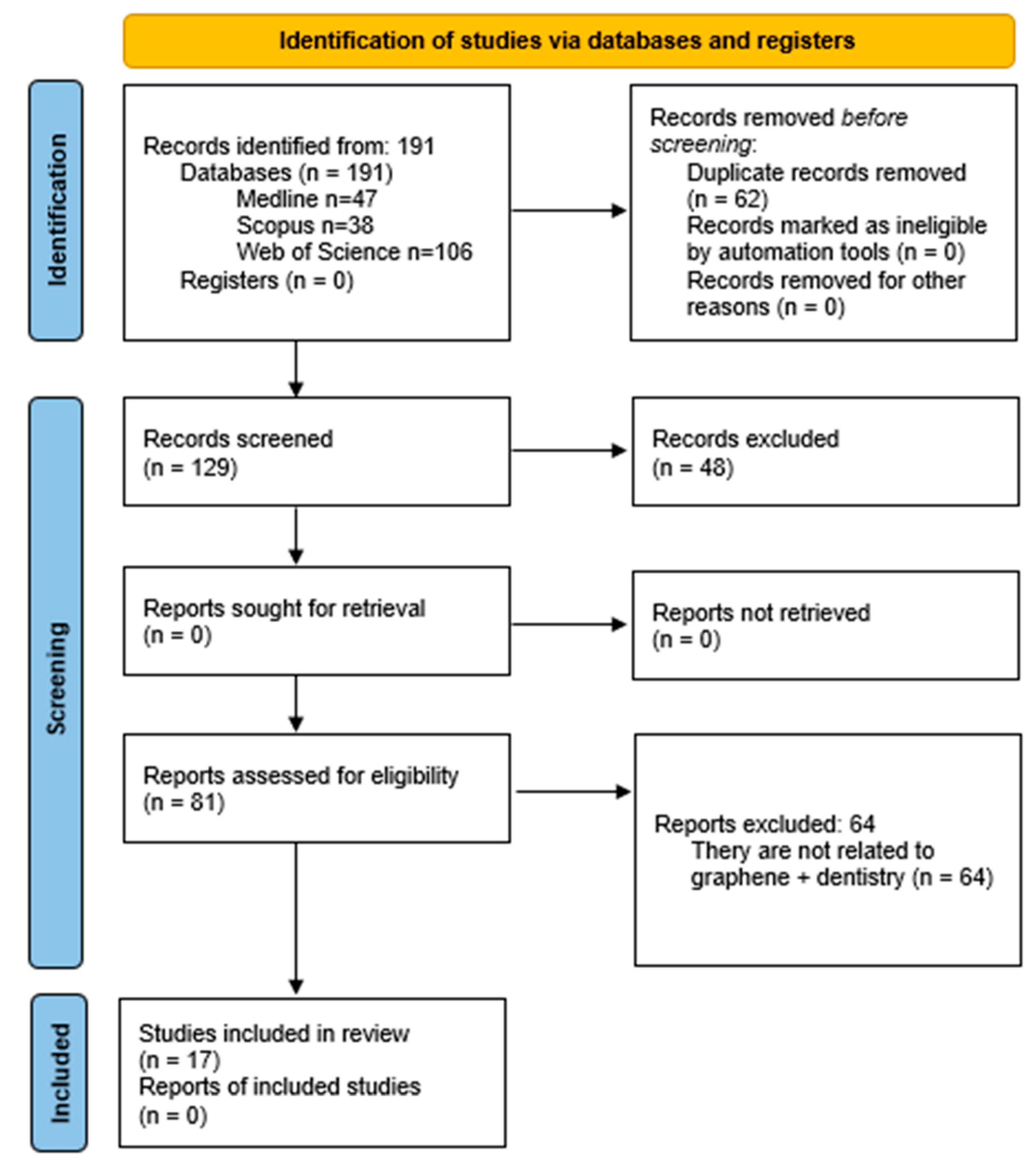
3.1. Study Selection and Flow Diagram
- Non-relevant Topics: excluded studies included those addressing periodontal nanoparticles, preventive dentistry, community dentistry, endodontics, and implantology.
- Material-Specific Exclusions: studies that investigated graphene as a sealant or exclusively as an antibacterial or angiogenic agent without reference to bone regeneration were excluded.
- Experimental Contexts: in vitro studies focusing on bone marrow cells without explicit links to dental applications were omitted.
- Non-Dental Regeneration Sites: research on bone regeneration in non-dental anatomical sites such as the skull, femur, or tibia was excluded.
- Cell Line Criteria: studies utilizing cell lines like MC3T3-E1, BMSCs, MG63, hMSCs, hADMSCs, or D1 cells unrelated to dentistry were not considered.
3.2. Data Extraction
3.3. Risk of Bias
4. Discussion
5. Conclusions
- The review has addressed concerns regarding the safety of graphene in dental settings. Graphene’s biocompatibility, coupled with its ability to enhance cellular interactions and reduce inflammation, highlights its suitability for regenerative applications.
- Composite materials, such as GO-poly-L-lactic acid (PLLA) scaffolds and GO/hydroxyapatite (HA) blends, have emerged as some of the most promising configurations for further exploration.
Author Contributions
Funding
Data Availability Statement
Conflicts of Interest
References
- Kawamoto, K.; Miyaji, H.; Nishida, E.; Miyata, S.; Kato, A.; Tateyama, A.; Furihata, T.; Shitomi, K.; Iwanaga, T.; Sugaya, T. Characterization and evaluation of graphene oxide scaffold for periodontal wound healing of class II furcation defects in dog. Int. J. Nanomed. 2018, 13, 2365–2376. [Google Scholar] [CrossRef] [PubMed]
- Siddiqui, Z.; Acevedo-Jake, A.M.; Griffith, A.; Kadincesme, N.; Dabek, K.; Hindi, D.; Kim, K.K.; Kobayashi, Y.; Shimizu, E.; Kumar, V. Cells and material-based strategies for regenerative endodontics. Bioact. Mater. 2022, 14, 234–249. [Google Scholar] [CrossRef]
- Apostu, A.M.; Sufaru, I.G.; Tanculescu, O.; Stoleriu, S.; Doloca, A.; Ciocan Pendefunda, A.A.; Solomon, S.M. Can Graphene Pave the Way to Successful Periodontal and Dental Prosthetic Treatments? A Narrative Review. Biomedicines 2023, 11, 2354. [Google Scholar] [CrossRef]
- Guazzo, R.; Gardin, C.; Bellin, G.; Sbricoli, L.; Ferroni, L.; Ludovichetti, F.S.; Piattelli, A.; Antoniac, I.; Bressan, E.; Zavan, B. Graphene-Based Nanomaterials for Tissue Engineering in the Dental Field. Nanomaterials 2018, 8, 349. [Google Scholar] [CrossRef] [PubMed]
- Park, C.; Park, S.; Lee, D.; Choi, K.S.; Lim, H.P.; Kim, J. Graphene as an Enabling Strategy for Dental Implant and Tissue Regeneration. Tissue Eng. Regen. Med. 2017, 14, 481–493. [Google Scholar] [CrossRef]
- Rygas, J.; Matys, J.; Wawrzyńska, M.; Szymonowicz, M.; Dobrzyński, M. The Use of Graphene Oxide in Orthodontics-A Systematic Review. J. Funct. Biomater. 2023, 14, 500. [Google Scholar] [CrossRef]
- Lei, Y.; Zhang, T.; Lin, Y.C.; Granzier-Nakajima, T.; Bepete, G.; Kowalczyk, D.A.; Lin, Z.; Zhou, D.; Schranghamer, T.F.; Dodda, A.; et al. Graphene and Beyond: Recent Advances in Two-Dimensional Materials Synthesis, Properties, and Devices. ACS Nanosci. Au 2022, 2, 450–485. [Google Scholar] [CrossRef] [PubMed]
- Lee, C.; Wei, X.; Kysar, J.W.; Hone, J. Measurement of the elastic properties and intrinsic strength of monolayer graphene. Science 2008, 321, 385–388. [Google Scholar] [CrossRef] [PubMed]
- Lee, J.H.; Park, S.J.; Choi, J.W. Electrical Property of Graphene and Its Application to Electrochemical Biosensing. Nanomaterials 2019, 9, 297. [Google Scholar] [CrossRef]
- Makwana, M.; Patel, A.M. Identification of microbes using single-layer graphene-based nano biosensors. J. Mol. Model. 2023, 29, 382. [Google Scholar] [CrossRef] [PubMed]
- Costa, A.C.; Alves, P.M.; Monteiro, F.J.; Salgado, C. Interactions between Dental MSCs and Biomimetic Composite Scaffold during Bone Remodeling Followed by In Vivo Real-Time Bioimaging. Int. J. Mol. Sci. 2023, 24, 1827. [Google Scholar] [CrossRef]
- Aryaei, A.; Jayatissa, A.H.; Jayasuriya, A.C. The effect of graphene substrate on osteoblast cell adhesion and proliferation. J. Biomed. Mater. Res. A 2014, 102, 3282–3290. [Google Scholar] [CrossRef] [PubMed]
- Qamar, Z.; Alghamdi, A.M.S.; Haydarah, N.K.B.; Balateef, A.A.; Alamoudi, A.A.; Abumismar, M.A.; Shivakumar, S.; Cicciù, M.; Minervini, G. Impact of temporomandibular disorders on oral health-related quality of life: A systematic review and meta-analysis. J. Oral. Rehabil. 2023, 50, 706–714. [Google Scholar] [CrossRef] [PubMed]
- Williams, A.G.; Moore, E.; Thomas, A.; Johnson, J.A. Graphene-Based Materials in Dental Applications: Antibacterial, Biocompatible, and Bone Regenerative Properties. Int. J. Biomater. 2023, 2023, 8803283. [Google Scholar] [CrossRef] [PubMed]
- Page, M.J.; McKenzie, J.E.; Bossuyt, P.M.; Boutron, I.; Hoffmann, T.C.; Mulrow, C.D.; Shamseer, L.; Tetzlaff, J.M.; Akl, E.A.; Brennan, S.E.; et al. The PRISMA 2020 statement: An updated guideline for reporting systematic reviews. Syst. Rev. 2021, 10, 89. [Google Scholar] [CrossRef]
- Faggion, C.M., Jr. Guidelines for reporting pre-clinical in vitro studies on dental materials. J. Evid. Based Dent. Pract. 2012, 12, 182–189. [Google Scholar] [CrossRef]
- Qiu, Z.; Lin, X.; Zou, L.; Fu, W.; Lv, H. Effect of graphene oxide/poly-L-lactic acid composite scaffold on the biological properties of human dental pulp stem cells. BMC Oral. Health 2024, 24, 413. [Google Scholar] [CrossRef] [PubMed]
- Lopez-Garcia, S.; Aznar-Cervantes, S.D.; Pagan, A.; Llena, C.; Forner, L.; Sanz, J.L.; Garcia-Bernal, D.; Sanchez-Bautista, S.; Ceballos, L.; Fuentes, V.; et al. 3D Graphene/silk fibroin scaffolds enhance dental pulp stem cell osteo/odontogenic differentiation. Dent. Mater. 2024, 40, 431–440. [Google Scholar] [CrossRef]
- Li, Y.; Huang, X.; Fu, W.; Zhang, Z.; Xiao, K.; Lv, H. Preparation of PDA-GO/CS composite scaffold and its effects on the biological properties of human dental pulp stem cells. BMC Oral. Health 2024, 24, 157. [Google Scholar] [CrossRef]
- Qasim, S.S.B.; Ahmed, J.; Karched, M.; Al-Asfour, A. The potential of nano graphene oxide and chlorhexidine composite membranes for use as a surface layer in functionally graded membranes for periodontal lesions. J. Mater. Sci. Mater. Med. 2023, 34, 63. [Google Scholar] [CrossRef]
- Souza, A.P.C.; Neves, J.G.; Navarro da Rocha, D.; Lopes, C.C.; Moraes, Â.M.; Correr-Sobrinho, L.; Correr, A.B. Chitosan/Xanthan membrane containing hydroxyapatite/Graphene oxide nanocomposite for guided bone regeneration. J. Mech. Behav. Biomed. Mater. 2022, 136, 105464. [Google Scholar] [CrossRef] [PubMed]
- Daulbayev, C.; Sultanov, F.; Korobeinyk, A.V.; Yeleuov, M.; Taurbekov, A.; Bakbolat, B.; Umirzakov, A.; Baimenov, A.; Daulbayev, O. Effect of graphene oxide/hydroxyapatite nanocomposite on osteogenic differentiation and antimicrobial activity. Surf. Interfaces 2022, 28, 101683. [Google Scholar] [CrossRef]
- Ferroni, L.; Gardin, C.; Rigoni, F.; Balliana, E.; Zanotti, F.; Scatto, M.; Riello, P.; Zavan, B. The Impact of Graphene Oxide on Polycaprolactone PCL Surfaces: Antimicrobial Activity and Osteogenic Differentiation of Mesenchymal Stem Cell. Coatings 2022, 12, 799. [Google Scholar] [CrossRef]
- Ketkar, G.N.; Malaiappan, S.; Rajeshkumar, S. Assessment of Cytotoxicity of Copper and Graphene Oxide Nano composite Synthesized Using Amla Extract Formulation-An In-Vitro Study. Res. Med. Dent. Sci. 2022, 10, 140–144. [Google Scholar]
- Mancinelli, R.; Di Filippo, E.S.; Tumedei, M.; Marrone, M.; Fontana, A.; Ettorre, V.; Giordani, S.; Baldrighi, M.; Iezzi, G.; Piattelli, A.; et al. Human Dental Pulp Stem Cell Osteogenic Differentiation Seeded on Equine Bone Block with Graphene and Melatonin. Appl. Sci. 2021, 11, 3218. [Google Scholar] [CrossRef]
- García-Contreras, H.; López, D.; Alvarez-Gayosso, C. Biological and physico-mechanical properties of poly (methyl methacrylate) enriched with graphene oxide as a potential biomaterial. J. Oral. Res. 2021, 10, 1–9. [Google Scholar] [CrossRef]
- Lee, J.J.; Shin, Y.C.; Song, S.-J.; Cha, J.M.; Hong, S.W.; Lim, Y.-J.; Jeong, S.J.; Han, D.-W.; Kim, B. Dicalcium Phosphate Coated with Graphene Synergistically Increases Osteogenic Differentiation In Vitro. Coatings 2018, 8, 13. [Google Scholar] [CrossRef]
- Vera-Sánchez, M.; Aznar-Cervantes, S.; Jover, E.; García-Bernal, D.; Oñate-Sánchez, R.E.; Hernández-Romero, D.; Moraleda, J.M.; Collado-González, M.; Rodríguez-Lozano, F.J.; Cenis, J.L. Silk-Fibroin and Graphene Oxide Composites Promote Human Periodontal Ligament Stem Cell Spontaneous Differentiation into Osteo/Cementoblast-Like Cells. Stem Cells Dev. 2016, 25, 1742–1754. [Google Scholar] [CrossRef] [PubMed]
- Golzar, H.; Mohammadrezaei, D.; Yadegari, A.; Rasoulianboroujeni, M.; Hashemi, M.; Omidi, M.; Yazdian, F.; Shalbaf, M.; Tayebi, L. Incorporation of functionalized reduced graphene oxide/magnesium nanohybrid to enhance the osteoinductivity capability of 3D printed calcium phosphate-based scaffolds. Compos. Part. B Eng. 2020, 185, 107749. [Google Scholar] [CrossRef]
- Zhang, W.; Yang, G.; Wang, X.; Jiang, L.; Jiang, F.; Li, G.; Zhang, Z.; Jiang, X. Magnetically Controlled Growth-Factor-Immobilized Multilayer Cell Sheets for Complex Tissue Regeneration. Adv. Mater. 2017, 29, 1703795. [Google Scholar] [CrossRef]
- Zhou, Q.; Yang, P.; Li, X.; Liu, H.; Ge, S. Bioactivity of periodontal ligament stem cells on sodium titanate coated with graphene oxide. Sci. Rep. 2016, 6, 19343. [Google Scholar] [CrossRef] [PubMed]
- Lee, J.H.; Shin, Y.C.; Lee, S.M.; Jin, O.S.; Kang, S.H.; Hong, S.W.; Jeong, C.M.; Huh, J.B.; Han, D.W. Enhanced Osteogenesis by Reduced Graphene Oxide/Hydroxyapatite Nanocomposites. Sci. Rep. 2015, 5, 18833. [Google Scholar] [CrossRef] [PubMed]
- Rodríguez-Lozano, F.J.; García-Bernal, D.; Aznar-Cervantes, S.; Ros-Roca, M.A.; Algueró, M.C.; Atucha, N.M.; Lozano-García, A.A.; Moraleda, J.M.; Cenis, J.L. Effects of composite films of silk fibroin and graphene oxide on the proliferation, cell viability and mesenchymal phenotype of periodontal ligament stem cells. J. Mater. Sci. Mater. Med. 2014, 25, 2731–2741. [Google Scholar] [CrossRef] [PubMed]
- Shin, S.R.; Li, Y.C.; Jang, H.L.; Khoshakhlagh, P.; Akbari, M.; Nasajpour, A.; Zhang, Y.S.; Tamayol, A.; Khademhosseini, A. Graphene-based materials for tissue engineering. Adv. Drug Deliv. Rev. 2016, 105, 255–274. [Google Scholar] [CrossRef]
- Nama, N.; Hennawy, M.; Barrowman, N.; O’Hearn, K.; Sampson, M.; McNally, J.D. Successful incorporation of single reviewer assessments during systematic review screening: Development and validation of sensitivity and work-saved of an algorithm that considers exclusion criteria and count. Syst. Rev. 2021, 10, 98. [Google Scholar] [CrossRef] [PubMed]
- Sayed, M.E. The Effect of Dentine Desensitizing Agents on the Retention of Cemented Fixed Dental Prostheses: A Systematic Review. Medicina 2023, 59, 515. [Google Scholar] [CrossRef] [PubMed]
- Hosseini, F.S.; Nair, L.S.; Laurencin, C.T. Inductive Materials for Regenerative Engineering. J. Dent. Res. 2021, 100, 1011–1019. [Google Scholar] [CrossRef] [PubMed]
- Govindarajan, D.; Saravanan, S.; Sudhakar, S.; Vimalraj, S. Graphene: A Multifaceted Carbon-Based Material for Bone Tissue Engineering Applications. ACS Omega 2024, 9, 67–80. [Google Scholar] [CrossRef]
- Zhang, H.; Xiang, Q.; Liu, Z.; Zhang, X.; Zhao, Y.; Tan, H. Supercritical mechano-exfoliation process. Nat. Commun. 2024, 15, 9329. [Google Scholar] [CrossRef]
- Inchingolo, F.; Inchingolo, A.M.; Latini, G.; Palmieri, G.; Di Pede, C.; Trilli, I.; Ferrante, L.; Inchingolo, A.D.; Palermo, A.; Lorusso, F.; et al. Application of Graphene Oxide in Oral Surgery: A Systematic Review. Materials 2023, 16, 6293. [Google Scholar] [CrossRef] [PubMed]
- Cao, K.; Tian, Z.; Zhang, X.; Wang, Y.; Zhu, Q. Green preparation of graphene oxide nanosheets as adsorbent. Sci. Rep. 2023, 13, 9314. [Google Scholar] [CrossRef] [PubMed]

| Database | Search Terms | Results |
|---|---|---|
| 1# graphene AND dent* AND regen* | 142 | |
| 2# graphene AND dent* AND regen* AND bone | 76 | |
| Pubmed | 3# graphene AND dent* AND regen* AND (bone OR osteo*) | 82 |
| 4# graphene AND dent* AND regen* AND (bone OR osteo*) NOT implant* | 47 | |
| 1# graphene AND dent* AND regen* | 119 | |
| Scopus | 2# graphene AND dent* AND regen AND bone | 85 |
| 3# graphene AND dent* AND regen* AND (bone OR osteo*) | 92 | |
| 4# graphene AND dent* AND regen* AND (bone OR osteo*) NOT implant* | 38 | |
| 1# graphene AND dent* AND regen* | 290 | |
| 2# graphene AND dent* AND regen* AND bone | 165 | |
| Web of Science | 3# graphene AND dent* AND regen* AND (bone or osteo*) | 182 |
| 4# graphene AND dent* AND regen* AND (bone or osteo*) NOT implant | 106 |
| Author | Objectives/Generalities | Material and Methods | Conclusions |
| Qiu, Z. et al., 2024 [17] | Dental pulp stem cells (DPSCs), capable of osteogenic differentiation under suitable conditions, require effective support materials for 3D tissue fabrication and bone regeneration. This study investigates the biological performance of human DPSCs on composite scaffolds made of graphene oxide (GO) and poly-L-lactic acid (PLLA). | -Cell line: hDPSCs -Scaffolds: 3D GO/PLLA scaffolds containing 0.15 wt%, 0.20 wt%, and 0.25 wt% GO | The data demonstrate that GO and PLLA integrate successfully, with GO/PLLA scaffolds exhibiting excellent bioactivity and biocompatibility with DPSCs. The 0.15% GO/PLLA scaffold significantly promoted DPSC proliferation and osteogenic differentiation, as shown by increased calcium nodule formation, enhanced ALP activity and staining, and upregulated expression of RUNX2 and COL1. These findings underscore the potential of GO/PLLA scaffolds as a promising material for cell culture and oral bone tissue engineering. |
| López-García, S. et al., 2024 [18] | To evaluate silk fibroin, with and without graphene, as a scaffold material for regenerative endodontics. | -Cell line: hDPSCs -Scaffolds: three types of scaffolds were studied. (1) Pure fibroin materials without incorporated GO designated as “SF”, (2) SF scaffolds containing GO adsorbed on their surface were termed as “SF/GO”, and (3) those in which the superficially adsorbed GO was reduced were termed “SF/rGO”. | The data indicate that SF/GO and SF/rGO scaffolds promote hDPSC differentiation by upregulating key osteo/odontogenic markers and facilitating extracellular matrix mineralization. However, further in vivo studies are required to validate their potential. |
| Li, Y. et al., 2024 [19] | Polydopamine (PDA) was used to reduce GO, forming the PDA-GO complex, which was combined with chitosan (CS) to develop PDA-GO/CS composite scaffolds for bone tissue engineering. PDA-GO improved the degradation rate of CS. The scaffolds’ physicochemical and antimicrobial properties were evaluated to determine the optimal composition, while biocompatibility was assessed using phalloidin and live/dead staining. Osteogenic differentiation of human dental pulp stem cells (hDPSCs) was analyzed via ALP staining, RT-qPCR, and alizarin red S staining. | -Cell line: hDPSCs -Scaffolds: A:CS;B:0.1%PDA-GO/CS;C:0.3%PDA-GO/CS;D:0.5%PDA-GO/CS;E:0.7%PDA-GO/CS -Biocompatibility and osteogenic assays: 0.3%PDA-GO/CS extracts. | The results confirmed the successful synthesis of the PDA-GO compound and its integration into PDA-GO/CS composite scaffolds. The 0.3% PDA-GO/CS scaffold showed improved antibacterial activity and hydrophilicity while reducing CS’s degradation rate. *In vitro*, it demonstrated excellent biocompatibility, promoting early hDPSC proliferation, migration, and osteogenic differentiation. These findings highlight PDA-GO/CS as a promising scaffold material for cell culture and bone tissue engineering applications. |
| Qasim, S. et al., 2023 [20] | Membranes are crucial for treating periodontal defects and guided bone regeneration. This study synthesized chitosan-based membranes with nanographene oxide, hydroxyapatite (HA), and chlorhexidine digluconate (CHX) via solvent casting. Four types (CH, CCG, 3511, and 3322) were evaluated for physicochemical, mechanical, barrier, degradation, and antimicrobial properties. | Scaffolds: four membranes. Chitosan (CH), CH/CHX/GO were used in a ratio of 50:25:25 (CCG). HA-based membranes were synthesized at a ratio of CH/HA/CHX/GO: 30:50:10:10 (CHCG 3511) and 30:30:20:20 (CHCG 3322). -No biocompatibility assays | The membranes demonstrated suitable mechanical properties and handling in both dry and wet conditions for clinical use. Chemical analysis indicated strong functional group interactions, while the nanocomposites’ unique physicochemical properties created an efficient antimicrobial system. Degradation studies, drug release profiles, and antimicrobial results suggest that 3511 could serve as a surface layer in a functionally graded membrane for guided bone regeneration. |
| Souza, A. et al., 2022 [21] | This study aimed to synthesize and characterize polymeric scaffolds of chitosan/xanthan gum/graphene oxide-hydroxyapatite (HA-GO) nanocomposites combined with mesenchymal stem cells for regenerative dentistry. | -Scaffolds: The chitosan-xanthan gum complex (CX) was blended with HA-GO at varying graphene oxide (GO) concentrations (0.5 wt%, 1.0 wt%, 1.5 wt%). Scaffolds were characterized using XRD, FTIR, Raman spectroscopy, TGA, SEM, and contact angle measurements. Mechanical properties were evaluated via compressive strength testing, and *in vitro* bioactivity and cytotoxicity (MTT assay) were assessed. Data were analyzed for normality and homogeneity. -Cell line: hDPSCs | XRD confirmed HA peaks, and FT-IR identified CX functional groups. Raman verified GO quality, while TGA showed CX degradation. SEM revealed porous scaffolds with HA adhesion. Hydrophilicity varied, with CXHA being most hydrophilic (p < 0.05). GO at 1.0 wt% enhanced compressive strength. Bioactivity tests showed apatite formation, and MTT assays indicated high cell viability in CXHAGO 1.0% and 1.5%. CXHAGO scaffolds demonstrate excellent properties for regenerative dentistry. |
| Daulbayev, C. et al., 2022 [22] | This article details the fabrication and characterization of an electrospun composite scaffold made from graphene oxide (GO), calcium hydroxyapatite (HAp), and polycaprolactone (PCL). PCL offers excellent medical properties and chemical resistance, while GO and HAp provide superior biocompatibility, mechanical strength, and conductivity. Synthesized from biowaste materials, the GO/HAp composite was incorporated into biodegradable PCL to develop a scaffold designed to enhance osteogenesis and osteoblast differentiation for medical applications. | -Scaffolds: GO, wt. % (0.01;0.05; 0,1; 0.5; 1.0) HAP, PCL and C2H5OH. -Cell Line: MC3T3-E1 | The GO/HAp/PCL composite scaffold demonstrates excellent biocompatibility and antimicrobial properties, making it a promising matrix for bone tissue regeneration and applications in medicine and clinical dentistry. |
| Ferroni, L. et al., 2022 [23] | Bone regeneration in dentistry requires osteoinductive, antibacterial biomaterials. Polycaprolactone (PCL) combined with reduced graphene oxide (rGO) was optimized in this study to create a biocompatible, antibacterial surface supporting MSC adhesion and differentiation. Composites with three rGO concentrations were tested. | Scaffolds: Pure PCL (Sigma-Aldrich, St. Louis, MO, USA) was mixed with rGO (Abalonyx AS, Oslo, Norway) at the percentages of 1.6% w/w, 3% w/w, or 5% w/w via melt compounding strategy assisted by a twin screw extruder (Themo Fisher Scientific, Waltham, MA, USA). -Cell line: 1. NCTC clone 929 (mouse fibroblast cell line; ATCC) 2. Human adipose-derived mesenchymal stem cells | The 5% rGO-PCL composite showed the highest bacteriostatic activity against Gram-positive bacteria and excellent biocompatibility. MSCs adhered, proliferated, and expressed extracellular matrix components more effectively on this surface. It also demonstrated superior osteoinductive properties, with increased alkaline phosphatase activity, mineralized matrix deposition, and osteogenic marker expression. These results highlight the potential of 5% rGO-PCL for bone tissue engineering. |
| Ketkar, G. et al., 2022 [24] | Conventional nanoparticle synthesis often involves toxic stabilizers, posing environmental risks. This study focused on the “green synthesis” of copper nanoparticles reinforced with graphene oxide and amla extract, evaluating their cytotoxicity. Copper’s antibacterial properties and graphene oxide’s structural strength make them ideal for nanocomposites. The objective was to develop an eco-friendly nanocomposite and assess its cytotoxicity. | Nanocomposite synthesis was achieved by mixing 50 mL of both 1M solutions of copper and graphene oxide nanoparticles. Cell Line: data not available | The copper and graphene oxide nanocomposite is safe for dental applications at concentrations up to 20 µL. Cytotoxic effects were noted at 40 µL, with significant toxicity at 80 µL. Within these limits, the nanocomposite is suitable for use as a periodontal dressing or in bone grafts for periodontal regeneration. |
| Mancinelli, R. et al., 2021 [25] | Equine bone blocks are biocompatible and osteoconductive, supporting bone regeneration and biomineralization when combined with hDPSCs, which differentiate into osteoblasts. In this study, collagenated equine bone blocks were coated with ammonia-functionalized graphene oxide (G-N) at 2 μg/mL (G-N2) and 10 μg/mL (G-N10). Raman spectroscopy confirmed GN coating homogeneity, and TGA measured GN deposition. The study aimed to evaluate *in vitro* the effect of GN-coated equine bone blocks with melatonin on hDPSC proliferation and differentiation. | Scaffolds: collagenated equine bone blocks were coated with ammonia-functionalized graphene oxide (G-N) at 2 μg/mL (G-N2) and 10 μg/mL (G-N10). Cell line: hDPSCs | Equine bone blocks, known for their osteogenic, biocompatible, and osteoconductive properties, support bone regeneration and biomineralization, particularly with hDPSCs, which differentiate into osteoblasts. This study coated collagenated equine bone blocks with ammonia-functionalized graphene oxide (G-N) at 2 μg/mL (G-N2) and 10 μg/mL (G-N10). Raman spectroscopy verified coating homogeneity, and TGA quantified GN deposition. The objective was to evaluate *in vitro* the impact of GN-coated equine bone blocks with melatonin on hDPSC proliferation and differentiation. |
| García-Contreras, R. et al., 2021 [26] | This study evaluated the cytotoxicity and cell proliferation of graphene oxide (GO) in cultures of gingival fibroblasts, dental pulp cells, and human osteoblasts, along with the physical, mechanical, and biological properties of GO-enriched polymethyl methacrylate (PMMA). GO was characterized via SEM, while cytotoxicity and proliferation were assessed using the MTT bioassay. Physical and mechanical properties, including flexural strength and elastic modulus, were measured with a universal testing machine. Adsorption, solubility, and porosity were analyzed via weight measurements and visual inspection. | -Scaffolds: The GO-enriched acrylic samples were prepared with a 50 ± 1 × 15 ± 1 × 3.0 ± 0.1 mm dimension. -Cell line: human gingival fibroblasts (HGF), human dental pulp cells (HPC), and human osteoblasts (HBC). | Graphene oxide (GO) displays a heterogeneous morphology with a particle size of 66.67 ± 64.76 μm and shows slight to no cytotoxicity (>50–75% cell viability) over 1–30 days. GO-enriched PMMA significantly enhances osteoblast proliferation (45 ± 8.2%, *p* < 0.01) after 24 h. PMMA with GO also improves physical and mechanical properties without affecting sorption, solubility, or porosity. In conclusion, GO, alone or in PMMA, demonstrates good biocompatibility, promotes cell proliferation and regeneration, and enhances PMMA’s material properties. |
| Lee, J. et al., 2018 [27] | This study prepared dicalcium phosphate (DCP) composites coated with reduced graphene oxide (rGO) at concentration ratios of 5:2.5, 5:5, and 5:10 g/mL (DCP-rGO composites) and analyzed their physicochemical properties. | Scaffolds: composites coated with reduced graphene oxide (rGO) at concentration ratios of 5:2.5, 5:5, and 5:10 g/mL Cell line: MC3T3-E1 preosteoblasts | This study suggests that hybrid DCP-rGO composites may be potent factors in accelerating bone tissue regeneration. |
| Vera-Sánchez, et al., 2016 [28] | This study explored various combinations of graphene and silk fibroin constructs for bioengineering applications in regenerative dentistry, focusing on their interaction with human periodontal ligament stem cells (hPDLSCs). | Scaffolds: The films were named as GO:SF (ratio expressed in dry weight). GO:SF (3:1) films were produced using a mixture of 750 mL of GO water dispersion (4 mg/mL) and 66.7 mL of SF water solution (1.5% w/v) leading to the formation of films composed of 3 mg of GO and 1 mg of SF. GO:SF (1:1) films were made by means of evaporation of 500 mL of GO water dispersion (4 mg/mL) mixed with 133.3 mL of SF water dissolution (1.5% w/v), being the final composition of each film, 2 mg of GO and 2 mg of SF. A blend of 250 mL of GO water dispersion (4 mg/mL) and 200 mL of SF water solution (1.5% w/v) was evaporated to produce GO:SF (1:3) films, whose composition was 1 mg of GO and 3 mg of SF per film. -Cell line: hPDLSCs | The results offer valuable insights for designing structures that support effective cell grafting, maintain cell viability and proliferation, promote spontaneous osteoblastic differentiation, and significantly enhance physiological cementum synthesis compared to existing biomaterials. |
| Golzar, H. et al., 2020 [29] | This study developed 3D-printed β-TCP scaffolds filled with a lyophilized gelatin/reduced graphene oxide/magnesium/arginine (GRMA) matrix to enhance DPSC osteogenic potential. RMA concentrations (0, 0.25%, 0.75%) were evaluated for effects on scaffold morphology, mechanics, and biological activity. | -Scaffold: hybrid constructs consisted of 3D-printed Beta-tricalcium phosphate (β-TCP)-based scaffolds filled with freeze-dried gelatin/reduced graphene oxide/magnesium/arginine (GRMA). -Cell line: DPSCs | The results show that incorporating RMA into scaffolds enhances mechanical properties and promotes cell proliferation and differentiation. The β-TCP/0.25 GRMA scaffold demonstrated the highest ALP activity, cell proliferation, and upregulation of bone-related genes and proteins (COL-I, RUNX2, OCN) after 14 days. Thus, β-TCP/0.25 GRMA scaffolds are promising candidates for bone tissue engineering. |
| Zhang, W. et al., 2017 [30] | This study introduces a novel “growth factor-immobilized cell sheet” design controlled magnetically using Fe3O4 magnetic nanoparticles coated with nanoscale graphene oxide (nGO@Fe3O4). These nanoparticles effectively label dental pulp stem cells (DPSCs) without compromising cell viability, enabling controlled delivery of growth factors. | Scaffolds: nanoparticles coated with nanoscale graphene oxide (nGO@Fe3O4) Cell line: hDPSCs | The results highlight the promising potential of cell sheet technology as an innovative tissue engineering approach for future regenerative medicine applications. |
| Zhou, Q. et al., 2016 [31] | This study evaluated the *in vitro* bioactivity of human periodontal ligament stem cells (PDLSCs) on a graphene oxide-coated titanium substrate (GO-Ti) compared to a sodium titanate substrate (Na-Ti). | Scaffolds: graphene oxide-coated titanium substrate (GO-Ti) compared to a sodium titanate substrate (Na-Ti). Cell line: PDLSCs | The results indicate that combining graphene oxide (GO) with periodontal ligament stem cells (PDLSCs) offers a promising approach for regenerative dentistry. |
| Lee, J. et al., 2015 [32] | This study investigated the potential of reduced graphene oxide (rGO) and hydroxyapatite (HAp) nanocomposites (rGO/HAp NC) to enhance osteogenesis in MC3T3-E1 preosteoblasts and stimulate new bone formation. | Scaffolds: rGO/HAp NC Cell line: MC3T3-E1 preosteoblasts | The results suggest that rGO/HAp nanocomposites (NCs) hold promise for developing dental and orthopedic bone grafts, leveraging their ability to stimulate osteogenesis and accelerate bone regeneration. |
| Rodríguez-Lozano, F.J. et al., 2014 [33] | This study aimed to examine the effects of graphene oxide (GO), silk fibroin (SF), and GO/SF composite films on the mesenchymal phenotype, viability, adhesion, and proliferation of periodontal ligament stem cells (PDLSCs). | Scaffolds: films of GO, SF, and GO/SF Cell line: PDLSCs | The results indicate that bioengineered constructs combining human dental stem cells, silk fibroin, and graphene oxide (GO) show significant potential for regenerative dentistry applications. |
| 1 | 2a | 2b | 3 | 4 | 5 | 6 | 7 | 8 | 9 | 10 | 11 | 12 | 13 | 14 | Risk of Bias | |
|---|---|---|---|---|---|---|---|---|---|---|---|---|---|---|---|---|
| Qiu Z et al., 2024 [17] | ✓ | ✓ | ✓ | ✓ | ✓ | ✓ | ✕ | ✕ | ✕ | ✕ | ✓ | ✓ | ✓ | ✓ | ✓ | 73.3% |
| López-García S et al., 2024 [18] | ✓ | ✓ | ✓ | ✓ | ✓ | ✓ | ✕ | ✕ | ✕ | ✕ | ✓ | ✓ | ✓ | ✓ | ✕ | 66.6% |
| Li Y et al., 2024 [19] | ✕ | ✓ | ✓ | ✓ | ✓ | ✓ | ✕ | ✕ | ✕ | ✕ | ✓ | ✓ | ✕ | ✓ | ✓ | 60% |
| Qasim S et al., 2023 [20] | ✕ | ✓ | ✓ | ✓ | ✓ | ✓ | ✕ | ✕ | ✕ | ✕ | ✓ | ✓ | ✕ | ✓ | ✕ | 53.3% |
| Souza A et al., 2022 [21] | ✕ | ✓ | ✓ | ✓ | ✓ | ✓ | ✕ | ✕ | ✕ | ✕ | ✓ | ✓ | ✕ | ✓ | ✕ | 53.3% |
| Ferroni L et al., 2022 [23] | ✕ | ✓ | ✓ | ✓ | ✓ | ✓ | ✕ | ✕ | ✕ | ✕ | ✓ | ✓ | ✕ | ✓ | ✕ | 53.5% |
| Daulbayev C. et al., 2022 [22] | ✕ | ✓ | ✓ | ✓ | ✓ | ✓ | ✕ | ✕ | ✕ | ✕ | ✕ | ✕ | ✕ | ✓ | ✕ | 40% |
| Ketkar G et al., 2022 [24] | ✓ | ✓ | ✓ | ✓ | ✓ | ✓ | ✕ | ✕ | ✕ | ✕ | ✕ | ✕ | ✓ | ✓ | ✕ | 53.3% |
| García-Contreras R et al., 2021 [26] | ✓ | ✓ | ✓ | ✓ | ✓ | ✓ | ✕ | ✕ | ✕ | ✕ | ✓ | ✓ | ✓ | ✓ | ✕ | 66.6% |
| Mancinelli R et al., 2021 [25] | ✕ | ✓ | ✓ | ✓ | ✓ | ✓ | ✕ | ✕ | ✕ | ✕ | ✓ | ✓ | ✕ | ✓ | ✓ | 60% |
| Golzar H et al., 2020 [29] | ✕ | ✓ | ✓ | ✓ | ✓ | ✓ | ✕ | ✕ | ✕ | ✕ | ✓ | ✓ | ✕ | ✓ | ✕ | 53.3% |
| Lee J et al., 2018 [27] | ✕ | ✓ | ✓ | ✓ | ✓ | ✓ | ✕ | ✕ | ✕ | ✕ | ✓ | ✓ | ✕ | ✓ | ✕ | 53.3% |
| Zhang W et al., 2017 [30] | ✕ | ✓ | ✓ | ✓ | ✓ | ✓ | ✕ | ✕ | ✕ | ✕ | ✓ | ✓ | ✕ | ✓ | ✓ | 60% |
| Vera-Sánchez M et al., 2016 [28] | ✕ | ✓ | ✓ | ✓ | ✓ | ✓ | ✕ | ✕ | ✕ | ✕ | ✓ | ✓ | ✕ | ✓ | ✕ | 53.3% |
| Zhou Q et al., 2016 [31] | ✕ | ✓ | ✓ | ✓ | ✓ | ✓ | ✕ | ✕ | ✕ | ✕ | ✓ | ✓ | ✕ | ✓ | ✓ | 60% |
| Lee J et al., 2015 [32] | ✕ | ✓ | ✓ | ✓ | ✓ | ✓ | ✕ | ✕ | ✕ | ✕ | ✓ | ✓ | ✕ | ✓ | ✕ | 60% |
| Rodriguez-Lozano FJ et al., 2014 [33] | ✕ | ✓ | ✓ | ✓ | ✓ | ✓ | ✕ | ✕ | ✕ | ✕ | ✓ | ✓ | ✕ | ✓ | ✕ | 53.3% |
Disclaimer/Publisher’s Note: The statements, opinions and data contained in all publications are solely those of the individual author(s) and contributor(s) and not of MDPI and/or the editor(s). MDPI and/or the editor(s) disclaim responsibility for any injury to people or property resulting from any ideas, methods, instructions or products referred to in the content. |
© 2025 by the authors. Licensee MDPI, Basel, Switzerland. This article is an open access article distributed under the terms and conditions of the Creative Commons Attribution (CC BY) license (https://creativecommons.org/licenses/by/4.0/).
Share and Cite
Narváez-Romero, A.M.; Rodríguez-Lozano, F.J.; Pecci-Lloret, M.P. Graphene-Based Materials for Bone Regeneration in Dentistry: A Systematic Review of In Vitro Applications and Material Comparisons. Nanomaterials 2025, 15, 88. https://doi.org/10.3390/nano15020088
Narváez-Romero AM, Rodríguez-Lozano FJ, Pecci-Lloret MP. Graphene-Based Materials for Bone Regeneration in Dentistry: A Systematic Review of In Vitro Applications and Material Comparisons. Nanomaterials. 2025; 15(2):88. https://doi.org/10.3390/nano15020088
Chicago/Turabian StyleNarváez-Romero, Azahara María, Francisco Javier Rodríguez-Lozano, and María Pilar Pecci-Lloret. 2025. "Graphene-Based Materials for Bone Regeneration in Dentistry: A Systematic Review of In Vitro Applications and Material Comparisons" Nanomaterials 15, no. 2: 88. https://doi.org/10.3390/nano15020088
APA StyleNarváez-Romero, A. M., Rodríguez-Lozano, F. J., & Pecci-Lloret, M. P. (2025). Graphene-Based Materials for Bone Regeneration in Dentistry: A Systematic Review of In Vitro Applications and Material Comparisons. Nanomaterials, 15(2), 88. https://doi.org/10.3390/nano15020088