Abstract
This review discusses several research studies that employed comet assay to evaluate the environmental impact of genotoxins in aquatic environments. It focuses on in vivo and in situ studies of aquatic animals. New chemicals are being added each year to the existing burden of toxic substances in the environment. Excessive agricultural and industrial activities adversely affect biodiversity, threatening the survival of species in a particular habitat, as well as posing disease risks to humans. Some of the chemicals, e.g., pesticides and heavy metals, may be genotoxic to the sentinel species and/or to non-target species, causing deleterious effects in somatic or germ cells. Comet assay is a quick, sensitive, and low-cost technique for detecting DNA strand breakage. However, the comet assay has much more to offer than being an assay for testing DNA strand breaks in animal organs. The use of repair enzymes increases the range of DNA lesions that can be detected with the assay. Comparing data from studies that employed different approaches, such as empirical scoring or comet tail lengths, comet assay is one of the challenging techniques to be utilized in environmental studies. The relative amount of DNA in the comet tail indicates DNA break intensity. The assay has been modified to detect various base alterations by including the digestion of nucleoids with a lesion-specific endonuclease. The determination of DNA damage in these indicator species using the comet test would thus offer information on the genotoxic potential of their habitat at an early stage. This would enable intervention techniques to prevent or mitigate adverse health impacts in sentinel animals and humans.
1. Introduction
The need for safe and reliable drinking, farming, and leisure water has increased over the past several decades. Enormous volumes of trash are deposited into receiving waterways such as rivers, marine coastal zones, and lakes, either directly from agriculture, urban populations and industry, or indirectly through the deposition of airborne contaminants. A complex environmental assemblage of well-known xenobiotics and a rising number of unknown pollutants are present in these streams, adversely affecting the aquatic ecosystems and human health and wellbeing [1,2]. Several compounds in the aquatic ecosystem are highly persistent and have mutagenic and clastogenic characteristics [3]. In the late 1970s, techniques based on Salmonella bioassay or sentinel species such as mussels and fish were developed for monitoring the presence of mutagens and genotoxicants in aquatic environments. These methods recognized the importance of detecting mutagenic/genotoxic risks associated with water pollution. Since then, various approaches for detecting DNA alterations in aquatic species have been developed. Many of these are based on potentially pre-mutagenic lesions such as DNA adducts, base modifications, DNA-DNA and DNA-protein cross-linking, and DNA strand breaks [4].
DNA alteration in aquatic organisms is an effective tool for assessing genotoxic contamination of ecosystems, with the capacity to identify exposure to low levels of pollutants in a wide range of species. In general, these techniques can detect and assess the genotoxic impact without requiring a thorough grasp of the identification and physical/chemical properties of the pollutants present. Assays directly quantifying DNA strand breakage or downstream changes following DNA strand damage are widely used to detect genotoxic effects in aquatic species. Early approaches for identifying DNA strand breaks relied on two factors: the separation of double-stranded DNA, as evaluated by centrifugation or filtration, and the denaturation rate, as determined by incorporating a fluorescent dye by the double-stranded DNA under alkaline circumstances.
2. Criteria for Selection of Papers
This is a narrative type of review article. We selected the papers using keywords, i.e., comet assay, genotoxicity, DNA strand breakage, and aquatic animals, from websites such as Pub Med, Springer, Sci Hub, Science Direct, and Google Scholar.
3. Evaluation of DNA Damage and Repair Processes
The concerns of evaluation techniques are that they must be sensitive, fast, simple, and capable of assessing damage in both proliferating and non-proliferating cells. There are numerous ways to assess DNA damage [5]. Table 1 depicts the benefits and drawbacks of each of these molecular procedures.

Table 1.
DNA damage assessment techniques.
Our main focus in this review is the comet assay which is briefly described as follows.
4. Comet Assay
Over the past few decades, the comet assay, or single-cell gel electrophoresis (SCGE) has become one of the standard methods for assessing DNA damage, with applications in genotoxicity testing, human biomonitoring, and molecular epidemiology, ecogenotoxicology, as well as fundamental research in DNA damage and repair [6]. The comet assay results may be categorized into four types: type 0, type 1, type 3, and type 4, when counted manually. These categories are classified based on the level of DNA damage. Type 0 indicates that no DNA damage has occurred. Type 1 explains that only minor DNA damage has occurred. Type 3 indicates extensive DNA damage, and Type 4 shows complete DNA destruction [7].
The best way to describe DNA break frequencies is suggested to be expressed as a percentage of tail DNA because the damage done by the comet mentioned may be seen clearly. However, according to Møller, et al. [8], many scholars still favor the usage of tail moments. In actuality, assay circumstances have a similar impact on the two descriptors [9,10].
The reporting of “essential” information for the primary comet assay descriptor (e.g., %DNA in tail, tail length, tail moment, or visual score), the number of comets that are analyzed per sample, and how the overall level of DNA migration is expressed, are necessary for scoring and data analysis of the comet assay (e.g., median or mean of comet scores). Reporting each comet’s unique result for each gel is “not important” [6].
4.1. History of Comet Assay
Ostling and Johanson [11] were the first to quantify DNA damage in individual mammalian cells after γ-irradiation, using a microgel electrophoresis technique known as the “single cell gel electrophoresis assay”, which was later known as the comet assay. The neutral conditions detect both single-strand and double-strand breaks in DNA but with less sensitivity than the alkaline version. Later, in 1988, Singh et al. [12] performed the assay under alkaline conditions, allowing the detection of alkali labile sites in addition to double- and single-strand breaks. According to several academics, Singh et al. [12] is unquestionably the source for the “original” alkaline comet assay. The length of DNA movement in an individual cell was determined by using photomicrographs. The term “comet” was initially used to characterize the form of the DNA in the agarose gels [13]. The moment of tail was also described as a DNA movement descriptor in the same article (i.e., the product of the tail length and percentage of fluorescence in the comet tail). The use of tail moments for more sophisticated measurement of comets necessitates the software to gauge the fluorescence intensity. Additionally, it showed a need to find more accurate methods for estimating the degree of DNA damage than just measuring the comet tail’s length. The use of tail moment as the primary comet assay descriptor for DNA damage is no longer recommended since its utility has remained disputed throughout time [8]. The DNA percentage in the comet tail is generally considered as the easiest descriptor among different types of descriptors reported through image analysis. Manual scoring is also a reliable method; however, there is disagreement over which descriptor best describes the degree of DNA damage in the comet assay [14].
Identification of DNA strand breaks and lesions after exposure to alkaline conditions is made through a common comet test (i.e., alkali labile sites). The first description of using bacterial repair enzymes to identify “silent” lesions was made by Gedik et al. [15]. They exposed Hela cells to UV-C light without triggering DNA strand breaks. Cyclobutane pyrimidine dimers were discovered as extra lesions when bacterial T4 endonuclease V was added to permeable Hela cells. Enzymes were then supplied immediately to the nucleoids caught in gel following cell lysis [14]. Different iterations of the enzyme-modified comet test have been used to identify alkylated bases, oxidized pyrimidines (i.e., endonuclease III-sensitive sites), and oxidized guanines (i.e., formamidopyrimidine DNA glycosylase (Fpg)-sensitive sites). Fpg-sensitive sites are also produced at high alkylating agent concentrations in cell cultures; however, this method has become quite well-liked for identifying oxidatively damaged DNA [16]. The Fpg enzyme is thought to be less accurate than human oxoguanine DNA glycosylase (hOGG1) at identifying DNA that has experienced oxidative damage [17]. The hOGG1-modified comet test has therefore taken the role of the Fpg-modified comet assay. The same experimental design has been upgraded to assess extracts’ ability to repair DNA in human cells and animal tissues [18,19].
The initial study of the comet test, published in 1993, included several assay techniques and emphasized the assay’s numerous uses [20]. Since then, the ranges of test methodologies and inter-laboratory variability in DNA damage levels have been challenging, particularly in biomonitoring investigations. It has been difficult to design standardized assay methods to lessen inter-laboratory variation. The first documented use of the comet test in occupational (i.e., styrene exposure in lamination workers) and environmental (i.e., air pollution) exposure investigations occurred in the mid-1990s [21]. During the same period, reports claimed that exposure to sunlight [22] and vigorous exercise [23] resulted in increased quantities of DNA strand breakage detected by the comet test. These are frequently regarded as confounding variables in biomonitoring investigations, although their influence must be assessed. In the mid-1990s, the comet test was introduced to phytochemical and antioxidant research [24,25]. The use of chromatographic tests for detecting oxidatively damaged DNA reduced rapidly during the next two decades compared to the enzyme-modified comet assay, which was popular in antioxidant and phytochemical research. An assay to detect DNA damage by nanoparticles in cell cultures was first demonstrated in 1997 [26]. During the late 1990s, DNA strand breaks in various mouse organs were studied after exposure to different chemicals obtained from the International Agency for Research on Cancer monographs (IARC) and the carcinogenicity database of National Toxicology Program (NTP) in the United States [27].
The word “comet assay” was formally created in the Medical Subject Headings used in the US National Libraries of Medicine and PubMed in 2000. The International Workshop on Genotoxicity Test Procedures launched the first set of recommendations for biomonitoring studies and the in vitro and in vivo comet test in genetic toxicological studies [28]. More recommendations for the in vivo comet test were released following the 4th International Comet Test Workshop in 2003 [29] and International Workgroup on Genotoxicity Testing workshops in 2007 [14] and 2013 [30]. The creation of a standardized technique for in vivo comet assay in genetic toxicology research coincided with the validation of comet assay in biomonitoring studies. In contrast to the design of other test methods for DNA damage through comet assay in animal models, the enzyme-modified assay for oxidatively damaged DNA has also been included in efforts to verify the in vivo comet assay in biomonitoring studies. Collins et al. [31] first reported this modification of technique to identify oxidatively damaged pyrimidine lesions in lymphocytes by digesting DNA with the DNA repair enzyme endonuclease III from Escherichia coli. Using E. coli, Fpg, a similar technique was used to discover 8-oxoguanine. In the 1990s, the quantity of 8-oxoguanine in mammalian DNA was discussed in detail, as the reported numbers differed depending on the chromatographic or Fpg-modified comet assay. For this purpose, the European Standards Committee on Oxidative DNA Damage (ESCODD) was created. Despite the efforts to standardize the assay procedure, three-ring trials demonstrated that the Fpg-modified comet assay exhibited considerable inter-laboratory variance in the degree of DNA damage in lymphocytes [32]. To reduce day-to-day variation, calibration curve samples—i.e., cryopreserved cells exposed to ionizing radiation—were then examined in intra-laboratory studies [33]. It was also the foundation for the European Comet Assay Validation Group (ECVAG) ring trials, which aimed to reduce inter-laboratory variance in DNA damage and repair activities by employing calibration curve samples to standardize the results [34]. Later, attempts were made to compile a database of comet assay results from biomonitoring investigations under the ongoing EU cost-action project hCOMET and the global network ComNet [35]. By 2013, articles on the comet assay were published in 78 different nations across five continents, according to a bibliometric analysis [36]. With the search term “comet assay” in PubMed, more than 10,000 entries have been found as of 2017. In the ongoing series of regular international workshops since 1995, the 13th International Comet Assay Workshop took place in 2019 [37].
4.2. Advantages of Comet assay
The comet assay has a proven sensitivity to detect low levels of DNA damage and has several advantages over other genotoxicity tests, including alkaline elution, sister chromatid exchanges, micronucleus assays and chromosomal aberrations. It also has demonstrated flexibility to use both proliferating and non-proliferating cells, is inexpensive, simple to use, and takes only a short time to research. Cells that come into touch with mutagenic or carcinogenic compounds for the first time might be used for the experiment (e.g., oral and nasal mucosal cells). Data obtained at the single-cell level enable robust statistical analysis [15].
The invention of the alkaline single-cell gel electrophoresis or comet test has transformed the area of genetic ecotoxicology in the lack of acceptable applicability for cytogenetic and molecular assays [38]. It integrates the single-cell approach typical of cytogenetic tests with the ease of use of biochemical methods for identifying DNA single-strand breaks (strand breaks and partial excision repair sites), cross-linking, and alkali-labile sites [39]. Despite previous knowledge of cell turnover rate and karyotype, this test has the advantage of analyzing DNA repair, cell and damage, and repair in many cell types of natural biota [40,41]. It has already been documented that this is crucial when cytogenetic and molecular tests are unavailable or challenging to use.
4.3. Disadvantages of Comet Assay
A limitation of the comet assay is its aneugenic effects, which could be a possible mechanism for carcinogenicity, and epigenetic mechanisms (indirectly) of DNA damage, such as, e.g., effects on cell cycle checkpoints, are not recorded. The disadvantages include single-cell data (which can limit the rate), small cell samples (resulting in sample bias), technical variability, and interpretation. However, its advantages far outweigh its disadvantages, which is why it is widely used in fields ranging from molecular epidemiology to genetic toxicology [42].
The comet assay’s results can vary widely because of its sensitivity, slight variations in the circumstances of different laboratories, and the impact of confounding variables in human research (age, diet, interindividual, lifestyle and seasonal variation). The lack of prospective cohort studies further constrains the comet assay’s application to determine its predictive value in human biomonitoring [43]. The comet assay’s drawback is that it can only identify strand breaks as a sign of DNA damage. Since base oxidation and DNA adduct formation are not detectable, the alkaline (pH > 13) variant of the test only evaluates direct DNA damage or alkali labile spots. Lesion-specific enzymes must be used for the accurate and sensitive identification of these lesions [44].
4.4. Applications of the Comet Assay
Due to their accessibility and simplicity, the neutral and alkaline versions of single-cell gel electrophoresis quickly gained enormous popularity. The comet assay is indispensable in biomonitoring and clinical investigations due to the small number of cells required for analysis, the flexibility to deal with cells of nearly any type, and the capacity to quantify diverse DNA damages with high accuracy. This method examined the mutagenicity of various chemicals, physical elements, nanoparticles, medicines, etc. [45,46]. Comet assay can be utilized to sort out particular types of genomic lesions, as evidenced by the fact that DNA strand breaks greatly aid in creating the electrophoretic track. For a very long time, it was widely believed that the neutral electrophoresis protocol, which occurs when double-stranded molecules migrate, only allowed for the estimation of double-stranded breaks. In contrast, the alkaline electrophoresis, which denatures DNA, allowed for estimating both single- and double-strand breaks. This oversimplified view is not entirely accurate, and there is ample proof that the neutral comet test is just as effective for detecting single-strand breaks [47]. The alkaline comet assay is also sensitive to alkali labile sites, such as apurinic/apyrimidinic, that are transformed into nicks at pH > 13; however, this must be specified [48]. A lesion-specific endonuclease is used to digest the nucleoid after cell lysis, which increases sensitivity and makes it possible to detect a wider spectrum of DNA damage [49]. As a result, the lesions are present because the enzyme causes strand breaks. For instance, T4 Endonuclease V, which identifies pyrimidine dimers, is beneficial for identifying UV-induced lesions, while Endonuclease III recognizes oxidized pyrimidines and transforms them into nicks. Ring-opened purines and 8-oxoguanine are the two main types of oxidative DNA damage that occur when DNA is damaged. Formamidopyrimidine DNA Glycosylase (FPG) and endonucleases are frequently used in the comet test to identify them [50]. Attempts to measure the global DNA methylation level in individual cells using methyl-sensitive restriction endonucleases represent another uncommon application of the comet test, which also uses particular enzymes [51]. The frequently used “methy-sens” comet assay involves nucleoid digestion with two restriction enzymes, i.e., HpaII and MspI, which recognize the same CCGG sites but have different sensitivities to DNA methylation of this tetranucleotide sequence (methylated cytosines are recognized by MspI, whereas HpaII digests unmethylated sites). A higher level of DNA methylation causes a more noticeable difference in the amounts of DNA in comet tails between HpaII- and MspI-digested nucleoids. As a result, the quantity of DNA cleavage by each endonuclease will reflect the level of DNA methylation in the cell. Although the methylation-sensitive comet assay has several technical drawbacks (difficulty in enzyme digestion of nucleoids, low sensitivity, etc.), it has gained favor as a technique for determining global DNA methylation primarily because it can investigate this characteristic in individual cells. Ostling and Johanson [11] hypothesized that the comet assay could detect DNA damage and its repair. They found that irradiated cells incubated in a culture medium for longer periods had lower DNA exit efficiencies than cells lysed immediately after irradiation. The DNA repair systems could identify and ligate strand breaks brought on by ionizing radiation during the incubation. The following more thorough tests were used to develop this straightforward concept for examining repair procedures at the level of individual cells [52]. Using the comet test, the kinetics of DNA repair and the impact of several variables on repair effectiveness were investigated. This technique combines fluorescence in situ hybridization (FISH) to identify specific gene repairs [53]. As a result, the comet test has demonstrated its effectiveness in comparing the rates of DNA breaks in cells after various treatments. Despite being quite popular, the comet assay has several restrictions and drawbacks, most of which are technical in nature. In particular, due to the comet assay’s great sensitivity to DNA damage, the method necessitates extremely careful sample manipulation to prevent the creation of DNA strand breaks during the fabrication of the slide. Another issue is that cells with well-structured cytoplasms, such as cells from the epithelium, do not respond well to the lysis method described above [54]. Proteases are utilized to acquire nucleoids, although this process could destroy nucleoid proteins and affect the outcomes. Standardization of the procedure is most likely an issue with single-cell gel electrophoresis that has to be addressed [55]. The lysis solution composition varies between laboratories, as do the combinations of lysis time (from several hours to a few days) and electrophoresis time (from 20 to 60 min), as well as the length of the alkaline incubation (in the case of the alkaline comet assay). This variation probably causes noticeable variations in the findings of studies examining the effects of mutagens, DNA repair systems, and DNA damage linked to certain disorders.
This assay has been successfully applied for model/sentinel species, i.e., organisms playing a unique function in their natural ecosystems [56]. It has been used on freshwater creatures such as Chironomus riparius [57]. Genotoxic substances, in particular, have the potential to have significant effects at extremely low doses and are better examined by effect-based tests. The presence of genotoxic xenobiotics in aquatic environments could negatively affect the fitness of the biota with a significant simultaneous risk to human health, according to the “one health” perspective [58,59].
The followings are the areas where comet assay is frequently used.
4.5. Genotoxicity Testing and Ecological Monitoring
The comet assay has become a standard test used to evaluate the safety of novel pharmaceuticals or chemicals [29]. It is readily applicable to in vivo research; the material comes from tissues that can be disaggregated into single-cell suspensions and white blood cells. The assay is typically used in its basic form to quantify strand breakage; however, adding repair endonucleases to the assay might boost its sensitivity and provide more details on its mechanisms of action. The microsomal “S9” fraction from the liver, which produces enzymes to convert substances into more reactive forms, can work alone or in conjunction with other liver components to assess genotoxicity in cell culture systems [60]. The comet test is ideally suited for evaluating the capacity of phytochemicals, e.g., to defend cells against genotoxic assault. Chemoprotection is the opposite side of the coin [24].
The in vivo genotoxicity assays can identify several types of damage, each of which has its own advantages and restrictions. Using a micronuclear test, erythrocyte precursors from bone marrow can be examined for clastogenic and aneuploidogenic processes. Its key drawback is that the substance or its metabolite(s) must reach the bone marrow in high enough concentrations to cause DNA damage. On the other hand, different organs, including the target organs for toxicity and carcinogenicity, can be used in both TGR and comet assays. The comet assay primarily identifies DNA lesions, including those created during DNA repair, whereas the TGR assay detects gene mutations. The comet assay indicates temporary lesions that may result in irreparable damage, while MN and TGR assays show irreversibly fixed DNA changes [61].
As a biosensor for genotoxic pollution of the environment, suitable organisms can be utilized in conjunction with the comet assay. Despite the early stage of this endeavor, promising findings have been reported. The mussel is the preferred organism for analyzing marine pollution [40]. Earthworm coelomocytes have effectively been utilized to detect genotoxic substances in soil [62], and tiny rodents living near waste sites indicated higher amounts of DNA damage in lymphocytes than animals residing on clean ground. The deployment of biomarkers is crucial to examine the causal relationship between exposure to environmental pollutants and observation of long-term consequences in people and ecosystems. Over the past 15 years, there has been an increase in the utilization of biomarkers for field monitoring. Genotoxicity biomarkers are now regarded as an essential component of this method because exposure to genotoxic substances can cause harm that extends beyond people and can be observed across multiple generations. In eco-genotoxicology, SCGE is now the most frequently used technique to identify DNA lesions. One of the initial effects of genotoxic substances on cellular DNA integrity is seen in organisms exposed to pollutants. The synthesis of DNA lesions is closely related to environmental contaminants’ mutagenic and carcinogenic effects, which have a wide range of structural variations. Chromosomal damage that manifests after cell replication is a cumulative impact of prolonged exposure, while the MN finds irreparable damage such as clastogenic and aneugenic lesions; SCGE detects recent repairable lesions such as fractures and alkali labile sites. Several studies have employed the comet assay, MN test, and/or other genotoxicity protocols to examine the impact of environmental pollutants [6,56]. Furthermore, it has been found that the comet test could be used to examine environmental genotoxicity in various aquatic organisms. Standardization, interlaboratory calibration, and combination with other biomarkers are advised to strengthen its utility in environmental assessment studies.
4.6. Protocol of Comet Assay
Singh et al. [12] used the comet assay to examine the existence of alkali-sensitive sites in both human and mouse cells. Human and mouse sperm cells contained more alkali-sensitive sites (106 to 107) than human lymphocytes or mouse bone marrow cells. These sites were connected to alkali labile sites created via functional condensation rather than existing single and/or double-strand breaks. This characteristic condensed chromatin, typical of nucleated erythrocytes, was also found in the DNA of chicken erythrocytes. An adaptation of SCGE for monitoring marine ecosystems was observed by comparing the basal levels of DNA movement in mussel gill cells, mussel hemocytes, and fish erythrocytes following mild alkaline (pH 12.1) and alkaline (pH > 13) versions of the comet assay [4]. The presence of alkali labile sites in fish erythrocytes was discovered, and experiments on trout erythrocytes provided further support for these findings. Similar results were obtained while testing human sperm, demonstrating that the mild alkaline version of the assay should be used to minimize increasing background levels of DNA strand breaking. Other researchers have reported high control values of fish erythrocytes compared to control values obtained for human blood samples when using the alkaline version of the comet test (pH > 13) [63]. This problem has also been raised in teleost species where DNA tail migration inhibits data interpretation.
The comet assay is a flexible technique for identifying nuclear DNA damage in distinct eukaryotic cells, ranging from yeast to human. The several kinds of damage seen include DNA strand breaks, alkali-labile sites (such as apurinic/apyrimidinic sites), alkylated and oxidized nucleobases, DNA-DNA crosslinks, UV-induced cyclobutane pyrimidine dimers, and a few chemically generated DNA adducts. The comet test technique must be modified significantly depending on the kind of specimen in order to prevent the creation of extra DNA damage during sample processing and to guarantee sufficient sensitivity to detect variations in DNA damage levels between sample groups.
Irrespective of the cell or tissue sample being investigated, the comet assay has up to nine steps, as follows:
- (i)
- Isolation of cells and preparation of single-cell suspensions;
- (ii)
- Embedding of the cells in agarose;
- (iii)
- Cell lysis;
- (iv)
- Incubation of the nucleoids with lesion-specific enzyme (for the enzyme-modified comet assay) or with cell or tissue extract (for the in vitro DNA repair assay);
- (v)
- Alkaline treatment;
- (vi)
- Electrophoresis;
- (vii)
- Neutralization;
- (viii)
- Staining and visualization;
- (ix)
- Scoring and data analysis [8].
Figure 1 briefly describes the general methodology for comet assay.
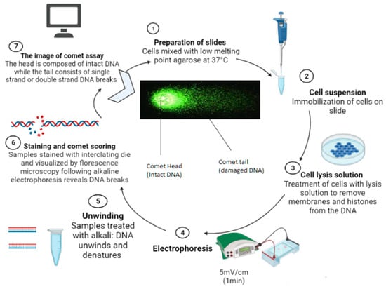
Figure 1.
Schematic diagram showing methodology of comet assay.
Many natural and human-made substances find a home in the aquatic environment. In the last decade, several in vivo and in situ studies have been carried out on both invertebrates and vertebrates. Our primary focus in this review is the study of DNA damage by comet assay in aquatic vertebrates, invertebrates, and organisms collected from contaminated sites.
4.7. Invertebrates
Comet assay has been used globally to assess DNA damage in terrestrial, aquatic invertebrates, and vertebrates. Figure 2 shows the target cells for assessing damage using comet assay.
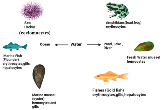
Figure 2.
Target cells for comet assay in aquatic organisms.
The assessment of DNA damage by comet assays after exposure of aquatic invertebrates to genotoxicants is listed in Table 2, while the detailed study is given as follows:
The genotoxic effect of gamma radiation was determined on aquatic fauna using two species of mussels [64]. It evaluated the possible use of comet assay to detect genetic damage in hemocytes and also compared the relative sensitivity of two species of mussels, namely Paphia malabarica and Meretrix casta, to gamma radiation. A significant increase in DNA damage was observed as indicated by an increase in percent tail DNA damage at various concentrations of EMS and all doses of gamma radiation compared to controls in both mussel species. This showed a dose-dependent increase in genetic damage induced by both EMS and gamma radiation in mussels. This study reported that gamma rays caused single-strand breaks in DNA measured by the alkaline comet assay in mussels. It was also revealed that comet assay is a sensitive and rapid method for detecting gamma-rays-induced genotoxicity. This study further indicates that M. casta and P. malabarica have almost identical sensitivity to gamma radiation measured by DNA damage. Li et al. [65] evaluated the genotoxic and physiological effects of acute hypoxia on Pacific white shrimp (L. vannamei). The comet assays in the gill and hepatopancreatic tissues showed an apparent time- and dose-dependent response to hypoxia, suggesting that the comet assay could be used as a sensitive biomarker to detect the occurrence of hypoxia in these two tissues [37,66].
We will focus on an overview of the genotoxicity data based on using the comet test in aquatic animals, published over more than 15 years. In addition, general statements are made regarding appropriate recommendations for standardizing comet assay to improve the comparability and interpretation of data.
4.8. Comet Assay in Mollusca
Comet assay on Mollusca has been studied in several research articles. Some of the studies regarding genotoxic effect of xenobiotics have been mentioned here. Xu et al. [67] aimed to investigate the possible adverse effects of Cu exposure at low and environmentally relevant concentrations. The in vivo activities at different levels of biological organization of the thick-shelled mussel Mytilus coruscus were exposed to two concentrations of copper. The exposure to copper led to DNA damage and an increase in the OTM value in a time- and concentration-dependent manner. In addition, exposure to copper could significantly induce the expression of MT-10, Hsp70, Hsp90, and C3. The present results deepen the suitability of mussels as a model species for marine invertebrates to study the significant adverse effects induced by potential toxins in combinations at different levels of biological organizations. Banni et al. [68] assessed DNA damage over time using the single-cell gel electrophoresis comet assay and the micronucleus test. The comet assay and micronuclei test in digestive gland cells indicated significant DNA damage with a maximal response after 72 h of exposure.
The use of the comet assay in hemocytes from caged non-native mussels was reported as a sensitive tool for monitoring freshwater genotoxicity [69]. The genotoxicity of a naturally contaminated deep-sea environment was investigated using DNA damage and repair studies in the ventricular mussel Bathymodiolus azoricus [70]. Riva et al. [69] reported the use of comet assay in hemocytes from caged alien mussels as a sensitive tool to monitor freshwater genotoxicity. The genotoxicity of a naturally contaminated deep-sea environment was investigated using DNA damage and repair studies in the ventricular clam Bathymodiolus azoricus [70]. The genotoxic effect of different heavy metal concentrations on gill cells of freshwater mussels (Anodonta anatina) exposed to lead (Pb), chromium (Cr), and copper (Cu) under laboratory conditions was estimated [71]. Mussel gill cells were used to determine DNA damage by comet assay. Tail DNA (%), comet tail length, and olive tail moment (OTM) were the parameters used to detect DNA damage. Assessment of the genotoxic effect of metals on freshwater mussels is essential for determining aquatic health and could be proposed as a biomarker. It is concluded that Cu and Pb caused more DNA damage than Cr and combined metal exposure (Pb + Cu + Cr). In addition, results showed that low-dose metal treatment has a more significant genotoxic effect than medium and high doses. Vasanthi et al. [72] reported the effects of heavy metals on DNA damage in the gills and hepatopancreas of Perna viridis collected from the Ennore estuary and Kovalam coastal waters. The tail DNA proportion in the Ennore estuary’s mussels was 12.44% in the gills and 10.14% in the hepatopancreas, respectively. Overall, viridis, the comet, and cytopathological assays have proven to be valuable biomarkers for assessing pollution levels and provide reliable information on coastal water ecotoxicology and genotoxicology. The comet assay could detect DNA damage in mussels (tapes semi decussates) as sediment biomonitor organisms [73,74]. Significant DNA strand breaks were observed in cells isolated from mussel hemolymph, gills, and digestive glands exposed to contaminated sediment [75].
The comet assay was used to assess the sperm DNA quality of cryopreserved Pacific oyster (Crassostrea gigas), as they are commonly used for artificial insemination [76]. Gielazyn et al. [77] demonstrated the use of the lesion-specific DNA repair enzyme formamidopyrimidine glycosylase (Fpg) to improve the usefulness and sensitivity of the comet assay in the study of oxidative diseases. DNA damage in isolated hemocytes from oysters (Crassostrea virginica) and mussels (Mercenaria mercenaria) was also studied. Studies in mussels have shown that comet assay is a sensitive but non-specific molecular biomarker of genotoxicity. One of the disadvantages of applying single-cell gel electrophoresis to field populations can be the animals’ adaptability to higher concentrations of pollutants (e.g., B[a]P), which can be a major issue [78]. In addition, seasonal variations and temperature altered both the baseline level of DNA damage in untreated animals and the sensitivity of cells to environmental pollutants under in vitro conditions [75]. The test has been used for various annelids, including polychaetes, oligochaetes, leeches, and tardigrades.
Understanding genotoxic responses in sediment-dwelling marine species such as polychaetes have become increasingly important as environmental chemicals that can damage DNA become more polluted in marine sediments [79]. Therefore, the comet assay was used to assess the effects of exposure to various pollutants on DNA damage in multiple cell types, including spermatozoa, coelomocytes, blood, and intestinal cells. Increased DNA damage was found in ragworms (Nereis diversicolor) [80,81]. For the investigation of PAHs such as fluoranthene, Capitella capitata showed differences in PAH tolerance between Capitella species [82].
4.9. Comet Assay in Arthropoda
Several scientists have tested the effects of different pollutants on arthropods. Essawy et al. [83] conducted a study to assess the harmful effects of CuO NPs in Lithophaga lithophaga after 28 days of exposure to sub-lethal concentrations. The findings showed that CuO NP deposition in gills increased with concentration and duration. Even in small amounts (5 g/L), CuO NPs damaged DNA in the gills. Malathion was utilized in Knapik and Ramsdorf’s comet experiment in 2020 to confirm its hazardous effects on aquatic species, including Daphnia magna. It was also noted that further research was needed to standardize the comet test method for Daphnia magna because their procedures differed significantly.
According to the Disciplinary of Integrated Production of the Province of Trento, a multi-level approach was utilized to assess the environmental stress generated by copper in a wild population of C. riparius from an agricultural region where copper was frequently used. The single-cell gel electrophoresis test, transcriptional and translational profiling, and enzyme activity assay, were used to demonstrate the role of stress-related genes in copper response. Four candidate genes from two protein families, the heat shock proteins and one cytochrome P450 monooxygenase (CYP4G), and molecular chaperones and oxidative enzymes involved in stress responses, were examined for changes in uploading on polyribosome of transcripts related to stress [36,84]. The stress-inducible proteins HSP70, which are frequently employed in molecular research as effective molecular indicators of overall stress, are encoded by the HSP70 gene [84,85]. After in vivo exposures to the aquatic larvae of the midge Chironomus riparius, DNA damage was observed [86]. The comet test was used to measure DNA-induced damage that led to DNA fragmentation following short-term (24 h) and long-term (96 h) exposures to various doses of the toxicants bisphenol A (BPA), nonylphenol (NP), pentachlorophenol (PCP), tributyltin (TBT), and triclosan (TCS). Massive increases in each of the comet metrics (percent of DNA in tail, tail length, tail moment, and Olive tail moment) for each of the five chemicals’ measured concentrations served as evidence of their genotoxic action. With the exception of TCS at the highest dosage, persistent exposure did not increase the severity of DNA damage, but rather a general decrease that is assumed to be related to the activation of DNA repair and detoxification systems. In addition to considerable time and concentration-dependent changes, comparative analysis revealed disparities in the genotoxic potential of the compounds, which most likely reflect differences in the capacity to repair DNA damage under the various treatments. A study indicated the sensitivity of the benthic larvae of C. riparius to various atmospheric genotoxins, showing its potential as a biomonitor organism in freshwater environments. The findings about the ability of various environmental pollutants to damage DNA highlight the necessity of future research on the genotoxicity of active endocrine compounds, which, by tying genotoxic activity to other biological responses, may help in better understanding the negative impacts in aquatic settings [83,85].
Table 2.
Comet test evaluation of DNA damage in aquatic invertebrates.
Table 2.
Comet test evaluation of DNA damage in aquatic invertebrates.
| Location | Phylum | Organism | Cell Type | Concentration Range | Exposure Time | Descriptor | Agent | Response | Reference |
|---|---|---|---|---|---|---|---|---|---|
| Siolim, Goa, India | Mollusca | Paphiamalabarica Meretrix casta | Hemolymph | Gama (2, 4, 6, 8, and 10 Gray Ethyl Methanesulfonate (18, 32, and 56 mg/L) | 24, 48, 72 h | % Tail DNA | Ethyl Methanesulfonate (EMS) Gamma radiation | Both EMS and gamma radiation indicated a dose-dependent manner. DNA damage for both species was seen in the following order: 24 > 48 and 72 h. | [64] |
| Wenchang, Hainan Province, China | Arthropoda | Litopenaeusvannamei | Gill, hepatopancreas and hemolymph | 3.0 and 1.5 ppm | 24 h | % Tail DNA | Hypoxia | Dose-dependent relation was observed, i.e., DNA damage increased at all concentrations. | [65] |
| Santa Barbara, California, United States of America | Mollusca | Mytilus galloprovincialis Mytilus californianus | Haemocytes | Heat stress: From 13 °C to 24 °C, 28 °C and 32 °C for Mytilus californianus For Mytilus galloprovincialis, 13 °C to 28 °C and 32 °C Cold stress: 2 °C, 6 °C | 0.5, 2 and 8 h | % Tail DNA | Temperature | At 32 °C single-strand breakage and double-strand breakage. DNA in tail length trend: Mytilus californianus > Mytilus galloprovincialis. Cold stress: temperature and time-dependent double-strand breakage in both species. | [87] |
| Zhoushan, Zhejiang Province, China | Mollusca | Mytilus coruscus | Haemocytes | 2 and 8 μg/L | 18 days | OTM | Copper | A time- and concentration-dependent pattern of DNA damage For 2 µ/L order of OTM value:18 > 12 > 6 days For 8 µ/L OTM value is greater after six days | [67] |
| Bizerta Lagoon, Tunisia | Mollusca | Mytilus galloprovincialis | Digestive gland cells | 75 Nm;19 µg/L per animal | 24, 48, and 72 h | % Tail DNA | Benzo[a]pyrene | DNA damage was maximum at 72 h after exposure | [68] |
| Unpolluted freshwater pond, Pakistan | Mollusca | Anodonta anatina | Gill cells | 120 µg/L 240 µg/L 360 µg/L | 15 days | OTM | Lead Chromium Copper | Concentration-wise DNA Damage: Low dose > medium dose > highest dose. Metal-wise DNA damage: Copper and Lead > Chromium and metal combination (Lead + Chromium + Copper) | [71] |
| Taishan, Guangzhou, China | Arthropoda | Scylla paramamosain | Haemocytes | 1.25, 2.5, 5 and 10 mg/L | 72 h | OTM | Cadmium | OTM value was highest at 10 mg/L | [88] |
| Local market in Xichang City, China | Arthropoda | Procambarusclarkii | Haemocytes | 7, 14, and 28 ng/L | 96 h | OTM | Deltamethrin | Olive tail moment (OTM) was highest at 28 ng/L | [89] |
| Ennore Estuary, Chennai city, India | Mollusca | Perna viridis | Gill and hepatopancreatic cells | Copper 65.1 µg/g Lead 0.123 µg/g Zinc 823 µg/g Cadmium 0.047 µg/g Manganese 10.3 µg/g Iron 191 µg/g | 48 h | TM, TL, % Tail DNA | Copper, Lead, Zinc, Cadmium, Manganese and Iron | Cd and Pb were negatively associated and accumulated at lower conc., while Fe showed a higher positive correlation compared to other metals, indicating enhanced accumulation. The trend of Comet Tail: Gills > hepatocreatic cells The trend of %Tail DNA was: Gills > hepatocreatic cells The trend of Tail Moment: Gills < hepatocreatic cells | [72] |
| Elanfoshy bay in Alexandria, Egypt | Mollusca | Lithophagalithophaga | Gill cells | 5 and 20 μg/L | 28 days | OTM, % Tail DNA | Silver oxide nanoparticles | Concentration and time-dependent DNA damage. The highest was at 20 μg/L | [83] |
| Ecotoxicology Laboratory of the Federal Technological University of Paraná, Brazil | Arthropoda | Daphnia magna | Whole organism (homogenized) cells | 0.23 and 0.47 μg/L | 48 h | % Tail DNA | Malathion | Results showed DNA damage at each concentration | [90] |
| Betanzos estuary (North coast of Galicia), Spain, Europe | Mollusca | Mytilus galloprovincialis | 2 gills per individual | 1:500 and 2:500 | 12 days | TL, %TDNA, and OTM | Spilled prestige crude oil | DNA Damage: 1:500 > 2:500 because of more accumulation of TPAH | [91] |
| Devon Great Consols (DGC), Britain | Annelid | Lumbricus rubellus, Dendrodrilus rubidus and Lumbricus terrestris | Soil sample, intestine | 204 mg/kg to 9025 mg/kg | 1, 3, 4, and 5 weeks | % Tail DNA | Arsenic | DNA damage in Earthworms living in As-contaminated soils at DGC < Non-Native Earthworms | [92] |
| Arabian Sea, Goa, India | Mollusca | Perna viridis | Gonad tissue | 2, 5, 10 and 15 µg per kg | 16 days | Percent comet nuclei and deformed nuclei | Cigar tobacco | Dose-response relationship was highest on day 12 | [93] |
| Rio de Janeiro, Brazil | Arthropoda | Amphipod Quadrivisio aff. lutzi (Gammaridea) | Blood cells | 6.25, 12.5, 25, 50 and 100% | 72 h | TL | Water-soluble fraction of heavy oil | DNA damage trend: 6 and 48 h > 24 and 72 h | [94] |
| The Bermuda Institute of Ocean Sciences | Echinodermata | Lytechinus variegatus, Lucunter lucunter, Tripneustes ventricosus and Isostichopus badionotus | Coelomocytes | (0–100 mM) (0–9999 J/m2) | 24 h | %DNA Damage | Hydrogen peroxide UV-C(Ultraviolet-C) | There was a definite concentration- and dose-dependent rise in the DNA damage for all of the examined echinoderm species. The trend of the % DNA damage levels in different coelomocytes of the tested species is as follows: I. badionotus < L. variegatus L. lucunter, T. ventricosus | [95] |
| Lake Trasimeno, Italy | Mollusca | Dreissena polymorpha | Haemocytes | (0.5 and 5 g/mL) (10 g/mL) 4, 18, 28 and 37 °C | 15 h | TL and OTM | Melphalan Hypochlorite Sodium hypochlorite Temperature | As the reaction is temperature dependent, at 4 °C, DNA damage from Hypochlorite > Melphalan and Sodium hypochlorite. At 18°C, DNA damage from Sodium Hypochlorite > Hypochlorite and Melphalan. At 28 °C, 5 g/mL, and 5 min electrophoresis DNA damage from Melphalan > Hypochlorite and Sodium hypochlorite. At 37 °C, DNA damage from Melphalan (5 g/mL) > 0.05 g/mL of Melpahlan and Hypochlorite. | [96] |
| Valencia, Spain | Arthropoda | Chironomus riparius | Fourth instars larvae | Bisphenol A (0.5 and 3 mg/L) Nonylphenol (1, 10 and 100 g/L) Pentachlorophenol (25 and 250 g/L) Tributyltin (0.1, 1 and 10 ng/L) Triclosan (10, 100 and 1000 g/L) | 24 and 96 h | TL, %TDNA, and OTM | Bisphenol A Nonylphenol Pentachlorophenol Tributyltin Triclosan | Time and concentration-dependent variations were significant, i.e., DNA damage was increased with an increase in concentration and time of exposure. | [86] |
| Northern Croatia | Mollusca | Dreissena polymorpha | Haemocytes | 10, 80, 100 and 150 mg/L | 7 days | TL, %TDNA, and OTM | Pentachlorophenol | DNA damage at 80, 100 and 150 mg/L > 10 mg/L | [97] |
| Pune, India | Annelid | Dichogaster curgensis | Coelomocytes | In vivo (1, 3, 10, 30, 70, 100 ppm) In vitro (1, 3, 10, 30, 70 and 100 ppm) | In vivo (14 days) In vitro (1 h) | TL | Cr (Chromium) (VI) | In vivo exposure revealed that DNA damage at 30 ppm > 70 and 100 ppm, while in vitro exposure showed dose-dependent DNA damage. It is hypothesized that a decrease in DNA mobility brought on by the formation of numerous cross-links may be the cause of loss in arbitrary units at greater concentrations. | [98] |
| Korea Institute of Toxicology, Daejeon, Korea | Arthropoda | Daphnia magna, and Chironomus riparius | Erythrocytes | Daphnia magna (0.3, 3, and 30 µg/L for Nonlyphenol and BPA) Chironomus riparius (1, 10 and 100 µg/L for Nonlyphenol and 5, 50 and 500 µg/L for bisphenol A) | 24 h | OTM | Nonlyphenol Bisphenol A | Only for Daphnia magna, OTM at 3 and 30 µg /L > 0.3 µg/L of Bisphenol A exposure. For Chronomus riparius, they statistically increased at all doses of both chemicals examined. | [99] |
| Rio Gola, Northeast-Italy | Arthropod | Chironomus riparius | Fourth-instar larvae | 0.05, 1, and 25 mg/L | 3 h | TL, %TDNA, and OTM | Copper | Significant increases in all the comet parameters at concentrations more than 1 mg/L serve as evidence that copper can cause DNA damage even at sublethal doses. | [100] |
| Anzali wetland, North of Iran | Mollusca | Anodonta cygnea | Haemolymph and gill cells | 0.25, 0.5, and 1.0 ppm | 10 days | TL | Crude oil | Statistically significant increases in DNA damage and micronuclei were reported with 0.25, 0.5, and 1.0 ppm of crude oil. | [101] |
| Randaberg, Norway | Echinodermata and mollusca | Strongylocentrotus droebachiensis and Mytilus edulis Lamellibranchiata | Sea urchin coelomocytes and mussel haemocytes | Sea urchins (0.06 and 0.25 mg/L) Mussels (0.015, 0.06, and 0.25 mg/L) | Sea urchin (4 weeks) Mussel (5 weeks) | Percentage of DNA in comet tail | Crude oil | DNA concentration in the comet tails increased significantly with oil concentrations of 0.06 and 0.25 mg/L. Mussels subjected to oil concentrations up to 0.06 mg/L revealed a considerable concentration-related rise in the comet tail’s DNA %. | [102] |
| Kat O, Hong Kong | Mollusca | Perna viridis | Haemocytes | 0, 0.3, 3, and 30 µg/L | 0, 1, 3, 6 and 12 days | TL, %TDNA, and OTM | Water-borne benzo[a]pyrene | For 0.3 µg/L TL, %TDNA and OTM at day 0–3 > 6 and 12 days. For 3 and 30µg/L TL, %TDNA and OTM at day 0–6 > 6–12 days. | [103] |
| Lake Ponikve Krk Island (Northern Adriatic Sea) | Annelid | Hirudo verbana | Leech haemocytes | 0.5 L | 30, 60, 180 days | TL, %TDNA, and OTM | Aluminium contaminated sediment and water | DNA damage at 180 > 60 > 30 days | [104] |
| BiAzerta Lagoon, Tunisia | Mollusca | Mytilus galloprovincialis | Digestive gland cells | 75 nM; 19 mg/L per animal | 24, 48, and 72 h | %TDNA | Benzo[a]pyrene | DNA damage at 72 h > 48 > 24 h | [105] |
| Marennes-Oléron Bay, France | Mollusca | Crassostrea gigas | Hemocytes, gills and spermatozoa | 0.4 and 0.6 μg/L 0.005% for solvent control | Two 7-day exposures | % tDNA | Diuron Acetonitrile | DNA damage in Gills, Hemocytes and Spermatozoa: Diuron > Control groups | [106] |
Abbreviations: Ethyl Methanesulfonate (EMS), Olive Tail Moment (OTM), Tail Length (TL), Percentage of DNA in the tail (%tDNA). Cadmium (Cd), Lead (Pb).
4.10. Vertebrates
Many fishes (freshwater and marine) have already been used for environmental biomonitoring. A summarized study of the assessment of DNA damage by comet assays is given in Table 3. These studies are discussed as follows.
4.11. Comet Assay in Fish
The comet test has been used to detect DNA strand breaks in aquatic animals exposed to various genotoxic chemicals in vitro and in vivo. Numerous contaminated estuaries and coastal bays possess high quantities of genotoxic pollutants such as polycyclic aromatic hydrocarbons, herbicides, and metals. Estuaries, in particular, are continually exposed to genotoxic compounds from industrial or municipal waste streams that might harm the biota [107]. In order to identify molecular genotoxicity biomarkers, it has been helpful to evaluate the DNA in individual cells after contamination [73,108]. Using a minimal number of cells, the single-cell gel electrophoresis (SCGE) or comet test looks to be a rapid, easy, consistent, and sensitive method to identify and evaluate genetic damage in nearly any kind of eukaryotic cell [31]. This method has demonstrated its usefulness for tracking the effects of DNA-damaging chemicals in various marine and freshwater fish [7,109,110,111,112]. DNA single-strand breaks (SSBs), alkali-labile sites, DNA-DNA/DNA-protein cross-linking, and SSBs associated with partial excision repair sites may be detected using the alkaline version created by [113].
Industrial trash and unprocessed municipal waste have contaminated the Sinos River in southern Brazil. Biomarkers for fish genotoxicity are useful indicators for evaluating environmental risk. The comet test is used to find genotoxicity caused by various pollution sources in the peripheral blood of a local fish species.
The comet assay has been conducted on cells from the animals taken from the sites (Table 4). The following is an overview of the findings from these sites:
Table 4.
Assessment of DNA damage by comet assay in different cells of aquatic animals obtained from polluted areas.
Table 3.
Comet test evaluation of DNA damage in aquatic vertebrates.
Table 3.
Comet test evaluation of DNA damage in aquatic vertebrates.
| Location | Phylum | Organism | Descriptor | Cell Type | Concentration Range | Exposure Time | Agent | Response | Reference |
|---|---|---|---|---|---|---|---|---|---|
| Gorukle, Bursa, Turkey | Chordata | Carassius auratus | TL | Erythrocytes | 5, 10, and 15 μg/L | 2, 4, and 6 days | Gesaprim Atrazine | Gesaprim increased proportion of damaged cells in the peripheral erythrocytes of Carassius auratus. However, no significant effect on erythrocytes was found. | [114] |
| Mandapam, southeast coast of India | Chordata | Therapon jarbua | % Tail DNA | Gill, kidney, and erythrocytes | 0.125, 0.25 ppm | 96 h | Mercuric chloride | At concentrations 0.125 and 025 ppm: Gill cells > kidneys and blood | [115] |
| CIFA’s (Central Institute of Freshwater Aquaculture) culture ponds in Bhubaneswar, India | Chordata | Labeo rohita | DNA damage (migration comet tail length and relative DNA content of the comet tail) | Blood, liver, and gill cells | 0.001, 0.002, 0.01 ppm | 96 h | Phorate | After 72 h, ↓DNA damage DNA breaks order: Liver > Gills and blood | [116] |
| Hatchery of the Freshwater Fisheries Institute of Lishui Zhejiang Province, China | Chordata | Cyprinus carpio | DNA damage percentage, TL, and TM | Liver cells | 0.41, 0.52, 0.69, 1.03 and 2.06 mg/L | 7 days | Cadmium | At concentrations (0.41 mg/L), ↓SOD activity, ↑ MDA and GSH levels These findings showed that cadmium (Cd) caused DNA damage in Cyprinus carpio liver. At concentration > 0.41mg/L all parameters were increased. | [117] |
| Commercial fish seed hatchery, India | Chordata | Catla catla | % Tail DNA TL TM | Erythrocytes | 30, 60, 90, 150 mg/L | 7, 14, and 21 days | Chromium | A significant (p 0.001) increase in % Tail DNA was observed after 21 days of exposure | [118] |
| Taian, Shandong, China | Chordata | Danio rerio | OTM | Liver cells | 0.3, 1.25, and 5 mg/L | 28 days | Imidacloprid | At the 21st day of exposure, at concentrations (of 1.25 and 5 mg/L), ROS generation and MDA contents were increased. Both time and dosage were factors in DNA damage. | [66] |
| Fish Seed Hatchery, Faisalabad | Chordata | Labeo rohita, Cirrhinus mrigala, Catla catla Ctenopharyngodon idella | Percent damaged cells, genetic damage index (GDI) and cumulative tail length | Erythrocytes | 17, 25, 33 and 50% LC50 | 30 days | Arsenic, Copper, and Zinc | The order of percentage damage in the erythrocytes of all four fish species is as follows: C. mrigala > L. rohita > C. idella > C. catla. L. rohita tail length revealed the only non-significant variation (C. mrigala > L. rohita > C. catla > C. idella) as exposure dramatically increased GDI, followed by exposures to Zn and Copper. | [119] |
| Rosario, Argentina laboratory | Chordata | Danio rerio | DNA damage (DNA damage score (Damage Index, DI)) | Gill cells | 0.3 or 0.6 µg/L | 12 days | Cypermethrin | The DNA damage index in the gill cells >dose- and time-dependent pathways. | [120] |
| China | Chordata | Gobio cyprisrarus | Tail DNA%, OTM | Liver cells | 0.1 mg/L 0.5 mg/L 2.0 mg/L | 60 days | Imidacloprid, Nitenpyram, and Dinotefuran | The DNA damage assay results showed that treatments with 2.0 mg/L imidacloprid significantly increased tail moments whereas treatments with 0.5, 2.0, 2.0 mg/L imidacloprid, 2.0 mg/L nitenpyram, and all treatments with dinotefuran significantly increased tail DNA%. Furthermore, treatments with 0.5 and 2.0 mg/L imidacloprid and 2.0 mg/L dinotefuran markedly increased the olive tail moments. | [121] |
| Water Treatment Station SANEAGO, in Goiânia city, GO, Brazil | Chordata | Poecilia reticulata | TL, TM, and OTM | Brain, Liver, and blood cells | 0.5, 1.0, and 1.5 μg/L | 24 and 96 h | Cylindrospermopsin | After 96 h, an increase in DNA damage was seen in peripheral blood cells using all three doses. | [122] |
| Snidjers Scientific, Tilburg, Nederland | Chordate | Oryzias latipes | TD% | Medaka cells | Acute toxicity (CdCl2 (0.1, 1, 10 and 100 μM) or Flu (2, 10 and 50 μM)) In vivo (CdCl2 (0.03, 0.1, 0.3, and 1 μM) or Flu (2, 10, and 50 μM)) In vitro (H2O2 (1, 10, and 100 μM) and (CdCl2 (0.03, 0.1, 0.3 and 1 μM) in 400 μL of Minimum Essential Medium (MEM) | 24 h | CdCl2 or Flu | Findings indicated that all tested chemicals significantly increased the proportion of damage in DNA of fragmented cells than the control one. | [123] |
| Eastern Croatia, Europe | Chordate | Cyprinus carpio | %TDNA | Erythrocytes | Nil | 3 weeks | Wastewaters, paper and pulp mill industry, chemicals (detergent and soap, textile) and metal industries | The results of comet assay revealed a statistically significant increase in the DNA damage in carp erythrocytes. | [124] |
| McElroy laboratory, The Ohio State University, United States | Chordate | Oryzias latipes | OTM | Embryos | 10 μg/L(final) | 7-day exposure | Polycyclic Aromatic Hydrocarbons (PAHs) Acenaphthenequinone and 7,12-Benz[a]anthracenquinone | After 48 h of exposure at the lowest tested dose (5 g/L), DNA damage was increased. | [125] |
| River Rhine, Germany | Chordate | Danio rerio | The tail moments, which are calculated as the ratio of the tail’s mean migration distance to its relative DNA content | Embryonic cells | 600 mg/mL = 100% (1:1), 300 mg/mL = 50% (1:2), and 150 mg/mL = 25% (1:4) 40, 20, 10 and 5 mg | 72 h | Exposure to whole (freeze-dried) sediment, exposure to organic sediment extracts | Only four native soils had genotoxic potential, compared to 17 out of 18 sediment extracts that significantly damaged the embryo cells’ DNA. The organic extracts, on the other hand, appear to have enhanced quantities of even barely soluble compounds | [126] |
| Coast of Gwangyang Bay, Korea | Chordate | Paralichthys olivaceus | TL | Whole blood | 5, 10, 50 and 100 ppb | 2 h for in vivo (0, 2, and 4 days | PAHs (Polycyclic Aromatic Hydrocarbons) BaP’s(benzo[a]pyrene, fluoranthene, anthracene, pyrene and phenanthrene) and sediments and also in vivo benzo[a]pyrene is used | Five groups treated with PAHs at concentrations of 50 and 100 ppb showed considerably shorter tail lengths than the control group. BaP’s genotoxic effects were associated with exposure time and concentration. Significant variations in DNA breakage between cells exposed to sediment extracts or PAHs and non-exposed control were seen throughout the investigation. | [127] |
| Zebrafish International Resource Center (ZIRC), United States | Chordate | Danio rerio | TL, %TDNA, and OTM | Embryos | 0 (control or vehicle), 250, 350, 625, 850, and 1250 μg/mL | 2, 4, 6, 12, 24, and 48 h | Monceren 250 SC | Caused DNA damage, as evidenced by the three genotoxicity parameters significantly increasing in zebrafish embryos. The survival percentage of larvae embryos was down to 40–45% after 48 h following fungicide treatment. | [128] |
| Fish Hatchery Station of the State University of Londrina, Brazil | Chordate | Prochilodus lineatus | TL | Erythrocytes and liver cells | 1.25, 12.5, 125, and 1250 mg /L | 120 h | Midacloprid (IMI) | Integrated Biomarker Response (IBR) index revealed: Liver and kidney > gills. | [129] |
| Santa Fe Province, Argentina | Chordate | Caiman latirostris | TL | Erythrocytes | 50, 100, 200, 300, 400, 500, 750, 1000, 1250 and 1750 µg/egg | 68 and 70 days in Experiment 1 and 66 and 77 days in Experiment 2 | Roundup | The MN test and the comet assay findings proved concentration-dependent and negative impacts. | [130] |
| Southern Brazil | Chordate | Gray mullet (Mugil sp.) and sea catfish (Netuma sp.) | Image length (IL), damage index (DI)and damage frequency (DF) | Erythrocytes | 2, 4, and 8 × 10⁻⁵ M | 1, 2, 6 and 24 h | Methyl methanesulfonate (MMS) | Baseline DNA damage enhanced at higher temperatures, as did MMS-induced DNA damage, and there was a definite dose/time response to MMS therapy. | [131] |
| Santa Fe city, Argentina | Chordate | Prochilodus lineatus | Comet tail and % of TDNA | Erythrocytes | 0.300, 0.150, 0.075 and 0.000 μg/L | 96 h | Cypermethrin | DNA damage was considerably higher at all Cypermethrin doses evaluated. | [132] |
| Siolim, Goa | Chordate | Channa punctatus (Bloch) | % tail DNA | The gills, kidneys, and lymphocytes | 1.48, 0.74, and 0.59 ppm | 0, 1, 3, 7, 15, 22 and 29 days (in vivo) Sub lethal (96 h) | Malathion | Up to day 3, DNA damage was seen in all of the tissues and increased as a function of concentration, followed by a nonlinear decline as exposure time increased. | [133] |
| Local outlets, India | Chordate | Clarias batrachus | TL | Erythrocytes | 2,4-D 25–75 ppm Butachlor 1–2.5 ppm | 48, 72, and 96 h | Herbicides: 2,4-D and Butachlor | The greatest tail length observed at the highest concentration and longer period of 2, 4-D (9.59 mm) and Butachlor indicated a concentration-related and time-dependent increase. | [134] |
| Toulouse, France’s Paul Sabatier University’s Department of Developmental Biology | Chordate | Xenopus laevis; Pleurodeles Waltl | tDNA, TL, OTM | Erythrocytes | Captan (fungicide) 2–125 mg/L; 62.5 mg/L | 1–12 days | Captan | From the first day of exposure, comet assay revealed that captan was genotoxic. | [135] |
| Toulouse, France’s Paul Sabatier University’s Department of Developmental Biology | Chordate | Xenopus laevis; Pleurodeles waltl | tDNA, TL, OTM | Erythrocytes | Cd 2–50 mg/L | 1–12 days | Cadmium | Comparing the findings of comet assay and micronuclei test, genotoxic effects were taken into account, along with the Cadmium concentrations used and the exposure duration. According to the comet assay, Cd was genotoxic from the first day of exposure. | [136] |
| Sweden | Chordate | Oncorhynchus mykiss | Tail DNA | Erythrocytes | 0.5% | 7 days | Algal extracts (Polysiphonia fucoides) | Results revealed substantial increases in DNA single-strand breaks equivalent to those caused by an in vivo exposure to 20 mg/kg B[a]P, but no changes in double-strand breaks or apoptotic cells were noted. | [137] |
| Uttar Pradesh, India | Chordate | Channapunctatus | %Tail DNA | Gill and kidney tissues | 4–10 ppb | 24–96 h | Endosulfan (pesticide) | In both tissues, there were dose-dependent reactions to DNA damage. Gill cells >kidney cells. | [138] |
| Byford, Stavanger, Norway | Chordate | Symphodus mellops | %Tail DNA | Erythrocytes | 2 mg/L | 7 days | Styrene | Fish and mussel blood cells’ comet tail DNA content was increased by styrene by 1.8 and 1.6 times, respectively | [139] |
SOD: Superoxide dismutase activity; CdCl2: Cadmium Chloride; Flu: Flourantthene; MDA: Malondialdehyde; GSH: Glutathione; ROS: Reactive Oxygen Species; Bisphenol A: BPA; Nonylphenol: NP; OTM: Olive Tail Moment; TL: Tail Length; %tDNA: percentage of DNA in tail; GDI: Genetic Damage Index; Cd: Cadmium; CA: comet assay; PAHs: Polycyclic Aromatic Hydrocarbons; MMS: Methyl methanesulfonate.
4.12. In Vivo and In Vitro Comet Assay
Genotoxicity testing increasingly uses in vivo comet assay (single-cell gel electrophoresis). The in vivo comet assay’s benefits include its adaptability to different tissues and/or unique cell types, sensitivity for the detection of minute amounts of DNA damage, need for few cells per sample, the overall comfort of test performance, quick research completion, and comparatively low cost [60]. International expert committees have produced recommendations detailing criteria aimed at high-quality methods to acquire accurate and dependable results, mirroring the rules for other modes of in vivo genotoxicity testing [29].
Mutagenicity testing is based on in vitro and in vivo experiments aiming at various genetic endpoints as per the recommendations of worldwide guidelines. The majority of the tests are mutagenicity assays, which look for changes to genes, chromosomes, or genomes. Mutations are thought to be a crucial element in the multi-step process of carcinogenesis and can cause hereditary illnesses. As a result, the findings of mutagenicity tests are primarily used to determine a mutagenic hazard. Extended genotoxicity testing may benefit from the findings of indicator tests, which measure consequences connected to the process of mutagenesis, such as DNA damage, recombination, and repair. The comet assay is generally used as an additional in vivo test for drugs that show positive findings from in vitro mutagenicity tests and/or for mechanistic investigations. It is an indicator test for the detection of DNA damage. The unscheduled DNA synthesis (UDS) test and the alkaline elution method are additional in vivo indicator tests with regulatory acceptance. The in vivo comet assay has several benefits over these tests. The comet assay may be used for almost any organ of interest given that an adequate cell preparation has been produced for each organ and cell type, in contrast to the in vivo UDS test, which is often exclusively done on hepatocytes. Additionally, the UDS test may not be sensitive enough to identify single-strand breaks and oxidative base damage since nucleotide excision repair does not repair these lesions. In contrast, the comet assay can detect a wider range of primary DNA lesions, including these [164]. Compared to the comet assay, the UDS test seemed to be relatively insensitive for identifying direct-acting or short-lived mutagens when utilized for the study of initial site-of-contact tissue, such as stomach epithelia [165].
4.13. Critical Factors Influencing Comet Assay Performance
The impact of agarose concentration is obvious. The least effective concentration was 0.4%, which resulted in relatively high tail DNA percentages (with cells containing DNA breaks) but was too fragile to be advised. The percentage of tail DNA continuously declines as the agarose concentration rises to 1.3%. It does not seem to matter how long the lysis took in the Triton and high salt solution. Although overnight lysis is frequent, the gel is sometimes held in lysis solution for weeks with no apparent influence on finding [166]. Nevertheless, the alkaline incubation period is crucial. Whenever changed across the time range of 10 to 60 min, there had been a continuous rise in the percentage of tail DNA during that period or an approach to a plateau after 40 min [9,167].
Changes in electrophoresis voltage and duration significantly impacted comets from treated and untreated cells [9]. With voltage and passing time, the relative tail intensity increases dramatically. The crucial factor is the voltage gradient, particularly the voltage gradient over the platform on which the slides are set. Due to the substantial depth of electrophoresis solution and thus low resistance compared to the depth of a few millimeters of solution over the slides, there is relatively little voltage change between the electrode and the edge of the platform. Within certain bounds, a shorter electrophoresis duration will be made up by a greater voltage gradient and vice versa.
4.14. The Comet Assay Modifications
Many DNA-damaging substances produce lesions other than just single-strand breaks (SBs), such as bulky adducts, intra- and inter-strand cross-links, oxidized or alkylated bases, or oxidized or alkylated bases, which tend to have more severe effects on the cell or organism than the easily repairable single SBs but are undetectable by the comet assay. The comet assay has been altered in many ways to overcome this limitation. Since cross-links prevent DNA that has been damaged by ionizing radiation from migrating, they can be used to test inter-strand cross-links. As a result, the less intense the comet tails, the more cross-linking is present [168]. Although not frequently used, the DNA synthesis inhibitors aphidicolin, hydroxyurea, and 1-D-arabinofuranosyl cytosine, or different combinations of these, have been used to detect bulky adducts or UV-induced lesions. These substances prevent DNA synthesis during nucleotide excision repair (NER), which results in the accumulation of breaks [169]. The comet assay can be applied to cultured cells, disaggregated tissue or white blood cells. This last application has been of great use in human biomonitoring, especially because cells can be assayed after prolonged frozen storage—an advantage in large-scale studies when it is impossible to assay samples immediately after collection. Occasionally, whole blood has been stored frozen and then used for comet assay analysis [170].
Any eukaryotic cell type that can be obtained as a single cell or nuclear suspension seems to be amenable to comet assay analysis. This includes cells in culture, blood cells taken from animals or humans, haemolymph cells from molluscs and insects, sperm, disaggregated animal tissues, yeast, and nuclei released from plant tissue. Even individual chromosomes can be used to create comets. By digesting the DNA following lysis with a lesion-specific enzyme, which causes a DNA break at the damage site, the method has been developed to measure DNA SBs as well as to detect damaged bases. This approach has been used to investigate bases that have been oxidized or alkylated as well as cyclobutane pyrimidine dimers produced by UV irradiation [171].
5. Conclusions
The comet assay is already a well-established technique. Due to its adaptability, toxicologists still consider it a sensitive method for assessing DNA damage. This has been demonstrated by the range of species, tissues, and cell types that can be analyzed to determine genotoxicity in aquatic vertebrates and invertebrates. As a result, the comet test can measure environmental exposure to genotoxins in a wide range of aquatic species, giving researchers and environmental managers a sensitive and rapid tool. However, the literature suggests that there are many different comet protocols. Methods must be standardized, and inter-laboratory calibration of the comet test is established where applicable to aquatic species. There is a need to improve water quality testing. Nevertheless, more in-depth studies are required to conduct more accurate genotoxicity testing and biomonitoring of aquatic species. The standards should be set up so they can easily be used among different international development agencies. This will more accurately monitor the impact of pollutants on the environment, natural ecosystems, food chains, and the environment.
Author Contributions
N.J., S.N., Y.M., Q.U., M.Z.K., A.M.M.C. and W.-D.B. designed the study and wrote the manuscript; A.M.M.C. and W.-D.B. supervised the manuscript; J.W., X.L., D.-Z.L. and S.T. helped in the collection of data resources and editing of the final version of the manuscript. All authors have read and agreed to the published version of the manuscript.
Funding
The present study was supported by State Key Laboratory of Hulless Barley and Yak Germplasm Resources and Genetic Improvement Project XZNKY-2021-C-014-Z04 and the Central Government Guided Local Science and Technology Development Fund Project XZ202101YD0017C.
Institutional Review Board Statement
Not applicable.
Informed Consent Statement
All authors agreed to publish this work.
Data Availability Statement
All the data are included in the main manuscript.
Conflicts of Interest
The authors declare no conflict of interest.
References
- Pollack, N.; Cunningham, A.R.; Rosenkranz, H.S. Environmental persistence of chemicals and their carcinogenic risks to humans. Mutat. Res. 2003, 528, 81–91. [Google Scholar] [CrossRef]
- Zubair, M.; Ahmad, M.; Saleemi, M.K.; Gul, S.T.; Ahmad, M.; Martyniuk, C.J.; Ullah, Q.; Umar, S. Sodium arsenite toxicity on hematology indices and reproductive parameters in Teddy goat bucks and their amelioration with vitamin C. Environ. Sci. Pollut. Res. 2020, 27, 15223–15232. [Google Scholar] [CrossRef]
- Naz, S.; Mansouri, B.; Chatha, A.M.M.; Ullah, Q.; Abadeen, Z.U.; Khan, M.Z.; Khan, A.; Saeed, S.; Bhat, R.A. Water quality and health risk assessment of trace elements in surface water at Punjnad Headworks, Punjab, Pakistan. Environ. Sci. Pollut. Res. 2022, 29, 61457–61469. [Google Scholar] [CrossRef]
- Frenzilli, G.; Nigro, M.; Lyons, B.P. The Comet assay for the evaluation of genotoxic impact in aquatic environments. Mutat. Res. 2009, 681, 80–92. [Google Scholar] [CrossRef]
- Evenson, D.P.; Kasperson, K.; Wixon, R.L. Analysis of sperm DNA fragmentation using flow cytometry and other techniques. Soc. Reprod. Fertil. Suppl. 2007, 65, 93–113. [Google Scholar]
- Møller, P.; Azqueta, A.; Boutet-Robinet, E.; Koppen, G.; Bonassi, S.; Milić, M.; Gajski, G.; Costa, S.; Teixeira, J.P.; Costa Pereira, C.; et al. Minimum Information for Reporting on the Comet Assay (MIRCA): Recommendations for describing comet assay procedures and results. Nat. Protoc. 2020, 15, 3817–3826. [Google Scholar] [CrossRef]
- Lee, R.F.; Steinert, S. Use of the single cell gel electrophoresis/comet assay for detecting DNA damage in aquatic (marine and freshwater) animals. Mutat. Res. 2003, 544, 43–64. [Google Scholar] [CrossRef]
- Møller, P.; Loft, S.; Ersson, C.; Koppen, G.; Dusinska, M.; Collins, A. On the search for an intelligible comet assay descriptor. Front. Genet. 2014, 5, 217. [Google Scholar] [CrossRef]
- Azqueta, A.; Gutzkow, K.B.; Brunborg, G.; Collins, A.R. Towards a more reliable comet assay: Optimising agarose concentration, unwinding time and electrophoresis conditions. Mutat. Res. 2011, 724, 41–45. [Google Scholar] [CrossRef]
- Azqueta, A.; Muruzabal, D.; Boutet-Robinet, E.; Milic, M.; Dusinska, M.; Brunborg, G.; Møller, P.; Collins, A.R. Technical recommendations to perform the alkaline standard and enzyme-modified comet assay in human biomonitoring studies. Mutat. Res. Genet. Toxicol. Environ. Mutagen. 2019, 843, 24–32. [Google Scholar] [CrossRef]
- Ostling, O.; Johanson, K.J. Microelectrophoretic study of radiation-induced DNA damages in individual mammalian cells. Biochem. Biophys. Res. Commun. 1984, 123, 291–298. [Google Scholar] [CrossRef] [PubMed]
- Singh, N.P.; McCoy, M.T.; Tice, R.R.; Schneider, E.L. A simple technique for quantitation of low levels of DNA damage in individual cells. Exp. Cell Res. 1988, 175, 184–191. [Google Scholar] [CrossRef]
- Olive, P.L.; Banáth, J.P.; Durand, R.E. Heterogeneity in radiation-induced DNA damage and repair in tumor and normal cells measured using the “comet” assay. Radiat. Res. 1990, 122, 86–94. [Google Scholar] [CrossRef]
- Burlinson, B.; Tice, R.R.; Speit, G.; Agurell, E.; Brendler-Schwaab, S.Y.; Collins, A.R.; Escobar, P.; Honma, M.; Kumaravel, T.S.; Nakajima, M.; et al. Fourth International Workgroup on Genotoxicity testing: Results of the in vivo Comet assay workgroup. Mutat. Res. 2007, 627, 31–35. [Google Scholar] [CrossRef]
- Gedik, C.M.; Ewen, S.W.; Collins, A.R. Single-cell gel electrophoresis applied to the analysis of UV-C damage and its repair in human cells. Int. J. Radiat. Biol. 1992, 62, 313–320. [Google Scholar] [CrossRef] [PubMed]
- Collins, A.R.; Duthie, S.J.; Dobson, V.L. Direct enzymic detection of endogenous oxidative base damage in human lymphocyte DNA. Carcinogenesis 1993, 14, 1733–1735. [Google Scholar] [CrossRef] [PubMed]
- Collins, A.R. The use of bacterial repair endonucleases in the comet assay. Methods Mol. Biol. 2011, 691, 137–147. [Google Scholar] [CrossRef]
- Speit, G.; Haupter, S.; Schütz, P.; Kreis, P. Comparative evaluation of the genotoxic properties of potassium bromate and potassium superoxide in V79 Chinese hamster cells. Mutat. Res. 1999, 439, 213–221. [Google Scholar] [CrossRef]
- Smith, C.C.; O’Donovan, M.R.; Martin, E.A. hOGG1 recognizes oxidative damage using the comet assay with greater specificity than FPG or ENDOIII. Mutagenesis 2006, 21, 185–190. [Google Scholar] [CrossRef]
- McKelvey-Martin, V.J.; Green, M.H.; Schmezer, P.; Pool-Zobel, B.L.; De Méo, M.P.; Collins, A. The single cell gel electrophoresis assay (comet assay): A European review. Mutat. Res. 1993, 288, 47–63. [Google Scholar] [CrossRef]
- Binková, B.; Lewtas, J.; Mísková, I.; Rössner, P.; Cerná, M.; Mrácková, G.; Peterková, K.; Mumford, J.; Meyer, S.; Srám, R. Biomarker studies in northern Bohemia. Environ. Health. Perspect. 1996, 104 (Suppl. S3), 591–597. [Google Scholar] [CrossRef][Green Version]
- Møller, P.; Knudsen, L.E.; Frentz, G.; Dybdahl, M.; Wallin, H.; Nexø, B.A. Seasonal variation of DNA damage and repair in patients with non-melanoma skin cancer and referents with and without psoriasis. Mutat. Res. 1998, 407, 25–34. [Google Scholar] [CrossRef]
- Hartmann, A.; Plappert, U.; Raddata, K.; Grünert-Fuchs, M.; Speit, G. Does physical activity induce DNA damage? Mutagenesis 1994, 9, 269–272. [Google Scholar] [CrossRef]
- Duthie, S.J.; Dobson, V.L. Dietary flavonoids protect human colonocyte DNA from oxidative attack in vitro. Eur. J. Nutr. 1999, 38, 28–34. [Google Scholar] [CrossRef]
- Pool-Zobel, B.L.; Bub, A.; Müller, H.; Wollowski, I.; Rechkemmer, G. Consumption of vegetables reduces genetic damage in humans: First results of a human intervention trial with carotenoid-rich foods. Carcinogenesis 1997, 18, 1847–1850. [Google Scholar] [CrossRef]
- Nakagawa, Y.; Wakuri, S.; Sakamoto, K.; Tanaka, N. The photogenotoxicity of titanium dioxide particles. Mutat. Res. 1997, 394, 125–132. [Google Scholar] [CrossRef]
- Sasaki, Y.F.; Sekihashi, K.; Izumiyama, F.; Nishidate, E.; Saga, A.; Ishida, K.; Tsuda, S. The comet assay with multiple mouse organs: Comparison of comet assay results and carcinogenicity with 208 chemicals selected from the IARC monographs and U.S. NTP Carcinogenicity Database. Crit. Rev. Toxicol. 2000, 30, 629–799. [Google Scholar] [CrossRef]
- Albertini, R.J.; Anderson, D.; Douglas, G.R.; Hagmar, L.; Hemminki, K.; Merlo, F.; Natarajan, A.T.; Norppa, H.; Shuker, D.E.; Tice, R.; et al. IPCS guidelines for the monitoring of genotoxic effects of carcinogens in humans. International Programme on Chemical Safety. Mutat. Res. 2000, 463, 111–172. [Google Scholar] [CrossRef]
- Hartmann, A.; Agurell, E.; Beevers, C.; Brendler-Schwaab, S.; Burlinson, B.; Clay, P.; Collins, A.; Smith, A.; Speit, G.; Thybaud, V.; et al. Recommendations for conducting the in vivo alkaline Comet assay. 4th International Comet Assay Workshop. Mutagenesis 2003, 18, 45–51. [Google Scholar] [CrossRef]
- Burlinson, P.; Studholme, D.; Cambray-Young, J.; Heavens, D.; Rathjen, J.; Hodgkin, J.; Preston, G.M. Pseudomonas fluorescens NZI7 repels grazing by C. elegans, a natural predator. ISME J. 2013, 7, 1126–1138. [Google Scholar] [CrossRef]
- Collins, A.R.; Dobson, V.L.; Dusinská, M.; Kennedy, G.; Stĕtina, R. The comet assay: What can it really tell us? Mutat. Res. 1997, 375, 183–193. [Google Scholar] [CrossRef]
- Gedik, C.M.; Collins, A. Establishing the background level of base oxidation in human lymphocyte DNA: Results of an interlaboratory validation study. FASEB J. 2005, 19, 82–84. [Google Scholar] [CrossRef]
- Møller, P.; Friis, G.; Christensen, P.H.; Risom, L.; Plesner, G.; Kjaersgaard, J.; Vinzents, P.; Loft, S.; Jensen, A.; Tved, M. Intra-laboratory comet assay sample scoring exercise for determination of formamidopyrimidine DNA glycosylase sites in human mononuclear blood cell DNA. Free Radic. Res. 2004, 38, 1207–1214. [Google Scholar] [CrossRef]
- Møller, P.; Möller, L.; Godschalk, R.W.; Jones, G.D. Assessment and reduction of comet assay variation in relation to DNA damage: Studies from the European Comet Assay Validation Group. Mutagenesis 2010, 25, 109–111. [Google Scholar] [CrossRef][Green Version]
- Neri, M.; Milazzo, D.; Ugolini, D.; Milic, M.; Campolongo, A.; Pasqualetti, P.; Bonassi, S. Worldwide interest in the comet assay: A bibliometric study. Mutagenesis 2015, 30, 155–163. [Google Scholar] [CrossRef]
- Koppen, G.; Azqueta, A.; Pourrut, B.; Brunborg, G.; Collins, A.R.; Langie, S.A.S. The next three decades of the comet assay: A report of the 11th International Comet Assay Workshop. Mutagenesis 2017, 32, 397–408. [Google Scholar] [CrossRef]
- Gajski, G.; Žegura, B.; Ladeira, C.; Novak, M.; Sramkova, M.; Pourrut, B.; Del Bo, C.; Milić, M.; Gutzkow, K.B.; Costa, S.; et al. The comet assay in animal models: From bugs to whales—(Part 2 Vertebrates). Mutat. Res. Rev. Mutat. Res. 2019, 781, 130–164. [Google Scholar] [CrossRef]
- Jha, A.N. Genotoxicological studies in aquatic organisms: An overview. Mutat. Res. Fundam. Mol. Mech. Mutagen. 2004, 552, 1–17. [Google Scholar] [CrossRef]
- Kumaravel, T.S.; Vilhar, B.; Faux, S.P.; Jha, A.N. Comet Assay measurements: A perspective. Cell Biol. Toxicol. 2009, 25, 53–64. [Google Scholar] [CrossRef]
- Dixon, D.R.; Pruski, A.M.; Dixon, L.R.; Jha, A.N. Marine invertebrate eco-genotoxicology: A methodological overview. Mutagenesis 2002, 17, 495–507. [Google Scholar] [CrossRef]
- Lorenzo, Y.; Costa, S.; Collins, A.R.; Azqueta, A. The comet assay, DNA damage, DNA repair and cytotoxicity: Hedgehogs are not always dead. Mutagenesis 2013, 28, 427–432. [Google Scholar] [CrossRef] [PubMed]
- Committee on Mutagenicity of Chemicals in Food, Consumer Products and the Environment (COM). Guidance on a Strategy for Testing of Chemicals for Mutagenicity; Department of Health: London, UK, 2000.
- Møller, P. The alkaline comet assay: Towards validation in biomonitoring of DNA damaging exposures. Basic. Clin. Pharmacol. Toxicol. 2006, 98, 336–345. [Google Scholar] [CrossRef]
- Collins, A.R. The comet assay for DNA damage and repair: Principles, applications, and limitations. Mol. Biotechnol. 2004, 26, 249–261. [Google Scholar] [CrossRef] [PubMed]
- Dhawan, A.; Bajpayee, M.; Parmar, D. Comet assay: A reliable tool for the assessment of DNA damage in different models. Cell. Biol. Toxicol. 2009, 25, 5–32. [Google Scholar] [CrossRef] [PubMed]
- Møller, P. The comet assay: Ready for 30 more years. Mutagenesis 2018, 33, 1–7. [Google Scholar] [CrossRef] [PubMed]
- Afanasieva, K.; Zazhytska, M.; Sivolob, A. Kinetics of comet formation in single-cell gel electrophoresis: Loops and fragments. Electrophoresis 2010, 31, 512–519. [Google Scholar] [CrossRef]
- Glei, M.; Schneider, T.; Schlörmann, W. Comet assay: An essential tool in toxicological research. Arch. Toxicol. 2016, 90, 2315–2336. [Google Scholar] [CrossRef] [PubMed]
- Collins, A.R. The Use of Bacterial Repair Endonucleases in the Comet Assay. In Drug Safety Evaluation: Methods and Protocols; Gautier, J.-C., Ed.; Springer: New York, NY, USA, 2017; pp. 173–184. [Google Scholar]
- Møller, P.; Jantzen, K.; Løhr, M.; Andersen, M.H.; Jensen, D.M.; Roursgaard, M.; Danielsen, P.H.; Jensen, A.; Loft, S. Searching for assay controls for the Fpg- and hOGG1-modified comet assay. Mutagenesis 2018, 33, 9–19. [Google Scholar] [CrossRef]
- Townsend, T.A.; Parrish, M.C.; Engelward, B.P.; Manjanatha, M.G. The development and validation of EpiComet-Chip, a modified high-throughput comet assay for the assessment of DNA methylation status. Environ. Mol. Mutagen. 2017, 58, 508–521. [Google Scholar] [CrossRef] [PubMed]
- Sykora, P.; Witt, K.L.; Revanna, P.; Smith-Roe, S.L.; Dismukes, J.; Lloyd, D.G.; Engelward, B.P.; Sobol, R.W. Next generation high throughput DNA damage detection platform for genotoxic compound screening. Sci. Rep. 2018, 8, 2771. [Google Scholar] [CrossRef]
- Henriksson, S.; Shaposhnikov, S.; Nilsson, M.; Collins, A. Study of gene-specific DNA repair in the comet assay with padlock probes and rolling circle amplification. Toxicol. Lett. 2011, 202, 142–147. [Google Scholar] [CrossRef]
- Rojas, E.; Lorenzo, Y.; Haug, K.; Nicolaissen, B.; Valverde, M. Epithelial cells as alternative human biomatrices for comet assay. Front. Genet. 2014, 5, 386. [Google Scholar] [CrossRef]
- Enciso, J.M.; Gutzkow, K.B.; Brunborg, G.; Olsen, A.K.; López de Cerain, A.; Azqueta, A. Standardisation of the in vitro comet assay: Influence of lysis time and lysis solution composition on the detection of DNA damage induced by X-rays. Mutagenesis 2018, 33, 25–30. [Google Scholar] [CrossRef] [PubMed]
- Gajski, G.; Žegura, B.; Ladeira, C.; Pourrut, B.; Del Bo, C.; Novak, M.; Sramkova, M.; Milić, M.; Gutzkow, K.B.; Costa, S.; et al. The comet assay in animal models: From bugs to whales—(Part 1 Invertebrates). Mutat. Res. Rev. Mutat. Res. 2019, 779, 82–113. [Google Scholar] [CrossRef] [PubMed]
- Lee, S.W.; Park, K.; Hong, J.; Choi, J. Ecotoxicological evaluation of octachlorostyrene in fourth instar larvae of Chironomus riparius (Diptera, Chironomidae). Environ. Toxicol. Chem. 2008, 27, 1118–1127. [Google Scholar] [CrossRef]
- Deutschmann, B.; Kolarevic, S.; Brack, W.; Kaisarevic, S.; Kostic, J.; Kracun-Kolarevic, M.; Liska, I.; Paunovic, M.; Seiler, T.B.; Shao, Y.; et al. Longitudinal profile of the genotoxic potential of the River Danube on erythrocytes of wild common bleak (Alburnus alburnus) assessed using the comet and micronucleus assay. Sci. Total. Environ. 2016, 573, 1441–1449. [Google Scholar] [CrossRef]
- Naz, S.; Hussain, R.; Ullah, Q.; Chatha, A.M.M.; Shaheen, A.; Khan, R.U. Toxic effect of some heavy metals on hematology and histopathology of major carp (Catla catla). Environ. Sci. Pollut. Res. 2021, 28, 6533–6539. [Google Scholar] [CrossRef]
- Tice, R.R.; Agurell, E.; Anderson, D.; Burlinson, B.; Hartmann, A.; Kobayashi, H.; Miyamae, Y.; Rojas, E.; Ryu, J.C.; Sasaki, Y.F. Single cell gel/comet assay: Guidelines for in vitro and in vivo genetic toxicology testing. Environ. Mol. Mutagen. 2000, 35, 206–221. [Google Scholar] [CrossRef]
- Bhagat, J. Combinations of genotoxic tests for the evaluation of group 1 IARC carcinogens. J. Appl. Toxicol. 2018, 38, 81–99. [Google Scholar] [CrossRef]
- Verschaeve, L.; Gilles, J. Single cell gel electrophoresis assay in the earthworm for the detection of genotoxic compounds in soils. Bull. Environ. Contam. Toxicol. 1995, 54, 112–119. [Google Scholar] [CrossRef] [PubMed]
- Langie, S.; Azqueta, A.; Collins, A. The comet assay: Past, present, and future. Front. Genet. 2015, 6, 266. [Google Scholar] [CrossRef] [PubMed]
- Kumar, P.M.K.; Shyama, S.K.; Sonaye, B.S.; Naik, U.R.; Kadam, S.B.; Bipin, P.D.; D’Costa, A.; Chaubey, R.C. Evaluation of γ-radiation-induced DNA damage in two species of bivalves and their relative sensitivity using comet assay. Aquat. Toxicol. 2014, 150, 1–8. [Google Scholar] [CrossRef]
- Li, Y.; Wei, L.; Cao, J.; Qiu, L.; Jiang, X.; Li, P.; Song, Q.; Zhou, H.; Han, Q.; Diao, X. Oxidative stress, DNA damage and antioxidant enzyme activities in the pacific white shrimp (Litopenaeus vannamei) when exposed to hypoxia and reoxygenation. Chemosphere 2016, 144, 234–240. [Google Scholar] [CrossRef]
- Ge, W.; Yan, S.; Wang, J.; Zhu, L.; Chen, A.; Wang, J. Oxidative stress and DNA damage induced by imidacloprid in zebrafish (Danio rerio). J. Agric. Food Chem. 2015, 63, 1856–1862. [Google Scholar] [CrossRef]
- Xu, K.; Tang, Z.; Liu, S.; liao, Z.; Xia, H.; Liu, L.; Wang, Z.; Qi, P. Effects of low concentrations copper on antioxidant responses, DNA damage and genotoxicity in thick shell mussel Mytilus coruscus. Fish Shellfish. Immunol. 2018, 82, 77–83. [Google Scholar] [CrossRef]
- Banni, M.; Negri, A.; Dagnino, A.; Jebali, J.; Ameur, S.; Boussetta, H. Acute effects of benzo[a]pyrene on digestive gland enzymatic biomarkers and DNA damage on mussel Mytilus galloprovincialis. Ecotoxicol. Environ. Saf. 2010, 73, 842–848. [Google Scholar] [CrossRef]
- Riva, C.; Binelli, A.; Cogni, D.; Provini, A. Evaluation of DNA damage induced by decabromodiphenyl ether (BDE-209) in hemocytes of Dreissena polymorpha using the comet and micronucleus assays. Environ. Mol. Mutagen. 2007, 48, 735–743. [Google Scholar] [CrossRef]
- Dixon, D.R.; Pruski, A.M.; Dixon, L.R.J. The effects of hydrostatic pressure change on DNA integrity in the hydrothermal-vent mussel Bathymodiolus azoricus: Implications for future deep-sea mutagenicity studies. Mutat. Res./Fundam. Mol. Mech. Mutagen. 2004, 552, 235–246. [Google Scholar] [CrossRef] [PubMed]
- Sohail, M.; Khan, M.N.; Qureshi, N.A.; Chaudhry, A.S. Monitoring DNA damage in gills of freshwater mussels (Anodonta anatina) exposed to heavy metals. Pak. J. Zool. 2017, 49, 305–311. [Google Scholar] [CrossRef]
- Vasanthi, L.A.; Revathi, P.; Babu Rajendran, R.; Munuswamy, N. Detection of metal induced cytopathological alterations and DNA damage in the gills and hepatopancreas of green mussel Perna viridis from Ennore Estuary, Chennai, India. Mar. Pollut. Bull. 2017, 117, 41–49. [Google Scholar] [CrossRef]
- Coughlan, B.M.; Hartl, M.G.J.; O’Reilly, S.J.; Sheehan, D.; Morthersill, C.; van Pelt, F.N.A.M.; O’Halloran, J.; O’Brien, N.M. Detecting genotoxicity using the Comet assay following chronic exposure of Manila clam Tapes semidecussatus to polluted estuarine sediments. Mar. Pollut. Bull. 2002, 44, 1359–1365. [Google Scholar] [CrossRef]
- Abdus Sallam, M.; Zubair, M.; Tehseen Gul, S.; Ullah, Q.; Idrees, M. Evaluating the protective effects of vitamin E and selenium on hematology and liver, lung and uterus histopathology of rabbits with cypermethrin toxicity. Toxin Rev. 2020, 39, 236–241. [Google Scholar] [CrossRef]
- Hartl, M.G.; Coughlan, B.M.; Sheehan, D.; Mothersill, C.; van Pelt, F.N.; O’Reilly, S.J.; Heffron, J.J.; O’Halloran, J.; O’Brien, N.M. Implications of seasonal priming and reproductive activity on the interpretation of Comet assay data derived from the clam, Tapes semidecussatus Reeves 1864, exposed to contaminated sediments. Mar. Environ. Res. 2004, 57, 295–310. [Google Scholar] [CrossRef]
- Gwo, J.C.; Wu, C.Y.; Chang, W.S.; Cheng, H.Y. Evaluation of damage in Pacific oyster (Crassostrea gigas) spermatozoa before and after cryopreservation using comet assay. Cryo letters 2003, 24, 171–180. [Google Scholar]
- Gielazyn, M.L.; Ringwood, A.H.; Piegorsch, W.W.; Stancyk, S.E. Detection of oxidative DNA damage in isolated marine bivalve hemocytes using the comet assay and formamidopyrimidine glycosylase (Fpg). Mutat. Res. 2003, 542, 15–22. [Google Scholar] [CrossRef]
- Large, A.T.; Shaw, J.P.; Peters, L.D.; McIntosh, A.D.; Webster, L.; Mally, A.; Chipman, J.K. Different levels of mussel (Mytilus edulis) DNA strand breaks following chronic field and acute laboratory exposure to polycyclic aromatic hydrocarbons. Mar. Environ. Res. 2002, 54, 493–497. [Google Scholar] [CrossRef]
- Lewis, C.; Galloway, T. Genotoxic damage in polychaetes: A study of species and cell-type sensitivities. Mutat. Res. Genet. Toxicol. Environ. Mutagen. 2008, 654, 69–75. [Google Scholar] [CrossRef]
- Palmqvist, A.; Rasmussen, L.J.; Forbes, V.E. Influence of biotransformation on trophic transfer of the PAH, fluoranthene. Aquat. Toxicol. 2006, 80, 309–319. [Google Scholar] [CrossRef][Green Version]
- Buffet, P.E.; Zalouk-Vergnoux, A.; Châtel, A.; Berthet, B.; Métais, I.; Perrein-Ettajani, H.; Poirier, L.; Luna-Acosta, A.; Thomas-Guyon, H.; Risso-de Faverney, C.; et al. A marine mesocosm study on the environmental fate of silver nanoparticles and toxicity effects on two endobenthic species: The ragworm Hediste diversicolor and the bivalve mollusc Scrobicularia plana. Sci. Total. Environ. 2014, 470–471, 1151–1159. [Google Scholar] [CrossRef]
- Bach, L.; Palmqvist, A.; Rasmussen, L.J.; Forbes, V.E. Differences in PAH tolerance between Capitella species: Underlying biochemical mechanisms. Aquat. Toxicol. 2005, 74, 307–319. [Google Scholar] [CrossRef]
- Essawy, A.E.; Sherif, S.S.E.; Osman, G.Y.; Morshedy, R.M.E.; Al-Nasser, A.S.; Sheir, S.K. Immune responses, DNA damage and ultrastructural alterations of gills in the marine mussel Lithophaga lithophaga exposed to CuO nanoparticles. Environ. Sci. Pollut. Res. 2022, 29, 15800–15815. [Google Scholar] [CrossRef]
- Gupta, S.C.; Sharma, A.; Mishra, M.; Mishra, R.K.; Chowdhuri, D.K. Heat shock proteins in toxicology: How close and how far? Life Sci. 2010, 86, 377–384. [Google Scholar] [CrossRef]
- Pauls, S.U.; Alp, M.; Bálint, M.; Bernabò, P.; Čiampor Jr, F.; Čiamporová-Zaťovičová, Z.; Finn, D.S.; Kohout, J.; Leese, F.; Lencioni, V.; et al. Integrating molecular tools into freshwater ecology: Developments and opportunities. Freshw. Biol. 2014, 59, 1559–1576. [Google Scholar] [CrossRef]
- Martínez-Paz, P.; Morales, M.; Martínez-Guitarte, J.L.; Morcillo, G. Genotoxic effects of environmental endocrine disruptors on the aquatic insect Chironomus riparius evaluated using the comet assay. Mutat. Res. Genet. Toxicol. Environ. Mutagen. 2013, 758, 41–47. [Google Scholar] [CrossRef]
- Yao, C.-L.; Somero, G.N. The impact of acute temperature stress on hemocytes of invasive and native mussels (Mytilus galloprovincialis and Mytilus californianus): DNA damage, membrane integrity, apoptosis and signaling pathways. J. Exp. Biol. 2012, 215, 4267–4277. [Google Scholar] [CrossRef]
- Cheng, C.H.; Ma, H.L.; Deng, Y.Q.; Feng, J.; Jie, Y.K.; Guo, Z.X. Oxidative stress, cell cycle arrest, DNA damage and apoptosis in the mud crab (Scylla paramamosain) induced by cadmium exposure. Chemosphere 2021, 263, 128277. [Google Scholar] [CrossRef] [PubMed]
- Hong, Y.; Huang, Y.; Yan, G.; Yin, H.; Huang, Z. DNA damage, immunotoxicity, and neurotoxicity induced by deltamethrin on the freshwater crayfish, Procambarus clarkii. Environ. Toxicol. 2020, 36, 16–23. [Google Scholar] [CrossRef] [PubMed]
- Knapik, L.F.O.; Ramsdorf, W. Ecotoxicity of malathion pesticide and its genotoxic effects over the biomarker comet assay in Daphnia magna. Environ. Monit. Assess. 2020, 192, 264. [Google Scholar] [CrossRef]
- Pérez-Cadahía, B.; Laffon, B.; Pásaro, E.; Méndez, J. Evaluation of PAH bioaccumulation and DNA damage in mussels (Mytilus galloprovincialis) exposed to spilled Prestige crude oil. Comp. Biochem. Physiol. C Toxicol. Pharmacol. 2004, 138, 453–460. [Google Scholar] [CrossRef]
- Button, M.; Jenkin, G.R.T.; Bowman, K.J.; Harrington, C.F.; Brewer, T.S.; Jones, G.D.D.; Watts, M.J. DNA damage in earthworms from highly contaminated soils: Assessing resistance to arsenic toxicity by use of the Comet assay. Mutat. Res./Genet. Toxicol. Environ. Mutagen. 2010, 696, 95–100. [Google Scholar] [CrossRef] [PubMed]
- Nagarajappa; Ganguly, A.; Goswami, U. DNA damage in male gonad cells of Green mussel (Perna viridis) upon exposure to tobacco products. Ecotoxicology 2006, 15, 365–369. [Google Scholar] [CrossRef] [PubMed]
- Weber, L.; Carvalho, L.; Sá, N.; Silva, V.; Beraldini, N.; Souza, V.; Conceição, M. Genotoxic effects of the water-soluble fraction of heavy oil in the brackish/freshwater amphipod Quadrivisio aff. lutzi (Gammaridea) as assessed using the comet assay. Ecotoxicology 2013, 22, 642–655. [Google Scholar] [CrossRef] [PubMed]
- El-Bibany, A.H.; Bodnar, A.G.; Reinardy, H.C. Comparative DNA Damage and Repair in Echinoderm Coelomocytes Exposed to Genotoxicants. PLOS ONE 2014, 9, e107815. [Google Scholar] [CrossRef]
- Buschini, A.; Carboni, P.; Martino, A.; Poli, P.; Rossi, C. Effects of temperature on baseline and genotoxicant-induced DNA damage in haemocytes of Dreissena polymorpha. Mutat. Res./Genet. Toxicol. Environ. Mutagen. 2003, 537, 81–92. [Google Scholar] [CrossRef] [PubMed]
- Pavlica, M.; Klobucar, G.I.; Mojas, N.; Erben, R.; Papes, D. Detection of DNA damage in haemocytes of zebra mussel using comet assay. Mutat. Res. 2001, 490, 209–214. [Google Scholar] [CrossRef] [PubMed]
- Manerikar, R.S.; Apte, A.A.; Ghole, V.S. In vitro and in vivo genotoxicity assessment of Cr(VI) using comet assay in earthworm coelomocytes. Environ. Toxicol. Pharmacol. 2008, 25, 63–68. [Google Scholar] [CrossRef] [PubMed]
- Park, S.Y.; Choi, J. Genotoxic effects of nonylphenol and bisphenol A exposure in aquatic biomonitoring species: Freshwater crustacean, Daphnia magna, and aquatic midge, Chironomus riparius. Bull. Environ. Contam. Toxicol. 2009, 83, 463–468. [Google Scholar] [CrossRef]
- Bernabò, P.; Gaglio, M.; Bellamoli, F.; Viero, G.; Lencioni, V. DNA damage and translational response during detoxification from copper exposure in a wild population of Chironomus riparius. Chemosphere 2017, 173, 235–244. [Google Scholar] [CrossRef]
- Eskandari, S.; Mozdarani, H.; Mashinchian Moradi, A.; Shahhosseiny, M. Cytogenetic damage induced by crude oil in Anodonta cygnea (mollusca, bivalvia) assessed by the comet assay and micronucleus test. Int. J. Mar. Sci. Eng. 2012, 2, 215–224. [Google Scholar]
- Taban, I.C.; Bechmann, R.K.; Torgrimsen, S.; Baussant, T.; Sanni, S. Detection of DNA damage in mussels and sea urchins exposed to crude oil using comet assay. Mar. Environ. Res. 2004, 58, 701–705. [Google Scholar] [CrossRef]
- Siu, W.H.; Cao, J.; Jack, R.W.; Wu, R.S.; Richardson, B.J.; Xu, L.; Lam, P.K. Application of the comet and micronucleus assays to the detection of B[a]P genotoxicity in haemocytes of the green-lipped mussel (Perna viridis). Aquat. Toxicol. 2004, 66, 381–392. [Google Scholar] [CrossRef]
- Mihaljević, Z.; Ternjej, I.; Stanković, I.; Kerovec, M.; Kopjar, N. Application of the comet assay and detection of DNA damage in haemocytes of medicinal leech affected by aluminium pollution: A case study. Environ. Pollut. 2009, 157, 1565–1572. [Google Scholar] [CrossRef] [PubMed]
- Banni, M.; Sforzini, S.; Arlt, V.M.; Barranger, A.; Dallas, L.J.; Oliveri, C.; Aminot, Y.; Pacchioni, B.; Millino, C.; Lanfranchi, G.; et al. Assessing the impact of Benzo[a]pyrene on Marine Mussels: Application of a novel targeted low density microarray complementing classical biomarker responses. PLOS ONE 2017, 12, e0178460. [Google Scholar] [CrossRef] [PubMed]
- Barranger, A.; Akcha, F.; Rouxel, J.; Brizard, R.; Maurouard, E.; Pallud, M.; Menard, D.; Tapie, N.; Budzinski, H.; Burgeot, T.; et al. Study of genetic damage in the Japanese oyster induced by an environmentally-relevant exposure to diuron: Evidence of vertical transmission of DNA damage. Aquat. Toxicol. 2014, 146, 93–104. [Google Scholar] [CrossRef] [PubMed]
- Rank, J.; Jensen, K. Comet assay on gill cells and hemocytes from the blue mussel Mytilus edulis. Ecotoxicol. Environ. Saf. 2003, 54, 323–329. [Google Scholar] [CrossRef]
- Mitchelmore, C.L.; Birmelin, C.; Livingstone, D.R.; Chipman, J.K. Detection of DNA strand breaks in isolated mussel (Mytilus edulis L.) digestive gland cells using the “Comet” assay. Ecotoxicol. Environ. Saf. 1998, 41, 51–58. [Google Scholar] [CrossRef]
- Nacci, D.E.; Cayula, S.; Jackim, E. Detection of DNA damage in individual cells from marine organisms using the single cell gel assay. Aquat. Toxicol. 1996, 35, 197–210. [Google Scholar] [CrossRef]
- Belpaeme, K.; Cooreman, K.; Kirsch-Volders, M. Development and validation of the in vivo alkaline comet assay for detecting genomic damage in marine flatfish. Mutat. Res. 1998, 415, 167–184. [Google Scholar] [CrossRef]
- Sumathi, M.; Kalaiselvi, K.; Palanivel, M.; Rajaguru, P. Genotoxicity of textile dye effluent on fish (Cyprinus carpio) measured using the comet assay. Bull. Environ. Contam. Toxicol. 2001, 66, 407–414. [Google Scholar] [CrossRef]
- Kim, I.-Y.; Hyun, C.-K. Comparative evaluation of the alkaline comet assay with the micronucleus test for genotoxicity monitoring using aquatic organisms. Ecotoxicol. Environ. Saf. 2006, 64, 288–297. [Google Scholar] [CrossRef]
- Singh, N.P.; Danner, D.B.; Tice, R.R.; Brant, L.; Schneider, E.L. DNA damage and repair with age in individual human lymphocytes. Mutat. Res. 1990, 237, 123–130. [Google Scholar] [CrossRef] [PubMed]
- Cavas, T. In vivo genotoxicity evaluation of atrazine and atrazine-based herbicide on fish Carassius auratus using the micronucleus test and the comet assay. Food Chem. Toxicol. 2011, 49, 1431–1435. [Google Scholar] [CrossRef] [PubMed]
- Nagarani, N.; Devi, V.J.; Kumaraguru, A.K. Identification of DNA damage in marine fish Therapon jarbua by comet assay technique. J. Environ. Biol. 2012, 33, 699–703. [Google Scholar] [PubMed]
- Mohanty, G.; Mohanty, J.; Nayak, A.K.; Mohanty, S.; Dutta, S.K. Application of comet assay in the study of DNA damage and recovery in rohu (Labeo rohita) fingerlings after an exposure to phorate, an organophosphate pesticide. Ecotoxicology 2011, 20, 283–292. [Google Scholar] [CrossRef]
- Jia, X.; Zhang, H.; Liu, X. Low levels of cadmium exposure induce DNA damage and oxidative stress in the liver of Oujiang colored common carp Cyprinus carpio var. color. Fish. Physiol. Biochem. 2011, 37, 97–103. [Google Scholar] [CrossRef]
- Arunachalam, K.D.; Annamalai, S.K.; Kuruva, J.K. In-Vivo Evaluation of Hexavalent Chromium Induced DNA Damage by Alkaline Comet Assay and Oxidative Stress in Catla catla. Am. J. Environ. Sci. 2013, 9, 470–482. [Google Scholar] [CrossRef]
- Kousar, S.; Javed, M. Diagnosis of metals induced DNA damage in fish using comet assay. Pak. Vet. J. 2015, 35, 168–172. [Google Scholar]
- Paravani, E.V.; Simoniello, M.F.; Poletta, G.L.; Casco, V.H. Cypermethrin induction of DNA damage and oxidative stress in zebrafish gill cells. Ecotoxicol. Environ. Saf. 2019, 173, 1–7. [Google Scholar] [CrossRef] [PubMed]
- Tian, X.; Hong, X.; Yan, S.; Li, X.; Wu, H.; Lin, A.; Yang, W. Neonicotinoids caused oxidative stress and DNA damage in juvenile Chinese rare minnows (Gobiocypris rarus). Ecotoxicol. Environ. Saf. 2020, 197, 110566. [Google Scholar] [CrossRef]
- Rabelo, J.C.S.; Hanusch, A.L.; de Jesus, L.W.O.; Mesquita, L.A.; Franco, F.C.; Silva, R.A.; Sabóia-Morais, S.M.T. DNA damage induced by cylindrospermopsin on different tissues of the biomonitor fish Poecilia reticulata. Environ. Toxicol. 2021, 36, 1125–1134. [Google Scholar] [CrossRef]
- Morin, B.; Filatreau, J.; Vicquelin, L.; Barjhoux, I.; Guinel, S.; Leray-Forget, J.; Cachot, J. Detection of DNA damage in yolk-sac larvae of the Japanese Medaka, Oryzias latipes, by the comet assay. Anal. Bioanal. Chem. 2011, 399, 2235–2242. [Google Scholar] [CrossRef] [PubMed]
- Klobucar, G.I.; Pavlica, M.; Erben, R.; Papes, D. Application of the micronucleus and comet assays to mussel Dreissena polymorpha haemocytes for genotoxicity monitoring of freshwater environments. Aquat. Toxicol. 2003, 64, 15–23. [Google Scholar] [CrossRef]
- Dasgupta, S.; Cao, A.; Mauer, B.; Yan, B.; Uno, S.; McElroy, A. Genotoxicity of oxy-PAHs to Japanese medaka (Oryzias latipes) embryos assessed using the comet assay. Environ. Sci. Pollut. Res. Int. 2014, 21, 13867–13876. [Google Scholar] [CrossRef]
- Kosmehl, T.; Krebs, F.; Manz, W.; Braunbeck, T.; Hollert, H. Differentiation between bioavailable and total hazard potential of sediment-induced DNA fragmentation as measured by the comet assay with Zebrafish embryos. J. Soils Sediments 2007, 7, 377–387. [Google Scholar] [CrossRef]
- Woo, S.; Kim, S.; Yum, S.; Yim, U.H.; Lee, T.K. Comet assay for the detection of genotoxicity in blood cells of flounder (Paralichthys olivaceus) exposed to sediments and polycyclic aromatic hydrocarbons. Mar. Pollut. Bull. 2006, 52, 1768–1775. [Google Scholar] [CrossRef]
- Ku-Centurión, M.; González-Marín, B.; Calderón-Ezquerro, M.C.; Martínez-Valenzuela, M.C.; Maldonado, E.; Calderón-Segura, M.E. DNA Damage Assessment in Zebrafish Embryos Exposed to Monceren(®) 250 SC Fungicide Using the Alkaline Comet Assay. Zebrafish 2016, 13, 442–448. [Google Scholar] [CrossRef]
- Vieira, C.E.D.; Pérez, M.R.; Acayaba, R.D.; Raimundo, C.C.M.; Dos Reis Martinez, C.B. DNA damage and oxidative stress induced by imidacloprid exposure in different tissues of the Neotropical fish Prochilodus lineatus. Chemosphere 2018, 195, 125–134. [Google Scholar] [CrossRef] [PubMed]
- Poletta, G.L.; Larriera, A.; Kleinsorge, E.; Mudry, M.D. Genotoxicity of the herbicide formulation Roundup (glyphosate) in broad-snouted caiman (Caiman latirostris) evidenced by the Comet assay and the Micronucleus test. Mutat. Res. 2009, 672, 95–102. [Google Scholar] [CrossRef]
- de Andrade, V.M.; de Freitas, T.R.; da Silva, J. Comet assay using mullet (Mugil sp.) and sea catfish (Netuma sp.) erythrocytes for the detection of genotoxic pollutants in aquatic environment. Mutat. Res. 2004, 560, 57–67. [Google Scholar] [CrossRef] [PubMed]
- Simoniello, M.F.; Gigena, F.; Poletta, G.; Loteste, A.; Kleinsorge, E.; Campana, M.; Scagnetti, J.; Parma, M.J. Alkaline comet assay for genotoxic effect detection in neotropical fish Prochilodus lineatus (Pisces, Curimatidae). Bull. Environ. Contam. Toxicol. 2009, 83, 155–158. [Google Scholar] [CrossRef]
- Kumar, R.; Nagpure, N.S.; Kushwaha, B.; Srivastava, S.K.; Lakra, W.S. Investigation of the genotoxicity of malathion to freshwater teleost fish Channa punctatus (Bloch) using the micronucleus test and comet assay. Arch. Environ. Contam. Toxicol. 2010, 58, 123–130. [Google Scholar] [CrossRef]
- Ateeq, B.; Abul Farah, M.; Ahmad, W. Detection of DNA damage by alkaline single cell gel electrophoresis in 2,4-dichlorophenoxyacetic-acid- and butachlor-exposed erythrocytes of Clarias batrachus. Ecotoxicol. Environ. Saf. 2005, 62, 348–354. [Google Scholar] [CrossRef]
- Mouchet, F.; Gauthier, L.; Mailhes, C.; Ferrier, V.; Devaux, A. Comparative evaluation of genotoxicity of captan in amphibian larvae (Xenopus laevis and Pleurodeles waltl) using the comet assay and the micronucleus test. Environ. Toxicol. 2006, 21, 264–277. [Google Scholar] [CrossRef]
- Mouchet, F.; Gauthier, L.; Baudrimont, M.; Gonzalez, P.; Mailhes, C.; Ferrier, V.; Devaux, A. Comparative evaluation of the toxicity and genotoxicity of cadmium in amphibian larvae (Xenopus laevis and Pleurodeles waltl) using the comet assay and the micronucleus test. Environ. Toxicol. 2007, 22, 422–435. [Google Scholar] [CrossRef]
- Del Barga, I.; Frenzilli, G.; Scarcelli, V.; Nigro, M.; Malmvärn, A.; Asplund, L.; Förlin, L.; Sturve, J. Effects of algal extracts (Polysiphonia fucoides) on rainbow trout (Oncorhynchus mykiss): A biomarker approach. Mar. Environ. Res. 2006, 62, S283–S286. [Google Scholar] [CrossRef] [PubMed]
- Pandey, S.; Nagpure, N.S.; Kumar, R.; Sharma, S.; Srivastava, S.K.; Verma, M.S. Genotoxicity evaluation of acute doses of endosulfan to freshwater teleost Channa punctatus (Bloch) by alkaline single-cell gel electrophoresis. Ecotoxicol. Environ. Saf. 2006, 65, 56–61. [Google Scholar] [CrossRef]
- Mamaca, E.; Bechmann, R.K.; Torgrimsen, S.; Aas, E.; Bjørnstad, A.; Baussant, T.; Floch, S.L. The neutral red lysosomal retention assay and Comet assay on haemolymph cells from mussels (Mytilus edulis) and fish (Symphodus melops) exposed to styrene. Aquat. Toxicol. 2005, 75, 191–201. [Google Scholar] [CrossRef]
- Scalon, M.C.; Rechenmacher, C.; Siebel, A.M.; Kayser, M.L.; Rodrigues, M.T.; Maluf, S.W.; Rodrigues, M.A.; Silva, L.B. Evaluation of Sinos River water genotoxicity using the comet assay in fish. Braz. J. Biol. 2010, 70, 1217–1222. [Google Scholar] [CrossRef][Green Version]
- Sahlmann, A.; Wolf, R.; Holth, T.F.; Titelman, J.; Hylland, K. Baseline and oxidative DNA damage in marine invertebrates. J. Toxicol. Environ. Health A 2017, 80, 807–819. [Google Scholar] [CrossRef] [PubMed]
- Augustyniak, M.; Orzechowska, H.; Kędziorski, A.; Sawczyn, T.; Doleżych, B. DNA damage in grasshoppers’ larvae—Comet assay in environmental approach. Chemosphere 2014, 96, 180–187. [Google Scholar] [CrossRef] [PubMed]
- Kolarević, S.; Knežević-Vukčević, J.; Paunović, M.; Kračun, M.; Vasiljević, B.; Tomović, J.; Vuković-Gačić, B.; Gačić, Z. Monitoring of DNA damage in haemocytes of freshwater mussel Sinanodonta woodiana sampled from the Velika Morava River in Serbia with the comet assay. Chemosphere 2013, 93, 243–251. [Google Scholar] [CrossRef]
- Kolarević, S.; Gačić, Z.; Kostić, J.; Sunjog, K.; Kračun-Kolarević, M.; Paunović, M.; Knežević-Vukčević, J.; Vuković-Gačić, B. Impact of Common Cytostatics on DNA Damage in Freshwater Mussels Unio pictorum and Unio tumidus. CLEAN-Soil, Air, Water 2016, 44, 1471–1476. [Google Scholar] [CrossRef]
- Buschini, A.; Martino, A.; Gustavino, B.; Monfrinotti, M.; Poli, P.; Rossi, C.; Santoro, M.; Dörr, A.J.; Rizzoni, M. Comet assay and micronucleus test in circulating erythrocytes of Cyprinus carpio specimens exposed in situ to lake waters treated with disinfectants for potabilization. Mutat. Res. 2004, 557, 119–129. [Google Scholar] [CrossRef]
- Marčić, M.M.; DoleNec, Z.; Mrakovčić, M. Assessment of DNA integrity in erythrocytes of Cobitis elongata affected by water pollution: The alkaline comet assay study. Folia Zool. 2008, 57, 120–130. [Google Scholar]
- Bonisoli-Alquati, A.; Voris, A.; Mousseau, T.A.; Møller, A.P.; Saino, N.; Wyatt, M.D. DNA damage in barn swallows (Hirundo rustica) from the Chernobyl region detected by use of the comet assay. Comp. Biochem. Physiol. C. Toxicol. Pharmacol. 2010, 151, 271–277. [Google Scholar] [CrossRef] [PubMed]
- Pereira, C.S.; Guilherme, S.I.; Barroso, C.M.; Verschaeve, L.; Pacheco, M.G.; Mendo, S.A. Evaluation of DNA damage induced by environmental exposure to mercury in Liza aurata using the comet assay. Arch. Environ. Contam. Toxicol. 2010, 58, 112–122. [Google Scholar] [CrossRef] [PubMed]
- Rajaguru, P.; Suba, S.; Palanivel, M.; Kalaiselvi, K. Genotoxicity of a polluted river system measured using the alkaline comet assay on fish and earthworm tissues. Environ. Mol. Mutagen. 2003, 41, 85–91. [Google Scholar] [CrossRef]
- Turan, F.; Ergenler, A. Assessment of DNA Damage by Comet Assay in Trachinotus ovatus Cells from Mersin Bay in the Northeastern Mediterranean. Nat. Eng. Sci. 2019, 4, 25–31. [Google Scholar]
- Erismis, U.C.; Ciğerci İ, H.; Konuk, M. Evaluation of DNA damage in Eurasian marsh frogs (Pelophylax ridibundus) by comet assay for determination of possible pollution in the different lakes in central Anatolia, Turkey. Bull. Environ. Contam. Toxicol. 2013, 90, 660–665. [Google Scholar] [CrossRef] [PubMed]
- Mihaljević, Z.; Ternjej, I.; Stanković, I.; Ivković, M.; Zelježić, D.; Mladinić, M.; Kopjar, N. Assessment of genotoxic potency of sulfate-rich surface waters on medicinal leech and human leukocytes using different versions of the Comet assay. Ecotoxicol. Environ. Saf. 2011, 74, 1416–1426. [Google Scholar] [CrossRef] [PubMed]
- Rank, J.; Lehtonen, K.K.; Strand, J.; Laursen, M. DNA damage, acetylcholinesterase activity and lysosomal stability in native and transplanted mussels (Mytilus edulis) in areas close to coastal chemical dumping sites in Denmark. Aquat. Toxicol. 2007, 84, 50–61. [Google Scholar] [CrossRef] [PubMed]
- Laffon, B.; Rábade, T.; Pásaro, E.; Méndez, J. Monitoring of the impact of Prestige oil spill on Mytilus galloprovincialis from Galician coast. Environ. Int. 2006, 32, 342–348. [Google Scholar] [CrossRef] [PubMed]
- Regoli, F.; Frenzilli, G.; Bocchetti, R.; Annarumma, F.; Scarcelli, V.; Fattorini, D.; Nigro, M. Time-course variations of oxyradical metabolism, DNA integrity and lysosomal stability in mussels, Mytilus galloprovincialis, during a field translocation experiment. Aquat. Toxicol. 2004, 68, 167–178. [Google Scholar] [CrossRef]
- Nigro, M.; Falleni, A.; Barga, I.D.; Scarcelli, V.; Lucchesi, P.; Regoli, F.; Frenzilli, G. Cellular biomarkers for monitoring estuarine environments: Transplanted versus native mussels. Aquat. Toxicol. 2006, 77, 339–347. [Google Scholar] [CrossRef]
- Halldórsson, H.P.; Ericson, G.; Svavarsson, J. DNA strand breakage in mussels (Mytilus edulis L.) deployed in intertidal and subtidal zone in Reykjavík harbour. Mar. Environ. Res. 2004, 58, 763–767. [Google Scholar] [CrossRef] [PubMed]
- Rank, J.; Jensen, K.; Jespersen, P.H. Monitoring DNA damage in indigenous blue mussels (Mytilus edulis) sampled from coastal sites in Denmark. Mutat. Res. 2005, 585, 33–42. [Google Scholar] [CrossRef] [PubMed]
- Frenzilli, G.; Scarcelli, V.; Barga, I.D.; Nigro, M.; Förlin, L.; Bolognesi, C.; Sturve, J. DNA damage in eelpout (Zoarces viviparus) from Göteborg harbour. Mutat. Res. Fundam. Mol. Mech. Mutagen. 2004, 552, 187–195. [Google Scholar] [CrossRef]
- Russo, C.; Rocco, L.; Morescalchi, M.A.; Stingo, V. Assessment of environmental stress by the micronucleus test and the Comet assay on the genome of teleost populations from two natural environments. Ecotoxicol Environ Saf. 2004, 57, 168–174. [Google Scholar] [CrossRef]
- Winter, M.J.; Day, N.; Hayes, R.A.; Taylor, E.W.; Butler, P.J.; Chipman, J.K. DNA strand breaks and adducts determined in feral and caged chub (Leuciscus cephalus) exposed to rivers exhibiting variable water quality around Birmingham, UK. Mutat. Res. Fundam. Mol. Mech. Mutagen. 2004, 552, 163–175. [Google Scholar] [CrossRef]
- Wirzinger, G.; Weltje, L.; Gercken, J.; Sordyl, H. Genotoxic damage in field-collected three-spined sticklebacks (Gasterosteus aculeatus L.): A suitable biomonitoring tool? Mutat. Res. 2007, 628, 19–30. [Google Scholar] [CrossRef]
- Huang, J.; Li, Q.; Sun, D.; Lu, Y.; Su, Y.; Yang, X.; Wang, H.; Wang, Y.; Shao, W.; He, N.; et al. Biosynthesis of silver and gold nanoparticles by novel sundried Cinnamomum camphora leaf. Nanotechnology 2007, 18, 105104. [Google Scholar] [CrossRef]
- Purschke, M.; Jacobi, H.; Witte, I. Differences in genotoxicity of H(2)O(2) and tetrachlorohydroquinone in human fibroblasts. Mutat. Res. 2002, 513, 159–167. [Google Scholar] [CrossRef]
- Kennelly, J.C.; Lane, M.P.; Barker, J.A.; Barber, G.; Tinwell, H.; Gallagher, J.E.; Pool-Zobel, B.; Schmezer, P.; Ashby, J. Genotoxic activity of 1-chloromethylpyrene in stomach epithelium in vivo: Insensitivity of the stomach scintillation UDS assay. Carcinogenesis 1993, 14, 637–643. [Google Scholar] [CrossRef] [PubMed]
- Azqueta, A.; Collins, A.R. The essential comet assay: A comprehensive guide to measuring DNA damage and repair. Arch. Toxicol. 2013, 87, 949–968. [Google Scholar] [CrossRef] [PubMed]
- Ersson, C.; Möller, L. The effects on DNA migration of altering parameters in the comet assay protocol such as agarose density, electrophoresis conditions and durations of the enzyme or the alkaline treatments. Mutagenesis 2011, 26, 689–695. [Google Scholar] [CrossRef]
- Danson, S.; Ranson, M.; Denneny, O.; Cummings, J.; Ward, T.H. Validation of the comet-X assay as a pharmacodynamic assay for measuring DNA cross-linking produced by the novel anticancer agent RH1 during a phase I clinical trial. Cancer Chemother. Pharmacol. 2007, 60, 851–861. [Google Scholar] [CrossRef] [PubMed]
- Ngo, L.P.; Owiti, N.A.; Swartz, C.; Winters, J.; Su, Y.; Ge, J.; Xiong, A.; Han, J.; Recio, L.; Samson, L.D.; et al. Sensitive CometChip assay for screening potentially carcinogenic DNA adducts by trapping DNA repair intermediates. Nucleic Acids Res. 2020, 48, e13. [Google Scholar] [CrossRef] [PubMed]
- Al-Salmani, K.; Abbas, H.H.; Schulpen, S.; Karbaschi, M.; Abdalla, I.; Bowman, K.J.; So, K.K.; Evans, M.D.; Jones, G.D.; Godschalk, R.W.; et al. Simplified method for the collection, storage, and comet assay analysis of DNA damage in whole blood. Free. Radic. Biol. Med. 2011, 51, 719–725. [Google Scholar] [CrossRef]
- Cortés-Gutiérrez, E.I.; Dávila-Rodríguez, M.I.; Fernández, J.L.; López-Fernández, C.; Gosálbez, A.; Gosálvez, J. New application of the comet assay: Chromosome-comet assay. J. Histochem. Cytochem. 2011, 59, 655–660. [Google Scholar] [CrossRef]


Disclaimer/Publisher’s Note: The statements, opinions and data contained in all publications are solely those of the individual author(s) and contributor(s) and not of MDPI and/or the editor(s). MDPI and/or the editor(s) disclaim responsibility for any injury to people or property resulting from any ideas, methods, instructions or products referred to in the content. |
© 2023 by the authors. Licensee MDPI, Basel, Switzerland. This article is an open access article distributed under the terms and conditions of the Creative Commons Attribution (CC BY) license (https://creativecommons.org/licenses/by/4.0/).