Kidney Involvement in Patients with Type 1 Autoimmune Pancreatitis

 , ,
, ,
Abstract
1. Introduction
2. Patients and Methods
2.1. Study Design
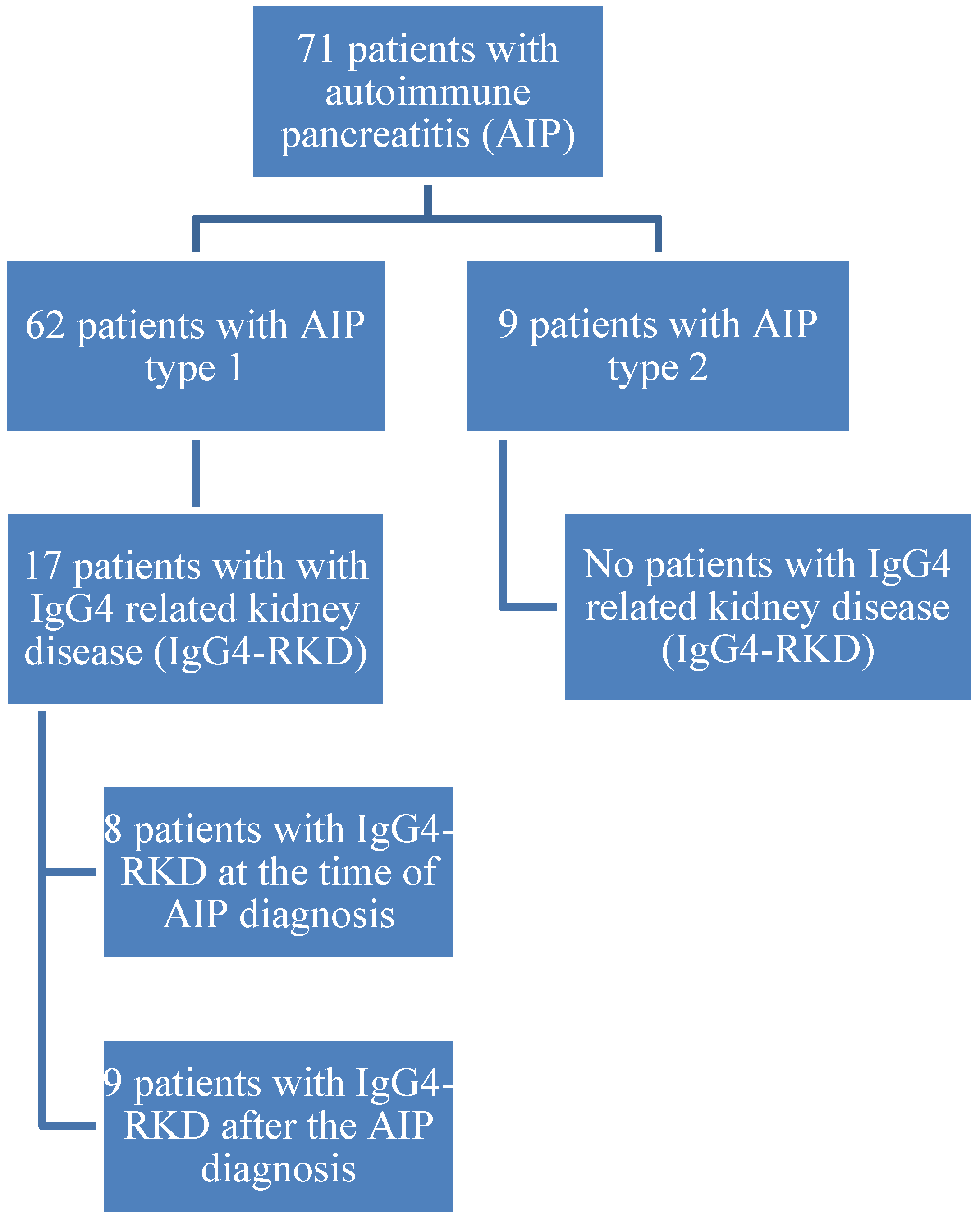
2.2. Cohort
2.3. Imaging
2.4. Ethics
3. Results
Imaging Features
4. Discussion
5. Conclusions
Author Contributions
Acknowledgments
Conflicts of Interest
Abbreviations
| AIP | Autoimmune Pancreatitis |
| OOI | Other Organ Involvement |
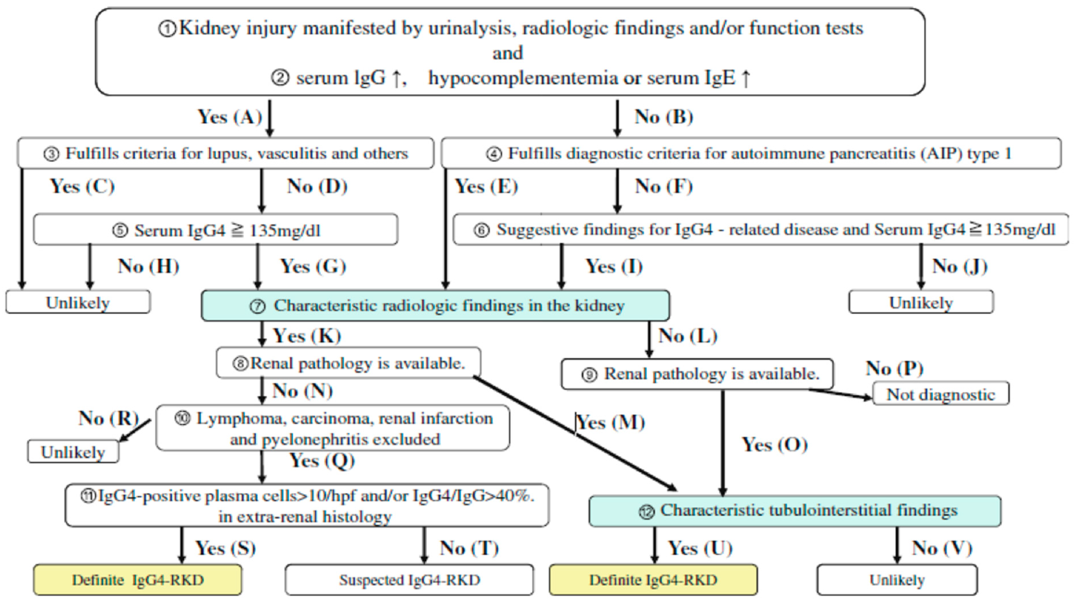
| IgG4-RKD | IgG4-Related Kidney Disease |
| LPSP | Lymphoplasmacytic Sclerosing Pancreatitis |
| IDCP | Idiopathic Duct-centric Pancreatitis |
| IAC | Immune Associated Cholangitis |
| ICDC | International Consensus Diagnostic Criteria |
| TIN | Tubulointerstitial Nephritis |
| JSN | Japanese Society of Nephrology |
| PACS | Picture Archiving and Communication System |
| SI | Signal Intensity |
| DWI | Diffusion-Weighted Imaging |
| ADC | Apparent Diffusion Coefficient |
| GFR | Glomerular Filtration Rate |
| DM | Diabetes Mellitus |
| AH | Arterial Hypertension |
References
- Klöppel, G.; Luttges, J.; Löhr, M.; Zamboni, G.; Longnecker, D. Autoimmune pancreatitis: Pathological, clinical, and immunological features. Pancreas 2003, 27, 14–19. [Google Scholar] [CrossRef] [PubMed]
- Hart, P.A.; Kamisawa, T.; Brugge, W.R.; Chung, J.B.; Culver, E.L.; Czakó, L.; Frulloni, L.; Go, V.L.W.; Gress, T.M.; Kim, M.H.; et al. Long-term outcomes of autoimmune pancreatitis: A multicentre, international analysis. Gut 2013, 62, 1771–1776. [Google Scholar] [CrossRef] [PubMed]
- Vujasinovic, M.; Valente, R.; Maier, P.; von Beckerath, V.; Haas, S.L.; Arnelo, U.; Del Chiaro, M.; Kartalis, N.; Pozzi-Mucelli, R.M.; Fernandez-Moro, C.; et al. Diagnosis, treatment and long-term outcome of autoimmune pancreatitis in Sweden. Pancreatology 2018, 18, 900–904. [Google Scholar] [CrossRef] [PubMed]
- Shimosegawa, T.; Chari, S.T.; Frulloni, L.; Kamisawa, T.; Kawa, S.; Mino-Kenudson, M.; Kim, M.H.; Klöppel, G.; Lerch, M.M.; Löhr, M.; et al. International consensus diagnostic criteria for autoimmune pancreatitis: Guidelines of the International Association of Pancreatology. Pancreas 2011, 40, 352–358. [Google Scholar] [CrossRef] [PubMed]
- Frulloni, L.; Scattolini, C.; Falconi, M.; Zamboni, G.; Capelli, P.; Manfredi, R.; Graziani, R.; D’onofrio, M.; Katsotourchi, A.M.; Amodio, A.; et al. Autoimmune pancreatitis: Differences between the focal and diffuse forms in 87 patients. Am. J. Gastroenterol. 2009, 104, 2288–2294. [Google Scholar] [CrossRef] [PubMed]
- Czakó, L.; Gyökeres, T.; Topa, L.; Sahin, P.; Takács, T.; Vincze, Á.; Dubravcsik, Z.; Szepes, A.; Pap, Á.; Földesi, I.; et al. Autoimmune pancreatitis in Hungary: A multicenter nationwide study. Pancreatology 2011, 11, 261–267. [Google Scholar] [CrossRef] [PubMed]
- Church, N.I.; Pereira, S.P.; Deheragoda, M.G.; Sandanayake, N.; Amin, Z.; Lees, W.R.; Gillams, A.; Rodriguez-Justo, M.; Novelli, M.; Seward, E.W.; et al. Autoimmune pancreatitis: Clinical and radiological features and objective response to steroid therapy in a UK series. Am. J. Gastroenterol. 2007, 102, 2417–2425. [Google Scholar] [CrossRef] [PubMed]
- López-Serrano, A.; Crespo, J.; Pascual, I.; Salord, S.; Bolado, F.; del-Pozo-García, A.J.; Ilzarbe, L.; de-Madaria, E.; Moreno-Osset, E.; Autoimmune Pancreatitis in Spain Study Group. Diagnosis, treatment and long-term outcomes of autoimmune pancreatitis in Spain based on the International Consensus Diagnostic Criteria: A multi-centre study. Pancreatology 2016, 16, 382–390. [Google Scholar] [CrossRef] [PubMed]
- Uchiyama-Tanaka, Y.; Mori, Y.; Kimura, T.; Sonomura, K.; Umemura, S.; Kishimoto, N.; Nose, A.; Tokoro, T.; Kijima, Y.; Yamahara, H.; et al. Acute tubulointerstitial nephritis associated with autoimmune-related pancreatitis. Am. J. Kidney Dis. 2004, 43, e18–e25. [Google Scholar] [CrossRef]
- Takeda, S.; Haratake, J.; Kasai, T.; Takaeda, C.; Takazakura, E. IgG4-associated idiopathic tubulointerstitial nephritis complicating autoimmune pancreatitis. Nephrol. Dial. Transplant. 2004, 19, 474–476. [Google Scholar] [CrossRef]
- Rudmik, L.; Trpkov, K.; Nash, C.; Kinnear, S.; Falck, V.; Dushinski, J.; Dixon, E. Autoimmune pancreatitis associated with renal lesions mimicking metastatic tumours. Cmaj 2006, 175, 367–369. [Google Scholar] [CrossRef]
- Shimoyama, K.; Ogawa, N.; Sawaki, T.; Karasawa, H.; Masaki, Y.; Kawabata, H.; Fukushima, T.; Wano, Y.; Hirose, Y.; Umehara, H. A case of Mikulicz’s disease complicated with interstitial nephritis successfully treated by high-dose corticosteroid. Mod. Rheumatol. 2006, 16, 176–182. [Google Scholar] [CrossRef] [PubMed]
- Tsubata, Y.; Akiyama, F.; Oya, T.; Ajiro, J.; Saeki, T.; Nishi, S.; Narita, I. IgG4-related chronic tubulointerstitial nephritis without autoimmune pancreatitis and the time course of renal function. Intern. Med. 2010, 49, 1593–1598. [Google Scholar] [CrossRef] [PubMed]
- Kawano, M.; Saeki, T.; Nakashima, H.; Nishi, S.; Yamaguchi, Y.; Hisano, S.; Yamanaka, N.; Inoue, D.; Yamamoto, M.; Takahashi, H.; Nomura, H. Proposal for diagnostic criteria for IgG4-related kidney disease. Clin. Exp. Nephrol. 2011, 15, 615–626. [Google Scholar] [CrossRef] [PubMed]
- Kim, B.; Kim, J.H.; Byun, J.H.; Kim, H.J.; Lee, S.S.; Kim, S.Y.; Lee, M.G. IgG4-related kidney disease: MRI findings with emphasis on the usefulness of diffusion-weighted imaging. Eur. J. Radiol. 2014, 83, 1057–1062. [Google Scholar] [CrossRef] [PubMed]
- Naitoh, I.; Nakazawa, T.; Ohara, H.; Ando, T.; Hayashi, K.; Tanaka, H.; Okumura, F.; Miyabe, K.; Yoshida, M.; Sano, H.; et al. Clinical significance of extrapancreatic lesions in autoimmune pancreatitis. Pancreas 2010, 39, e1–e5. [Google Scholar] [CrossRef] [PubMed]
- Hirth, M.; Vujasinovic, M.; Münch, M.; Weiss, C.; Löhr, M.; Ebert, M.P.; Schneider, A. Monitoring and predicting disease activity in autoimmune pancreatitis with the M-ANNHEIM-AiP-Activity-Score. Pancreatology 2018, 18, 29–38. [Google Scholar] [CrossRef]
- Saeki, T.; Nishi, S.; Imai, N.; Ito, T.; Yamazaki, H.; Kawano, M.; Yamamoto, M.; Takahashi, H.; Matsui, S.; Nakada, S.; et al. Clinicopathological characteristics of patients with IgG4-related tubulointerstitial nephritis. Kidney Int. 2010, 78, 1016–1023. [Google Scholar] [CrossRef]
- Zhang, L.; Notohara, K.; Levy, M.J.; Chari, S.T.; Smyrk, T.C. IgG4-positive plasma cell infiltration in the diagnosis of autoimmune pancreatitis. Mod. Pathol. 2007, 20, 23–28. [Google Scholar] [CrossRef]
- Aalberse, R.C.; Schuurman, J. IgG4 breaking the rules. Immunology 2002, 105, 9–19. [Google Scholar] [CrossRef] [PubMed]
- Sporek, M.; Dumnicka, P.; Gala-Bladzinska, A.; Ceranowicz, P.; Warzecha, Z.; Dembinski, A.; Stepien, E.; Walocha, J.; Drozdz, R.; Kuzniewski, M.; et al. Angiopoietin-2 Is an Early Indicator of Acute Pancreatic-Renal Syndrome in Patients with Acute Pancreatitis. Mediat. Inflamm. 2016, 2016, 5780903. [Google Scholar] [CrossRef] [PubMed]
- Sporek, M.; Gala-Błądzińska, A.; Dumnicka, P.; Mazur-Laskowska, M.; Kielczewski, S.; Walocha, J.; Ceranowicz, P.; Kuźniewski, M.; Mituś, J.; Kuśnierz-Cabala, B. Urine NGAL is useful in the clinical evaluation of renal function in the early course of acute pancreatitis. Folia Med. Cracov. 2016, 56, 13–25. [Google Scholar] [PubMed]
- Cornell, L.D. IgG4-related tubulointerstitial nephritis. Kidney Int. 2010, 78, 951–953. [Google Scholar] [CrossRef] [PubMed]
- Raissian, Y.; Nasr, S.H.; Larsen, C.P.; Colvin, R.B.; Smyrk, T.C.; Takahashi, N.; Bhalodia, A.; Sohani, A.R.; Zhang, L.; Chari, S.; et al. Diagnosis of IgG4-related tubulointerstitial nephritis. J. Am. Soc. Nephrol. 2011, 22, 1343–1352. [Google Scholar] [CrossRef] [PubMed]


| N | Gender | Age | Treatment | OOI (Other than Kidney/Pancreas) | Imaging | Type of Kidney Involvement | Unilateral vs. Bilateral Involvement | Onset of Kidney Involvement |
|---|---|---|---|---|---|---|---|---|
| 1 | M | 74 | steroids, surgery | cholangitis | CEMR | multiple lesions | bilateral | 6 y after AIP |
| vasculitis (aorta) | ||||||||
| retroperitoneal fibrosis | ||||||||
| 2 | F | 73 | steroids | cholangitis | CECT CEMR | multiple lesions | bilateral | 3 m after AIP |
| Sjögren’s Syndrome enlarge mediastinal LN | ||||||||
| 3 | M | 52 | steroids, biliary stent | cholangitis | CECT MRw/o c | multiple lesions | bilateral | synchronous |
| 4 | M | 49 | steroids, surgery | cholangitis | CECT CEMR | soft tissue in the perinephric space, diffuse swelling | bilateral | 6 m after AIP |
| hepatitis | ||||||||
| enlarge abdominal LN | ||||||||
| 5 | M | 60 | steroids | cholangitis | CEMR | multiple lesions | unilateral (left) | 11 m after AIP |
| 6 | M | 57 | steroids, azathioprine | cholangitis | CECT CEMR | solitary lesion | unilateral (right) | synchronous |
| enlarge abdominal LN vasculitis (aorta) | ||||||||
| 7 | M | 42 | steroids | cholangitis | CECT CEMR | multiple lesions | bilateral | synchronous |
| 8 | F | 39 | none | cholangitis | CECT CEMR | multiple lesions | bilateral | synchronous |
| lung involvement | ||||||||
| 9 | M | 39 | none | cholangitis | CECT CEMR | multiple lesions | bilateral | synchronous |
| 10 | M | 73 | none | cholangitis | CEMR | multiple lesions | bilateral | synchronous |
| 11 | M | 68 | steroids | none | CECT CEMR | multiple lesions | bilateral | synchronous |
| 12 | M | 68 | steroids, surgery | cholangitis | CECT CEMR | focal thinning of renal cortex | bilateral | synchronous |
| 13 | M | 85 | biliary stent | cholangitis | CECT CEMR | multiple lesions | bilateral | synchronous |
| lung involvement | ||||||||
| vasculitis (aorta) | ||||||||
| 14 | M | 71 | steroids | cholangitis | CECT * | multiple lesions | bilateral | 2 y after AIP |
| 15 | M | 65 | steroids | cholangitis | CEMR | multiple lesions | bilateral | 8 y after AIP |
| vasculitis (aorta) | ||||||||
| 16 | M | 52 | none | cholangitis | CEMR | multiple lesions | bilateral | 4 y after AIP |
| 17 | M | 64 | none | cholangitis | CEMR | solitary lesion | unilateral (right) | synchronous |
| Signal Intensity (SI) | MRI Sequences | |||||
|---|---|---|---|---|---|---|
| T2-Weigthed | T1-Weighted (w/o Contrast Agent) | T1-Weighted Arterial Phase | T1-Weighted Venous Phase | T1-Weighted Delayed Phase | DWI * | |
| Hypointense | 10/15 (66.6%) | 2/15 (13.4%) | 9/15 (60%) | 8/15 (53.4%) | 9/15 (60%) | 1/15 (6.6%) |
| Isointense | 5/15 (33.4%) | 13/15 (86.6%) | 6/15 (40%) | 7/15 (46.6%) | 6/15 (40%) | 0 |
| Hyperintense | 0 | 0 | 0 | 0 | 0 | 14/15 (93.4%) |
| Restricted SI | - | - | - | - | - | 10/11 (90.9%) |
| Parameter | Present Study | Saeki et al. [18] | Kawano et al. [14] |
|---|---|---|---|
| Number of patients | 17 | 23 | 41 |
| Gender | 15 (88.2%) male and 2 (11.8%) female | 20 (86.9%) male and 3 (13.1%) female | 30 (73.2%) male and 11 (26.8%) female |
| Age at diagnosis (years) | 60.6 ± 13.1 (range 39–85) | 65.2 ± 10.1 (range 40–83) | 63.7 ± 12.3 years (range 27–83) |
| OOI % | 94.1 | 95.7 | 95.1 |
| Haematuria % | 23.5 | 34.8 | 41.7 |
| Proteinuria % | 23.5 | 8.7 | 58.3 |
| Elevated creatinine values % | 47.1 | 56.5 | 58.5 |
| Elevated IgG4 values % | 58.8 | 100 | 100 |
| Treatment with corticosteroids % | 76.5 | 91.3 | 92.7 |
| Improvement after steroid therapy % | 100 | 94.7 | 92.1 |
| Haemodialysis after steroid therapy % | 0 | 5.2 | 2.6 |
© 2019 by the authors. Licensee MDPI, Basel, Switzerland. This article is an open access article distributed under the terms and conditions of the Creative Commons Attribution (CC BY) license (http://creativecommons.org/licenses/by/4.0/).
Share and Cite
Vujasinovic, M.; Pozzi Mucelli, R.M.; Valente, R.; Verbeke, C.S.; Haas, S.L.; Löhr, J.-M. Kidney Involvement in Patients with Type 1 Autoimmune Pancreatitis. J. Clin. Med. 2019, 8, 258. https://doi.org/10.3390/jcm8020258
Vujasinovic M, Pozzi Mucelli RM, Valente R, Verbeke CS, Haas SL, Löhr J-M. Kidney Involvement in Patients with Type 1 Autoimmune Pancreatitis. Journal of Clinical Medicine. 2019; 8(2):258. https://doi.org/10.3390/jcm8020258
Chicago/Turabian StyleVujasinovic, Miroslav, Raffaella Maria Pozzi Mucelli, Roberto Valente, Caroline Sophie Verbeke, Stephan L. Haas, and J.-Matthias Löhr. 2019. "Kidney Involvement in Patients with Type 1 Autoimmune Pancreatitis" Journal of Clinical Medicine 8, no. 2: 258. https://doi.org/10.3390/jcm8020258
APA StyleVujasinovic, M., Pozzi Mucelli, R. M., Valente, R., Verbeke, C. S., Haas, S. L., & Löhr, J.-M. (2019). Kidney Involvement in Patients with Type 1 Autoimmune Pancreatitis. Journal of Clinical Medicine, 8(2), 258. https://doi.org/10.3390/jcm8020258