Non-Pharmacological Therapeutic Options for Liver Metastases in Advanced Neuroendocrine Tumors

 ,
,  ,
,
Abstract
1. Therapeutic Options for LM
1.1. Surgery
1.1.1. Techniques
1.1.2. Results
1.1.3. Complications
1.2. Liver Transplantation
1.2.1. Techniques
1.2.2. Results
1.2.3. Complications
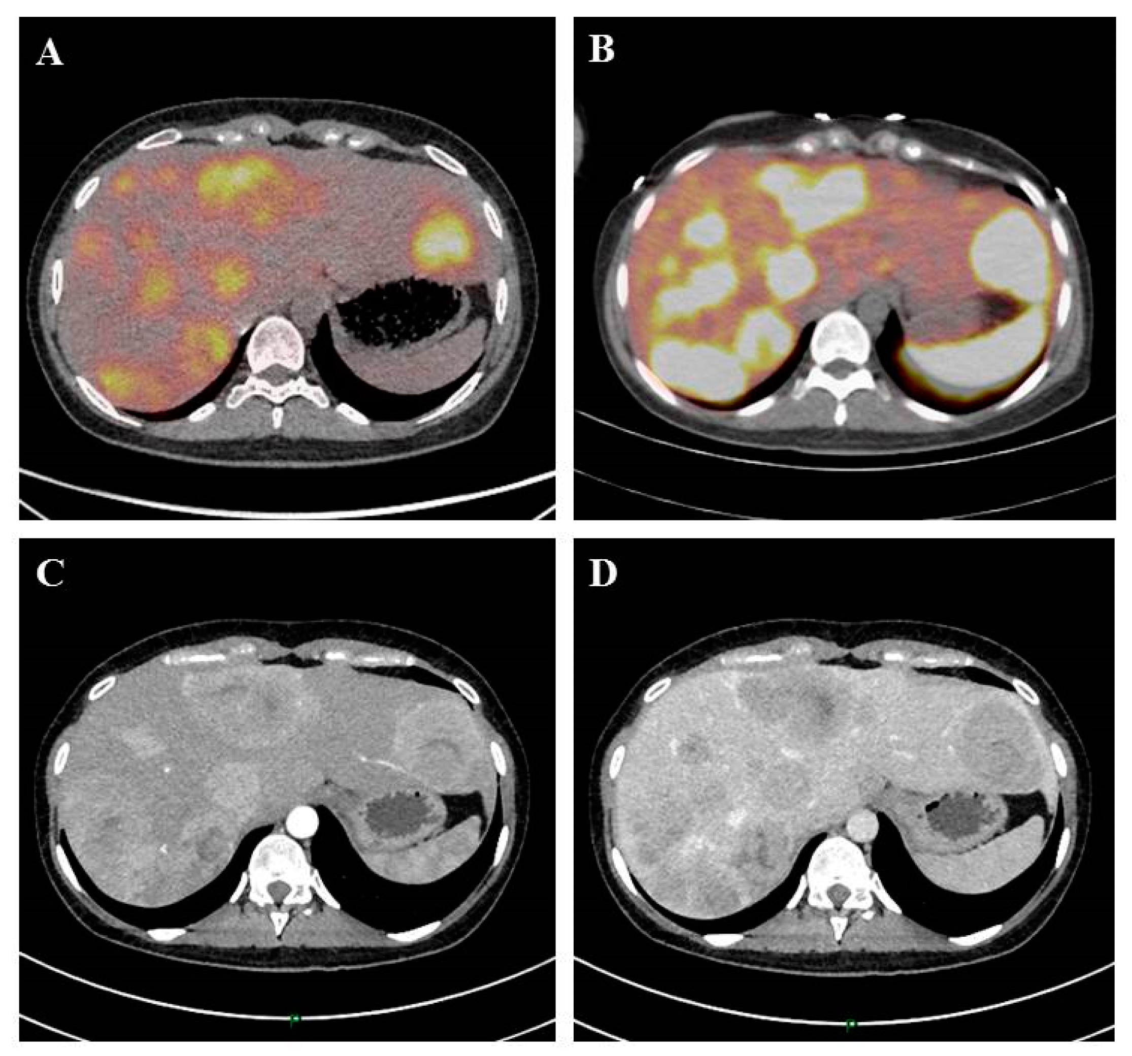
1.3. Ablative Techniques (Radiofrequency Ablation and Other Ablative Techniques)
Techniques
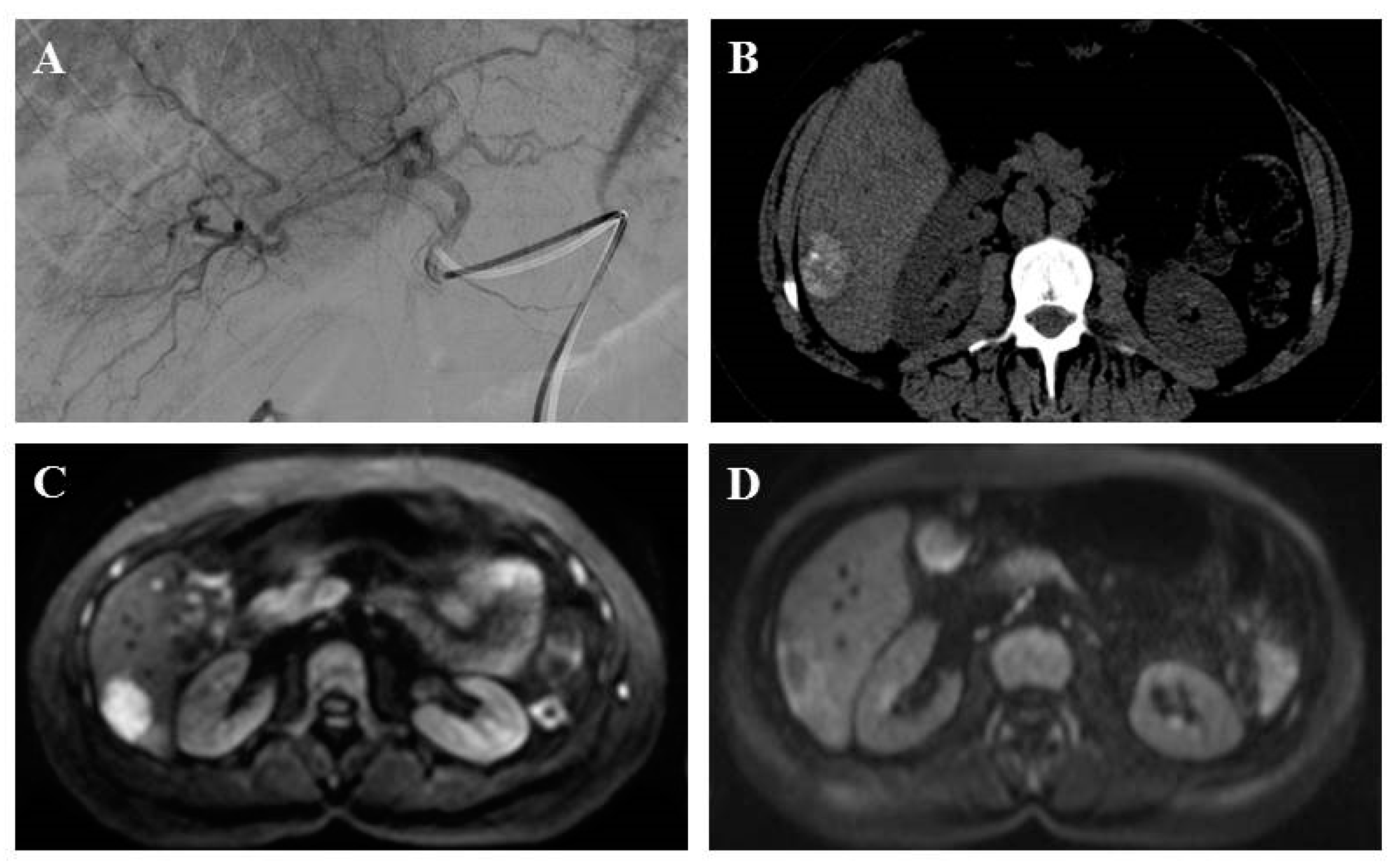
1.4. Trans-Arterial Embolization and Chemoembolization
1.4.1. Techniques
1.4.2. Results
1.4.3. Complications
1.5. Trans-Arterial Radio-Embolization
1.5.1. Techniques
1.5.2. Results
1.5.3. Complications
1.6. Peptide Receptor Radionuclide Therapy
1.6.1. Techniques
1.6.2. Results
1.6.3. Complications
2. Conclusions
Author Contributions
Funding
Conflicts of Interest
References
- Dasari, A.; Shen, C.; Halperin, D.; Zhao, B.; Zhou, S.; Xu, Y.; Shih, T.; Yao, J.C. Trends in the Incidence, Prevalence, and Survival Outcomes in Patients with Neuroendocrine Tumors in the United States. JAMA Oncol. 2017, 3, 1335–1342. [Google Scholar] [CrossRef] [PubMed]
- Lepage, C.; Bouvier, A.M.; Faivre, J. Endocrine tumours: Epidemiology of malignant digestive neuroendocrine tumours. Eur. J. Endocrinol. 2013, 168, R77–R83. [Google Scholar] [CrossRef] [PubMed]
- Scoazec, J.-Y.; Couvelard, A.; Monges, G.; Guyétant, S.; Bisot-Locard, S.; Parot, X.; Lepage, C.; PRONET Study Group. Professional Practices and Diagnostic Issues in Neuroendocrine Tumour Pathology: Results of a Prospective One-Year Survey among French Pathologists (the PRONET Study). Neuroendocrinology 2017, 105, 67–76. [Google Scholar] [CrossRef] [PubMed]
- Rindi, G. Nomenclature and classification of neuroendocrine neoplasms of the digestive system. In WHO Classification of Tumors of the Digestive System, 4th ed.; Bosman, F.T., Carneiro, F., Hruban, R.H., Theise, N.D., Eds.; International Agency for Research on Cancer: Lyon, France, 2017. [Google Scholar]
- Coriat, R.; Walter, T.; Terris, B.; Couvelard, A.; Ruszniewski, P. Gastroenteropancreatic Well-Differentiated Grade 3 Neuroendocrine Tumors: Review and Position Statement. Oncologist 2016, 21, 1191–1199. [Google Scholar] [CrossRef] [PubMed]
- Frilling, A.; Sotiropoulos, G.C.; Li, J.; Kornasiewicz, O.; Plöckinger, U. Multimodal management of neuroendocrine liver metastases. HPB 2010, 12, 361–379. [Google Scholar] [CrossRef]
- Pavel, M.; O’Toole, D.; Costa, F.; Capdevila, J.; Gross, D.; Kianmanesh, R.; Krenning, E.; Knigge, U.; Salazar, R.; Pape, U.F.; et al. ENETS Consensus Guidelines Update for the Management of Distant Metastatic Disease of Intestinal, Pancreatic, Bronchial Neuroendocrine Neoplasms (NEN) and NEN of Unknown Primary Site. Neuroendocrinology 2016, 103, 172–185. [Google Scholar] [CrossRef]
- Frilling, A.; Modlin, I.M.; Kidd, M.; Russell, C.; Breitenstein, S.; Salem, R.; Kwekkeboom, D.; Lau, W.Y.; Klersy, C.; Vilgrain, V.; et al. Recommendations for management of patients with neuroendocrine liver metastases. Lancet Oncol. 2014, 15, e8–e21. [Google Scholar] [CrossRef]
- Yao, J.C.; Hassan, M.; Phan, A.; Dagohoy, C.; Leary, C.; Mares, J.E.; Abdalla, E.K.; Fleming, J.B.; Vauthey, J.N.; Rashid, A.; et al. One hundred years after ‘carcinoid’: Epidemiology of and prognostic factors for neuroendocrine tumors in 35,825 cases in the United States. J. Clin. Oncol. 2008, 26, 3063–3072. [Google Scholar] [CrossRef]
- Ronot, M.; Clift, A.K.; Baum, R.P.; Singh, A.; Kulkarni, H.R.; Frilling, A.; Vilgrain, V. Morphological and Functional Imaging for Detecting and Assessing the Resectability of Neuroendocrine Liver Metastases. Neuroendocrinology 2018, 106, 74–88. [Google Scholar] [CrossRef]
- Ronot, M.; Clift, A.K.; Vilgrain, V.; Frilling, A. Functional imaging in liver tumours. J. Hepatol. 2016, 65, 1017–1030. [Google Scholar] [CrossRef]
- Dromain, C.; de Baere, T.; Lumbroso, J.; Caillet, H.; Laplanche, A.; Boige, V.; Ducreux, M.; Duvillard, P.; Elias, D.; Schlumberger, M.; et al. Detection of Liver Metastases From Endocrine Tumors: A Prospective Comparison of Somatostatin Receptor Scintigraphy, Computed Tomography, and Magnetic Resonance Imaging. J. Clin. Oncol. 2005, 23, 70–78. [Google Scholar] [CrossRef] [PubMed]
- Ruf, J.; Heuck, F.; Schiefer, J.; Denecke, T.; Elgeti, F.; Pascher, A.; Pavel, M.; Stelter, L.; Kropf, S.; Wiedenmann, B.; et al. Impact of Multiphase 68 Ga-DOTATOC-PET/CT on Therapy Management in Patients with Neuroendocrine Tumors. Neuroendocrinology 2010, 91, 101–109. [Google Scholar] [CrossRef] [PubMed]
- Nigri, G.; Petrucciani, N.; Debs, T.; Mangogna, L.M.; Crovetto, A.; Moschetta, G.; Persechino, R.; Aurello, P.; Ramacciato, G. Treatment options for PNET liver metastases: A systematic review. World J. Surg. Oncol. 2018, 16. Available online: https://wjso.biomedcentral.com/articles/10.1186/s12957-018-1446-y (accessed on 15 July 2019). [CrossRef] [PubMed]
- Sarmiento, J.M.; Heywood, G.; Rubin, J.; Ilstrup, D.M.; Nagorney, D.M.; Que, F.G. Surgical treatment of neuroendocrine metastases to the liver. J. Am. Coll. Surg. 2003, 197, 29–37. [Google Scholar] [CrossRef]
- Sarmiento, J.M.; Que, F.G.; Grant, C.S.; Thompson, G.B.; Farnell, M.B.; Nagorney, D.M. Concurrent resections of pancreatic islet cell cancers with synchronous hepatic metastases: Outcomes of an aggressive approach. Surgery 2002, 132, 976–983. [Google Scholar] [CrossRef]
- Yuan, C.; Wang, J.; Xiu, D.; Tao, M.; Ma, Z.; Jiang, B.; Li, Z.F.; Li, L.; Wang, L.; Wang, H.; et al. Meta-analysis of Liver Resection Versus Nonsurgical Treatments for Pancreatic Neuroendocrine Tumors with Liver Metastases. Ann. Surg. Oncol. 2016, 23, 244–249. [Google Scholar] [CrossRef]
- Watzka, F.M.; Fottner, C.; Miederer, M.; Schad, A.; Weber, M.M.; Otto, G.; Lang, H.; Musholt, T.J. Surgical therapy of neuroendocrine neoplasm with hepatic metastasis: Patient selection and prognosis. Langenbecks Arch. Surg. 2015, 400, 349–358. [Google Scholar] [CrossRef]
- Boudreaux, J.P.; Klimstra, D.S.; Hassan, M.M.; Woltering, E.A.; Jensen, R.T.; Goldsmith, S.J.; Nutting, C.; Bushnell, D.L.; Caplin, M.E.; Yao, J.C. The NANETS consensus guideline for the diagnosis and management of neuroendocrine tumors: Well-differentiated neuroendocrine tumors of the Jejunum, Ileum, Appendix, and Cecum. Pancreas 2010, 39, 753–766. [Google Scholar] [CrossRef]
- Kulke, M.H.; Anthony, L.B.; Bushnell, D.L.; de Herder, W.W.; Goldsmith, S.J.; Klimstra, D.S.; Marx, S.J.; Pasieka, J.L.; Pommier, R.F.; Yao, J.C.; et al. NANETS treatment guidelines: Well-differentiated neuroendocrine tumors of the stomach and pancreas. Pancreas 2010, 39, 735–752. [Google Scholar] [CrossRef]
- Gedaly, R. Liver Transplantation for the Treatment of Liver Metastases from Neuroendocrine Tumors: An Analysis of the UNOS Database. Arch. Surg. 2011, 146, 953–958. [Google Scholar] [CrossRef]
- Le Treut, Y.P.; Grégoire, E.; Klempnauer, J.; Belghiti, J.; Jouve, E.; Lerut, J.; Castaing, D.; Soubrane, O.; Boillot, O.; Mantion, G.; et al. Liver Transplantation for Neuroendocrine Tumors in Europe—Results and Trends in Patient Selection: A 213-Case European Liver Transplant Registry Study. Ann. Surg. 2013, 257, 807–815. [Google Scholar] [CrossRef] [PubMed]
- Clift, A.K.; Frilling, A. Liver transplantation and multivisceral transplantation in the management of patients with advanced neuroendocrine tumours. World J. Gastroenterol. 2018, 24, 2152–2162. [Google Scholar] [CrossRef] [PubMed]
- Moris, D.; Tsilimigras, D.I.; Ntanasis-Stathopoulos, I.; Beal, E.W.; Felekouras, E.; Vernadakis, S.; Fung, J.J.; Pawlik, T.M. Liver transplantation in patients with liver metastases from neuroendocrine tumors: A systematic review. Surgery 2017, 162, 525–536. [Google Scholar] [CrossRef] [PubMed]
- Nobel, Y.R.; Goldberg, D.S. Variable Use of Model for End-Stage Liver Disease Exception Points in Patients With Neuroendocrine Tumors Metastatic to the Liver and Its Impact on Patient Outcomes. Transplantation 2015, 99, 2341–2346. [Google Scholar] [CrossRef]
- Sposito, C.; Droz dit Busset, M.; Citterio, D.; Bongini, M.; Mazzaferro, V. The place of liver transplantation in the treatment of hepatic metastases from neuroendocrine tumors: Pros and cons. Rev. Endocr. Metab. Disord. 2017, 18, 473–483. [Google Scholar] [CrossRef]
- Zappa, M.; Abdel-Rehim, M.; Hentic, O.; Vullierme, M.-P.; Ruszniewski, P.; Vilgrain, V. Liver-directed therapies in liver metastases from neuroendocrine tumors of the gastrointestinal tract. Target. Oncol. 2012, 7, 107–116. [Google Scholar] [CrossRef]
- Elias, D.; Goéré, D.; Leroux, G.; Dromain, C.; Leboulleux, S.; de Baere, T.; Ducreux, M.; Baudin, E. Combined liver surgery and RFA for patients with gastroenteropancreatic endocrine tumors presenting with more than 15 metastases to the liver. Eur. J. Surg. Oncol. 2009, 35, 1092–1097. [Google Scholar] [CrossRef][Green Version]
- Akyildiz, H.Y.; Mitchell, J.; Milas, M.; Siperstein, A.; Berber, E. Laparoscopic radiofrequency thermal ablation of neuroendocrine hepatic metastases: Long-term follow-up. Surgery 2010, 148, 1288–1293. [Google Scholar] [CrossRef]
- Berber, E.; Siperstein, A.E. Perioperative outcome after laparoscopic radiofrequency ablation of liver tumors: An analysis of 521 cases. Surg. Endosc. 2007, 21, 613–618. [Google Scholar] [CrossRef]
- Martin, R.C.G.; Scoggins, C.R.; McMasters, K.M. Safety and Efficacy of Microwave Ablation of Hepatic Tumors: A Prospective Review of a 5-Year Experience. Ann. Surg. Oncol. 2010, 17, 171–178. [Google Scholar] [CrossRef]
- Bilchik, A.J.; Sarantou, T.; Foshag, L.J.; Giuliano, A.E.; Ramming, K.P. Cryosurgical palliation of metastatic neuroendocrine tumors resistant to conventional therapy. Surgery 1997, 122, 1040–1048. [Google Scholar] [CrossRef]
- Livraghi, T.; Bolondi, L.; Buscarini, L.; Cottone, M.; Mazziotti, A.; Morabito, A.; Torzilli, G.; Italian Cooperative HCC Study Group. No treatment, resection and ethanol injection in hepatocellular carcinoma: A retrospective analysis of survival in 391 patients with cirrhosis. Italian Cooperative HCC Study Group. J. Hepatol. 1995, 22, 522–526. [Google Scholar] [CrossRef]
- Gaba, R.C.; Lewandowski, R.J.; Hickey, R.; Baerlocher, M.O.; Cohen, E.I.; Dariushnia, S.R.; Janne, D.O.; Padia, S.A.; Salem, R.; Wang, D.S.; et al. Transcatheter Therapy for Hepatic Malignancy: Standardization of Terminology and Reporting Criteria. J. Vasc. Interv. Radiol. 2016, 27, 457–473. [Google Scholar] [CrossRef] [PubMed]
- Kennedy, A.; Bester, L.; Salem, R.; Sharma, R.A.; Parks, R.W.; Ruszniewski, P. Role of hepatic intra-arterial therapies in metastatic neuroendocrine tumours (NET): Guidelines from the NET-Liver-Metastases Consensus Conference. HPB 2015, 17, 29–37. [Google Scholar] [CrossRef]
- de Baere, T.; Deschamps, F.; Tselikas, L.; Ducreux, M.; Planchard, D.; Pearson, E.; Berdelou, A.; Leboulleux, S.; Elias, D.; Baudin, E. GEP-NETS UPDATE: Interventional radiology: Role in the treatment of liver metastases from GEP-NETs. Eur. J. Endocrinol. 2015, 172, R151–R166. [Google Scholar] [CrossRef]
- Del Prete, M.; Fiore, F.; Modica, R.; Marotta, V.; Marciello, F.; Ramundo, V.; Di Sarno, A.; Carratù, A.; Di Roseto, C.D.; Tafuto, S.; et al. Hepatic arterial embolization in patients with neuroendocrine tumors. J. Exp. Clin. Cancer Res. 2014, 33, 43. [Google Scholar] [CrossRef]
- Orgera, G.; Krokidis, M.; Cappucci, M.; Gourtsoyianni, S.; Tipaldi, M.A.; Hatzidakis, A.; Rebonato, A.; Rossi, M. Current Status of Interventional Radiology in the Management of Gastro-Entero-Pancreatic Neuroendocrine Tumours (GEP-NETs). Cardiovasc. Interv. Radiol. 2015, 38, 13–24. [Google Scholar] [CrossRef]
- Carrasco, C.; Charnsangavej, C.; Ajani, J.; Samaan, N.; Richli, W.; Wallace, S. The carcinoid syndrome: Palliation by hepatic artery embolization. Am. J. Roentgenol. 1986, 147, 149–154. [Google Scholar] [CrossRef]
- Therasse, E.; Breittmayer, F.; Roche, A.; De Baere, T.; Indushekar, S.; Ducreux, M.; Rougier, P. Transcatheter chemoembolization of progressive carcinoid liver metastasis. Radiology 1993, 189, 541–547. [Google Scholar] [CrossRef]
- Diamandidou, E.; Ajani, J.A.; Yang, D.J.; Chuang, V.P.; Brown, C.A.; Carrasco, H.C.; Lawrence, D.D.; Wallace, S. Two-phase study of hepatic artery vascular occlusion with microencapsulated cisplatin in patients with liver metastases from neuroendocrine tumors. Am. J. Roentgenol. 1998, 170, 339–344. [Google Scholar] [CrossRef][Green Version]
- Yao, K.A.; Talamonti, M.S.; Nemcek, A.; Angelos, P.; Chrisman, H.; Skarda, J.; Benson, A.B.; Rao, S.; Joehl, R.J. Indications and results of liver resection and hepatic chemoembolization for metastatic gastrointestinal neuroendocrine tumors. Surgery 2001, 130, 677–685. [Google Scholar] [CrossRef] [PubMed]
- Loewe, C.; Schindl, M.; Cejna, M.; Niederle, B.; Lammer, J.; Thurnher, S. Permanent Transarterial Embolization of Neuroendocrine Metastases of the Liver Using Cyanoacrylate and Lipiodol: Assessment of Mid- and Long-Term Results. Am. J. Roentgenol. 2003, 180, 1379–1384. [Google Scholar] [CrossRef] [PubMed]
- Osborne, D.A.; Zervos, E.E.; Strosberg, J.; Boe, B.A.; Malafa, M.; Rosemurgy, A.S.; Yeatman, T.J.; Carey, L.; Duhaine, L.; Kvols, L.K. Improved Outcome With Cytoreduction Versus Embolization for Symptomatic Hepatic Metastases of Carcinoid and Neuroendocrine Tumors. Ann. Surg. Oncol. 2006, 13, 572–581. [Google Scholar] [CrossRef] [PubMed]
- Strosberg, J.R.; Choi, J.; Cantor, A.B.; Kvols, L.K. Selective Hepatic Artery Embolization for Treatment of Patients with Metastatic Carcinoid and Pancreatic Endocrine Tumors. Cancer Control. 2006, 13, 72–78. [Google Scholar] [CrossRef] [PubMed]
- Bloomston, M.; Al-Saif, O.; Klemanski, D.; Pinzone, J.J.; Martin, E.W.; Palmer, B.; Guy, G.; Khabiri, H.; Ellison, E.C.; Shah, M.H. Hepatic Artery Chemoembolization in 122 Patients with Metastatic Carcinoid Tumor: Lessons Learned. J. Gastrointest. Surg. 2007, 11, 264–271. [Google Scholar] [CrossRef]
- Granberg, D.; Eriksson, L.-G.; Welin, S.; Kindmark, H.; Janson, E.T.; Skogseid, B.; Öberg, K.; Eriksson, B.; Nyman, R. Liver embolization with trisacryl gelatin microspheres (embosphere) in patients with neuroendocrine tumors. Acta Radiol. 2007, 48, 180–185. [Google Scholar] [CrossRef]
- Ho, A.S.; Picus, J.; Darcy, M.D.; Tan, B.; Gould, J.E.; Pilgram, T.K.; Brown, D.B. Long-Term Outcome After Chemoembolization and Embolization of Hepatic Metastatic Lesions from Neuroendocrine Tumors. Am. J. Roentgenol. 2007, 188, 1201–1207. [Google Scholar] [CrossRef]
- Marrache, F.; Vullierme, M.P.; Roy, C.; Assoued, Y.E.; Couvelard, A.; O’Toole, D.; Mitry, E.; Hentic, O.; Hammel, P.; Levy, P.; et al. Arterial phase enhancement and body mass index are predictors of response to chemoembolisation for liver metastases of endocrine tumours. Br. J. Cancer 2007, 96, 49–55. [Google Scholar] [CrossRef]
- Varker, K.A.; Martin, E.W.; Klemanski, D.; Palmer, B.; Shah, M.H.; Bloomston, M. Repeat Transarterial Chemoembolization (TACE) for Progressive Hepatic Carcinoid Metastases Provides Results Similar to First TACE. J. Gastrointest. Surg. 2007, 11, 1680–1685. [Google Scholar] [CrossRef]
- Christante, D.; Pommier, S.; Givi, B.; Pommier, R. Hepatic artery chemoinfusion with chemoembolization for neuroendocrine cancer with progressive hepatic metastases despite octreotide therapy. Surgery 2008, 144, 885–894. [Google Scholar] [CrossRef]
- de Baere, T.; Deschamps, F.; Teriitheau, C.; Rao, P.; Conengrapht, K.; Schlumberger, M.; Leboulleux, S.; Baudin, E.; Hechellhammer, L. Transarterial Chemoembolization of Liver Metastases from Well Differentiated Gastroenteropancreatic Endocrine Tumors with Doxorubicin-eluting Beads: Preliminary Results. J. Vasc. Interv. Radiol. 2008, 19, 855–861. [Google Scholar] [CrossRef] [PubMed]
- Kamat, P.P.; Gupta, S.; Ensor, J.E.; Murthy, R.; Ahrar, K.; Madoff, D.C.; Wallace, M.J.; Hicks, M.E. Hepatic Arterial Embolization and Chemoembolization in the Management of Patients with Large-Volume Liver Metastases. Cardiovasc. Interv. Radiol. 2008, 31, 299–307. [Google Scholar] [CrossRef] [PubMed]
- Pitt, S.C.; Knuth, J.; Keily, J.M.; McDermott, J.C.; Weber, S.M.; Chen, H.; Rilling, W.S.; Quebbeman, E.J.; Agarwal, D.M.; Pitt, H.A. Hepatic Neuroendocrine Metastases: Chemo- or Bland Embolization? J. Gastrointest. Surg. 2008, 12, 1951–1960. [Google Scholar] [CrossRef] [PubMed]
- Dong, X.D.; Carr, B.I. Hepatic artery chemoembolization for the treatment of liver metastases from neuroendocrine tumors: A long-term follow-up in 123 patients. Med. Oncol. 2011, 28, 286–290. [Google Scholar] [CrossRef] [PubMed]
- Gaur, S.K.; Friese, J.L.; Sadow, C.A.; Ayyagari, R.; Binkert, C.A.; Schenker, M.P.; Kulke, M.; Baum, R. Hepatic Arterial Chemoembolization Using Drug-Eluting Beads in Gastrointestinal Neuroendocrine Tumor Metastatic to the Liver. Cardiovasc. Interv. Radiol. 2011, 34, 566–572. [Google Scholar] [CrossRef] [PubMed]
- Maire, F.; Lombard-Bohas, C.; O’Toole, D.; Vullierme, M.-P.; Rebours, V.; Couvelard, A.; Pelletier, A.L.; Zappa, M.; Pilleul, F.; Hentic, O.; et al. Hepatic Arterial Embolization versus Chemoembolization in the Treatment of Liver Metastases from Well-Differentiated Midgut Endocrine Tumors: A Prospective Randomized Study. Neuroendocrinology 2012, 96, 294–300. [Google Scholar] [CrossRef]
- Hur, S.; Chung, J.W.; Kim, H.-C.; Oh, D.-Y.; Lee, S.-H.; Bang, Y.-J.; Kim, W.H. Survival Outcomes and Prognostic Factors of Transcatheter Arterial Chemoembolization for Hepatic Neuroendocrine Metastases. J. Vasc. Interv. Radiol. 2013, 24, 947–956. [Google Scholar] [CrossRef]
- Fiore, F.; Del Prete, M.; Franco, R.; Marotta, V.; Ramundo, V.; Marciello, F.; Di Sarno, A.; Carratù, A.C.; Di Roseto, C.D.; Colao, A.; et al. Transarterial embolization (TAE) is equally effective and slightly safer than transarterial chemoembolization (TACE) to manage liver metastases in neuroendocrine tumors. Endocrine 2014, 47, 177–182. [Google Scholar] [CrossRef]
- Dhir, M.; Shrestha, R.; Steel, J.L.; Marsh, J.W.; Tsung, A.; Tublin, M.E.; Amesur, N.B.; Orons, P.D.; Santos, E.; Geller, D.A. Initial Treatment of Unresectable Neuroendocrine Tumor Liver Metastases with Transarterial Chemoembolization using Streptozotocin: A 20-Year Experience. Ann. Surg. Oncol. 2017, 24, 450–459. [Google Scholar] [CrossRef]
- Kim, Y.H.; Ajani, J.A.; Carrasco, C.H.; Dumas, P.; Richli, W.; Lawrence, D.; Chuang, V.; Wallace, S. Selective Hepatic Arterial Chemoembolization for Liver Metastases in Patients with Carcinoid Tumor or Islet Cell Carcinoma. Cancer Investig. 1999, 17, 474–478. [Google Scholar] [CrossRef]
- Kress, O.; Wagner, H.-J.; Wied, M.; Klose, K.J.; Arnold, R.; Alfke, H. Transarterial Chemoembolization of Advanced Liver Metastases of Neuroendocrine Tumors—A Retrospective Single-Center Analysis. Digestion 2003, 68, 94–101. [Google Scholar] [CrossRef] [PubMed]
- King, J.; Quinn, R.; Glenn, D.M.; Janssen, J.; Tong, D.; Liaw, W.; Morris, D.L. Radioembolization with selective internal radiation microspheres for neuroendocrine liver metastases. Cancer 2008, 113, 921–929. [Google Scholar] [CrossRef] [PubMed]
- Ezziddin, S.; Meyer, C.; Kahancova, S.; Haslerud, T.; Willinek, W.; Wilhelm, K.; Biersack, H.J.; Ahmadzadehfar, H. 90Y Radioembolization After Radiation Exposure from Peptide Receptor Radionuclide Therapy. J. Nucl. Med. 2012, 53, 1663–1669. [Google Scholar] [CrossRef] [PubMed]
- Memon, K.; Lewandowski, R.J.; Mulcahy, M.F.; Riaz, A.; Ryu, R.K.; Sato, K.T.; Gupta, R.; Nikolaidis, P.; Miller, F.H.; Yaghmai, V.; et al. Radioembolization for Neuroendocrine Liver Metastases: Safety, Imaging, and Long-Term Outcomes. Int. J. Radiat. Oncol. 2012, 83, 887–894. [Google Scholar] [CrossRef] [PubMed]
- Paprottka, P.M.; Hoffmann, R.-T.; Haug, A.; Sommer, W.H.; Raeßler, F.; Trumm, C.G.; Schmidt, G.P.; Ashoori, N.; Reiser, M.F.; Jakobs, T.F. Radioembolization of Symptomatic, Unresectable Neuroendocrine Hepatic Metastases Using Yttrium-90 Microspheres. Cardiovasc. Interv. Radiol. 2012, 35, 334–342. [Google Scholar] [CrossRef] [PubMed]
- Fidelman, N.; Kerlan, R.K., Jr.; Hawkins, R.A.; Pampaloni, M.; Taylor, A.G.; Kohi, M.P.; Kolli, K.P.; Atreya, C.E.; Bergsland, E.K.; Kelley, R.K.; et al. Radioembolization with 90Y glass microspheres for the treatment of unresectable metastatic liver disease from chemotherapy-refractory gastrointestinal cancers: Final report of a prospective pilot study. J. Gastrointest. Oncol. 2016, 7, 860–874. [Google Scholar] [CrossRef]
- Rajekar, H.; Bogammana, K.; Stubbs, R.S. Selective Internal Radiation Therapy for Gastrointestinal Neuroendocrine Tumour Liver Metastases: A New and Effective Modality for Treatment. Int. J. Hepatol. 2011, 2011, 404916. [Google Scholar] [CrossRef]
- Kennedy, A.S.; Dezarn, W.A.; McNeillie, P.; Coldwell, D.; Nutting, C.; Carter, D.; Murthy, R.; Rose, S.; Warner, R.R.; Liu, D.; et al. Radioembolization for Unresectable Neuroendocrine Hepatic Metastases Using Resin 90Y-Microspheres: Early Results in 148 Patients. Am. J. Clin. Oncol. 2008, 31, 271–279. [Google Scholar] [CrossRef]
- Murthy, R.; Kamat, P.; Nunez, R.; Madoff, D.C.; Gupta, S.; Salem, R.; Yao, J.C. Yttrium-90 Microsphere Radioembolotherapy of Hepatic Metastatic Neuroendocrine Carcinomas after Hepatic Arterial Embolization. J. Vasc. Interv. Radiol. 2008, 19, 145–151. [Google Scholar] [CrossRef]
- Rhee, T.K.; Lewandowski, R.J.; Liu, D.M.; Mulcahy, M.F.; Takahashi, G.; Hansen, P.D.; Benson, A.B., III; Kennedy, A.S.; Omary, R.A.; Salem, R. 90Y Radioembolization for Metastatic Neuroendocrine Liver Tumors: Preliminary Results from a Multi-institutional Experience. Ann. Surg. 2008, 247, 1029–1035. [Google Scholar] [CrossRef]
- Kalinowski, M.; Dressler, M.; König, A.; El-Sheik, M.; Rinke, A.; Höffken, H.; Gress, T.M.; Arnold, R.; Klose, K.J.; Wagner, H.J. Selective Internal Radiotherapy with Yttrium-90 Microspheres for Hepatic Metastatic Neuroendocrine Tumors: A Prospective Single Center Study. Digestion 2009, 79, 137–142. [Google Scholar] [CrossRef] [PubMed]
- Cao, D.; Wu, G.; Zhang, B.; Quan, Y.; Wei, J.; Jin, H.; Jiang, Y.; Yang, Z.A. Resting energy expenditure and body composition in patients with newly detected cancer. Clin. Nutr. Edinb. Scotl. 2010, 29, 72–77. [Google Scholar] [CrossRef] [PubMed]
- Saxena, A.; Chua, T.C.; Bester, L.; Kokandi, A.; Morris, D.L. Factors Predicting Response and Survival After Yttrium-90 Radioembolization of Unresectable Neuroendocrine Tumor Liver Metastases: A Critical Appraisal of 48 Cases. Ann. Surg. 2010, 251, 910–916. [Google Scholar] [CrossRef] [PubMed]
- Lacin, S.; Oz, I.; Ozkan, E.; Kucuk, O.; Bilgic, S. Intra-Arterial Treatment with 90 Yttrium Microspheres in Treatment-Refractory and Unresectable Liver Metastases of Neuroendocrine Tumors and the Use of 111 In-Octreotide Scintigraphy in the Evaluation of Treatment Response. Cancer Biother. Radiopharm. 2011, 26, 631–637. [Google Scholar] [CrossRef] [PubMed]
- Peker, A.; Cicek, O.; Soydal, C.; Kucuk, N.O.; Bilgic, S. Radioembolization with yttrium-90 resin microspheres for neuroendocrine tumor liver metastases. Diagn. Interv. Radiol. 2015, 21, 54–59. [Google Scholar] [CrossRef] [PubMed]
- Hirmas, N.; Jadaan, R.; Al-Ibraheem, A. Peptide Receptor Radionuclide Therapy and the Treatment of Gastroentero-pancreatic Neuroendocrine Tumors: Current Findings and Future Perspectives. Nucl. Med. Mol. Imaging 2018, 52, 190–199. [Google Scholar] [CrossRef]
- Hope, T.A.; Abbott, A.; Colucci, K.; Bushnell, D.L.; Gardner, L.; Graham, W.S.; Lindsay, S.; Metz, D.C.; Pryma, D.A.; Stabin, M.G.; et al. NANETS/SNMMI Procedure Standard for Somatostatin Receptor-Based Peptide Receptor Radionuclide Therapy with 177 Lu-DOTATATE. J. Nucl. Med. 2019, 60, 937–943. [Google Scholar] [CrossRef]
- Bushnell, D.L.; O’Dorisio, T.M.; O’Dorisio, M.S.; Menda, Y.; Hicks, R.J.; Van Cutsem, E.; Baulieu, J.L.; Borson-Chazot, F.; Anthony, L.; Benson, A.B.; et al. 90 Y-Edotreotide for Metastatic Carcinoid Refractory to Octreotide. J. Clin. Oncol. 2010, 28, 1652–1659. [Google Scholar] [CrossRef]
- Cwikla, J.B.; Sankowski, A.; Seklecka, N.; Buscombe, J.R.; Nasierowska-Guttmejer, A.; Jeziorski, K.G.; Mikolajczak, R.; Pawlak, D.; Stepien, K.; Walecki, J. Efficacy of radionuclide treatment DOTATATE Y-90 in patients with progressive metastatic gastroenteropancreatic neuroendocrine carcinomas (GEP-NETs): A phase II study. Ann. Oncol. 2010, 21, 787–794. [Google Scholar] [CrossRef]
- Kwekkeboom, D.J.; de Herder, W.W.; Kam, B.L.; van Eijck, C.H.; van Essen, M.; Kooij, P.P.; Feelders, R.A.; van Aken, M.O.; Krenning, E.P. Treatment With the Radiolabeled Somatostatin Analog [177 Lu-DOTA 0, Tyr 3] Octreotate: Toxicity, Efficacy, and Survival. J. Clin. Oncol. 2008, 26, 2124–2130. [Google Scholar] [CrossRef]
- Valkema, R.; Pauwels, S.; Kvols, L.K.; Barone, R.; Jamar, F.; Bakker, W.H.; Kwekkeboom, D.J.; Bouterfa, H.; Krenning, E.P. Survival and response after peptide receptor radionuclide therapy with [90Y-DOTA0,Tyr3]octreotide in patients with advanced gastroenteropancreatic neuroendocrine tumors. Semin. Nucl. Med. 2006, 36, 147–156. [Google Scholar] [CrossRef] [PubMed]
- Pfeifer, A.K.; Gregersen, T.; Grønbæk, H.; Hansen, C.P.; Müller-Brand, J.; Herskind Bruun, K.; Krogh, K.; Kjær, A.; Knigge, U. Peptide Receptor Radionuclide Therapy with 90Y-DOTATOC and 177Lu-DOTATOC in Advanced Neuroendocrine Tumors: Results from a Danish Cohort Treated in Switzerland. Neuroendocrinology 2011, 93, 189–196. [Google Scholar] [CrossRef] [PubMed]
- Bodei, L.; Cremonesi, M.; Grana, C.M.; Fazio, N.; Iodice, S.; Baio, S.M.; Bartolomei, M.; Lombardo, D.; Ferrari, M.E.; Sansovini, M.; et al. Peptide receptor radionuclide therapy with 177Lu-DOTATATE: The IEO phase I-II study. Eur. J. Nucl. Med. Mol. Imaging 2011, 38, 2125–2135. [Google Scholar] [CrossRef] [PubMed]
- Ezziddin, S.; Attassi, M.; Yong-Hing, C.J.; Ahmadzadehfar, H.; Willinek, W.; Grunwald, F.; Guhlke, S.; Biersack, H.J.; Sabet, A. Predictors of Long-Term Outcome in Patients with Well-Differentiated Gastroenteropancreatic Neuroendocrine Tumors After Peptide Receptor Radionuclide Therapy with 177Lu-Octreotate. J. Nucl. Med. 2014, 55, 183–190. [Google Scholar] [CrossRef]
- Gabriel, M.; Nilica, B.; Kaiser, B.; Virgolini, I.J. Twelve-Year Follow-up After Peptide Receptor Radionuclide Therapy. J. Nucl. Med. 2019, 60, 524–529. [Google Scholar] [CrossRef]
- Strosberg, J.; El-Haddad, G.; Wolin, E.; Hendifar, A.; Yao, J.; Chasen, B.; Mittra, E.; Kunz, P.L.; Kulke, M.H.; Jacene, H.; et al. Phase 3 Trial of 177Lu-Dotatate for Midgut Neuroendocrine Tumors. N. Engl. J. Med. 2017, 376, 125–135. [Google Scholar] [CrossRef]
- Brabander, T.; van der Zwan, W.A.; Teunissen, J.J.M.; Kam, B.L.R.; Feelders, R.A.; de Herder, W.W.; van Eijck, C.H.; Franssen, G.J.; Krenning, E.P.; Kwekkeboom, D.J. Long-Term Efficacy, Survival, and Safety of [177 Lu-DOTA 0, Tyr 3] octreotate in Patients with Gastroenteropancreatic and Bronchial Neuroendocrine Tumors. Clin. Cancer Res. 2017, 23, 4617–4624. [Google Scholar] [CrossRef]
- Kesavan, M.; Turner, J.H. Myelotoxicity of Peptide Receptor Radionuclide Therapy of Neuroendocrine Tumors: A Decade of Experience. Cancer Biother. Radiopharm. 2016, 31, 189–198. [Google Scholar] [CrossRef]
- Kaçmaz, E.; Heidsma, C.M.; Besselink, M.G.H.; Dreijerink, K.M.A.; Klümpen, H.-J.; Nieveen van Dijkum, E.J.M.; Engelsman, A.F. Treatment of Liver Metastases from Midgut Neuroendocrine Tumours: A Systematic Review and Meta-Analysis. J. Clin. Med. 2019, 8, 403. [Google Scholar] [CrossRef]
- Kianmanesh, R.; Sauvanet, A.; Hentic, O.; Couvelard, A.; Lévy, P.; Vilgrain, V.; Ruszniewski, P.; Belghiti, J. Two-step surgery for synchronous bilobar liver metastases from digestive endocrine tumors: A safe approach for radical resection. Ann. Surg. 2008, 247, 659–665. [Google Scholar] [CrossRef]
- Raoul, J.-L.; Gilabert, M.; Sarran, A.; Giovannini, M.; Niccoli, P. Modalités et Indications des Traitements Locorégionaux dans les Tumeurs Neuroendocrines. Cancéro Dig. 2012. Available online: http://hdl.handle.net/2042/47389 (accessed on 27 June 2019).
- Liu, D.M.; Kennedy, A.; Turner, D.; Rose, S.C.; Kee, S.T.; Whiting, S.; Murthy, R.; Nutting, C.; Heran, M.; Lewandowski, R.; et al. Minimally invasive techniques in management of hepatic neuroendocrine metastatic disease. Am. J. Clin. Oncol. 2009, 32, 200–215. [Google Scholar] [CrossRef] [PubMed]


| Author, Years | N | Tumor Type | Treatment | Methods | Symptom Response (%) | Imaging Response | Survival | ||||
|---|---|---|---|---|---|---|---|---|---|---|---|
| Small Bowel | Pancreas | Other | Tumor Response Rates (%) * | Stable Disease | Progression-Free Survival (Month) | Overall Survival (Month) | |||||
| Carrasco, 1986 | 25 | 16 | - | 9 | TAE | Sponge | 87 | 94 | - | 11 | 16 |
| Therasse, 1993 | 23 | 23 | - | - | TACE | Doxo + sponge | 100 | 35 | 24 | 29 | 24 |
| Diamandidou, 1998 | 20 | 17 | 3 | - | TACE | Cispl | 67 | 78 | 22 | - | - |
| Kim, 1999 | 30 | 16 | 14 | - | TACE | Cisp + doxo | - | 25 | - | 24 | 15 |
| 5FU + STZ | 50 | ||||||||||
| Yao, 2001 | 20 | 10 | 10 | - | TACE | Doxo + mito + cispl | 50 | 25 | 10 | 32 | 40 |
| Kress, 2003 | 26 | 12 | 10 | 4 | TACE | Doxo | 8 | 53 | |||
| Loewe, 2003 | 23 | 23 | - | - | TAE | Cyanoacrylate | 56 | 73 | 23 | - | 69 |
| Roche, 2003 | 14 | 14 | - | - | TACE | Doxo + sponge | 90 | 72 | 14 | 47 | |
| Osborne, 2006 | 59 | 42 | 17 | - | TAE | PVA or embosphères | 91 | 22 | 24 | ||
| Strosberg, 2006 | 84 | 59 | 20 | 5 | TAE | PVA or embosphères | 80 | 48 | 52 | - | 36 |
| Bloomston, 2007 | 122 | 122 | - | - | TACE | Doxo + mito + cispl | 92 | 82 | 12 | 19 | 33 |
| Granberg, 2007 | 15 | 7 | - | 8 | TAE | Embosphères | 42 | 35 | 56 | 6 | - |
| Ho, 2007 | 46 | 31 | 15 | - | TAE or | Sponge or PVA | 78 | 45 | 32 | 23$ | 42$ |
| TACE | Doxo + mito + cispl | 75 | 45 | 45 | 16$ | 44$ | |||||
| Marrache, 2007 | 67 | 48 | 19 | - | TACE | STZ or doxo | 91 | 37 | 36 | 15 | - |
| Varker, 2007 | 27 | 13 | 4 | 10 | TACE | Doxo + mito + cispl | 77 | 61 | - | 5 | 28 |
| Christante, 2008 | 77 | 37 | 15 | 25 | TAE or TACE | 5FU | 61 | 58 | 22 | 19 | 39 |
| De Baere, 2008 | 20 | 20 | - | TACE | DEB | - | 80 | 15 | 15 | - | |
| Kamat, 2008 | 38 | 7 | 10 | 21 | TAE or TACE | PVA or sponge Multiple chemotherapy | 65 | 44 | - | 9 | 19 |
| Pitt, 2008 | 100 | 56 | 44 | - | TAE or TACE | Sponge, PVA, embosphere | TAE 76 | 26 | |||
| Cispl, adriamycin, mitomycin C | TACE 69 | ||||||||||
| Dong, 2011 | 123 | 21 | 61 | 41 | TACE | Doxo, STZ | - | 62 | 24 | - | 65$ |
| Gaur, 2011 | 18 | 18 | - | - | TACE | DEB doxo | - | 58 | 42 | 14 | |
| Maire, 2012 | 26 | 26 | - | - | TAE or TACE | Sponge | - | 65 | 30 | 24 | |
| Doxo | 19 | ||||||||||
| Hur, 2013 | 46 | 22 | - | - | TACE | Doxo | - | 58 | - | 16 | 39 |
| Fiore, 2014 | 30 | 16 | 12 | 2 | TAE | Lipiodol | - | 30 | - | 36 | 60 |
| TACE | Epirubicin | 38 | - | ||||||||
| Dhir, 2017 | 91 | 35 | 22 | 34 | TACE | STZ | 54 | 23 | 47 | 18 | 44 |
| Author, Years | N | Tumor Type | Treatment | Line | Symptom Response (%) | Imaging Response | Survival | ||||
|---|---|---|---|---|---|---|---|---|---|---|---|
| Small Bowel | Pancreas | Other | Tumor Response Rates (%) * | Stable Disease (%) | Progression -Free Survival (Month) | Overall Survival (Month) | |||||
| Kennedy, 2008 | 148 | 100 | 28 | 20 | SIR-Spheres | 1st | - | 70 | 25 | - | 70 |
| King, 2008 | 34 | 15 | 8 | 11 | SIR-Spheres + 5FU infusion | 1st or more | 55 | 50 | 15 | - | 24 |
| Murthy, 2008 | 8 | 1 | 6 | 1 | SIR-Spheres | Last | - | 12 | 50 | - | 14 |
| Rhee, 2008 | 42 | 31 | 11 | SIR-Spheres | - | - | 52 | 41 | - | 28 | |
| Theraspheres | 22 | ||||||||||
| Kalinowski, 2009 | 9 | 4 | 3 | 2 | SIR-Spheres | - | - | 67 | 33 | 11 | - |
| Cao, 2010 | 58 | 21 | 14 | 23 | SIR-Spheres | - | - | 39 | 27 | - | 36 |
| Saxena, 2010 | 41 | 22 | 15 | 4 | SIR-Spheres | 1st | - | 54 | 23 | - | 35 |
| Lacin, 2011 | 13 | 3 | 3 | 7 | SIR-Spheres | 2nd or more | - | 50 | 40 | - | 18 |
| Rajekar, 2011 | 14 | 14 | SIR-Spheres SIR-Spheres + 5FU infusion | - | 100 | 100 | - | 25 | |||
| Ezzidin, 2012 | 23 | 14 | 9 | SIR-Spheres Therasheres | Last | 80 | 30 | 61 | - | 29 | |
| Memon, 2012 | 40 | 10 | 9 | 21 | Theraspheres | 1st | 84 | 64 | - | - | 34 |
| Paprottka, 2012 | 42 | 23 | 9 | 10 | SIR-Spheres | Last | 95 | 22.5 | 75 | - | - |
| Ozao-Choy, 2013 | 18 | 14 | 3 | 1 | SIR-Spheres | - | - | 58 | 32 | - | - |
| Peker, 2015 | 30 | 6 | 7 | 17 | SIR-Spheres | 2nd or more | - | 46 | 67 | - | 39 |
| Fidelman, 2016 | 11 | 6 | 3 | 2 | TheraSpheres | Last | 100 | 73 | 27 | 9 | - |
| Study Title | Identifier | Type of Study | Recruiting Status | Sponsor |
|---|---|---|---|---|
| Selective Intra-arterial Injection of PRRT in Neuroendocrine Tumor Patients with Liver Metastases | NCT03724409 | Early Phase 1 | Recruiting | Sandeep Laroia |
| Randomized Embolization Trial for NeuroEndocrine Tumor Metastases to The Liver | NCT02724540 | Phase 2 | Recruiting | University of Pennsylvania |
| Pembrolizumab and Liver-Directed Therapy in Well-Differentiated Neuroendocrine Tumors with Liver Metastases | NCT03457948 | Phase 2 | Recruiting | Nicholas Fidelman, MD |
| Neo-adjuvant Peptide Receptor Mediated Radiotherapy With 177 Lutetium in Front of Curative Intended Liver Transplantation in Patients with Hepatic Metastasis of Neuroendocrine Tumors (NEO-LEBE) | NCT01201096 | Observational | Unknown | University of Jena |
| Phase II Study of Sunitinib Malate Following Hepatic Artery Embolization | NCT00434109 | Phase 2 | Completed | H. Lee Moffitt Cancer Center and Research Institute |
| DEBOXA for Inoperable NET Liver Metastases | NCT03881306 | Phase 1 | Recruiting | Xiangya Hospital of Central South University |
| Phase 2 | ||||
| Stereotactic Body Radiation Therapy (SBRT) for Unresectable Liver Metastases | NCT02185443 | Phase 2 | Recruiting | University of Sao Paulo |
| Everolimus After (Chemo)Embolization for Liver Metastases from Digestive Endocrine Tumors (EVACEL) | NCT01678664 | Phase 2 | Active, not recruiting | Federation Francophone de Cancerologie Digestive |
| Efficacy and Safety of 177Lu-edotreotide PRRT in GEP-NET Patients (COMPETE) | NCT03049189 | Phase 3 | Recruiting | ITM Solucin GmbH |
| Antitumor Efficacy of Peptide Receptor Radionuclide Therapy With 177Lutetium -Octreotate Randomized vs Sunitinib in Unresectable Progressive Well-Differentiated Neuroendocrine Pancreatic Tumor: First Randomized Phase II (OCCLURANDOM) | NCT02230176 | Phase 2 | Recruiting | Gustave Roussy, Cancer Campus, Grand Paris |
© 2019 by the authors. Licensee MDPI, Basel, Switzerland. This article is an open access article distributed under the terms and conditions of the Creative Commons Attribution (CC BY) license (http://creativecommons.org/licenses/by/4.0/).
Share and Cite
Dermine, S.; Palmieri, L.-J.; Lavolé, J.; Barré, A.; Dohan, A.; Abou Ali, E.; Cottereau, A.-S.; Gaujoux, S.; Brezault, C.; Chaussade, S.; et al. Non-Pharmacological Therapeutic Options for Liver Metastases in Advanced Neuroendocrine Tumors. J. Clin. Med. 2019, 8, 1907. https://doi.org/10.3390/jcm8111907
Dermine S, Palmieri L-J, Lavolé J, Barré A, Dohan A, Abou Ali E, Cottereau A-S, Gaujoux S, Brezault C, Chaussade S, et al. Non-Pharmacological Therapeutic Options for Liver Metastases in Advanced Neuroendocrine Tumors. Journal of Clinical Medicine. 2019; 8(11):1907. https://doi.org/10.3390/jcm8111907
Chicago/Turabian StyleDermine, Solène, Lola-Jade Palmieri, Julie Lavolé, Amélie Barré, Antony Dohan, Einas Abou Ali, Anne-Ségolène Cottereau, Sébastien Gaujoux, Catherine Brezault, Stanislas Chaussade, and et al. 2019. "Non-Pharmacological Therapeutic Options for Liver Metastases in Advanced Neuroendocrine Tumors" Journal of Clinical Medicine 8, no. 11: 1907. https://doi.org/10.3390/jcm8111907
APA StyleDermine, S., Palmieri, L.-J., Lavolé, J., Barré, A., Dohan, A., Abou Ali, E., Cottereau, A.-S., Gaujoux, S., Brezault, C., Chaussade, S., & Coriat, R. (2019). Non-Pharmacological Therapeutic Options for Liver Metastases in Advanced Neuroendocrine Tumors. Journal of Clinical Medicine, 8(11), 1907. https://doi.org/10.3390/jcm8111907