Tips and Pitfalls of Surgical Techniques for Scoliotic Deformities in Neurofibromatosis Type 1
 , ,
, ,  ,
,  and
and
Abstract
1. Introduction
2. Materials and Methods
2.1. Systematic Literature Review
2.2. Inclusion and Exclusion Criteria
2.3. Data Extraction and Quality Assessment
2.4. Evaluation of Study Quality
3. Results
4. Discussion
4.1. Treatment of Non-Dystrophic Scoliosis
4.2. Treatment of Dystrophic Scoliosis
4.2.1. Spinal Fusion Techniques
4.2.2. Growth-Friendly Methods
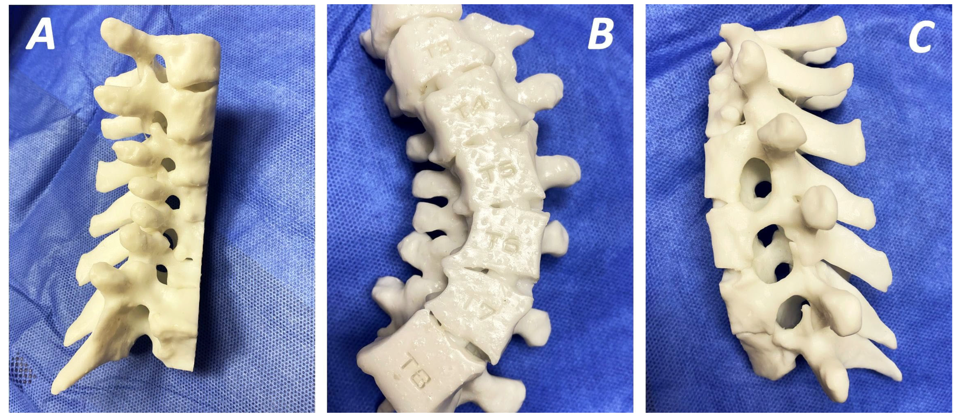
4.2.3. Preoperative Planning—Three-Dimensional Printing Technology
4.2.4. Intraoperative Assistant Methods
4.2.5. Post-Operative Management
4.3. Strengths and Limitations
5. Conclusions
Supplementary Materials
Author Contributions
Funding
Institutional Review Board Statement
Informed Consent Statement
Data Availability Statement
Conflicts of Interest
Abbreviations
References
- Delucia, T.A.; Yohay, K.; Widmann, R.F. Orthopaedic Aspects of Neurofibromatosis: Update. Curr. Opin. Pediatr. 2011, 23, 46–52. [Google Scholar] [CrossRef]
- Barxer, D.; Wright, E.; Nguyen, K.; Cannon, L.; Fain, P.; Goldgar, D.; Bishop, D.T.; Carey, J.; Baty, B.; Kivlin, J.; et al. Gene for von Recklinghausen Neurofibromatosis Is in the Pericentromeric Region of Chromosome. Science 1987, 236, 1100–1102. [Google Scholar] [CrossRef]
- Krygowska, A.A.; Castellano, E. PI3K: A Crucial Piece in the RAS Signaling Puzzle. Cold Spring Harb. Perspect. Med. 2018, 8, a031450. [Google Scholar] [CrossRef]
- Kaspiris, A.; Savvidou, O.D.; Vasiliadis, E.S.; Hadjimichael, A.C.; Melissaridou, D.; Iliopoulou-Kosmadaki, S.; Iliopoulos, I.D.; Papadimitriou, E.; Chronopoulos, E. Current Aspects on the Pathophysiology of Bone Metabolic Defects during Progression of Scoliosis in Neurofibromatosis Type 1. J. Clin. Med. 2022, 11, 444. [Google Scholar] [CrossRef]
- Young, H.; Hyman, S.; North, K. Neurofibromatosis 1: Clinical Review and Exceptions to the Rules. J. Child Neurol. 2002, 17, 613–621. [Google Scholar] [CrossRef]
- Jett, K.; Friedman, J.M. Clinical and Genetic Aspects of Neurofibromatosis 1. Genet. Med. 2010, 12, 1–11. [Google Scholar] [CrossRef]
- Akbarnia, B.; Gabriel, K.; Beckmann, E.; Chalk, D. Prevalence of Scoliosis in Neurofibromatosis. Spine 1992, 17, 244–248. [Google Scholar] [CrossRef]
- Gutmann, D.H.; Ferner, R.E.; Listernick, R.H.; Korf, B.R.; Wolters, P.L.; Johnson, K.J. Neurofibromatosis Type 1. Nat. Publ. Gr. 2017, 3, 17004. [Google Scholar] [CrossRef] [PubMed]
- Well, L.; Careddu, A.; Stark, M.; Farschtschi, S.; Bannas, P.; Adam, G.; Mautner, V.F.; Salamon, J. Phenotyping Spinal Abnormalities in Patients with Neurofibromatosis Type 1 Using Whole—Body MRI. Sci. Rep. 2021, 11, 16889. [Google Scholar] [CrossRef] [PubMed]
- Khoshhal, K.I.; Ed, F.R.C.S.; Ellis, R.D. Paraparesis After Posterior Spinal Fusion in Neurofibromatosis Secondary to Rib Displacement: Case Report and Literature Review. J. Pediatr. Orthop. 2000, 20, 799–801. [Google Scholar] [PubMed]
- Carbone, M.; Vittoria, F.; Del Sal, A. Treatment of Early-Onset Scoliosis with Growing Rods in Patients with Neurofibromatosis-1. J. Pediatr. Orthop. Part B 2019, 28, 278–287. [Google Scholar] [CrossRef]
- Lykissas, M.G.; Schorry, E.K.; Crawford, A.H.; Gaines, S.; Rieley, M.; Jain, V.V. Does the Presence of Dystrophic Features in Patients with Type 1 Neurofibromatosis and Spinal Deformities Increase the Risk of Surgery? Spine 2013, 38, 1595–1601. [Google Scholar] [CrossRef]
- Durrani, A.A.; Crawford, A.H.; Chouhdry, S.N.; Saifuddin, A.; Morley, T.R. Modulation of Spinal Deformities in Patients with Neurofibromatosis Type 1. Spine 2000, 25, 69–75. [Google Scholar] [CrossRef] [PubMed]
- Halmai, V.; Domán, I.; De Jonge, T.; Illés, T. Surgical Treatment of Spinal Deformities Associated with Neurofibromatosis Type 1: Report of 12 Cases. J. Neurosurg. 2002, 97, 310–316. [Google Scholar]
- Tsirikos, A.I.; Saifuddin, A.; Noordeen, M.H. Spinal Deformity in Neurofibromatosis Type-1: Diagnosis and Treatment. Eur. Spine J. 2005, 14, 427–439. [Google Scholar] [CrossRef]
- Yalcin, N.; Bar-On, E.; Yazici, M. Impingement of Spinal Cord by Dislocated Rib in Dystrophic Scoliosis Secondary to Neurofibromatosis Type 1: Radiological Signs and Management Strategies. Spine 2008, 33, 881–886. [Google Scholar] [CrossRef]
- Li, M.; Fang, X.; Li, Y.; Ni, J.; Gu, S.; Zhu, X. Successful Use of Posterior Instrumented Spinal Fusion Alone for Scoliosis in 19 Patients with Neurofibromatosis Type-1 Followed up for at Least 25 Months. Arch. Orthop. Trauma Surg. 2009, 129, 915–921. [Google Scholar] [CrossRef] [PubMed]
- Koptan, W.; Elmiligui, Y. Surgical Correction of Severe Dystrophic Neurofibromatosis Scoliosis: An Experience of 32 Cases. Eur. Spine J. 2010, 19, 1569–1575. [Google Scholar] [CrossRef]
- Sun, D.; Dai, F.; Liu, Y.Y.; Xu, J.Z. Posterior-Only Spinal Fusion without Rib Head Resection for Treating Type I Neurofibromatosis with Intracanal Rib Head Dislocation. Clinics 2013, 68, 1521–1527. [Google Scholar] [CrossRef] [PubMed]
- Heflin, J.A.; Cleveland, A.; Ford, S.D.; Morgan, J.V.; Smith, J.T. Use of Rib-Based Distraction in the Treatment of Early-Onset Scoliosis Associated with Neurofibromatosis Type 1 in the Young Child. Spine Deform. 2015, 3, 239–245. [Google Scholar] [CrossRef]
- Wang, Z.; Fu, C.; Leng, J.; Qu, Z.; Xu, F.; Liu, Y. Treatment of Dystrophic Scoliosis in Neurofibromatosis Type 1 with One-Stage Posterior Pedicle Screw Technique. Spine J. 2015, 15, 587–595. [Google Scholar] [CrossRef]
- Zhao, X.; Li, J.; Shi, L.; Yang, L.; Wu, Z.X.; Zhang, D.W.; Lei, W.; Jie, Q. Surgical Treatment of Dystrophic Spinal Curves Caused by Neurofibromatosis Type 1. Medicine 2016, 95, e3292. [Google Scholar] [CrossRef]
- Cai, S.; Zhang, J.; Shen, J.; Zhao, H.; Weng, X.; Qiu, G. Posterior Correction Without Rib-Head Resection for Patients with Neurofibromatosis Type 1, Dystrophic Scoliosis, and Rib-Head Protrusion into the Spinal Canal. Clin. Spine Surg. 2017, 30, 32–37. [Google Scholar] [CrossRef]
- Deng, A.; Zhang, H.Q.; Tang, M.X.; Liu, S.H.; Wang, Y.X.; Gao, Q. Le Posterior-Only Surgical Correction of Dystrophic Scoliosis in 31 Patients with Neurofibromatosis Type 1 Using the Multiple Anchor Point Method. J. Neurosurg. Pediatr. 2017, 19, 96–101. [Google Scholar] [CrossRef] [PubMed]
- Jain, V.V.; Berry, C.A.; Crawford, A.H.; Emans, J.B.; Sponseller, P.D. Growing Rods Are an Effective Fusionless Method of Controlling Early-Onset Scoliosis Associated with Neurofibromatosis Type 1 (NF1): A Multicenter Retrospective Case Series. J. Pediatr. Orthop. 2017, 37, e612–e618. [Google Scholar] [CrossRef] [PubMed]
- Lyu, Q.; Zhou, C.; Song, Y.; Liu, L.; Wang, L.; Zhou, Z. Does Spinal Deformity Correction of Non-Dystrophic Scoliosis in Neurofibromatosis Type I with One-Stage Posterior Pedicle Screw Technique Produce Outcomes Similar to Adolescent Idiopathic Scoliosis ? Spine J. 2017, 17, 1850–1858. [Google Scholar] [CrossRef]
- Wang, J.Y.; Lai, P.L.; Chen, W.J.; Niu, C.C.; Tsai, T.T.; Chen, L.H. Pedicle Screw versus Hybrid Posterior Instrumentation for Dystrophic Neurofibromatosis Scoliosis. Medicine 2017, 96, e6977. [Google Scholar] [CrossRef] [PubMed]
- Yao, Z.; Li, H.; Zhang, X.; Li, C.; Qi, X. Incidence and Risk Factors for Instrumentation-Related Complications after Scoliosis Surgery in Pediatric Patients with NF-1. Spine 2018, 43, 1719–1724. [Google Scholar] [CrossRef]
- Yao, Z.; Guo, D.; Li, H.; Bai, Y.; Sun, B.; Zhang, X.; Li, C.; Qi, X. Surgical Treatment of Dystrophic Scoliosis in Neurofibromatosis Type 1: Outcomes and Complications. Clin. Spine Surg. 2019, 32, E50–E55. [Google Scholar] [CrossRef]
- Xu, E.; Gao, R.; Jiang, H.; Lin, T.; Shao, W.; Zhou, X. Combined Halo Gravity Traction and Dual Growing Rod Technique for the Treatment of Early Onset Dystrophic Scoliosis in Neurofibromatosis Type 1. World Neurosurg. 2019, 126, e173–e180. [Google Scholar] [CrossRef]
- Cai, S.; Cui, L.; Qiu, G.; Shen, J.; Zhang, J. Comparison between Surgical Fusion and the Growing-Rod Technique for Early-Onset Neurofibromatosis Type-1 Dystrophic Scoliosis. BMC Musculoskelet. Disord. 2020, 21, 455. [Google Scholar] [CrossRef]
- Cai, S.; Li, Z.; Qiu, G.; Shen, J.; Zhao, H.; Zhao, Y.; Wang, Y.; Zhang, J. Posterior Only Instrumented Fusion Provides Incomplete Curve Control for Early-Onset Scoliosis in Type 1 Neurofibromatosis. BMC Pediatr. 2020, 20, 63. [Google Scholar] [CrossRef] [PubMed]
- Cai, S.; Tian, Y.; Qiu, G.; Zhang, J.; Shen, J.; Zhao, H.; Zhao, Y. Neurofibromatosis Type 1 with Severe Dystrophic Kyphosis: Surgical Treatment and Prognostic Analysis of 27 Patients. Orthop. Surg. 2020, 12, 1923–1940. [Google Scholar] [CrossRef]
- Li, S.; Mao, S.; Ma, Y.; Shi, B.L.; Liu, Z.; Zhu, Z.Z.; Qiao, J.; Qiu, Y. Could Screw/Hook Insertion at the Apical Vertebrae with Rib Head Dislocation Effectively Retract the Corresponding Rib Head from Spinal Canal in Dystrophic Scoliosis Secondary to Type 1 Neurofibromatosis? BMC Musculoskelet. Disord. 2022, 23, 285. [Google Scholar] [CrossRef]
- Mladenov, K.V.; Spiro, A.S.; Krajewski, K.L.; Stücker, R.; Kunkel, P. Management of Spinal Deformities and Tibial Pseudarthrosis in Children with Neurofibromatosis Type 1 (NF-1). Child’s Nerv. Syst. 2020, 36, 2409–2425. [Google Scholar] [CrossRef]
- Tauchi, R.; Kawakami, N.; Castro, M.A.; Ohara, T.; Saito, T.; Morishita, K.; Yamauchi, I. Long-Term Surgical Outcomes After Early Definitive Spinal Fusion for Early-Onset Scoliosis With Neurofibromatosis Type 1 at Mean Follow-up of 14 Years. J. Pediatr. Orthop. 2020, 40, 42–47. [Google Scholar] [CrossRef]
- Tauchi, R.; Kawakami, N.; Suzuki, T.; Uno, K.; Yamaguchi, T.; Yanagida, H.; Yamamoto, T.; Murakami, H.; Demura, S.; Kotani, T. Comparison of Early Definitive Fusion and Traditional Growing Rods in Early-Onset Dystrophic Scoliosis in Neurofibromatosis Type 1: A Preliminary Report. J. Pediatr. Orthop. 2020, 40, 569–574. [Google Scholar] [CrossRef] [PubMed]
- Li, S.; Mao, S.; Du, C.; Zhu, Z.; Shi, B.; Liu, Z.; Qiao, J.; Qiu, Y. Assessing the Unique Characteristics Associated with Surgical Treatment of Dystrophic Lumbar Scoliosis Secondary to Neurofibromatosis Type 1: A Single-Center Experience of More than 10 Years. J. Neurosurg. Spine 2021, 34, 413–423. [Google Scholar] [CrossRef]
- Marrache, M.; Suresh, K.V.; Miller, D.J.; Hwang, S.; Schorry, E.K.; Rios, J.J.; Sponseller, P.D. Early-Onset Spinal Deformity in Neurofibromatosis Type 1: Natural History, Treatment, and Imaging Surveillance. JBJS Rev. 2021, 9, e20. [Google Scholar] [CrossRef]
- Pushpa, B.T.; Rajasekaran, S.; Anand, K.S.S.V.; Shetty, A.P.; Kanna, R.M. Anatomical Changes in Vertebra in Dystrophic Scoliosis Due to Neurofibromatosis and Its Implications on Surgical Safety. Spine Deform. 2022, 10, 159–167. [Google Scholar] [CrossRef] [PubMed]
- Mao, S.; Li, S.; Ma, Y.; Shi, B.; Liu, Z.; Zhu, Z.; Qiao, J.; Qiu, Y. How to Rectify the Convex Coronal Imbalance in Patients with Unstable Dystrophic Scoliosis Secondary to Type I Neurofibromatosis: Experience from a Case Series. BMC Musculoskelet. Disord. 2022, 23, 368. [Google Scholar] [CrossRef]
- Neifert, S.N.; Khan, H.A.; Kurland, D.B.; Kim, N.C.; Yohay, K.; Segal, D.; Samdani, A.; Hwang, S.; Lau, D. Management and Surgical Outcomes of Dystrophic Scoliosis in Neurofibromatosis Type 1: A Systematic Review. Neurosurg. Focus 2022, 52, E7. [Google Scholar] [CrossRef]
- Li, H.; Zhang, W.; Yao, Z.; Guo, R.; Hao, C.; Zhang, X. Genotypes and Clinical Intervention of Patients with Neurofibromatosis Type 1 Associated Dystrophic Scoliosis. Front. Pediatr. 2022, 10, 918136. [Google Scholar] [CrossRef] [PubMed]
- Price, G.; Martini, M.L.; Caridi, J.M.; Lau, D.; Oermann, E.K.; Neifert, S.N. A Nationwide Study Characterizing the Risk and Outcome Profiles of Multilevel Fusion Procedures in Neuromuscular Scoliosis Patients with Neurofibromatosis Type 1. World Neurosurg. 2023, 171, e620–e630. [Google Scholar] [CrossRef] [PubMed]
- Wu, J.; Tao, Z.; Jiang, H.; Lin, T.; Ma, J.; Zhou, X.; Wang, C. A Novel Hybrid Technique in the Treatment of Dystrophic Scoliosis Secondary to Neurofibromatosis Type 1 Lacking Pedicles in the Apical Area. World Neurosurg. 2023, 169, e171–e180. [Google Scholar] [CrossRef]
- Zhao, J.; Meng, Y.; Ma, J.; Zhou, X.; Jiang, H. Sectional Correction Technique in Dystrophic Scoliosis Secondary to Neurofibromatosis Type 1: A Comparison with Traditional 2-Rod Correction Technique. World Neurosurg. 2022, 167, e507–e514. [Google Scholar] [CrossRef]
- Xu, A.L.; Suresh, K.V.; Gomez, J.A.; Emans, J.B.; Larson, A.N.; Cahill, P.J.; Andras, L.M.; White, K.K.; Miller, D.J.; Murphy, J.S.; et al. Consensus-Based Best Practice Guidelines for the Management of Spinal Deformity and Associated Tumors in Pediatric Neurofibromatosis Type 1: Screening and Surveillance, Surgical Intervention, and Medical Therapy. J. Pediatr. Orthop. 2023, 43, E531–E537. [Google Scholar] [CrossRef] [PubMed]
- Dastagirzada, Y.; Neifert, S.; Kurland, D.B.; Kim, N.C.; Panicucci-Roma, T.; Frempong-Boadu, A.; Lau, D. Vertebral Column Subluxation in Neurofibromatosis Type 1-Associated Dystrophic Scoliosis: A Report of Two Cases and Narrative Review. Oper. Neurosurg. 2024, 28, 762–771. [Google Scholar] [CrossRef]
- Liang, M.; Cao, J.; Zhang, X.; Guo, D.; Yao, Z.; Gao, R.; Bai, Y. Safety and Effectiveness of Halo Gravity Traction Combined with Traditional Growing Rods in Severe Early-Onset Scoliosis with Neurofibromatosis Type 1. J. Pediatr. Orthop. B 2024, 34, 74–82. [Google Scholar] [CrossRef]
- Wang, D.; Zhang, B.H.; Wen, X.; Chen, K.H.; Xiao, H.T.; Xu, X.W.; Li, Q.F. Clinical Features and Surgical Treatments of Scoliosis in Neurofibromatosis Type 1: A Systemic Review and Meta-Analysis. Eur. Spine J. 2024, 33, 2646–2665. [Google Scholar] [CrossRef] [PubMed]
- Lo, Y.S.; Dai, Y.T.; Qiu, Y.; Lin, E.T.E.; Hsieh, H.L.; Wei, X.P.; Tsai, C.H.; Fong, Y.C.; Chen, H.T.; Tzeng, S.T.; et al. Three-Column Osteotomy versus Halo-Gravity Traction Combined with Posterior Column Osteotomy in the Treatment of Dystrophic Neurofibromatosis Type 1 Kyphoscoliosis: A Retrospective Comparative Cohort Study. J. Orthop. Surg. Res. 2025, 20, 470. [Google Scholar] [CrossRef]
- Yun, C.; Shen, C.L. Anterior Release for Scheuermann’s Disease: A Systematic Literature Review and Meta-Analysis. Eur. Spine J. 2017, 26, 921–927. [Google Scholar] [CrossRef] [PubMed]
- Jin, M.; Liu, Z.; Liu, X.; Yan, H.; Han, X.; Qiu, Y.; Zhu, Z. Does Intraoperative Navigation Improve the Accuracy of Pedicle Screw Placement in the Apical Region of Dystrophic Scoliosis Secondary to Neurofibromatosis Type I: Comparison between O-Arm Navigation and Free-Hand Technique. Eur. Spine J. 2016, 25, 1729–1737. [Google Scholar] [CrossRef]
- Li, Y.; Yuan, X.; Sha, S.; Liu, Z.; Zhu, W.; Qiu, Y.; Wang, B.; Yu, Y.; Zhu, Z. Effect of Higher Implant Density on Curve Correction in Dystrophic Thoracic Scoliosis Secondary to Neurofbromatosis Type 1. J. Neurosurg. Pediatr. 2017, 20, 371–377. [Google Scholar] [CrossRef]
- Qiu, J.; Li, Y.; Liu, W.; Zhu, Z.; Shi, B.; Liu, Z.; Sun, X.; Qiu, Y. Intra-Operative Neurophysiological Monitoring in Patients with Dystrophic Neurofibromatosis Type 1 Scoliosis. Somatosens. Mot. Res. 2021, 38, 95–100. [Google Scholar] [CrossRef]
- Shao, X.; Huang, Z.; Yang, J.; Deng, Y.; Yang, J.; Sui, W. Efficacy and Safety for Combination of T-EMG with O-Arm Assisted Pedicle Screw Placement in Neurofibromatosis Type I Scoliosis Surgery. J. Orthop. Surg. Res. 2021, 16, 16–21. [Google Scholar] [CrossRef]
- Wang, Y.; Li, Z.; Liu, L.; Qi, L. Halo-pelvic traction for thoracic spine dislocation in neurofibromatosis type 1: A case series. Arch. Orthop. Trauma Surg. 2022, 142, 571–577. [Google Scholar] [CrossRef]
- Singh, K.; Samartzis, D.; Ebhc, D.; An, H.S. Neurofibromatosis Type I with Severe Dystrophic Kyphoscoliosis and Its Operative Management via a Simultaneous Anterior-Posterior Approach: A Case Report and Review of the Literature. Spine J. 2005, 5, 461–466. [Google Scholar] [CrossRef]
- Samdani, A.F. Learning Curve for Placement of Thoracic Pedicle Screws in the Deformed Spine. Neurosurgery 2010, 66, 12–15. [Google Scholar] [CrossRef] [PubMed]
- Chen, Q.; Luo, C.; Lu, T.; Yao, S.; Pu, X.; Yang, M.; Chen, L.; Wang, L. Three-Dimensional Printing Technology Based on Digital Orthopedics: 5-Point Positioning Point- Contact Pedicle Navigation Template in the Case of Scoliosis and Complex Pedicle. Orthop. Surg. 2024, 16, 2976–2983. [Google Scholar] [CrossRef] [PubMed]
- Pan, A.; Ding, H.; Hai, Y.; Liu, Y.; Hai, J.J.; Yin, P.; Han, B. The Value of Three-Dimensional Printing Spine Model in Severe Spine Deformity Correction Surgery. Glob. Spine J. 2023, 13, 787–795. [Google Scholar] [CrossRef] [PubMed]
- Katiyar, P.; Boddapati, V.; Coury, J.; Roye, B.; Vitale, M.; Lenke, L. Three-Dimensional Printing Applications in Pediatric Spinal Surgery: A Systematic Review. Glob. Spine J. 2024, 14, 718–730. [Google Scholar] [CrossRef]
- Luo, M.; Wang, W.; Mmed, N.Y.; Xia, L. Does Three-Dimensional Printing Plus Pedicle Guider Technology in Severe Congenital Scoliosis Facilitate Accurate and E Ffi Cient Pedicle Screw Placement? Clin. Orthop. Relat. Res. 2019, 477, 1904–1912. [Google Scholar] [CrossRef]
- McMaster, M.J.; James, J.I. Pseudoarthrosis after Spinal Fusion for Scoliosis. J. Bone Jt. Surg Br. 1976, 58, 305–312. [Google Scholar] [CrossRef] [PubMed]
- Welborn, M.; Tambe, A.; Adeyemi, A.; Dupuis, M.; Simoneau, D.; Brandi, M.L. Burden of Disease and Unmet Needs Associated with Scoliosis in Neurofibromatosis Type 1: A Systematic Literature Review. JBMR Plus 2025, 9, ziaf072. [Google Scholar] [CrossRef]
- Bobyn, J.D.; Deo, N.; Little, D.G.; Schindeler, A. Modulation of Spine Fusion with BMP-2, MEK Inhibitor (PD0325901), and Zoledronic Acid in a Murine Model of NF1 Double Inactivation. J. Orthop. Sci. 2021, 26, 684–689. [Google Scholar] [CrossRef]
- Birke, O.; Schindeler, A.; Ramachandran, M.; Bellemore, M.; Little, D.G. Preliminary Experience with the Combined Use of Recombinant Bone Morphogenetic Protein and Bisphosphonates in the Treatment of Congenital Pseudarthrosis of the Tibia. J. Child. Orthop. 2010, 4, 507–517. [Google Scholar] [CrossRef]
- Han, C.; Zhou, C.; Zhang, H.; Yin, P.; Guo, R.; Wang, W.; Zhang, Y. Evaluation of Bone Mineral Density in Adolescent Idiopathic Scoliosis Using a Three—Dimensional Finite Element Model: A Retrospective Study. J. Orthop. Surg. Res. 2023, 18, 938. [Google Scholar] [CrossRef]
- Page, M.J.; McKenzie, J.E.; Bossuyt, P.M.; Boutron, I.; Hoffmann, T.C.; Mulrow, C.D.; Shamseer, L.; Tetzlaff, J.M.; Akl, E.A.; Brennan, S.E.; et al. The PRISMA 2020 statement: An updated guideline for reporting systematic reviews. BMJ 2021, 29, 372. [Google Scholar] [CrossRef]



| Author (Year) | Type of Article | Number of Patients | Purpose of Study | Subtypes of NF1 | Method of Treatment | Newcastle–Ottawa Score | Key Findings |
|---|---|---|---|---|---|---|---|
| Halmai et al., 2002 [14] | Case series | 12 | Present results from surgical treatment of dystrophic and non-dystrophic deformities in patients with NF1 | Non-dystrophic and dystrophic | In non-dystrophic posterior-only fusion. Halo-gravity traction in dystrophic and then two stages (anterior and then posterior fusion with interval of halo traction) | 07 (Good) | 62.9% correction of curvature in non-dystrophic cases. No dysplasia or pseudarthrosis. 4° correction loss in the frontal plane, 5° in the sagittal plane at final fusion. For dystrophic cases, mean correction loss of 5.4° in the sagittal plane and 4.8° in the frontal, at 4.4 years follow up. No pseudarthrosis and one case with neurological complications reported. |
| Tsirikos et al., 2004 [15] | Review | Not applicable | Review diagnosis, clinical manifestations, and treatment of spinal deformities in NF1 | Non-dystrophic and dystrophic | Non-dystrophic curves treated as idiopathic scoliosis, dystrophic with aggressive treatment | - | Stabilisation of the vertebral column should be the priority rather than extensive correction, which may be accompanied by neurological impairment. |
| Yalcin et al., 2007 [16] | Cases series | 3 | Diagnosis and management of rib dislocation during deformity correction surgery in patients with NF1 dystrophic scoliosis | Dystrophic | Rib head resection in two of the cases presented, rib dislocated away from the canal during reduction manoeuvres in the other case | 07 (Good) | No postoperative complications reported. Rib heads of apical convex can be dislocated in the spinal canal of these patients. Treatment with spinal instrumentation techniques (including growing rods) achieved with preventative measures. |
| Li et al., 2009 [17] | Research article | 19 | Evaluate whether extension of posterior-only fusion at one level beyond conventional one enables as efficient fusion as combined anterior–posterior approach in patients with NF1 scoliosis | Non-dystrophic and dystrophic | Posterior fusion alone in all the patients (hybrid constructs with hooks, screws and sublaminar wires) | 08 (Good) | Three patients with non-dystrophic subtype. Similar results with those with combined anterior–posterior fusion (scoliosis from 40° to 90°). Correction loss greater than those treated for adolescent scoliosis but comparable with patients with NF1. One case of revision surgery presented. |
| Coptan and Elmiligui, 2010 [18] | Original article | 32 | Evaluate clinical radiological outcomes from multisegmental instrumented fusion for scoliosis with NF1 | Dystrophic | Two-stage anterior release/fusion and posterior correction/fusion | 08 (Good) | Correction comparable with literature results, greater corrective loss in greater kyphotic deformities. |
| Sun et al., 2013 [19] | Clinical article | 6 | Analyse results from surgical treatment of patients with NF1 deformity and intraspinal rib head insertion | Dystrophic | Posterior spinal fusion (including multilevel Smith-Petersen osteotomies) without rod head resection | 07 (Good) | No neurological complications, dural tear, intracanal haematoma were reported postoperatively. Rib heads retracted away from spinal canal (5.18 × 2.6 cm). Solid spinal fusion achieved and clinical correction maintained one year after surgery. |
| Heflin et al., 2015 [20] | Research article | 12 | Evaluate results from rib-based distraction in treating patients with NF1 scoliosis and concomitant complications | Dystrophic | Vertical Expandable Prosthetic Titanium Rib implants under spinal erector fascia with rib cradles proximally inline or parallel, distally in non-ambulatory system attach pelvis with Dunne McCarthy-style pelvic S hooks. In ambulatory children, either down-going lamina hooks or pedicle screws used | 08 (Good) | 17 complications in 8 children. In most cases, maintenance or improvement of preop Cobb angle, progression more than 10° only 3 patients. |
| Wang et al., 2015 [21] | Clinical study | 16 | Present results from patients with dystrophic NF1 scoliosis treated with one stage posterior pedicle screw fixation | Dystrophic | Posterior one-stage pedicle screw fixation (posterior vertebral column resection in some patients). Allogeneic graft or iliac crest used | 08 (Good) | No revision surgery required. One patient with transient weakness of lower extremity and one with weakness necessitating surgical exploration and intraspinal haematoma removal. Satisfactory improvement in coronal and sagittal balance. Overall, spinal balance needs to be improved. No progression of the deformity was reported. |
| Zhao et al., 2016 [22] | Observational study | 26 | Analyse radiological outcome of surgical management of dystrophic NF1-associated scoliosis | Dystrophic | Posterior column resection with intracanal rib head resection. Pedicle of hook fixation | 08 (Good) | Hook dislodgement and pseudarthrosis in two patients. Extension of fusion and solid instrumentation for successful fusion results. Complete resection of intracanal rib heads (6 patients). One patient with mild transient paraparesis, postoperatively. |
| Cai et al., 2017 [23] | Research article | 8 | Evaluate results from NF1 dystrophic scoliosis with rib head protrusion in canal with posterior spinal fusion without rib head resection | Dystrophic | Posterior correction of the deformity with a pedicle screw–rod system (three-dimensional). Spinal fusion in 7 cases, one with growing rod system. No rib head resection | 07 (Good) | Median spinal canal space involving intraspinal rib head was significantly lower postoperatively than preoperatively. No neurological complications at one-year follow up and maintenance of the correction. |
| Deng et al., 2017 [24] | Clinical article | 31 | Evaluate clinical efficiency of posterior-only fusion system with a multiple anchor point method | Non-dystrophic and dystrophic | Posterior-only fusion with a multiple anchor point method (as many as possible pedicle screws or hooks in key vertebrae) | 09 (Good) | Significant correction at mean coronal Cobb angle and apical vertebral rotation, with only 2.3% correction loss rate of the Cobb angle. Revision required in only one case with hook dislodgement and pseudarthrosis. |
| Jain et al., 2017 [25] | Original article | 14 | Evaluate results from use of growing rods in patients with early-onset scoliosis with NF1 | Dystrophic | Traditional growing rod instrumentation used (none with magnetically controlled rod) | 07 (Good) | Four patients with definite fusion treatment. The correction rate was 50.1% (final follow-up), and yearly T1-S1 growth rate around 11.2 mm. 1.4 complication rate per patient (mainly proximal junctional kyphosis and proximal construct failure). |
| Lyu et al., 2017 [26] | Clinical study | 15 | Evaluate clinical outcomes from one-stage posterior fusion in patients with non-dystrophic NF1 scoliosis and compare with adolescents’ idiopathic types | Non-dystrophic | Posterior correction and posterior fusion with segmental instrumentation | 07 (Good) | Similar spinal correction in both groups. Results showed the curve correction in the NF-1 group was better than patients with non-dystrophic scoliosis treated by hook–rod-based instrumentation. No apparent progression of deformity with the pedicle screw system and similar correction loss. |
| Wang et al., 2017 [27] | Observational study | 9 | Present results from management of severe rigid dystrophic scoliotic curves in NF1 | Dystrophic | Combined anterior release fusion with posterior correction and instrumentation (single or two-stage procedure) | 07 (Good) | No statistically significant difference in coronal and sagittal curve correction between pedicle screw and hybrid instrumentation construct. Less reduction in coronal correction in the pedicle screw group, but without statistically significant difference in loss of sagittal correction. |
| Yao et al., 2018 [28] | Research article | 59 | Evaluate incidence and risk factors for complications after surgical treatment for NF1-associated scoliosis | Dystrophic | Instrumented-based surgical treatment (pedicle screw–rod and growing rod methods) | 09 (Good) | Seventeen patients with a total of nineteen complications (no neurological complications referred). Age less than nine years, kyphosis exceeding 50°, and growing rod implantation are risk factors for complications. |
| Carbone et al., 2019 [11] | Original article | 7 | Study results of growth-friendly instrumentation in management of early-onset NF1-associated scoliosis and evaluate “law of diminishing returns” effect | Dystrophic | Posterior double growing rod implantation without fusion applied in patients with dystrophic type of NF1. Lengthening performed every twelve months | 07 (Good) | Three patients treated with initial instrumentation removal and final fusion. Several complications, mainly rod breakages. Comparable results to literature with the advantage of one lengthening operation annually. Law of diminishing returns not confirmed. |
| Yao et al., 2019 [29] | Research article | 59 | Evaluate outcomes and instrumentation complications in patients for NF1 dystrophic scoliosis | Dystrophic | Posterior fusion only (instrumented) or combined with anterior fusion, compared to the growing rod approach | 09 (Good) | Early fusion (definitive) in patients approaching skeletal maturity or with short and sharp curves. Use of growing rod shows higher complication rate and lower corrective one. Final fusion when more instrument-related complications presented and less growth feasible after distraction. |
| Xu et al., 2019 [30] | Original article | 11 | Present results from combined use of halo-gravity traction and dual growing rod in dystrophic NF1 scoliosis | Dystrophic | Use of preoperative halo-gravity traction up to 50% of body weight and placement of 2 rods. Lengthening of 1–2 cm achieved every 6 months, until no traction feasible | 08 (Good) | 3.9 times of lengthening with average distance of 1.6 cm. One patient with hook dislodgement. No autofusion reported. |
| Cai et al., 2020 [31] | Research article | 16 | Compare outcomes of posterior fusion with growth-friendly treatment of dystrophic NF1 scoliosis | Dystrophic | Posterior fusion with rod derotation and in situ reduction (translational) with compression or distraction manoeuvres, growth-friendly applied with 2 rods, hooks or pedicle screws, and autologous bone and allograft at foundation area | 08 (Good) | Major curve of 3 patients decreased postoperatively in growth-friendly group, while 4 patients in the other group with major curve progression. No neurological complication, no statistically significant complications between the groups. Favourable T1-S1 growth in the first group. |
| Cai et al., 2020 [32] | Research article | 10 | Compare mid–long-term results from posterior-only instrumented fusion in NF1 patients with early-onset dystrophic scoliosis | Dystrophic | All patients with screw-based instrumentation | 07 (Good) | High incidence of alignment complications (only one required revision surgery). Major curve correction from 66.1° ± 16.2° preoperatively, to 31.1° ± 14.6° postoperatively. No neurological complication or lung function deterioration. |
| Cai et al., 2020 [33] | Research article | 27 | Present surgical treatment and prognosis in 27 patients with NF1-associated dystrophic deformities | Dystrophic | Various procedures related to the grade of deformity/pontodestomy or lower articular surface resection and fusion or apical vertebral body or discectomy (upper) and fusion | 08 (Good) | Obvious correction in many cases, instrument (case) dislocation or rod breakage described (required revision surgery). Major curve correction without significant difference between two or three groups (certain correction of the surgical programme). |
| Li at al., 2020 [34] | Research article | 37 | Report impact of screw/hook insertion in retraction of rib head from spinal canal in patients with dystrophic NF1 scoliosis | Dystrophic | All pedicle screws constructs for 21 patients and hybrid hook screw for 16 patients. | 09 (Good) | No significant different results regarding kyphosis correction and spinal height between screw/hook and non-screw/hook group. Screw/hook placement associated with higher Cobb angle and vertebral translation correction, three-dimensional relationship between spinal canal and rib head could be changed through traction and derotational withdrawal. |
| Mladenov et al., 2020 [35] | Annual issue article | 33 (3 of them with cervical deformities) | Report outcomes from surgical treatment of skeletal deformities in patients with NF1 and spinal deformities | Dystrophic | 11 patients treated with definite fusion, 11 with growth-preserving techniques, 7 with combination, 5 with preserving methods convert to fusion | 08 (Good) | Good results regarding curve correction (mean 54%). Preservation of annual thoracic spine growth. Posterior approach in curves less than 60°, combination of approaches in greater curves. Use of laminar hooks or sublaminar brands in dystrophic areas. |
| Tauchi et al., 2020 [36] | Original article | 11 | Present long-term results from definitive spinal fusion for early-onset scoliosis in NF1 patients | Dystrophic | Halo traction for 2 to 3 weeks in curves larger than 80°, then anterior release bone grafting and intervertebral disc removal (via thoracotomy). Finally, posterior spinal fusion. 7 cases with subtotal tumour resection on concave side and rib strut grafting | 07 (Good) | Patients shorter than general population (early definitive fusion and large scale of fusion applied). Stable instrumentation achieved (longer fusion levels). Circumferential approach, with subtotal tumour resection, rib strut grafting. |
| Tauchi et al., 2020 [37] | Original article | 26 | Compare results from early and growing rods in patients with NF1 dystrophic scoliosis | Dystrophic | Early fusion (anterior and posterior or posterior-only) versus growing rod (final fusion at mean age of 12.7 years) | 08 (Good) | Greater correction of curvature in the early fusion group, with fewer surgical procedures, growing rod allows continued growth in the thorax and spine. |
| Li et al., 2021 [38] | Clinical article | 39 | Present specific features related to treatment of dystrophic NF1-associated lumbar scoliosis | Dystrophic | Posterior-only fusion approach or combined posterior–anterior/anterior–posterior | 09 (Good) | No difference in pain or function scores between two groups. Anterior approach to enhance spinal fusion and stability, while reducing rod breakage and revision. Reduces growth asymmetry and prevents crankshaft phenomenon. |
| Marrache et al., 2021 [39] | Review article | Not applicable | Highlight natural history, management and imaging surveillance of spinal deformities in NF1 scoliosis | Dystrophic | Skeletally immature patients with non-dystrophic NF1 scoliosis treated with brace, with 20° to 40° curves. Patients with curves under 20°, only observation follow-up every six months, curves over 45° with early fusion or growth-friendly instrumentation. Patients with dystrophic curves under 20° clinical observation every six months, surgically treated upon progression (relative indication). | - | Annual clinical and scoliosis examination for children aged 1 to 5 years with NF1. Growing rods compared to early fusion allow spinal lengthening, but early fusion can result in similar correction with fewer procedures. Magnetically controlled growing rods contribute to growth lengthening by an external magnet, avoiding additional surgical procedures. |
| Pushpa et al., 2021 [40] | Case series | 10 | Present a radiological evaluation of morphological alterations of ten dystrophic scoliotic curves and impact on surgical management | Dystrophic | Not applicable | 07 (Good) | A wide spectrum of anatomical changes, with gross variations even with small curves, 34% of pedicles in apex and three adjacent segments (above and below) safe for instrument insertion. Need for CT-guided preoperative planning. |
| Mao et al., 2022 [41] | Case series | 15 | Present risk factors for convex coronal imbalance and improve manoeuvres for postoperative coronal balance | Dystrophic | 4 patients with combined staged treatment, 11 patients with posterior-only fusion | 07 (Good) | For thoracolumbar/lumbar convex coronal imbalance limited and unreliable distal screw purchases with poor correction of lumbosacral curve, leave residual take of angle, risk of failure of coronal rebalance. |
| Neifert et al., 2022 [42] | Systematic review | 30 studies (761 patients) | Present natural history, treatment options and outcomes in patients with dystrophic scoliosis related to NF1 | Dystrophic | Different treatment: posterior-only fusion, anterior and posterior approach, growth-friendly methods (hybrid constructs, pedicle screw-only constructs, and hook-based constructs) | - | Immediate postoperative neurological complication was 2.1%, rate of permanent neurological deficits was 1.2%. Revision surgery rate was 21.5%. |
| Li et al., 2022 [43] | Research article | 14 | Analyse genotype, outcomes in patients with NF1-associated dystrophic scoliosis | Dystrophic | Growing rods (lengthening at 6–12 months) or posterior spinal fusion | 07 (Good) | Twelve patients with pathogenic variants. No clear association between genotype and phenotype, no mutation hotspot on NF1 gene. |
| Price et al., 2022 [44] | Original article | 533 | Report outcomes from multilevel fusion surgery | Dystrophic | Multilevel spinal fusion | 09 (Good) | NF1 patients with higher risk of neurological impairment. No statistically significant difference in mortality (in-hospital), resource utilisation and quality-based outcome. |
| Wu et al., 2022 [45] | Original article | 46 | Compare results of a hybrid method of segmented correction (lacking pedicle screws in apical area) with these of the traditional in patients with dystrophic NF1 scoliosis | Dystrophic | Halo-gravity traction preoperatively in both groups. After pedicle screw placement and second level osteotomy of the apical area performed, two rods placed on concave side connected with tulip connectors and locked after distraction | 08 (Good) | No statistically significant differences regarding spinal flexibility, level of fusion, blood loss, age at operation or time of operation. Higher average Cobb angle correction with lower postoperative loss of correction in the group with segmented correction. No pseudarthrosis or fixation failure reported in either group. |
| Zhao et al., 2022 [46] | Original article | 53 | Compare radiographical and clinical results from a 2 rod and sectional correction technique in patients with dystrophic NF1 scoliosis | Dystrophic | Sectional procedure included two short rods at concave side of curve connected with domino, compression manoeuvre of rods to correct concave curve, installation of long rod at the convex. Traditionally performed with two rods (known method) | 09 (Good) | Better results regarding coronal balance distance in sectional group, with less correction loss postoperatively. No implant failure reported, due to prevention of pedicle screw removal throughout translation, rod rotation or screw loosening as a result of excessive stress. |
| Xu et al., 2023 [47] | Original article | Not applicable | Emphasise the role of magnetically controlled growing rods (MCGRs) and definitive spinal fusion on treatment of children with NF1. | Dystrophic | Recommendations on thoracolumbar and cervical deformity correction (four contraindications for use of magnetically controlled growing rods) | - | Use of magnetically controlled growing rods associated with improvements in curve magnitude and spinal height. Diminished visualisation when imaging the postoperative spine. |
| Dastagirzanda et al., 2024 [48] | Research article (narrative review) | 2 | Present two cases of NF1 dystrophic scoliosis with vertebral subluxation | Dystrophic | Use of halo-gravity traction in the first case without change in subluxation and then fusion. In the second case, neck haematoma and respiratory failure, necessitating embolization while patient was on halo-gravity traction. The same patient, while on rehabilitation, led to paraplegia, as a result of a fall, which was treated with four-rod construct accompanied with C7 to T2 laminectomy and adequate spinal cord decompression | 07 (Good) | Unpredictable progression and complication of curves in both cases (following skeletal maturity). Rare complications of halo-gravity traction, which can be unsuccessful as in the first case or cause artery injuries. Preoperative planning of deformity correction should include plexiform tumour resection, tissue defects, and dural ectasia. |
| Liang et al., 2024 [49] | Original article | 15 | Evaluate safety, effectiveness of use of halo-gravity traction and growing rod system in the treatment of patients with early-onset NF1-associated scoliosis | Dystrophic | Traction duration for more than 12 h. Traditional growing rods applied with lengthening interval of 9–12 months | 07 (Good) | No complications associated with halo-gravity traction, one-rod dislocation and one breakage of rod reported. Correction rate of Cobb angle with growing rod surgery was 47.67%. Better results regarding trunk balance, correction rate, and spine height. |
| Wang et al., 2024 [50] | Review article | 37 articles (1032 patients) | Review current literature on natural history, clinical features and surgical managements of NF1 spinal deformities | Dystrophic | 24 studies with bone-grafted or instrument-based spinal fusion, growing rod used in two of them. 4 comparative studies of both treatment methods | - | Satisfactory results for Cobb angle, sagittal kyphosis, and the T1-S1 length of the spinal in patients treated with spinal fusion. Acceptable results from the use of growing rods, but showed lower curve correction, higher incidence of complications not capable of correcting the sagittal kyphosis. Small population of study on this treatment method. |
| Lo et al., 2025 [51] | Research article | 126 | Compare outcomes of patients with NF1 dystrophic scoliosis after three column osteotomies, halo-gravity traction, and posterior column osteotomy | Dystrophic | Posterior column osteotomy performed in all three patient groups | 09 (Good) | Statistically significant postoperative results observed in main curve Cobb angle, apical vertebral translation, segmental kyphosis, and deformity angular ratio in all three groups. No significant loss of correction reported, and coronal imbalance showed significant improvement in both the halo-gravity traction and posterior column osteotomy group. |
| Author (Year) | Type of Article | Number of Patients | Purpose of Study | Subtypes of NF1 | Method of Treatment | Newcastle- Ottawa Score | Key Findings |
|---|---|---|---|---|---|---|---|
| Jin et al., 2015 [53] | Original article | 32 | Compare accuracy of insertion pedicle screw with O-arm navigation with free-hand technique in dystrophic NF1-associated scoliosis | Dystrophic | Pedicle screw posterior procedures | 07 (Good) | Statistically greater correction of the main curve in the O-arm group. Higher accuracy in screw position in the O-arm group. Lower incidence of medial screw perforation and increased implant density in the apical region in this group. |
| Li et al., 2017 [54] | Clinical article | 41 | Evaluate implant density, radiological, clinical outcomes in NF1 dystrophic thoracic scoliosis | Dystrophic | Posterior pedicle screw or hybrid instrumentation with hook (free-hand or O-arm navigation technique) | 08 (Good) | Better immediate postoperative coronal correction rate with implant density higher than 1.35, less loss of correction at follow-up. |
| Qiu et al., 2021 [55] | Clinical article | 92 | Evaluate data from intraoperative neurophysiological monitoring during management of NF1 dystrophic scoliosis and risks from failure | Dystrophic | Posterior spinal deformity correction and fusion | 09 (Good) | 17 patients with failed intraoperative neurophysiological monitoring, associated with more dystrophic features and preoperative neurological deficits. The overall success rates of SEP (somatosensory evoked potentials) and MEP (motor evoked potentials) were 87.0 and 94.6%. |
| Shao et al., 2021 [56] | Research article | 65 | Report results from triggered electromyography (t-EMG) with O-arm-assisted pedicle screw placement in treatment of NF1 scoliosis | Dystrophic | Posterior thoracolumbar spinal fusion with (T1-S1) with t-EMG and O-arm-assisted screw placement | 08 (Good) | 3 malpositioned screws (2 patients) were not detected with O-arm, but only with t-EMG (all of them in the periapical area). Combination of methods associated with 100% sensitivity, positive predictive value of 66.7% and specificity of 96.2%. |
| Author (Year) | Type of Article | Number of Patients | Purpose of Study | Method of Treatment | Key Findings |
|---|---|---|---|---|---|
| Cai et al., 2017 [23] | Research article | 8 | Evaluate results from patients with NF1 dystrophic scoliosis with rib head protrusion in spinal canal treated posterior spinal fusion without rib head resection | Posterior correction of deformity with pedicle screw–rod system (three-dimensional). Spinal fusion performed in 7 cases, one with growing rod system. No procedure involved rib head resection | Median spinal canal space involving intraspinal rib head significantly lower postoperatively than preoperatively. No neurological complications at 1-year follow-up correction maintenance |
| Jain et al., 2017 [25] | Original article | 14 | Evaluate results from the use of growing rods in patients with early-onset NF1-associated scoliosis | Traditional growing rod instrumentation used (none with magnetically controlled rod) | Four patients with definite fusion. Correction rate 50.1% (final follow-up), yearly T1-S1 growth rate 11.2 mm. 1.4 complication rate per patient (mainly proximal junctional kyphosis and proximal construct failure) |
| Yao et al., 2018 [28] | Research article | 59 | Evaluate incidence and risk factors for complications after surgical treatment for NF1-associated scoliosis | Instrument-based surgical treatment (pedicle screw–rod and growing rod methods) | 17 patients with total of 19 complications (no neurological). Age under 9 years, kyphosis > 50° |
| Carbone et al., 2019 [11] | Original article | 7 | Study results of growth-friendly instrumentation in the management of early-onset NF1-associated scoliosis and evaluate the “law of diminishing returns” effect | Posterior double growing rod implantation without fusion applied in patients with dystrophic NF1. Lengthening performed every twelve months | 3 patients with initial instrumentation, removal and final fusion. Rod breakages. Advantage of 1 lengthening operation annually |
| Xu et al., 2019 [30] | Original article | 11 | Present results from combined use of halo-gravity traction and a dual growing rod method in dystrophic NF1 scoliosis | Use of preoperative halo traction up to 50% of body weight and surgical placement of 2 rods. Lengthening of 1–2 cm achieved every 6 months, until no traction feasible | 3.9 times of lengthening with average distance of 1.6 cm. One patient with hook dislodgement. No autofusion reported |
| Cai et al., 2020 [31] | Research article | 16 | Compare outcomes of posterior fusion and with growth-friendly treatment of dystrophic NF1 scoliosis | Posterior fusion with rod derotation and in situ reduction (translational) with compression or distraction manoeuvres, growth-friendly applied with 2 rods, hooks or pedicle screws and autologous bone and allograft at the foundation area | Major curve of 3 patients decreased postoperatively in growth-friendly group, while 4 patients in the other group with major curve progression. No neurological complications, no statistically significant complications between two groups. Favourable T1-S1 growth in the first group |
| Mladenov et al., 2020 [35] | Annual issue article | 33 (3 of them with cervical deformities | Report outcomes from surgical treatment of skeletal deformities in paediatric patients with NF1 and spinal deformities | 11 patients treated with definite fusion, 11 with growth preserving techniques, 7 with a combination of both, and 5 treated with preserving methods convert to fusion | Good results regarding curve correction (mean 54%). Preservation of annual thoracic spine growth. Posterior approach in curves under 60°, combination of approaches in greater curves. Use of laminar hooks or sublaminar brands in dystrophic areas |
| Tauchi, et al., 2020 [37] | Original article | 26 | Compare results from early and growing rods in patients with NF1 dystrophic scoliosis | Early fusion (anterior and posterior or posterior-only) versus growing rod (final fusion at mean age of 12.7 years) | Greater correction of curvature in the early fusion group, with fewer surgical procedures, growing rod allows continued growth in the thorax and spine |
| Marrache et al., 2021 [39] | Review article | Not applicable | Highlight natural history, management and imaging surveillance of spinal deformities in NF1 scoliosis | Skeletally immature patients with non-dystrophic NF1 scoliosis treated with brace when curves 20° to 40°. Curves under 20° only observation every six months, exceeding 45° early fusion or growth-friendly instrumentation. Dystrophic curves under 20° observation every 6 months, treated when curvature progressing | Annual examination for children aged 1 to 5 years. Growing rods compared to early fusion spinal lengthening can result in similar correction. Magnetically controlled growing rods contribute to growth lengthening, avoiding surgical procedures |
| Neifert et al., 2022 [42] | Systematic review | 30 studies (761 patients) | Present natural history, treatment options and outcomes in patients with NF1-associated dystrophic scoliosis | Different treatment options: posterior-only fusion, anterior and posterior approach, growth-friendly methods | Immediate postoperative neurological complication was 2.1%, rate of permanent neurological deficits 1.2%. Revision rate 21.5% |
| Li et al., 2022 [43] | Research article | 14 | Analyse genotype and surgery outcomes in NF1-associated dystrophic scoliosis | Growing rods (lengthening at 6–12 months) or posterior spinal fusion | 12 patients with pathogenic variants. No clear association genotype and phenotype, no mutation hotspot on NF1 gene |
| Xu et al., 2023 [47] | Original article | Not applicable | Emphasise the role of magnetically controlled growing rods (MCGRs) and definitive spinal fusion in the treatment of children with NF1 | Recommendations on thoracolumbar and cervical deformity correction (four contraindications for the use of magnetically controlled growing rods) | Use of magnetically controlled growing rods with improvements in curve magnitude and spinal height. Diminished visualisation imaging postoperative spine |
| Wang et al., 2024 [50] | Review article | 37 articles (1032 patients) | Review current literature on the natural history, clinical features and surgical managements of NF1 spinal deformities | 24 studies with bone-grafted or instrument-based spinal fusion, growing rod used in two of them. 4 comparative studies of both treatment methods | Satisfactory results of Cobb angle, sagittal kyphosis, and T1-S1 length in spinal fusion. Acceptable results from growing rods, lower curve correction, higher incidence of complications, not capable of correcting sagittal kyphosis |
Disclaimer/Publisher’s Note: The statements, opinions and data contained in all publications are solely those of the individual author(s) and contributor(s) and not of MDPI and/or the editor(s). MDPI and/or the editor(s) disclaim responsibility for any injury to people or property resulting from any ideas, methods, instructions or products referred to in the content. |
© 2025 by the authors. Licensee MDPI, Basel, Switzerland. This article is an open access article distributed under the terms and conditions of the Creative Commons Attribution (CC BY) license.
Share and Cite
Kaspiris, A.; Lianou, I.; Marouglianis, V.; Afrati, R.-S.; Sakellariou, E.; Morakis, A.; Karampinas, P.; Vasilisadis, E.S.; Pneumaticos, S.G. Tips and Pitfalls of Surgical Techniques for Scoliotic Deformities in Neurofibromatosis Type 1. J. Clin. Med. 2026, 15, 104. https://doi.org/10.3390/jcm15010104
Kaspiris A, Lianou I, Marouglianis V, Afrati R-S, Sakellariou E, Morakis A, Karampinas P, Vasilisadis ES, Pneumaticos SG. Tips and Pitfalls of Surgical Techniques for Scoliotic Deformities in Neurofibromatosis Type 1. Journal of Clinical Medicine. 2026; 15(1):104. https://doi.org/10.3390/jcm15010104
Chicago/Turabian StyleKaspiris, Angelos, Ioanna Lianou, Vasileios Marouglianis, Roberta-Spyridoula Afrati, Evangelos Sakellariou, Andreas Morakis, Panagiotis Karampinas, Elias S. Vasilisadis, and Spiros G. Pneumaticos. 2026. "Tips and Pitfalls of Surgical Techniques for Scoliotic Deformities in Neurofibromatosis Type 1" Journal of Clinical Medicine 15, no. 1: 104. https://doi.org/10.3390/jcm15010104
APA StyleKaspiris, A., Lianou, I., Marouglianis, V., Afrati, R.-S., Sakellariou, E., Morakis, A., Karampinas, P., Vasilisadis, E. S., & Pneumaticos, S. G. (2026). Tips and Pitfalls of Surgical Techniques for Scoliotic Deformities in Neurofibromatosis Type 1. Journal of Clinical Medicine, 15(1), 104. https://doi.org/10.3390/jcm15010104