Systematic Review

The Use of Tissue Grafts Associated with Immediate Implant Placement to Achieve Better Peri-Implant Stability and Efficacy: A Systematic Review and Meta-Analysis

by
Enrico Maria Rondone
1,
Bruno Leitão-Almeida
1,2,
Miguel Silva Pereira
1,2,
Gustavo Vicentis Oliveira Fernandes
2,3,* and
Tiago Borges
1,2,*
1
Faculty of Dental Medicine, Universidade Católica Portuguesa, 3504-505 Viseu, Portugal
2
Centre for Interdisciplinary Research in Health (CIIS), Universidade Católica Portuguesa, 3504-505 Viseu, Portugal
3
A. T. Still University—Missouri School of Dentistry & Oral Health, St. Louis, MO 63104, USA
*
Authors to whom correspondence should be addressed.
J. Clin. Med. 2024, 13(3), 821; https://doi.org/10.3390/jcm13030821
Submission received: 4 January 2024 / Revised: 17 January 2024 / Accepted: 26 January 2024 / Published: 31 January 2024
(This article belongs to the Special Issue Current and Emerging Treatment Options in Dental Implatology)

Abstract

:
Background: The goal of this systematic study and meta-analysis was to evaluate the efficacy of hard and/or soft tissue grafts associated with type-1 implants on healing and treatment outcomes. The primary outcomes studied were implant survival rate, pocket depth, marginal peri-implant recession, bone loss, bone thickness (volumetric change), interproximal bone level, mesial and distal papilla migration, and radiographic evaluation; and the secondary parameters were Pink Esthetic Score (PES), vertical distance from implant shoulder and bone, Visual Analogue Score (VAS), Implant Stability Quotient (ISQ), and biological complications (fistulas, pain, mucositis, and peri-implantitis). Methods: The PICO strategy was used to formulate the hypothesis under study: “For patients who underwent extraction and immediate implant placement, what is the efficacy of using any type of graft (bone or soft tissue) compared to non-grafting regarding the peri-implant parameters?” The electronic search process was performed on the MedLine/PubMed and Cochrane databases. It included randomized controlled trials (RCTs) from the last 11 years (from 2012 to November 2023), which were identified and analyzed. Results: Nine RCTs (κ = 0.98) were selected (403 patients and 425 implants); they were divided into three groups: bone graft (75 patients and 75 implants inserted), bone graft and membrane (213 patients and 235 implants inserted), and without bone graft (115 patients and 115 implants inserted). Three studies calculated the mid-facial mucosa level and two reported better results when a connective tissue graft was combined with the xenograft, whereas another study found better results in the combination of a dual-zone technique with a xenograft. Three studies evaluated the total Pink Esthetic Score (PES) at 12 months, where the authors found no significant difference in using a xenogeneic graft with or without a membrane. In the same period, the facial bone thickness was assessed in two articles; the authors reported better results in graft-treated and flapless groups. The risk-of-bias assessment found four studies with low risk, four with moderate risk, and one with a high risk of bias. The meta-analysis showed a medium level of heterogeneity for the mid-facial mucosa level analysis (I2 = 46%) and an overall effect size of 0.79 (95% CI [0.18; 1.40]), a statistically significant results (p = 0.01), with a tendency to favor the experimental group. Also, there was a medium level of heterogeneity among studies regarding total PES (I2 = 45%), with no significant differences between studies (p = 0.91). Homogeneous results (I2 = 0%) were found among studies analyzing facial bone thickness, favoring the experimental group; the forest plot showed an effect of 0.37 (95% CI [0.25; 0.50]), which was statistically significant (p < 0.00001) for this parameter. Conclusions: Then, it was possible to conclude that using bone and soft tissue grafting techniques associated with immediate implant placement (IIP), even though they are not fundamental, was a valuable resource to prevent significant tissue reduction, reaching greater bone stability and higher levels in the Pink Esthetic Score (PES) and Visual Analogue Score (VAS).

1. Introduction

Implant rehabilitation techniques involve using biomaterials [1,2] and titanium/zirconia to replace one or more teeth [3]. The classification of implant surgery techniques was developed based on alveolar healing times and included the following: type-1 protocol, immediate implantation (IIP), extraction and insertion in the same surgical protocol; type-2 protocol, early implant, insertion after 4–8 weeks after extraction, soft tissue healing; type-3 protocol, early-delayed implant, insertion after 12–16 weeks, partial healing of the alveolar bone component; and type-4 protocol, late implant, insertion after six months, complete healing of the alveolar bone component [4].
The type-1 protocol is a predictable treatment modality with success rates comparable to the type-4 technique [5,6,7,8]. This protocol brings several advantages, such as shorter procedures, reduced number of procedures, reduced vertical and horizontal resorption, and ideal gingival tissue esthetics [9,10,11]. The surgical criteria advocated for type-1 implants are an intact facial bone wall with a thick phenotype (greater than 1 mm), thick gingival biotype, absence of acute infection, and apical and palatal bone volume suitable for implant placement with sufficient primary stability [12].
As a result of post-extraction implant insertion into the socket, a gap is formed between the inner surface of the buccal cortical plate and the implant, named the jumping gap. Management of the gap is a critical decision for the clinician, who must choose to fill it with a graft or leave it clear with the blood clot alone [13]. In the 1990s, guided bone regeneration (GBR) was introduced, and alternatives were included, including using different grafts associated with a membrane placement [13]. The GBR technique has been increasingly indicated in type-1 (immediate implant) rehabilitation treatments claiming the purpose to compensate for volumetric changes in hard and soft tissues by using autogenous bone tissue grafts, deproteinized bovine bone mineral (DBBM) as fillers, and connective tissue grafts (CTG) and xenogeneic collagen grafts for post-placement alveolus closure [14].
Buser et al. [9] argued that the implant should be placed 2 mm from the inner surface of the buccal cortical plate to facilitate appropriate gap filling with the bone graft. Preclinical studies suggest that a smaller gap results in greater vertical resorption of the cortical bone [9]. The use of xenogeneic collagen material and a connective tissue graft to seal the socket and promote increased peri-implant keratinized mucosal volume is also well documented in the literature [15]. The xenogeneic resorbable matrix provides advantages such as faster healing and fewer surgeries since no surgical procedure is required to harvest the connective tissue graft [16,17]. In addition, DBBM placed in the marginal gap area reduces the amount of horizontal and vertical bone resorption associated with type-1 implant treatment [16].
It shows that a 4-walled defect is more favorable and presents reduced evidence of post-extraction ridge resorption because of the capability of containing the graft and greater effective capacity to incorporate the graft material [17]. Buser et al. [9] concluded that GBR surgical techniques are indeed effective in promoting bone filling and partial or complete resolution of cortical defects; they are more successful when associated with type-1 and -2 implants than late implants [12]. Therefore, there is a lack of uniformity about using or not tissue graft associated with IIP, and the type utilized if applicable. Thus, the objective of this systematic study was to review the literature in order to evaluate the efficacy of hard and/or soft tissue grafts associated with type-1 implants on healing and treatment outcomes. The null hypothesis was that the use of any graft material did not change or improve the healing process and esthetic result. The primary outcome variables were implant survival rate, pocket depth, marginal peri-implant recession, bone loss, bone thickness (volumetric change), interproximal bone level, mesial and distal papilla migration, and radiographic evaluation. The secondary parameters were Pink Esthetic Score (PES), vertical distance from implant shoulder and bone, Visual Analogue Score (VAS), Implant Stability Quotient (ISQ), and biological complications (fistulas, pain, mucositis, and peri-implantitis).

2. Materials and Methods

2.1. Protocol and PICO Strategy

The protocol of this systematic study was performed according to the PRISMA (Preferred Reporting Items for Systematic Reviews and Meta-Analyses) guidelines [18,19] and registered in the PROSPERO platform (International Prospective Register of Systematic Reviews, www.prospero.org, accessed on 10 January 2024; CRD42023383620).
The PICO (Population, Intervention, Comparison, and Outcome) strategy was used as the research model. The following question was posed to formulate the hypothesis under study: “For patients who underwent extraction and immediate implant placement, what is the efficacy of using any type of graft (bone or soft tissue) compared to non-grafting regarding the peri-implant parameters?” Population (P): Patients with a hopeless maxillary/mandibular tooth in the posterior or anterior areas who have received a type-1 implant with or without hard and soft tissue grafting; Intervention (I): Type-1 implant placement with or without hard and soft tissue grafting; Comparison (C): Hard and/or soft tissue grafting and standard healing; Outcome (O): Soft and hard tissue response as measured with the Pink Esthetic Score (PES), midfacial mucosa height, marginal bone loss (MBL), papilla index (PI), linear buccal change, volumetric change, bleeding on probing (BOP), and plaque index.

2.2. Eligibility Criteria

The inclusion criteria established for this review were as follows: randomized clinical trials (RCTs) that enrolled a minimum of 20 patients, had a follow-up of at least six months, and were published in the English language; studies that evaluated the efficacy of hard and/or soft tissue grafts on peri-implant tissue healing in the anterior or posterior sites in the maxilla or mandible.
The exclusion criteria established were animal or in vitro studies; any type of review; cohort studies; randomized clinical trials published before 2012; studies that included patients with uncontrolled systemic disorder; and editorials, abstracts in Congress, case reports, and case series.

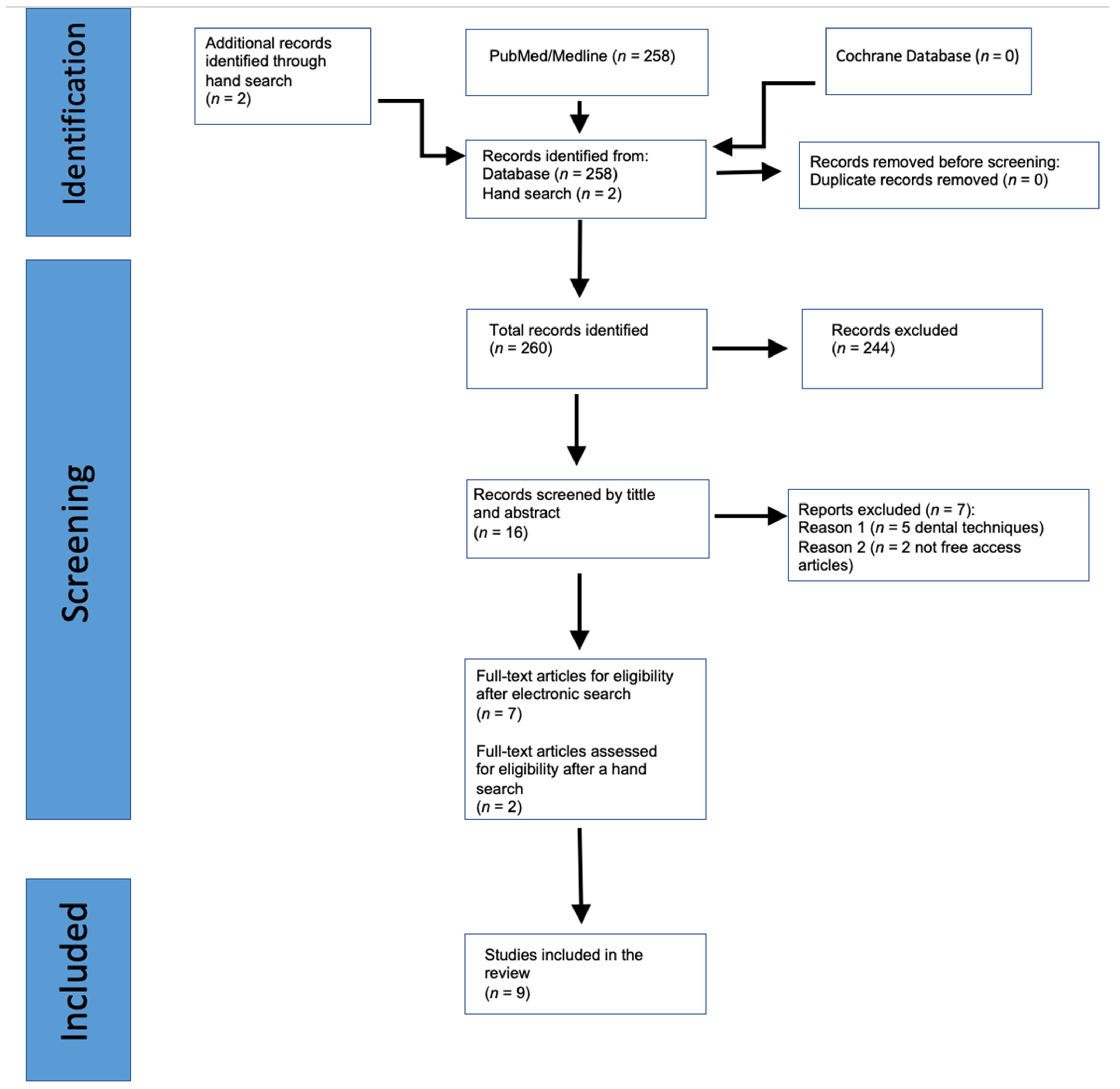
2.3. Search Strategy and Data Extraction

Two calibrated researchers (E.M.R. and T.B.) executed the search strategy independently on the MedLine/PubMed and Cochrane Database platforms using the English language, human studies, and publications made since 2012 as search filters. Any disputes were resolved via confrontation and discussion between the two reviewers. The interrater reliability (Cohen’s kappa coefficient, κ) was performed to verify the degree of agreement between evaluators.
The bibliographic search consisted of a combination of MeSH terms and free-text words combined through Boolean Operators (AND or OR). The keywords used were the following: (1) PubMed/Medline: Dental implant [Mesh] OR dental implantation [Mesh] AND immediate implant placement [text word] AND graft [Mesh] AND bone [Mesh] OR bone graft [text word] OR buccal gap [text word]; filters: RCT-studies; 10 years studies; Human studies; English studies. (2) Cochrane: Dental Implant [Mesh] OR dental implantation [Mesh] AND immediate implant placement [text word] AND graft [Mesh] AND bone [Mesh] OR bone graft [text word] OR buccal gap [text word]; filter: None.
Data were extracted based on the general study characteristics, population characteristics, graft, and implant technique characteristics. Any discrepancy was solved with discussion and collaboration. The data were collected in predefined tables: general information, including study design, year of publication, number of patients, and patient information; information related to implant surgery and type of graft, including number of implants, implant location in the mouth, type of graft for both groups, follow-up, and follow-up intervals; information related to the surgical protocol; information related with the studies outcome variables; main outcomes: Pink Esthetic Score (PES), midfacial mucosa height, marginal bone loss (MBL), papilla index (PI), linear buccal change, and volumetric change; and secondary outcomes, including bleeding on probing (BoP) and plaque index.

2.4. Quality Assessment and Risk of Bias

The quality of the study was independently assessed by two reviewers (T.B. and E.M.R.). The risk of bias for RCTs was performed by using a revised Cochrane risk-of-bias tool for randomized trials (RoB2) [20]. The included parameters addressed with the tool (RoB2) were the following: the randomization process, deviation from intended interventions, missing outcome data, outcome measure, and selection of reported outcomes. If all parameters were filled with low risk (green) or up until there were two unclear (yellow), the overall result was Low Risk of Bias (green). For results with only one high risk (red) and up to two unclear (yellow), the result was Moderate Risk of Bias. Whereas, if filled with 2 or more High Risk (red) and/or more than 2 unclear risks (yellow), the overall result was High Risk of Bias.
A meta-analysis gathered the studies according to the similar analysis performed. A forest plot was developed using the random effect model to evaluate the effect size measures of standardized mean differences (95% confidence interval). Heterogeneity analysis was performed using Cochran’s Q test and Higgins’ I2, to verify if the existence of heterogeneity was the manifestation of differences between studies in relation to effect estimation. Percentages for I2 of 0–40%, 41–75%, and 76–100% of the mean were considered, respectively, as low, medium, and high heterogeneity. All statistical analyses were performed using the software Review Manager (v. 5.4).

3. Results

The initial electronic search identified 258 articles. Duplicate and triplicate articles were removed. The titles and abstracts of the potentially eligible articles concerning hard and soft tissue grafts in immediate implant placement (IIP) were carefully reviewed for eligibility. Two hundred forty-four publications were excluded, resulting in 14 articles (κ = 0.83). They were added to two hand-searched items, resulting in 16 articles chosen by title and abstract. The reasons for exclusion were studies not specific to dentistry (involved bone grafts in general surgery). Finally, the remaining 16 articles were examined via full-text evaluation. Nine articles were excluded (κ = 0.98). The reasons for the exclusion were as follows: (1) dental techniques did not meet the chosen criteria for the study and (2) inability to access the article. Finally, nine articles were included in the study [21,22,23,24,25,26,27,28,29]. The flow chart of the screening process is shown in Figure 1.

3.1. Study Characteristics

The characteristics of the included studies are described in Table 1, Table 2, Table 3, Table 4 and Table 5. Nine randomized clinical trials were analyzed with a total of 403 patients and 425 implants that we divided into three groups: bone graft (75 patients and 75 implants inserted), bone graft and membrane (213 patients and 235 implants inserted), and without bone graft (115 patients and 115 implants inserted). Regarding gender (Table 1), data were extracted only from seven out of the nine articles, totaling 156 men and 175 women [21,22,23,24,25,27,28].
The implant sites are detailed in Table 2. In three studies, implants were placed in the posterior area [24,26,27], whereas, in two RCTs, implants were placed in the anterior area, including premolars [25,28]. Regarding the arch, one RCT studied implants placed in the mandible (number of implants = 43) [27]; on the other hand, three articles placed implants in the maxilla (numbers of implants = 186) [24,25,28]. For the other included studies, it was not possible to obtain this information because of the lack of information.
Current smoking habit was considered as an exclusion criterion in six studies [21,22,23,25,28,29]. Regarding periodontal status, untreated/uncontrolled periodontal disease was an exclusion criterion in three studies [23,27,28]. No adverse effects were reported related to smoking or periodontal disease. For the alveolar bone condition, an intact buccal bone wall was considered as an inclusion criterion in four articles [22,23,25,28].

3.2. Characteristics and Results of Interventions (Table 4)

3.2.1. Bone Grafting versus Extractive Technique without Bone Grafting

Four studies [21,22,23,24] compared the use of a bone graft and various extraction techniques without a bone graft.

3.2.2. Alloplastic Graft with Membrane versus Extraction Technique (Naji et al., 2021 [22])

The authors reported a 100% implant success rate at the sixth-month follow-up. They found a significant reduction in the buccal bone plate at the 6-month observation in the flap extraction group without a graft, compared with the flap and flapless extraction group with a graft. No significant differences between the flap extraction with graft and flapless groups were found: the group with a graft presented changes of −0.37 ± 0.09 mm, the flap group −0.91 ± 0.54 mm, and the flapless group −0.24 ± 0.11 mm.

3.2.3. Xenograft versus Socket Shield Technique (Atef et al., 2021 [23])

The authors reported a 100% implant success rate at the 12-month follow-up. They did not find a significant difference in terms of the total PES at 12 months. They did find, however, a statistically significant difference in MFR at 12 months: the socket shield group had an apical gingival migration of −0.45 (±0.75) mm compared with −0.466 (±0.58) mm in the xenograft group (p = 0.017). They also computed a significant difference in the horizontal dimensional change of the buccal bone at 6 months: the socket shield group 0.29 ± 0.34 mm and the xenograft group 1.45 ± 0.72 mm (p = 0.002). At the vertical bone level, the authors presented marginal bone changes of 0.36 ± 0.62 mm in the socket shield group and 1.71 ± 1.02 mm in the xenograft group (p = 0.008).

3.2.4. Xenograft with Autogenous Graft and Membrane + VST Technique versus VST Technique without Grafting (Elaskary et al., 2022 [21])

The authors reported a 100% survival rate of the inserted implants. They found significant differences in terms of buccal bone thickness at the midpoint and apical level of the implant, but not at the crestal level, in favor of the VST technique with xenograft + autograft graft 1 year after implant insertion. The group treated with a VST technique with a graft presented middle bone mean values of 2.95 ± 0.97 mm and 3.75 ± 1.30 mm at the apical area, while the group treated without a graft showed a bone thickness of 1.82 ± 0.64 mm at the middle area and 2.03 ± 0.81 mm apically (p = 0.003 and p = 0.002, respectively). Similarly, the study identified a significant difference in the overall buccal alveolar ridge thickness level in favor of the grafted group at 1 year after surgery. An average bone increase of 2.95 ± 0.97 mm was noticed in this group, whereas in the non-graft group, only 1.98 ± 0.56 mm was computed (p = 0.003).

3.2.5. Xenograft with Membrane versus Extraction (Mastrangelo et al., 2018 [24])

The authors reported a 99.1% implant success rate at the 1-year follow-up and 98.3% 3 years after implant placement. They found no significant association between the two groups regarding BoP measurements at 1 year and mucositis presence at the 3-year implant follow-up. On the other hand, the authors found a significant difference in distal and mesial bone levels at the third-year implant evaluation but not between the two groups. Similarly, they found significant PD differences at this point but not between the two groups. Also, the authors identified a statistically significant difference between the two groups in terms of PES score in favor of the graft-treated group: 9.7 ± 2.023 and 8.14 ± 1.895, respectively.

3.3. Different Types of Bone Grafting and/or Different Surgical Techniques

Five studies [25,26,27,28,29] compared different types of bone grafts associated with different surgical techniques.

3.3.1. Xenograft with Dual-Zone Technique versus Xenograft (Wanis et al., 2022 [25])

The authors reported the failure of three implants (one in the test group and two in the control group). No significant differences in PES at 6 and 12 months were reported between the two groups. Similarly, they found no significant differences between the two groups in buccal bone changes, gingival recession, vestibular gingival thickness, and keratinized tissue.

3.3.2. Xenograft versus Autogenous Graft (Noelken et al., 2020 [26]; Li et al., 2018 [27])

Noelken et al. [28] reported the failure of one implant from the xenograft group. They found no significant differences in implant survival, interproximal bone, buccal, and PD levels between the two groups at the 3-year follow-up period. Nevertheless, they found a statistically significant difference in favor of the xenograft with respect to buccal ridge thickness variation at 1 mm depth: the autogenous bone group decreased from 9.65 to 9.56 mm (−0.08 mm/−0.9%) while the xenograft group increased from 9.91 to 10.63 mm (+0.72 mm/+7.3%).
Li et al. [29] reported the failure of two implants but did not mention which group they belonged to. The authors found no significant difference in the ISQ of implant stability and marginal bone resorption between the two groups at 1 year. According to the authors, particulate derived from granulation of the extracted tooth appears to be a viable alternative for the GBR technique in immediate implant placement.

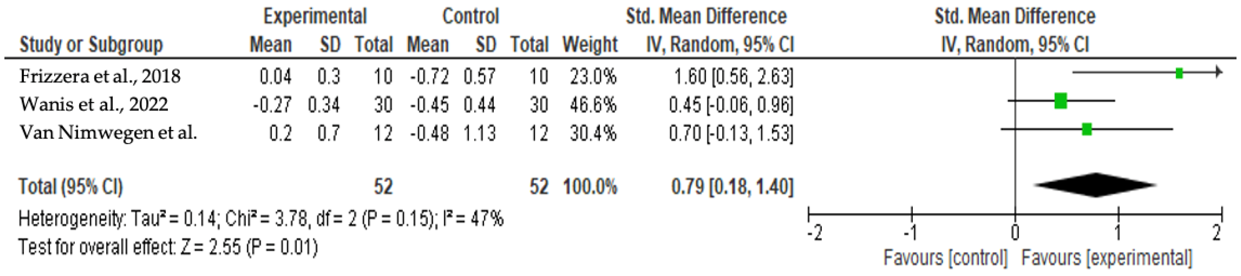
3.3.3. Xenograft with Autogenous Graft and Connective Tissue Graft (CTG) versus Xenograft with Autogenous Graft (Van Nimwegen et al., 2018 [28])

The authors reported the failure of two implants (one per group), with a 1-year survival rate of 96.7%. Tissue volume loss was found in both groups, but no significant difference was identified between the two groups. However, a statistically significant difference was identified at the buccal mucosa level at 1 year in favor of the xenograft + autograft + CTG treated group. The group with the soft tissue graft presented a mean increase of +0.20 ± 0.70 mm compared with a mean loss of −0.48 ± 1.13 mm in the group treated without using the CTG. This may be translated to a nonsignificant difference in terms of soft tissue volume reduction in the post-extraction phase but to greater tissue stability when using a connective tissue membrane. No significant difference was found between the two groups in total PES, PD, and Plaque Index at 1-year follow-up.

3.3.4. Xenograft versus Xenograft with Collagen Matrix versus Xenograft with Autogenous CTG (Frizzera et al., 2018 [29])

The authors reported a 100% success rate of the inserted implants. The total PES found no significant differences between the groups at 6 months and 1 year. Nevertheless, they stated that one of the PES scores, the alveolar process, was significantly better in the group treated without a membrane or CTG. In contrast, the PES score regarding gingival recession favors the group treated with graft and CTG. The authors found no significant differences in bone thickness and bone resorption between the three groups at 6 months and 1 year after implant insertion.

3.4. Clinical Outcomes

3.4.1. Mid-Facial Mucosa Level at 12 Months

Gingival recession at 1 year was calculated in the work of Frizzera et al. [29], Van Nimwegen et al. [28], and Wanis et al. [25]. In Frizzera et al.’s [29] and Van Nimwegen et al.’s [28] studies, the authors found better results when a connective tissue membrane is combined with the xenogeneic graft. Wanis et al.’s [25] study reported better results with a combination of the dual-zone technique with a xenogeneic graft.

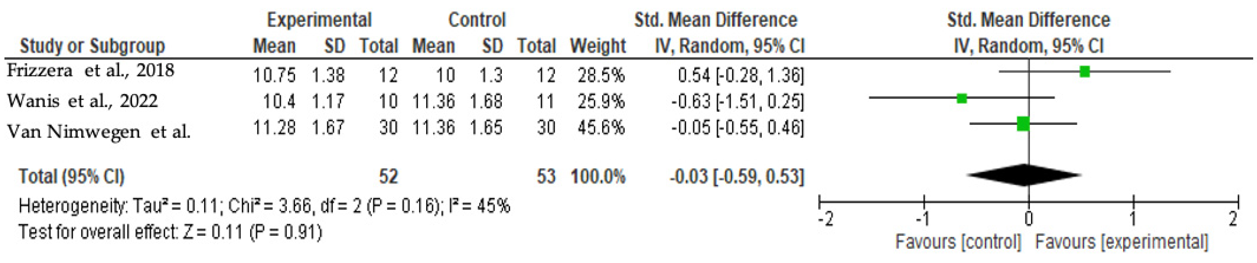
3.4.2. Total PES at 12 Months

Total PES at 1 year was considered by Frizzera et al. [29], Wanis et al. [25], and Van Nimwegen et al. [28]. The authors found no significant difference in using a xenogeneic graft with or without a membrane.

3.4.3. Facial Bone Thickness at 12 Months

Facial bone thickness was studied by Naji et al. [22] and Elaskary et al. [21]. The authors found better yields in the graft-treated and flapless groups.

3.5. Quality Assessment and Risk of Bias
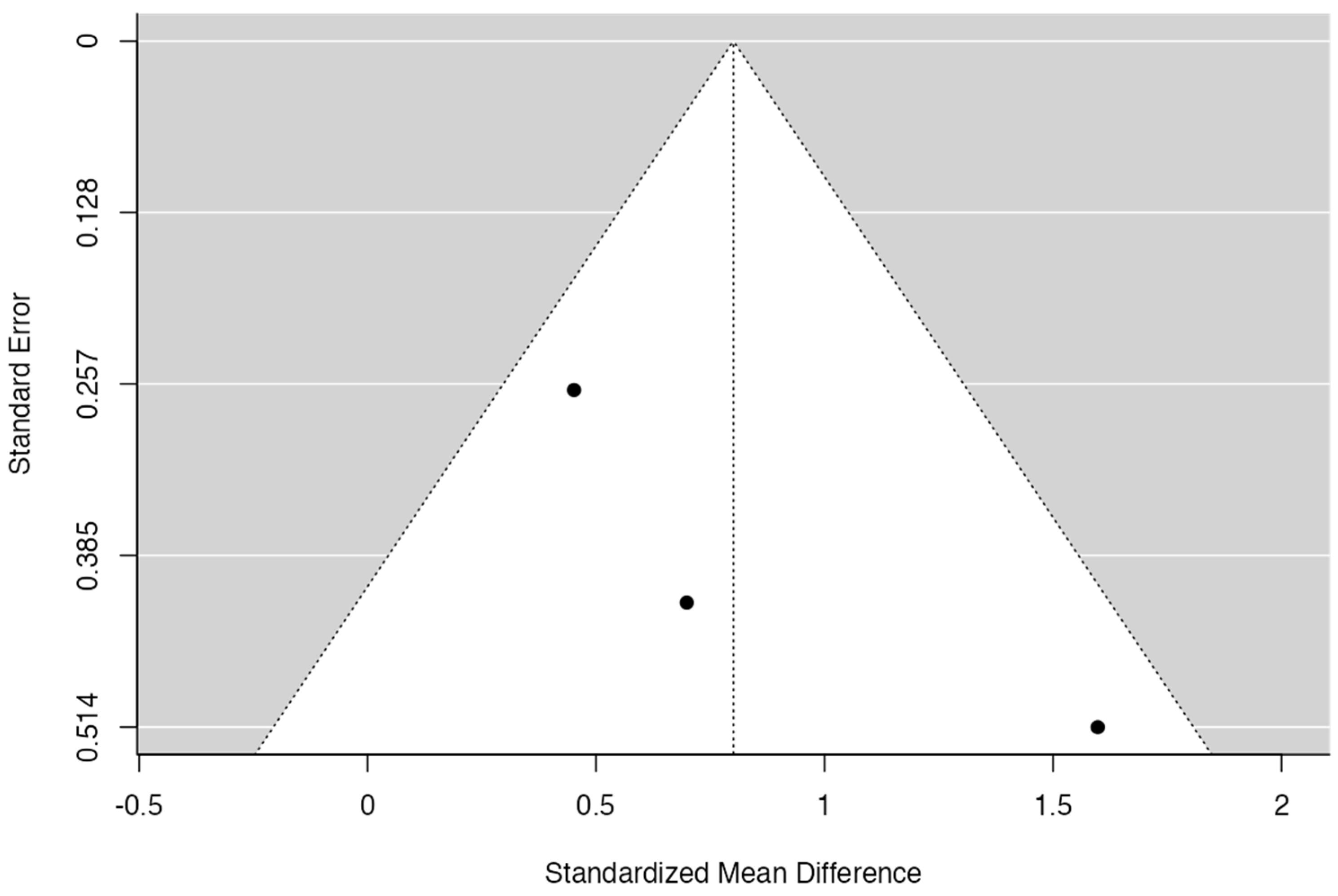
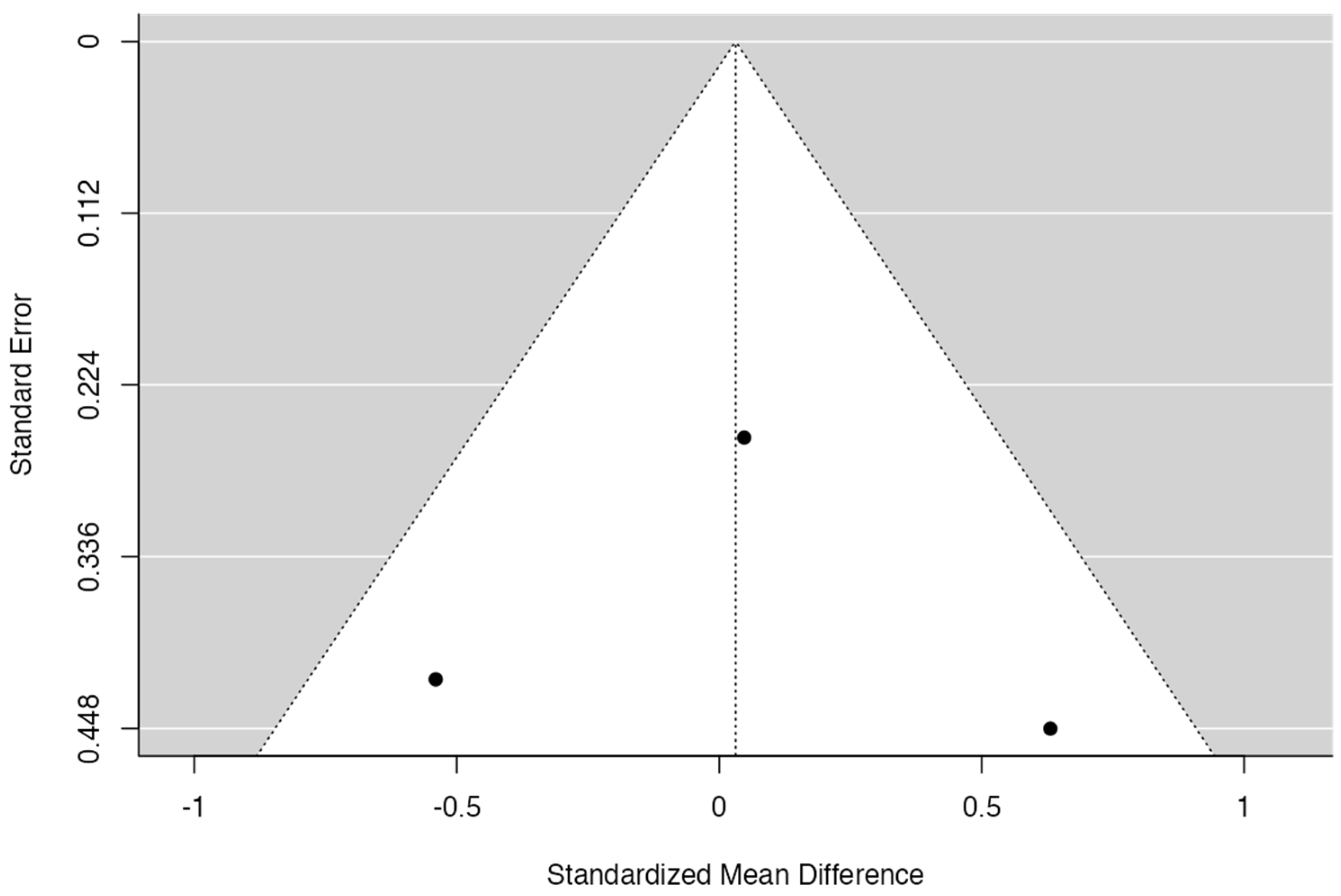
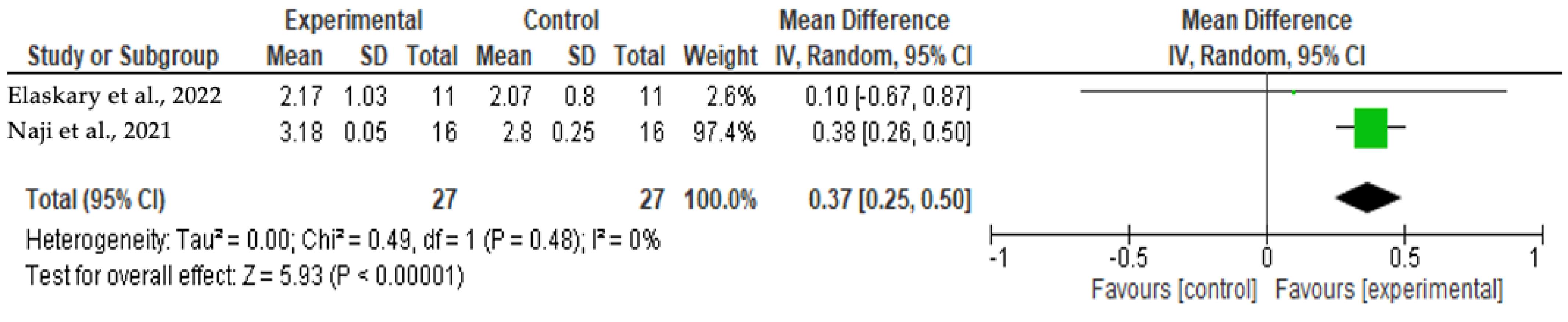
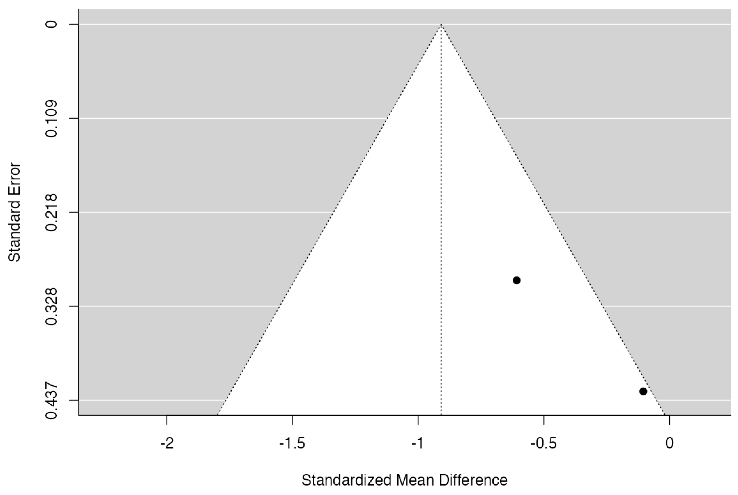
The risk of bias was evaluated by using a revised Cochrane risk-of-bias tool for randomized trials (RoB2). Four studies had low risk of bias, four had moderate risk of bias, and one had high risk of bias (Table 5).

3.6. Meta-Analysis

Given the results of Figure 2 and Figure 3, Cochran’s Q had a p-value of 0.15 and I2 = 46%; then, it was possible to verify a medium level of heterogeneity for the mid-facial mucosa level analysis. The forest plot shows that the meta-analysis effect of 0.79 (95% CI [0.18; 1.40]) was statistically significant (p = 0.01). There was a tendency to favor the experimental group in all articles included for this analysis.
Considering the results of Figure 4 and Figure 5, Cochran’s Q had a p-value of 0.16 and I2 = 45%; thus, it was possible to verify a medium level of heterogeneity among studies regarding total PES. The forest plot shows no significant differences between studies (p = 0.91), although Frizzera et al. [29] presented favoring toward the experimental group, and Wanis et al. [25] toward the control group.
Figure 6 and Figure 7 show homogeneity (I2 = 0%) among the facial bone thickness analysis studies. The Cochran’s Q analysis had a p-value of 0.48, presenting no statistical differences between the studies evaluated, with both favoring the experimental group. Naji et al.’s [22] study had a higher weight (97.4%) in this analysis. The forest plot shows that the meta-analysis effect of 0.37 (95% CI [0.25;0.50]) was statistically significant (p < 0.00001).

4. Discussion

The aim of this systematic study was to evaluate the efficacy of hard and/or soft tissue grafts associated with type-1 implants (IIP) on healing and treatment outcomes to provide a more predictable result. Our study included only RCTs that evaluated IIP with at least one group using hard and/or soft tissue grafts.
Buser et al. [9,30] identified some factors that may increase the risk of esthetic predictability in IIP cases: (1) thin gingival biotype, (2) thin buccal bone wall, and (3) implant surgical procedure by itself. Also, Buser et al. [30] showed a greater risk of having a 1 mm gingival recession and greater variability in esthetic results when using the immediate implant technique. This was also stated by Bakkali et al. [31], who argued that there is less esthetic predictability using this approach. On the other hand, Siqueira et al. [32] showed a case report of IIP combined with CTG, demineralized bovine bone mineral with 10% collagen, and immediate provisional crown adjusted intra- and extra-orally (establishing the ideal critical and subcritical contour), with high tissue stability after 4 years. Among the included studies, only two considered and used the CTG associated with IIP [28,29], with improved clinical results compared to the control group. Even though it has been shown that a membrane promotes bone neoformation by positively influencing bone remodeling as a scaffold between implant and bone [33], two studies [22,24] that considered the utilization of collagen membranes did not have significant results.
Borges et al. [34] pointed out that a buccal bone ridge thinner than 1 mm is the primary determinant of buccal tissue reduction at 1-year post-treatment with immediate dental implants. The authors showed that pretreatment of this anatomic condition is important in cases where the individual is also diagnosed with a thin gingival biotype [34]. Due to this concern (the presence of buccal bone ridge thinner than 1 mm), several authors have advocated using bone grafts, which could significantly reduce peri-implant tissue reduction [34,35,36]. Moreover, bone grafts contributed to horizontal bone preservation and soft tissue stability at the midfacial aspect of immediate implants, which should be considered as an adjunct to IIP in clinical practice [7]. Elaskary et al. [21] reported improved clinical yield regarding the buccal bone ridge when the post-extraction gap was treated with a graft. The experimental group, treated with a VST technique and a graft, presented a facial bone thickness of 2.95 ± 0.97 mm at the mid-level and 3.75 ± 1.30 mm at the apical level of the alveolar ridge, in contrast with the control group (without a graft) which presented values for the buccal bone of 1.82 ± 0.64 mm and 2.03 ± 0.81 mm, respectively. Therefore, the results must be carefully interpreted due to the limited number of studies included and the heterogeneity found.
It is essential to highlight that if the remnant post-extraction buccal gap size exceeds 1.5 mm, it might achieve incomplete bone regeneration if left to spontaneous heal [37]. This fact agrees with Naji et al.’s results [22], which indicated that the group with the flap technique and a graft had better buccal bone maintenance (buccal bone changes of −0.37 ± 0.09 mm) compared to the group with a flap without a graft (buccal bone changes of −0.91 ± 0.54 mm). The same authors also advocated that, if possible, applying the flapless technique can substantially improve the tissue healing performance compared to the use of a flap, preventing or reducing the buccal bone resorption; this can be explained by a reduction in the breaking of the local vascularization; the blood perfusion for the buccal bone ridge comes essentially from the periodontal ligaments, periosteum, and bone marrow [38]. In addition, they reported better results using a membrane than first intention closure, which agrees with a systematic review [31] that supported using bone grafts to reduce the buccal bone wall resorption after tooth extraction. However, the results presented in our study must be carefully interpreted because of the small sample size present in some of the included articles and the moderate/high risk of bias observed.
Clearly, esthetic factors are influenced by the presence or absence of a substantial buccal bone volume. Without a sufficient buccal bone plate, marginal bone resorption will result in volumetric alteration of the peri-implant soft tissue [39]. This theory entirely agrees with Guarnieri et al.’s [10] arguments, which include the believe that bone loss and the likelihood of soft tissue volume reduction are directly proportional: greater bone loss will likely cause a gingival recession. Fernandes and collaborators [40] stated that the predictability of these outcomes in IIP is related to correct patient selection criteria. However, they also argued that there is a lack of objectivity in the methods used in studies to evaluate esthetic outcomes, which often depend on the observer, reducing reproducibility among different observers and studies [40]. Wanis et al.’s study [25] agrees with these findings; they claimed that both groups revealed a PES value between 10 and 12, which is considered a good result. In contrast, Abd-Elrahman et al. [41], who observed similar groups to those within Wanis et al.’s [25] study, but without the use of bone grafts, reported a significantly lower PES at 6-month follow-up (8.85 ± 1.81). Wanis et al. [25] reported that using a surgical technique without a graft at the post-extraction buccal gap was responsible for the reduced total PES value; they stated that no real prevention of the buccal bone loss was observed in either of the groups at 6 and 12 months after implant insertion: −0.88 mm ± 0.41 in the dual-zone technique group and −1.08 mm ± 0.28 in the group with the graft alone.
The literature suggests some variability in terms of the mean MBL associated with type-1 implants with various surgical techniques and grafts: Siqueira et al. [42] reported a −0.66 ± 0.38 mm mean of marginal bone reduction while Pardal-Pelaez et al. [43] reported −0.42 ± 0.78 mm and Mazzocchi et al. [44] reported −0.48 ± 0.76 mm. These slight changes could be due to the different assessment techniques, which are unequal and operator-dependent [25]. Sanz et al. [45] had previously reported a significant reduction in MBL when IIP was associated with using a bone graft: −1.1 mm (graft group) compared to −1.6 mm in the non-graft group. However, several authors consider the intact buccal bone plate and gingival biotype beyond other factors, such as the flapless technique and the distance between implant shoulder and cortical bone, as determinants for the buccal bone healing at IIP sites, regardless of the presence of large gaps and the use of different grafts [46]. Furthermore, Elaskary et al. [21] argued that the first six months after surgery is the most critical period concerning post-extraction bone resorption. This is also supported by Borges et al. [34] and Lops et al. [47].
In addition, the literature has limitations regarding studies on compromised post-extraction sockets [11]. Most studies report positive data for IIP techniques in fresh and intact sockets [29,48,49] but strongly question such outcomes when the technique is used in damaged/compromised post-extraction ones [50,51,52,53]. Elaskary et al. [21] argued that when faced with such eventuality, the clinician should always opt for bone regeneration with a membrane in conjunction with immediate implantation to achieve better esthetic and functional results. These authors reported better results when the xenograft is combined with a CTG (from the patient’s palate). They also reported a higher PES score regarding the level of marginal mucosal tissue [21]. This fact was also highlighted by Van Nimwegen et al. [28], who hypothesized the association between a xenograft and CTG might provide greater stability to peri-implant tissues, namely at the marginal mucosal level. Nevertheless, the authors concluded that using a CTG associated with a xenogeneic bone graft may not counteract the volumetric tissue changes after IIP. The data presented did not show any significant differences in terms of volumetric reduction of the soft tissue, but it had positive effects on peri-implant marginal mucosa stability one year after treatment: the test group presented an average increase of 0.20 ± 0.70 mm compared to an average loss of −0.48 ± 1.13 mm in the control group [28]. They also reported significantly higher marginal gingival PES results than those without connective tissue grafts, even though total PES values showed no significant statistical differences between the two groups. Then, the authors concluded that CTG should be associated with IIP to reduce the possibility of asymmetry between peri-implant soft tissue and adjacent teeth [28].
The limitations of this review can be assigned to different items. We included only articles published in English; given the relatively recent nature of these techniques, the literature is still sparse, and we only included nine RCTs, following the inclusion criteria initially established. In addition, one of the databases consulted had no search results. Also, for the selected articles, after careful independent analysis by the two independent researchers (EMR and TB), we found considerable variability in terms of the chosen variables included in the studies and evaluation techniques that were not always objective, repeatable, and comparable; some articles had a small sample size, the presence of heterogeneity was found, and moderate/high RoB was observed, which suggests a careful interpretation of the data obtained. These reasons explain why we were able to conduct a meta-analysis of a low number of variables, selecting a total of five articles, which were divided among three comparable parameters. Moreover, because of the variability of the evaluation techniques and variables, it was necessary to work on interpretation and equivalence to summarize the data into clear and universal parameters that could be used to group the highest number of publications.

5. Conclusions

Within the limitations of this study, it was possible to conclude that using bone and soft tissue grafting techniques associated with IIP, even though they are not fundamental, were a valuable resource to prevent significant tissue reduction, reaching greater bone stability and higher levels in the Pink Esthetic Score (PES) and Visual Analogue Score (VAS). Results also may depend on the professional’s surgical and clinical ability/experience. In addition, the use of CTG combined with a xenogeneic bone graft brought advantages to the mid-facial mucosa position around immediate implants. It is important to state that standardized clinical assessment techniques and objective criteria are needed for comparisons in future studies.

Author Contributions

Conceptualization, E.M.R. and T.B.; methodology, E.M.R., B.L.-A., M.S.P., G.V.O.F. and T.B.; software, E.M.R., B.L.-A., M.S.P. and T.B.; validation, E.M.R., B.L.-A., M.S.P. and T.B.; formal analysis, E.M.R., B.L.-A., M.S.P. and T.B.; investigation, E.M.R., B.L.-A., M.S.P., G.V.O.F. and T.B.; resources, E.M.R., B.L.-A., M.S.P., G.V.O.F. and T.B.; data curation, E.M.R., B.L.-A., M.S.P., G.V.O.F. and T.B.; writing-original draft preparation, E.M.R., B.L.-A., M.S.P., G.V.O.F. and T.B.; writing—review and editing, E.M.R., B.L.-A., M.S.P., G.V.O.F. and T.B.; visualization, E.M.R., B.L.-A., M.S.P., G.V.O.F. and T.B.; supervision, T.B.; project administration, E.M.R., B.L.-A., M.S.P. and T.B. All authors have read and agreed to the published version of the manuscript.

Funding

This research received no external funding.

Institutional Review Board Statement

Not applicable.

Informed Consent Statement

Not applicable.

Data Availability Statement

All data obtained were included in this article.

Conflicts of Interest

The authors declare no conflicts of interest.

References

  1. Gasperini, F.M.; Fernandes, G.V.O.; Mitri, F.F.; Calasans-Maia, M.D.; Mavropoulos, E.; Rossi, A.M.; Granjeiro, J.M. Histomorphometric evaluation, SEM, and synchrotron analysis of the biological response of biodegradable and ceramic hydroxyapatite-based grafts: From the synthesis to the bed application. Biomed. Mater. 2023, 18, 065023. [Google Scholar] [CrossRef]
  2. Bonato, R.S.; Fernandes, G.V.d.O.; Calasans-Maia, M.D.; Mello, A.; Rossi, A.M.; Carreira, A.C.O.; Sogayar, M.C.; Granjeiro, J.M. The Influence of rhBMP-7 Associated with Nanometric Hydroxyapatite Coatings Titanium Implant on the Osseointegration: A Pre-Clinical Study. Polymers 2022, 14, 4030. [Google Scholar] [CrossRef]
  3. Borges, H.; Correia, A.R.M.; Castilho, R.M.; Fernandes, G.V.O. Zirconia implants and marginal bone loss: A systematic review and meta-analysis of clinical studies. Int. J. Oral Maxillofac. Implants 2020, 35, 707–720. [Google Scholar] [CrossRef]
  4. Kan, J.Y.; Rungcharassaeng, K. Immediate placement and provisionalization of maxillary anterior single implants: A surgical and prosthodontic rationale. Pract. Periodontics Aesthet. Dent. 2000, 12, 817–824. [Google Scholar]
  5. Pitman, J.; Seyssens, L.; Christiaens, V.; Cosyn, J. Immediate implant placement with or without immediate provisionalization: A systematic review and meta-analysis. J. Clin. Periodontol. 2022, 49, 1012–1023. [Google Scholar] [CrossRef]
  6. Ragucci, G.M.; Elnayef, B.; Criado-Cámara, E.; Del Amo, F.S.-L.; Hernández-Alfaro, F. Immediate implant placement in molar extraction sockets: A systematic review and meta-analysis. Int. J. Implant Dent. 2020, 6, 40. [Google Scholar] [CrossRef]
  7. Seyssens, L.; Eeckhout, C.; Cosyn, J. Immediate implant placement with or without socket grafting: A systematic review and meta-analysis. Clin. Implant Dent. Relat. Res. 2022, 24, 339–351. [Google Scholar] [CrossRef]
  8. Pitman, J.; Christiaens, V.; Callens, J.; Glibert, M.; Seyssens, L.; Blanco, J.; Cosyn, J.J. Immediate implant placement with flap or flapless surgery: A systematic review and meta-analysis. J. Clin. Periodontol. 2023, 50, 755–764. [Google Scholar] [CrossRef]
  9. Buser, D.; Chappius, V.; Belser, U.C.; Chen, S. Implant placement post extraction in esthetic single tooth sites: When immediate, when early, when late? Periodontology 2000 2017, 73, 84–102. [Google Scholar] [CrossRef]
  10. Guarnieri, R.; Ceccherini, A.; Grande, M. Single-tooth replacement in the anterior maxilla by means of immediate implantation and early loading: Clinical and aesthetic results at 5 years. Clin. Imp. Dent. Relat. Res. 2015, 17, 314–326. [Google Scholar] [CrossRef]
  11. Martins, S.C.R.; da Costa Marques, M.; Gomes Vidal, M.; Tolentino, P.H.M.P.; Dinelli, R.G.; Fernandes, G.V.O.; Shibli, J.A. Is the facial bone wall critical to achieving esthetic outcomes in immediate implant placement with immediate restoration? A systematic review. Adv. Clin. Exp. Med, 2024; ahead of print. [Google Scholar] [CrossRef]
  12. Tarnow, D.P.; Magner, A.W.; Fletcher, P. The effect of the distance from the contact point to the crest of bone on the presence or absence of the interproximal dental papilla. J. Periodontol. 1992, 63, 995–996. [Google Scholar] [CrossRef]
  13. Hammerle, C.H.; Araujo, M.G.; Simion, M.; Osteology Consensus, G. Evidence-based knowledge on the biology and treatment of extraction sockets. Clin. Oral Implants Res. 2012, 23 (Suppl. S5), 80–82. [Google Scholar] [CrossRef]
  14. Elgali, I.; Omar, O.; Dahlin, C.; Thomsen, P. Guided bone regeneration: Materials and biological mechanisms revisited. Eur. J. Oral Sci. 2017, 125, 315–337. [Google Scholar] [CrossRef]
  15. Maffei, S.H.; Fernandes, G.V.O.; Fernandes, J.C.H.; Orth, C.; Joly, J.C. Clinical and histomorphometric soft tissue assessment comparing free gingival graft and a collagen matrix as alveolar-sealer materials: A randomized controlled pilot clinical trial. Quintessence Int. 2023, 54, 756–769. [Google Scholar] [CrossRef]
  16. Sanz, M.; Cecchinato, D.; Ferrus, J.; Salvi, G.E.; Ramseier, C.; Lang, N.P.; Lindhe, J. Implants placed in fresh extraction sockets in the maxilla: Clinical and radiographic outcomes from 3-year follow-up examination. Clin. Oral Implants Res. 2014, 25, 321–327. [Google Scholar] [CrossRef]
  17. Blanco, J.; Carral, C.; Argibay, O.; Linares, A. Implant placement in fresh extraction sockets. Periodontology 2000 2019, 79, 151–167. [Google Scholar] [CrossRef]
  18. Page, M.J.; Moher, D.; Bossuyt, P.M.; Boutron, I.; Hoffmann, T.C.; Mulrow, C.D.; Shamseer, L.; Tetzlaff, J.M.; Akl, E.A.; Brennan, S.E.; et al. PRISMA 2020 explanation and elaboration: Updated guidance and exemplars for reporting systematic reviews. BMJ 2021, 372, n160. [Google Scholar] [CrossRef]
  19. Page, M.J.; McKenzie, J.E.; Bossuyt, P.M.; Boutron, I.; Hoffmann, T.C.; Mulrow, C.D.; Shamseer, L.; Tetzlaff, J.M.; Akl, E.A.; Brennan, S.E.; et al. The PRISMA 2020 statement: An updated guideline for reporting systematic reviews. BMJ 2021, 372, n712021. [Google Scholar] [CrossRef]
  20. Methods for Risk of Bias 2 (RoB 2) Tool. Available online: https://methods.cochrane.org/risk-bias-2 (accessed on 3 January 2024).
  21. Elaskary, A.; Abdelrahman, H.; Elsabagh, H.H.; El-Kimary, G.I. Does grafting the jumping gap in immediately placed anterior implants using vestibular socket therapy influence the labial bone thickness? J. Oral Maxillofac. Surg. 2022, 80, 1398–1407. [Google Scholar] [CrossRef]
  22. Naji, B.M.; Abdelsameaa, S.S.; Alqutaibi, A.Y.; Said Ahmed, W.M. Immediate dental implant placement with a horizontal gap more than two millimeters: A randomized clinical trial. Int. J. Oral Maxillofac Surg. 2021, 50, 683–690. [Google Scholar] [CrossRef]
  23. Atef, M.; El Barbary, A.; El-D Dahrous, M.S.; Zahran, A.F. Comparison of the soft and hard peri-implant tissue dimensional changes around single immediate implants in the esthetic zone with socket shield technique versus using xenograft: A randomized controlled clinical trial. Clin. Implant Dent. Res. 2021, 23, 456–465. [Google Scholar] [CrossRef] [PubMed]
  24. Mastrangelo, F.; Gastaldi, G.; Vinci, R.; Troiano, G.; Tettamanti, L.; Gherlone, E.; Lo Muzio, L. Immediate Postextractive Implants With and Without Bone Graft: 3-year Follow-up Results From a Multicenter Controlled Randomized Trial. Imp. Den. 2018, 27, 6. [Google Scholar] [CrossRef]
  25. Wanis, R.W.; Hosny, M.M.; El Nahass, H. Clinical evaluation of the buccal aspect around immediate implant using dual zone therapeutic concept versus buccal gap fill to bone level: A randomized controlled clinical trial. Clin. Implant Dent. Res. 2022, 24, 307–319. [Google Scholar] [CrossRef]
  26. Noelken, R.; Pausch, T.; Wagner, W.; Al-Nawas, B. Peri-implant defect grafting with autogenous bone or bone graft material in immediate implant placement in molar extraction sites 1 to 3-year results of a prospective randomized study. Clin. Oral Implants Res. 2020, 31, 1138–1148. [Google Scholar] [CrossRef] [PubMed]
  27. Li, P.; Zhu, H.C.; Huang, D.H. Autogenous DDM versus Bio-Oss granules in GBR for immediate implantation in periodontal postextraction sites: A prospective clinical study. Clin. Implant Dent Relat Res. 2018, 20, 923–928. [Google Scholar] [CrossRef] [PubMed]
  28. Van Nimwegen, W.; Raghoebar, G.M.; Zuiderveld, E.G.; Jung, R.E.; Meijer, H.J.A.; Mühlemann, S. Immediate placement and provisionalization of implants in the aesthetic zone with or without a connective tissue graft: A 1-year randomized controlled trial and volumetric study. Clin. Oral Impl Res. 2018, 29, 671–678. [Google Scholar] [CrossRef] [PubMed]
  29. Frizzera, F.; De Freitas, R.M.; Munoz-Chavez, O.F.; Cabral, G.; Marcantonio, E. Impact of soft tissue graft to reduce peri-implant alterations after immediate implant placement and provisionalization in compromised sockets. Int. J. Periodontics Restor. Dent. 2018, 39, 381–389. [Google Scholar] [CrossRef]
  30. Buser, D.; Martin, W.; Belser, U.C. Optimizing esthetics for implant restorations in the anterior maxilla: Anatomic and surgical considerations. Int. J. Oral Maxillofac. Implants 2004, 19, 43–61. [Google Scholar] [PubMed]
  31. Bakkali, S.; Rizo-Gorrita, M.; Romero-Ruiz, M.M.; Gutierrez-Perez, J.L.; Torres-Lagares, D.; Serrera-Figallo, M.A. Efficacy of different surgical techniques for peri-implant tissue preservation in immediate implant placement: A systematic review and meta-analysis. Clin. Oral Investig. 2021, 25, 1655–1675. [Google Scholar] [CrossRef]
  32. Siqueira, G.R.C.; Tavares, J.R.; Pedrosa, R.F.; Siqueira, R.A.C.; Fernandes, G.V.O. Immediate Implant with Provisionalization and Soft Tissue Grafting After 4-Year Follow-up. Clin. Adv. Periodontics 2022, 12, 32–38. [Google Scholar] [CrossRef]
  33. Tarnow, D.; Chu, S.; Gotta, S.; Saito, H. Flapless postextraction socket implant placement in the esthetic zone: Part 1. The effect of bone grafting and/or provisional restoration on facial–palatal ridge dimensional change—A retrospective cohort study. Int. J. Periodontics Restor. Dent. 2014, 34, 323–331. [Google Scholar] [CrossRef]
  34. Borges, T.; Fernandes, D.; Almeida, B.; Pereira, M.; Martins, D.; Azevedo, L.; Marques, T. Correlation between alveolar bone morphology and volumetric dimensional changes in immediate maxillary implant placement: A 1-year prospective cohort study. J. Periodontol. 2020, 91, 1167–1176. [Google Scholar] [CrossRef]
  35. Spray, J.R.; Black, C.G.; Morris, H.F.; Ochi, S. The influence of bone thickness on facial marginal bone response: Stage 1 placement through stage 2 uncovering. Ann. Periodontol. 2000, 5, 119–128. [Google Scholar] [CrossRef]
  36. Kazor, C.E.; Al-Shammari, K.; Sarment, D.P.; Misch, C.E.; Wang, H.L. Implant plastic surgery: A review and rationale. J. Oral Implantol. 2004, 30, 240–254. [Google Scholar] [CrossRef] [PubMed]
  37. Wilson, T.G.; Schenk, R.; Buser, D.; Cochran, D. Implants placed in immediate extraction sites: A report of histologic and histometric analyses of human biopsies. Int. J. Oral Maxillofac. Implants 1998, 13, 333–341. [Google Scholar] [PubMed]
  38. Chu, S.J.; Salama, M.A.; Garber, D.A.; Salama, H.; Sarnachiaro, G.O.; Sarnachiaro, E.; Gotta, S.L.; Reynolds, M.A.; Saito, H.; Tarnow, D.P. Flapless postextraction socket implant placement, part 2: The effects of bone grafting and provisional restoration on peri-implant soft tissue height and thickness—A retrospective study. Int. J. Periodontics Restor. Dent. 2015, 35, 803–809. [Google Scholar] [CrossRef] [PubMed]
  39. Chappuis, V.; Engel, O.; Reyes, M.; Shahim, K.; Nolte, L.P.; Buser, D. Ridge alterations post-extraction in the esthetic zone: A 3D analysis with CBCT. J. Dent. Res. 2013, 12, 195–201. [Google Scholar] [CrossRef] [PubMed]
  40. Fernandes, D.; Nunes, S.; Lopez-Castro, G.; Marques, T.; Montero, J.; Borges, T. Effect of customized healing abutments on the peri-implant linear and volumetric tissue changes at maxillary immediate implant sites: A 1-year prospective randomized clinical trial. Clin. Implant Dent. Relat. Res. 2021, 23, 745–757. [Google Scholar] [CrossRef] [PubMed]
  41. Abd-Elrahman, A.; Shaheen, M.; Askar, N.; Atef, M. Socket shield technique vs conventional immediate implant placement with immediate temporization. Randomized clinical trial. Clin. Implant Dent. Relat Res. 2020, 22, 602–611. [Google Scholar] [CrossRef]
  42. Siqueira, R.A.C.; Fontao, F.; Sartori, I.A.M.; Santos, P.G.F.; Bernardes, S.R.; Tiossi, R. Effect of different implant placement depths on crestal bone levels and soft tissue behavior: A randomized clinical trial. Clin. Oral Implants Res. 2017, 28, 1227–1233. [Google Scholar] [CrossRef]
  43. Pardal-Peláez, B.; Flores-Fraile, J.; Pardal-Refoyo, J.L.; Montero, J. Implant loss and crestal bone loss in immediate versus delayed load in edentulous mandibles: A systematic review and meta-analysis. J. Prosthet. Dent. 2021, 125, 437–444. [Google Scholar] [CrossRef]
  44. Mazzocco, F.; Jimenez, D.; Barallat, L.; Paniz, G.; Del Fabbro, M.; Nart, J. Bone volume changes after immediate implant placement with or without flap elevation. Clin. Oral Implants Res. 2017, 28, 495–501. [Google Scholar] [CrossRef] [PubMed]
  45. Sanz, M.; Lindhe, J.; Alcaraz, J.; Sanz-Sanchez, I.; Cecchinato, D. The effect of placing a bone replacement graft in the gap at immediately placed implants: A randomized clinical trial. Clin. Oral Implants Res. 2017, 28, 902–910. [Google Scholar] [CrossRef] [PubMed]
  46. Tarnow, D.P.; Chu, S.J. Human histologic verification of osseointegration of an immediate implant placed into a fresh extraction socket with excessive gap distance without primary flap closure, graft, or membrane: A case report. Int. J. Periodontics Restor. Dent. 2011, 31, 515–521. [Google Scholar]
  47. Lops, D.; Chiapasco, M.; Rossi, A.; Bressan, E.; Romeo, E. Incidence of inter-proximal papilla between a tooth and an adjacent immediate implant placed into a fresh extraction socket: 1-year prospective study. Clin. Oral Implant Res. 2008, 19, 1135–1140. [Google Scholar] [CrossRef] [PubMed]
  48. Crespi, R.; Capparè, P.; Gherlone, E. Immediate loading of dental implants placed in periodontally infected and non-infected sites: A 4-years follow-up clinical study. J. Periodontol. 2010, 81, 1140–1146. [Google Scholar] [CrossRef] [PubMed]
  49. Lin, G.H.; Chan, H.L.; Wang, H.L. Effects of currently available surgical and restorative interventions on reducing midfacial mucosal recession of immediately placed single-tooth implants: A systematic review. J. Periodontol. 2014, 85, 92–102. [Google Scholar] [CrossRef] [PubMed]
  50. Braut, V.; Bornstein, M.M.; Belser, U.; Buser, D. Thickness of the anterior maxillary facial bone wall: A retrospective radiographic study using cone-beam computed tomography. Int. J. Periodontics Restor. Dent. 2011, 31, 125–131. [Google Scholar]
  51. Noelken, R.; Neffe, B.A.; Kunkel, M.; Wagner, W. Maintenance of marginal bone support and soft tissue esthetics at immediately provisionalized OsseoSpeed implants placed into extraction sites: 2-year results. Clin. Oral Implants Res. 2014, 25, 214–220. [Google Scholar] [CrossRef]
  52. Amid, R.; Kadkhodazadeh, M.; Moscowchi, A. Immediate implant placement in compromised sockets: A systematic review and meta-analysis. J. Prosthet. Dent. 2023, 130, 307–317. [Google Scholar] [CrossRef]
  53. Zhao, L.; Hu, W.; Liu, Y.; Chung, K.-H. Evaluation of implant placement following ridge preservation in periodontally compromised molar extraction sockets: Three-year results of a prospective cohort study. Clin. Oral Implants Res. 2022, 33, 735–744. [Google Scholar] [CrossRef] [PubMed]
Figure 1. The flow diagram for the selection process is according to the PRISMA report (Preferred Reporting Items for Systematic Reviews and Meta-Analyses).
Jcm 13 00821 g001
Figure 2. Results of the meta-analysis for the mid-facial mucosa level (12 months) [25,28,29].
Jcm 13 00821 g002
Figure 3. Funnel plot for the mid-facial mucosa level (12 months).
Jcm 13 00821 g003
Figure 4. Results of meta-analysis for the total PES (12 months) [25,28,29].
Jcm 13 00821 g004
Figure 5. Funnel plot for the total PES (12 months).
Jcm 13 00821 g005
Figure 6. Results of meta-analysis for facial bone thickness (12 months) [21,22].
Jcm 13 00821 g006
Figure 7. Funnel plot for facial bone thickness (12 months).
Jcm 13 00821 g007
Table 1. Demographic information.
AuthorYearNAge RangeGender (Male/Female)
Elaskary et al. [21]202222Mean 45M: 8 F: 14
Group I (intervention): 11Group I: 44.63Group I: M: 5 (45.5%) F: 6 (54.5%)
Group II (control): 11Group II: 45.81Group II: M: 3 (27.3%) F: 8 (72.7%)
Naji et al. [22]20214828–55F: 30 M: 18
Group I (intervention): 16Group I: 40.2Group I: M: 5 (31.25%) F: 11 (68.75%)
Group II (control I): 16Group II: 43.3Group II: M: 7 (43.75%) F: 9 (56.25%)
Group III (control II): 16Group III: 41.1Group III: M: 6 (37.5%) F: 10 (62.5%)
Atef et al. [23]202142>18M: 25% F: 75%
Intervention Group: 21mean 36Test group: M: 5 (25%) F: 15 (75%)
Control group: 21Control Group: M: 5 (25%) F: 15 (75%)
Mastrangelo et al. [24]201810218–72M: 63 F: 39
Group A (intervention): 51Mean 44Group A: M: 31 (60.7%) F: 20 (39.2%)
Group B (control): 51Group B: M: 32 (62.7%) F: 19 (37.2%)
Wanis et al. [25]20222420–45M: 7 F: 17
DZ Group (intervention): 12DZ Group: 34.27DZ Group: M: 4 (36%) F: 7 (63.4%)
BCG Group (control): 12BCG Group: 30.30BCG Group: M: 3 (30%) F: 7 (70%)
Noelken et al. [26]20205023–73M: 18 F: 32
AB Group (control): 25Mean 47
BBGM Group (intervention): 25
Li et al. [27]20184020–60M: 24 F: 16
DDM Group (control): 20DDM Group: 36.6DDM Group: M: 11 (57.8%) F: 8 (42.10%)
BIO Group (intervention): 20BIO Group: 34.9BIO Group: M: 11 (43.75%) F: 8 (56.25%)
van Nimwegen et al. [28]20186019–82M: 28 F: 32
Intervention Group (CTG): 30Test Group: 19.5–67.84 (mean 45.5)Test Group: M: 13 (43.3%) F: 17 (56.5%)
Control Group: 30Control Group: 20.9–82.2 (mean 47.8)Control Group: M: 15 (50%) F: 15 (50%)
Frizzera et al. [29]20182423–65M: 7 F: 17
CTL Group (control): 8
CM Group (Intervention I): 8
CTG Group (Intervention II): 8
M—male; F—female; AB—autogenous bone; BBGM—biphasic bone graft material; DDM—demineralized dentin matrix; BIO—Bio-Oss; CTL—control; CM—collagen matrix; CTG—connective tissue graft; DZ—dual-zone therapeutic concept; BCG—buccal bone crest with immediate temporization.
Table 2. Graft and implant information.
Author Follow-Up Intervals Type of Graft Implant Placement (Site) Implant (n)Outcome
Elaskary et al. [21] 1 yearT0: baseline preextraction
T1: 1 year
Group I: particulate bone graft 75% autogenous bone chips harvested form local surgical sites and 25% deproteinized bovine bone mineral (DBBM) of bovine origin (MinerOss X Cortical Particle Size, 500–1000 microns) (Biohorizons Implant Systems, Birmingham, Alabama, USA)

GROUP II: no graft
Esthetic zone22Buccal bone thickness
Naji et al. [22] 6 monthsT0: before extraction

T1: immediately after implant placement

T2: 6 months
GROUP I: alloplastic nanocrystalline calcium sulphate bone graft (Orthogen LLC, Springfield, New Jersey, USA) and an absorbable collagen membrane (Bioimplon GmbH, Gießen, Germany)

GROUP II–III: without graft and membrane
Upper premolar tooth52CBCT bone examination
Pain intensity
Atef et al. [23] 1 yearT0: casts before the extraction

T1: CBCT immediately post placement of implant

T2: CBCT after 6 months

T3: photos, casts and patients satisfaction 12 months
Test group: without graft + collagen plug;

Control group: with bovine cancellous xenograft (Tutobone, Tutogen Medical GmbH, Neunkirchen a. Brand, Germany) +
collagen plug
26: premolar tooth

16: upper incisors and canine area
42Peri-implant soft tissue
PES
Midfacial mucosa alteration
Change in the buccal bone I-C (vertical)
Change in the buccal bone I-OS (horizontal)
Patient satisfaction
Mastrangelo et al. [24] 3 yearsRadiographic and clinical periodontal assessment
T0: 3 months
T1: 1 year
T2: 3 years
Group A: granular bone grafting was inserted (BioOss, Geistlich, Germany), which completely covered the pericardium membrane (Osteobiol Evolution, Tecnoss, Italy)

Group B: no graft and barrier
Upper premolar:
36: 1.4
26: 1.5
30: 2.4
23: 2.5
115Implants failure
Marginal bone loss
PES
Pocket depth
Biological complications
Wanis et al. [25] 1 yearT0: baseline

T1: 6 months

T2: 1 year
DZ Group—BCG Group: bone graft cortico-cancellous collagenated bone grafting material of porcine origin pre-hydrated and collagenated cortico-cancellous porcine bone, 250–1000 μm, Gen-Os® (Osteobiol, Technoss Dental S.r.l.)

DZ Group: dual technique zone

BCG Group: flapless technique
6: upper central
5: upper lateral
1: canines
5: first premolar
4: second premolar
24PES
BBL: Buccal bone changes (horizontal) via probe
MFR: The midfacial recession
STT: The soft tissue thickness at 2–4–6 mm
KTW: The keratinized tissue
VAS for POS
PS
Noelken et al. [26] 3 yearsT0: baseline

T1: placement implant

T2: 1 year (n = 8 implants) T3: 2 years (n = 16 implants) T4: 3 years (n = 24 implants)
AB Group: autogenous bone grafts were harvested at the mandibular ramus by collecting bone particles with a disposable bone scraper (Micross, META).

BBGM Group: a resorbable, biphasic, and anorganic graft material of plant origin derived from red algae (BBGM) (Symbios, Dentsply Sirona).
Molar of the maxilla and the mandible

34: mandibular implants

16: maxillary implants
50Implant survival rate
Marginal bone level changes
Buccal bone level
Buccal width of the alveolar crest
Pocket depths
Implant success rate
Plaque index
BoP
Li et al. [27] 18 monthsRadiographic
T0: baseline
T1: 6 months
T2: 18 months
DDM Group: autogenous
DDM granules from the extracted tooth

BIO Group: Bio-Oss (Geistlich Pharma AG, Wolhusen, Switzerland) cancellous granules
Lower premolar: 19
Lower molar: 25
45ISQ
Measurements of marginal bone resorption
Van Nimwegen et al. [28] 1 yearT0: preextraction clinical parameters, photos, and impression

T1: 1-year, clinical parameters, photos, and impression
Test and Control Group: a 1:1 mixture of autogenous bone and anorganic bovine bone (Geistlich Bio-Oss®; Geistlich Pharma AG, Wolhusen, Switzerland)

Test Group received connective tissue graft (CTG), which was harvested from the tuberosity region
Maxilla Incisor: 47
Maxilla Canine: 10
Maxilla Premolar: 3
60Volumetric change: thickness
Midfacial mucosa recession
Gingival biotype
Implant probing depths
Plaque scores
Bleeding scores
Mucosa inflammation
PES
Patient satisfaction: VAS
Frizzera et al. [29] 12 monthsT0: baseline
T1: 6 months
T2: 12 months
CTL Group: no soft tissue graft
CM Group: graft of collagen matrix (Mucograft, Geistlich)
CTG Group: CTG from palate

The facial space was filled with bovine bone mineral containing 10% porcine collagen (Bio-Oss Collagen, Geistlich) placed between the membrane and the dental implant
13: 1.1/2.1
11: 1.2/2.2
24MPR
Implant success rate
Papilla migration
PES
Soft tissue thickness
Bone thickness
CBCT—Cone Beam Computed Tomography; CTL—control; CM—collagen matrix; CTG—connective tissue graft; BIO—Bio-Oss; AB—autogenous bone; BBGM—biphasic bone graft material; DDM—demineralized dentin; DZ—dual-zone therapeutic concept; BCG—buccal bone crest with immediate temporization; PES—Pink Esthetics Score; MPR—marginal peri-implant recession; VAS—visual analogue scale; ISQ—implant stability quotient; BoP—bleeding on probing; PS—patient satisfaction; POS—postoperative swelling.
Table 3. Surgical protocol.
AuthorSurgical Protocol
Elaskary et al. [21]Atraumatic tooth extraction and the VST protocol. Then, a cortical membrane shield was made of heterologous origin and introduced through the tunnel apically.
Group I: using the graft
Group II: not using the graft.
Naji et al. [22]For group I and II a full thickness flap.
The junction gap was filled.
Group II was treated without bone graft or membrane.
Group III healing was free.
Atef et al. [23]Test Group: the socket shield technique.
Control Group: atraumatic extraction following implant placement; the junction gap was filled with bovine cancellous xenograft.
A piece of a collagen plug was placed to close the entrance of the extraction socket in both groups.
Mastrangelo et al. [24]Tooth extraction with mucoperiosteal flap. The immediate implant was inserted.
Group A: graft and barrier healing.
Group B: no graft and barrier.
Wanis et al. [25]A flapless minimally traumatic extraction technique. The immediate implants were inserted.
DZ Group: the bone graft filled the junction gap to wall up to the free gingival margin.
BCG Group: the bone graft filled the junction gap; the graft was packed just reaching the buccal bone crestal level.
Noelken et al. [26]Atraumatic flapeless extraction technique. The immediate implants were inserted.
The junction gap was filled with AB or BBGM graft.
The graft was additionally covered with a platelet-rich fibrin (PRF) membrane.
Li et al. [27]Tooth extraction with a mucoperiosteal flap. Immediate implant was inserted.
The junction gap was filled with a graft and injectable PRF and membrane barrier for healing.
Van Nimwegen et al. [28]Atraumatic flapless extraction technique. The junction gap was filled with xenograft inorganic bovine before the insertion of the immediate implant. In the test group, a connective autogenous graft was utilized.
Frizzera et al. [29]Atraumatic tooth extraction and implant placement with immediate loading of a provisional crown. A bovine graft was utilized in every group.
CTL Group: no soft tissue graft.
CM Group: graft of collagen matrix.
CTG Group: tissue autogenous graft from the palate
CTL—control; CM—collagen matrix; CTG—connective tissue graft; AB—autogenous bone; BBGM—biphasic bone graft material; DZ—dual-zone therapeutic concept; BCG—buccal bone crest with immediate temporization; VST—Vestibular Socket Therapy; PRF—platelet-rich fibrin.
Table 4. Clinical outcomes of selected studies.
AuthorOutcome
Elaskary et al. [21]Comparison of the overall bone thickness:
Baseline
Group I: 1.45 ± 0.92 mmGroup II: 0.79 ± 0.49 mm
12 months
Group I: 2.95 ± 0.97 mmGroup II: 1.98 ± 0.56 mm
Naji et al. [22]CBCT Bone examinations
Mean value of the buccal bone plate thickness + horizontal gap width at T1 was:
Group I:Group II:Group III:
T1: 3.56 ± 0.10 mmT1: 3.71 ± 0.57 mmT1: 3.43 ± 0.33 mm
T2: 3.18 ± 0.05 mmT2: 2.80 ± 0.25 mmT2: 3.19 ± 0.28 mm
T2–T1 = −0.37 ± 0.09 mmT2–T1 = −0.91 ± 0.54 mmT2–T1 = −0.24 ± 0.11 mm
PAIN INTENSITY
Group I:Group II:Group III:
5.14 ± 0.693.71 ± 0.760.71 ± 0.49
Atef et al. [23]Mid-facial mucosal alteration
Control group(xenograft)Test group(socket shield)
−0.466 ± 0.58 mm0.45 ± 0.75 mm
Radiographic outcomes
The change in the buccal(I-C):
Control groupTest group
1.71 ± 1.02 mm0.36 ± 0.62 mm
The change in the buccal(I-OS):
Control groupTest group
1.45 ± 0.72 mm0.29 ± 0.34 mm
Patient satisfaction vas score(12 months):
Control groupTest group
9.25 (±0.70)9.37 (±0.80)
PES
Control groupTest group
11.86 ± 0.3512.12 ± 0.64
Mastrangelo et al. [24]Implants failure:
Group A: 1Group B: 1
Marginal bone level
T0–T2
Group A: −0.25 ± 0.362 mmGroup B: −0.28 ± 0.3 mm
PES
Group A: 8.14Group B: 9.7
Probing depth
T0–T2
Group A: 1.69 ± 1.34 mmGroup B: 1.4 ± 1.61 mm
Biological complications like fistulas, mucositis, and periimplantitis: 58 patients
Wanis et al. [25]Two implants failed osteointegration after 2 months post-surgery (one from each group).
PES
Baseline:6 months12 months:
DZ Group: 10.82 (±1.54)DZ Group: 11.09 (±1.58)DZ Group: 11.36 (±1.69)
BCG Group: 10.10 (±1.20)BCG Group: 10.40 (±1.17)BCG Group: 10.80 (±1.55)
BBL (at 0 mm):
6 months12 months:
DZ Group: 0.67 (±0.43) mmDZ Group: 0.88 (±0.41) mm
BCG Group: 0.84 (±0.26) mmBCG Group: 1.08 (±0.28) mm
BBL (at 2 mm):
6 months12 months:
DZ Group: 0.59 (±0.32) mmDZ Group: 0.82 (±0.32) mm
BCG Group: 0.51 (±0.27) mmBCG Group: 0.79 (±0.30) mm
Noelken et al. [26]Implant survival rate
AB Group:BBGM Group:
100%96%
Mean interproximal bone level
AB Group T1:BBGM Group T1:
Min: −13.2 mmMin: −11.86 mm
Max: −2.19 mmMax: −3.80 mm
Mean: −7.36 mmMean: −7.6 mm
AB Group final:BBGM Group final:
Min: −0.87 mmMin: −1.83 mm
Max: −1.85 mmMax: 1.93 mm
Mean: 0.38 ± 0.78 mmMean: 0.1 ± 0.78 mm
Mean vertical distance from implant shoulder to the bottom of the buccal bone defect
AB Group T1:BBGM Group T1:
−7.18 ± 3.43 mmT1: −6.59 ± 2.65 mm
Li et al. [27]ISQ
DDM Group
T0:T1:T3:
53.6 ± 11.9 mm77.6 ± 7.979.5 ± 6.0 mm
BIO Group
T0:T1:T3:
54.1 ± 13.0 mm78.1 ± 4.280.2 ± 4.3 mm
Marginal bone resorption around implant
DDM Group
T1:T2:
1.7 ± 0.3 mm1.9 ± 0.6 mm
BIO Group
T1:T2:
1.8 ± 0.1 mm2.0 ± 0.5 mm
Van Nimwegen et al. [28]Volumetric change
  A. Thickness (T0–final):
Control group:Test group:
−0.49 ± 0.54 mm0.68 ± 0.59 mm
  B. Mid-facial mucosa (T0–final):
Control group:Test group:
−0.48 ± 1.13 mm0.20 ± 0.70 mm
PD at 1 year
Control group:Test group:
2.44 ± 1.19 mm2.28 ± 0.79 mm
PES
Control group:Test group:
11.36 ± 1.6511.28 ± 1.67
Frizzera et al. [29]PES
Baseline:12 months:
(CTL Group) 10.75 (±2.05)mm(CTL Group) 9.87 (±1.64) mm
(CM Group) 10.63(±1.84) mm(CM Group) 10 (±1.3) mm
(CTG Group) 9.37(±1.9) mm(CTG Group) 10.75 (±1.38) mm
MP (mesial papilla migration)
6 months:12 months:
(CTL Group) 0.64 (±0.41) mm(CTL Group) 0.36 (±0.7) mm
(CM Group) 0.39 (±0.45) mm(CM Group) 0.41 (±0.47) mm
(CTG Group) 0.53(±0.28) mm(CTG Group) 0.56 (±0.57) mm
DP(distal papilla migration)
6 months:12 months:
(CTL Group) 0.69 (±0.62) mm(CTL Group) 0.74 (±0.68) mm
(CM Group) 0.64 (±0.80) mm(CM Group) 0.52 (±0.67) mm
(CTG Group) 0.44 (±0.79) mm(CTG Group) 0.47 (±0.53) mm
MPR(marginal peri-implant recession)
6 months:12 months:
(CTL Group) 0.41 (±0.40) mm(CTL Group) 0.72 (±0.57) mm
(CM Group) 0.14 (±0.37) mm(CM Group) 0.42 (±0.60) mm
(CTG Group) −0.41 (±0.75) mm(CTG Group) −0.04 (±0.3) mm
CBCT—Cone Beam Computed Tomography; CTL—control; CM—collagen matrix; CTG—connective tissue graft; BIO—Bio-Oss; AB—autogenous bone; BBGM—biphasic bone graft material; DDM—demineralized dentin; DZ—dual-zone therapeutic concept; BCG—buccal bone crest with immediate temporization; MPR—marginal peri-implant recession; BBL—buccal bone loss.
Table 5. Overall risk-of-bias assessment using the Cochrane risk-of-bias 2 (RoB2) tool.
ArticlesRandomization ProcessDeviations fromThe Intended InterventionsMissing Outcome DataMeasurement of the OutcomeSelection of the Reported ResultOverall
Naji et al. [22]Jcm 13 00821 i001Jcm 13 00821 i001Jcm 13 00821 i002Jcm 13 00821 i002Jcm 13 00821 i001Jcm 13 00821 i001
Atef er al. [23]Jcm 13 00821 i001Jcm 13 00821 i001Jcm 13 00821 i002Jcm 13 00821 i002Jcm 13 00821 i001Jcm 13 00821 i001
Wanis et al. [25]Jcm 13 00821 i001Jcm 13 00821 i001Jcm 13 00821 i003Jcm 13 00821 i002Jcm 13 00821 i001Jcm 13 00821 i002
Noelken et al. [26]Jcm 13 00821 i001Jcm 13 00821 i001Jcm 13 00821 i001Jcm 13 00821 i001Jcm 13 00821 i001Jcm 13 00821 i001
van Nimwegen et al. [28]Jcm 13 00821 i001Jcm 13 00821 i002Jcm 13 00821 i003Jcm 13 00821 i003Jcm 13 00821 i003Jcm 13 00821 i003
Elaskary et al. [21]Jcm 13 00821 i001Jcm 13 00821 i002Jcm 13 00821 i002Jcm 13 00821 i001Jcm 13 00821 i001Jcm 13 00821 i001
Mastrangelo et al. [24]Jcm 13 00821 i001Jcm 13 00821 i001Jcm 13 00821 i002Jcm 13 00821 i002Jcm 13 00821 i002Jcm 13 00821 i002
Li et al. [27]Jcm 13 00821 i001Jcm 13 00821 i001Jcm 13 00821 i002Jcm 13 00821 i002Jcm 13 00821 i002Jcm 13 00821 i002
Frizzera et al. [29]Jcm 13 00821 i001Jcm 13 00821 i001Jcm 13 00821 i003Jcm 13 00821 i001Jcm 13 00821 i002Jcm 13 00821 i002
Disclaimer/Publisher’s Note: The statements, opinions and data contained in all publications are solely those of the individual author(s) and contributor(s) and not of MDPI and/or the editor(s). MDPI and/or the editor(s) disclaim responsibility for any injury to people or property resulting from any ideas, methods, instructions or products referred to in the content.

