Abstract
To summarize the evidence on non-hemorrhagic adrenal infarction (NHAI) and identify questions arising in diagnosis and management, cases in the PubMed database were merged with our case series. A total of 92 publications were retrieved, 15 of which reported on NHAI during pregnancy. Including the four in our case series, 24 cases have been described so far. Severe, unremitting pain requiring opioid analgesia was the leading symptom, often combined with nausea and vomiting. Laboratory results were non-contributory in most cases. Diagnosis was established via MRI in nine cases (37.5%) and via CT in six (25%); nine patients (37.5%) underwent both investigations. Location was predominantly on the right side (n = 16, 66.7%). In addition to analgesia, anticoagulation with heparin was commenced in 18 cases (75%). When thrombophilia screening was performed, major thrombogenic polymorphisms were detected in six cases (33.3%). One woman developed signs of adrenal insufficiency. The reported perinatal outcome was unremarkable. Unilateral NHAI has emerged as a rare but important cause of severe abdominal pain in pregnancy. The threshold to perform an MRI in pregnant women with characteristic clinical findings should be low. To prevent fetal radiation exposure, diagnostic imaging via CT should be avoided. In addition to symptomatic treatment with opioid analgesia, initiation of anticoagulant treatment should be strongly considered.
1. Introduction
Abdominal pain during pregnancy can have various causes, and establishing the correct diagnosis is challenging for several reasons. First, symptoms may be caused by obstetric complications. Second, changes in the position of intra-abdominal organs make the clinical assessment difficult. Finally, pregnancy-associated changes of reference values for several laboratory tests need to be taken into consideration [1].
If diagnostic imaging is required, ultrasonography is the mode of choice. However, its application for deep-lying abdominal soft tissue structures is limited. Additionally, the expanding uterus reduces visibility and therefore the diagnostic yield.
In case ultrasonography fails to establish a diagnosis, CT (computed tomography) and MRI (magnetic resonance imaging) are available. Both methods are particularly suitable for the imaging of soft tissue structures within the abdomen and pelvis.
Low-dose abdominal CT is associated with fetal radiation of some 1.5–35 mGy; the corresponding dose for pelvic CT amounts to 10–50 mGy [2]. The exposure depends on the number and spacing of adjacent image sections. Dose and timing during pregnancy determine the effects of ionizing radiation on the embryo/fetus.
From conception to the early second trimester, the teratogenic effect of ionizing radiation is of concern. At this gestational age, the recommended maximum permissible radiation dose is 5 mSv [3]. Another issue of concern is the effect of ionizing radiation on the developing brain, particularly if exposure occurs between 8 and 15 weeks of gestation. The minimum dose which may contribute to the development of microcephaly and intellectual disability is estimated to be 60–310 mGy. Below a radiation exposure threshold of 50 mGy, fetal anomalies and growth restriction may not occur [2].
Last, the stochastic effects of ionizing radiation need to be considered. This pertains especially to carcinogenesis. Fetal radiation exposure of 10–20 mGy may increase the risk for the development of childhood cancer (hematologic malignancies in particular) 1.5- to 2-fold [3]. More recent investigations calculated a lower effect (excess risk of a 10 mGy fetal dose producing an excess risk of 1 in 1667 to 1 in 4545) [4]. Due to the inevitable fetal radiation exposition, CT examinations should be avoided during pregnancy whenever possible.
In case CT needs to be performed, the application of iodinated contrast media does not seem to have a negative impact on the developing fetus with respect to teratogenesis or mutagenesis. Likewise, negative effects on the fetal thyroid have not been detected [5] Compared to CT, data on exposure to MRI during pregnancy are reassuring. Concerns regarding teratogenesis and potential effects of heat and acoustics have been expressed, but deleterious effects have not been detected. This also applies to MRI examinations with ≤3 Tesla field strength [6].
MRI has therefore emerged as the diagnostic imaging procedure of choice during pregnancy if ultrasonographic investigations fail to establish a diagnosis [1,4,5]. However, this statement only applies for non-enhanced MRI. Gadolinium-based contrast agents are known to pass the placenta. Mutagenic effects are unlikely, but the persistence of dissociated-free gadolinium within the fetus may increase the risk of stillbirth and neonatal death as well as the development of rheumatologic, inflammatory, or infiltrative skin conditions [7]. In cases where MRI is performed during pregnancy, the application of gadolinium-based contrast agents should therefore be avoided.
Since the first publication of unilateral non-hemorrhagic adrenal infarction (NHAI) diagnosed via MRI [8], further reports have been published [8,9,10,11,12,13]. NHAI during pregnancy needs to be differentiated from an acute bilateral adrenal hemorrhage. The latter usually occurs in patients with severe infections, coagulopathy, or after physical trauma [14]. To date, epidemiologic data on NHAIs are lacking. Likewise, etiology and pathogenesis are unknown. The adrenal perfusion is characterized by a rich arterial supply and drainage by a singular central vein. Pregnancy-associated hypercoagulability and the effect of the expanding uterus on the intra- and retroperitoneal structures may contribute to the development of NHAI [9,10,11,15,16,17,18].
Stipulated by our own series of four cases of NHAI during pregnancy at our institution within the past three years, we performed a literature search to summarize the published evidence on NHAI and to identify and address questions arising in diagnosis and management.
2. Materials and Methods
Pregnant women who presented for care at our center, a level IV university hospital, between the years 2018 and 2023 in whom a diagnosis of NHAI was established were prospectively followed. Results of laboratory tests and other investigations were collected. We recorded details of the maternal treatment, the delivery, and newborn data.
A literature review was performed using PubMed database from PubMed inception (January 1996) through May 2023. The search was restricted to publications in English. The following search terms were applied: ((adrenal) AND (infarction) AND (pregnancy)). In addition, references from original papers were manually searched for relevant citations. The exposure for our review was NHAI in pregnancy. Inclusion criteria were an observational study design, and a report of the maternal and perinatal outcome. We excluded reviews, editorials, and letters without sufficient data. Studies describing adrenal hemorrhage were excluded.
The extracted information included author, publication year, number of women, obstetric and medical history, medication, symptoms, types of investigations and results, treatment, delivery, newborn outcome, and long-term outcome.
Standard methods of descriptive statistics (median, percentage) were applied.
3. Results
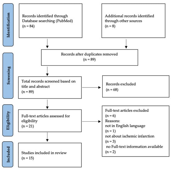
A total of 92 publications were retrieved, 15 of which reported on NHAI during pregnancy [8,9,10,11,12,13,15,16,19,20,21,22,23,24,25]. Only case reports and small case series were found. Figure 1 illustrates the identification, selection and exclusion process of our search. Including our own patients and excluding cases published twice [8,9,19,23,24,25], 24 cases are described [8,9,11,12,13,15,16,19,20,21,22,25].

Figure 1.
Flowchart: study identification, selection, and exclusions.
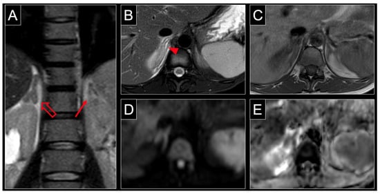
Symptoms, investigations, management and outcome are listed in Table 1, Table 2 and Table 3. Typical MRI-findings are depicted in Figure 2 (case no 1, Table 1, Table 2 and Table 3) [10]. The median age was 29 years (IQR 24–31). No patient had a history of thromboembolic or ischemic events. The median gestational age (GA) at the onset of symptoms was 30 weeks of gestation (IQR 28–33). Laboratory results were non-contributory in most cases. Mildly increased markers of inflammation (leucocytosis, elevated C-reactive protein) or ketonuria were reported in eight cases (35%) (n = 3, 13% respectively).

Table 1.
Overview: patient-related characteristics and history.

Table 2.
Overview: symptoms and diagnostic procedures.

Table 3.
Overview: treatment and outcome.

Figure 2.
MRI of a 31-year-old patient at 34 weeks of pregnancy with non-hemorrhagic infarction of the right adrenal gland. Note the characteristic enlargement of the right adrenal gland (hollow arrow) compared to the normal contralateral adrenal gland (solid arrow) in the coronal plane of a T2 weighted sequence (A). Axial T2 weighting with fat saturation shows central and surrounding hyperintensity (arrowheads) reflecting organ edema with inflammatory response of retroperitoneal fat (B). The lack of intraparenchymal hyperintensity (arrowhead) in T1 weighting (C) indicates the absence of acute hemorrhage. Similar to other body regions, organ infarction is associated with restricted diffusion, which manifests as hyperintensity at high b-values, such as 800 s/mm2 (D) and low values in the ADC map (E) of DWI. ADC = apparent diffusion coefficient, DWI = diffusion weighted imaging.
Severe, unremitting pain requiring opioid analgesia was the leading symptom. The type of analgesia was not mentioned in three patients; one woman received an epidural analgesia. Diagnosis was established via MRI in nine cases and via CT in six; nine patients underwent both investigations. Location was predominantly on the right side (n = 16, 66.7%). In two cases (8.3%). both adrenal glands were affected. In addition to analgesia, 18 cases (75%) received anticoagulation with heparin in various dosages. Thrombophilia screening was performed in 18 cases (75%); major thrombogenic polymorphisms were detected in six cases (33.3%) (Factor V Leiden Mutation n = 2, Lupus anticoagulant n = 1, Methylenetetrahydrofolate (MTHFR) mutation n = 3).
Screening for adrenal insufficiency was performed in 17 cases (70.8%), predominantly postpartum (n = 11, 64.7%). One woman developed signs of adrenal insufficiency and required substitution. Of the reported perinatal outcomes, all were unremarkable. The majority of women (n = 14, 58.3%) received postpartum anticoagulation, with heparin being the preferred drug.
4. Discussion
We summarized existing evidence on unilateral NHAI during pregnancy. We retrieved only 24 cases with our literature search. Therefore, it seems to be an extremely rare condition. The reluctance to perform diagnostic imaging during pregnancy, and abdominal CT in particular, along with the steady increase of publications with the advent of high-resolution MRI leads us to suggest that that NHAI has gone and still may go unnoticed in a number of cases. The fact that the course of the disease is characterized by improvement and even remission with symptomatic therapy further lends support to this assumption. The high number of cases diagnosed at our institution (n = 4, 16.7% of all published cases) may be a result of a low threshold to perform non-enhanced MRI in pregnant symptomatic women if ultrasound and laboratory tests fail to establish a diagnosis.
Increased diagnostic yield associated with the widespread utilization of MRI may allow for a more detailed understanding of the pathophysiology and help to establish an evidence-based treatment approach. The sequence of events resulting in NHAI is not yet fully elucidated [9,10,11,15,16,17,18]. Based on the peculiarity of the adrenal perfusion with a rich arterial supply and a singular central vein an initial venous thrombotic event—either microvascular or of the adrenal vein—is favoured. This initial thrombotic event may be followed either by hemorrhage during reperfusion or by bypass of the thrombosed vessel(s) without hemorrhage. A spasm of the cortical arteries resulting in ischemic necrosis is another suggested pathomechanism. Pregnancy-induced hypercoagulability increases the risk of venous thromboembolism and thrombophilic risk factors further increase that risk. An initial thrombotic event is therefore the favoured pathomechanism. Accordingly, thrombophilia screening was performed in the majority of cases, showing a positive screening result in 6 out of 18 patients (33.3%).
The majority of events occurred on the right side. However bilateral NHAI occurred in two cases (8.3%). Preference of the right side may be the consequence of the anatomic features of the right adrenal vein: compared to the left adrenal vein it is very short and thin and enters the inferior vena cava directly and from dorsolaterally, thus increasing the chance of venous stasis [26,27]. A higher pressure of the gravid uterus on the retroperitoneal vessels, particularly with advanced gestational age (GA) further increases the chance of venous stasis. The advanced GA at the onset of symptoms supports our assumption.
No other risk factors were reported. Larger numbers are required to identify specific risk factors.
Due to the assumption of an initial thrombotic event initiation of anticoagulation is consistent. Various dosage and durations were chosen. With the impending delivery where hemorrhage is a major concern this question requires further attention. Dosage and duration of anticoagulation and/or antiaggregation need to be analyzed, aiming to avoid both, over- and undertreatment.
Only one case of adrenal insufficiency was reported. The necessity of screening for adrenal insufficiency on a regular basis, particularly in asymptomatic individuals, needs to be called into question.
Limitations of our study result from the small number of reported cases which precludes the calculation of prevalence and incidence. Likewise, recommendations regarding treatment and follow-up are based on very limited evidence.
5. Conclusions
In conclusion, unilateral NHAI has emerged as a rare but important cause of severe abdominal pain in pregnancy. The threshold to perform MRI in pregnant women with characteristic clinical findings should be low. Diagnostic imaging by CT should be abandoned to avoid fetal radiation exposure. In addition to symptomatic treatment with opioid analgesia, initiation of anticoagulant treatment at a therapeutic dosage should be strongly considered.
More data are required to identify risk factors, determine adequate treatment, and decide on endocrine follow-up. For a better understanding of this excruciating condition an international registry may be beneficial.
Author Contributions
Conceptualization, N.T. and W.M.M.; methodology, N.T.; investigation, N.T.; data curation, N.T.; writing—original draft preparation, N.T.; writing—review and editing, N.T., W.M.M., B.P., U.G. and P.K.; visualization, N.T., P.K. and W.M.M.; supervision, W.M.M. All authors have read and agreed to the published version of the manuscript.
Funding
This research received no external funding.
Institutional Review Board Statement
Ethical review and approval was waived due to the retrospective nature of the study.
Informed Consent Statement
Patient consent was waived due to the retrospective nature of the study. Patients where not contacted, and data collection was within the scope of routine patient care.
Data Availability Statement
The data sets used and/or analyzed during the current study are available from the corresponding author on reasonable request.
Conflicts of Interest
The authors declare no conflict of interest.
References
- Abbassi-Ghanavati, M.; Greer, L.G.; Cunningham, F.G. Pregnancy and laboratory studies: A reference table for clinicians. Obstet. Gynecol. 2009, 114, 1326–1331. [Google Scholar] [CrossRef] [PubMed]
- Committee Opinion No. 723: Guidelines for Diagnostic Imaging During Pregnancy and Lactation. Obstet. Gynecol. 2017, 130, e210–e216. [CrossRef]
- Gjelsteen, A.C.; Ching, B.H.; Meyermann, M.W.; Prager, D.A.; Murphy, T.F.; Berkey, B.D.; Mitchell, L.A. CT, MRI, PET, PET/CT, and ultrasound in the evaluation of obstetric and gynecologic patients. Surg. Clin. N. Am. 2008, 88, 361–390.vii. [Google Scholar] [CrossRef] [PubMed]
- Lowe, S.A. Ionizing radiation for maternal medical indications. Prenat. Diagn. 2020, 40, 1150–1155. [Google Scholar] [CrossRef]
- American College of Radiology. ACR Manual On Contrast Media. 2023. Available online: https://www.acr.org/-/media/acr/files/clinical-resources/contrast_media.pdf (accessed on 30 June 2023).
- American College of Radiology. ACR Manual on MR Safety. 2020. Available online: https://www.acr.org/-/media/ACR/Files/Radiology-Safety/MR-Safety/Manual-on-MR-Safety.pdf (accessed on 30 June 2023).
- Ray, J.G.; Vermeulen, M.J.; Bharatha, A.; Montanera, W.J.; Park, A.L. Association Between MRI Exposure During Pregnancy and Fetal and Childhood Outcomes. Jama 2016, 316, 952–961. [Google Scholar] [CrossRef]
- Guenette, J.P.; Tatli, S. Nonhemorrhagic Adrenal Infarction With Magnetic Resonance Imaging Features During Pregnancy. Obstet. Gynecol. 2015, 126, 775–778. [Google Scholar] [CrossRef]
- Glomski, S.A.; Guenette, J.P.; Landman, W.; Tatli, S. Acute Nonhemorrhagic Adrenal Infarction in Pregnancy: 10-Year MRI Incidence and Patient Outcomes at a Single Institution. Am. J. Roentgenol. 2018, 210, 785–791. [Google Scholar] [CrossRef] [PubMed]
- Chagué, P.; Marchi, A.; Fechner, A.; Hindawi, G.; Tranchart, H.; Carrara, J.; Vivanti, A.J.; Rocher, L. Non-Hemorrhagic Adrenal Infarction during Pregnancy: The Diagnostic Imaging Keys. Tomography 2021, 7, 533–544. [Google Scholar] [CrossRef]
- Agarwal, K.A.; Soe, M.H. Cryptogenic adrenal infarction: A rare case of unilateral adrenal infarction in a pregnant woman. BMJ Case Rep. 2019, 12, e228795. [Google Scholar] [CrossRef]
- Reichman, O.; Keinan, A.; Weiss, Y.; Applbaum, Y.; Samueloff, A. Non-hemorrhagic adrenal infarct in pregnancy—A rare clinical condition diagnosed by non-contrast magnetic resonance image. Eur. J. Obstet. Gynecol. Reprod. Biol. 2016, 198, 173–174. [Google Scholar] [CrossRef] [PubMed]
- Molière, S.; Gaudineau, A.; Koch, A.; Leroi, T.; Roedlich, M.N.; Veillon, F. Usefulness of diffusion-weighted imaging for diagnosis of adrenal ischemia during pregnancy: A preliminary report. Emerg. Radiol. 2017, 24, 705–708. [Google Scholar] [CrossRef] [PubMed]
- Elhassan, Y.S.; Ronchi, C.L.; Wijewickrama, P.; Baldeweg, S.E. Approach to the Patient With Adrenal Hemorrhage. J. Clin. Endocrinol. Metab. 2023, 108, 995–1006. [Google Scholar] [CrossRef] [PubMed]
- Chasseloup, F.; Bourcigaux, N.; Christin-Maitre, S. Unilateral nonhaemorrhagic adrenal infarction as a cause of abdominal pain during pregnancy. Gynecol. Endocrinol. 2019, 35, 941–944. [Google Scholar] [CrossRef] [PubMed]
- Sormunen-Harju, H.; Sarvas, K.; Matikainen, N.; Sarvilinna, N.; Laitinen, E.K. Adrenal infarction in a healthy pregnant woman. Obstet. Med. 2016, 9, 90–92. [Google Scholar] [CrossRef]
- Fox, B. Venous infarction of the adrenal glands. J. Pathol. 1976, 119, 65–89. [Google Scholar] [CrossRef]
- Galatola, R.; Gambardella, M.; Mollica, C.; Calogero, A.; Magliulo, M.; Romeo, V.; Maurea, S.; Mainenti, P.P. Precocious ischemia preceding bilateral adrenal hemorrhage: A case report. Radiol. Case Rep. 2020, 15, 803–807. [Google Scholar] [CrossRef]
- Aljenaee, K.Y.; Ali, S.A.; Cheah, S.K.; Alroomi, M.J. Unilateral adrenal infarction in pregnancy secondary to elevated factor VIII. Saudi Med. J. 2017, 38, 654–656. [Google Scholar] [CrossRef]
- Green, P.A.; Ngai, I.M.; Lee, T.T.; Garry, D.J. Unilateral adrenal infarction in pregnancy. BMJ Case Rep. 2013, 2013, bcr2013009997. [Google Scholar] [CrossRef]
- Shah, N.; Deshmukh, H.; Akbar, M.; Malik, S.; Saeed, Y.; Allan, B. Unilateral adrenal infarction in pregnancy with associated acute hypoadrenalism and subsequent spontaneous biochemical and radiological resolution. Authorea 2021, 10, e05442. [Google Scholar] [CrossRef]
- Jerbaka, M.; Slaiby, T.; Farhat, Z.; Diab, Y.; Toufayli, N.; Rida, K.; Diab, T. Left flank pain during pregnancy with an unpredictable etiology: Think of nonhemorrhagic adrenal infarction. Future Sci. OA 2021, 7, Fso718. [Google Scholar] [CrossRef]
- Hynes, D.; Jabiev, A.; Catanzano, T. Nonhemorrhagic Adrenal Infarction in Pregnancy: Magnetic Resonance Imaging and Computed Tomography Evaluation. J. Comput. Assist. Tomogr. 2019, 43, 884–886. [Google Scholar] [CrossRef]
- Mathew, R.; Ali, A.; Sanders, K.; Flint, A.; Lamsal, S.; DeReus, H.; Cueno, M.; Jacob, R. Adrenal Infarction in Pregnancy Secondary to Elevated Plasma Factor VIII Activity. Cureus 2021, 13, e19491. [Google Scholar] [CrossRef] [PubMed]
- Warda, F.; Soule, E.; Gopireddy, D.; Gandhi, G.Y. Acute Unilateral Nonhemorrhagic Adrenal Infarction in Pregnancy. AACE Clin. Case Rep. 2021, 7, 228–229. [Google Scholar] [CrossRef]
- Cesmebasi, A.; Du Plessis, M.; Iannatuono, M.; Shah, S.; Tubbs, R.S.; Loukas, M. A review of the anatomy and clinical significance of adrenal veins. Clin. Anat. 2014, 27, 1253–1263. [Google Scholar] [CrossRef]
- Omura, K.; Ota, H.; Takahashi, Y.; Matsuura, T.; Seiji, K.; Arai, Y.; Morimoto, R.; Satoh, F.; Takase, K. Anatomical Variations of the Right Adrenal Vein: Concordance Between Multidetector Computed Tomography and Catheter Venography. Hypertension 2017, 69, 428–434. [Google Scholar] [CrossRef] [PubMed]
Disclaimer/Publisher’s Note: The statements, opinions and data contained in all publications are solely those of the individual author(s) and contributor(s) and not of MDPI and/or the editor(s). MDPI and/or the editor(s) disclaim responsibility for any injury to people or property resulting from any ideas, methods, instructions or products referred to in the content. |
© 2023 by the authors. Licensee MDPI, Basel, Switzerland. This article is an open access article distributed under the terms and conditions of the Creative Commons Attribution (CC BY) license (https://creativecommons.org/licenses/by/4.0/).