Case Report

Myeloid Sarcoma as a Presentation of Acute Myeloid Leukemia and Blastic Phase in the Course of Chronic Myeloid Leukemia: A Case Report and Literature Review

1
Department of Nephrology and Transplant Medicine, University Clinical Hospital in Wroclaw, 50-556 Wrocław, Poland
2
Faculty of Medicine, Wroclaw Medical University, 50-367 Wroclaw, Poland
3
Lower Silesian Centre for Oncology, Pulmonology and Hematology in Wrocław, 53-439 Wroclaw, Poland
4
Department of Nuclear Medicine and Endocrine Oncology, M. Sklodowska-Curie National Research Institute of Oncology Gliwice Branch, 44-102 Gliwice, Poland
*
Author to whom correspondence should be addressed.
J. Clin. Med. 2023, 12(14), 4845; https://doi.org/10.3390/jcm12144845
Submission received: 14 June 2023 / Revised: 10 July 2023 / Accepted: 17 July 2023 / Published: 23 July 2023
(This article belongs to the Section Hematology)

Abstract

:
Background: Myeloid sarcoma (MS), also known as granulocytic sarcoma or chloroma, is a rare disease entity characterized by the emergence of an extramedullary tumor, which may be antecedent, coexisting, or manifest secondarily to an ongoing malignancy of lymphoid origin. Owing to its low prevalence, scientific reports addressing this matter comprise mainly retrospective studies with a limited number of participants, rather low-quality research, and only few case reports. Despite MS’s rarity, the need for enhancing their diagnostic tools and refinement of their therapeutic regimens is broadly recognized among physicians. Case summary: In this case series, we present the clinical histories of two patients diagnosed with MS. The former (Case 1) exhibited MS of the sternum alongside chronic myeloid leukemia (CML), while in case of the latter (Case 2) MS presented as the initial manifestation of a current acute myeloid leukemia (AML). Treatment for both patients included chemotherapy (CHTH) and radiation (RT); however, patient 1 with CML died due to cardiorespiratory insufficiency secondary to an infection, while patient 2 is in clinical remission (CR) for 16 months since their MS diagnosis. Furthermore, a comprehensive analysis of previously reported cases was conducted which incorporated MS in patients with AML and CML. Conclusion: The objective of this report was to emphasize the heterogeneity among the clinical manifestations of MS, to underline the relevance of the histopathological and molecular diagnostic tools in opting for the appropriate therapy, and that, in spite of it occurring rather uncommonly, physicians should think of MS in the presence of pathological masses in patients under risk of hematological malignancies.

1. Introduction

According to the World Health Organization (WHO) classification of hematolymphoid tumors, MS presents as a distinctive tissue-based manifestation of AML, transformed myelodysplastic syndrome (MDS), myeloproliferative neoplasms (MPN) or MDS/myeloproliferative neoplasms (MPN) [1]. The majority of individuals with MPN are typically identified in the chronic phase (CP), during which there is a possibility of progressing onto the blast phase (BP). The BP is characterized by the presence of additional cytogenetic and/or molecular abnormalities. In view of the 5th edition of the WHO Classification of Hematolymphoid Tumors, MS can be interpreted as a manifestation of the BP in CML, as one of the diagnostic criteria of the BP is the presence of an abnormal proliferation of blasts outside the bone marrow [1]. Similarly, according to the latest International Consensus Classification of Myeloid Neoplasms and Acute Leukemias, one of the diagnostic criteria of the BP in CML is MS, which has been defined as an extramedullary blast proliferation. There have also been reports in which MS ensued without bone marrow (BM) involvement, in which case it is termed as isolated myeloid sarcoma [2]. Cases of newly occurring myeloid sarcoma should undergo a thorough investigation that includes cytogenetic and molecular analyses to permit accurate classification and effective treatment planning. Approximately 70% of patients exhibit concordant molecular alterations in both myeloid sarcoma and concurrent bone marrow disease, implying a potential origin from a shared hematopoietic stem cell or precursor [3,4]. In some individuals with a morphologically normal BM, relevant gene mutations can be still detected, indicating the presence of a low-level clonal myeloid disease or clonal hematopoiesis within the bone marrow [3,5]. It has been found that there is a prevalence of males over females in patients affected by MS, of whom approximately 9% are at risk of developing AML or CML [6]. Due to its rarity and lack of prospective and randomized controlled studies, data concerning the prognosis is conflicting, and there is no consensus on the treatment. In addition, MS is a source of significant diagnostic burden, which results in frequent misdiagnosis with various malignancies, such as Non-Hodgkin’s Lymphoma (NHL), small round cell tumors, or thymoma, which significantly delays treatment institution [7]. MS can be interpreted in various manners depending on its time of manifestation in regard to that of AML or CML. If it presents primarily or concomitantly, MS may be perceived as a manifestation of an ongoing hematological malignancy outside of the BM. However, if it is secondary, it could be seen a sign of disease progression and development of resistance to therapy. As for the applied regimen, it is worth mentioning in that it could undergo modifications over time due to the presence of mutations that may appear as the disease progresses. As a result, the initially administered medications become ineffective. Through an in-depth analysis of our cases along with previous studies concerning MS in patients with AML or CML, we sought to provide further insights into both the diagnostic and therapeutic approaches for the management of patients afflicted by MS, and emphasize the fundamental role played by molecular testing in opting for the adequate, most efficacious therapy. Moreover, we explored whether MS occurs rather primarily or secondarily to myeloid malignancies, the most frequently affected sites, and how its manifestation may influence our proceedings.

2. Materials and Methods
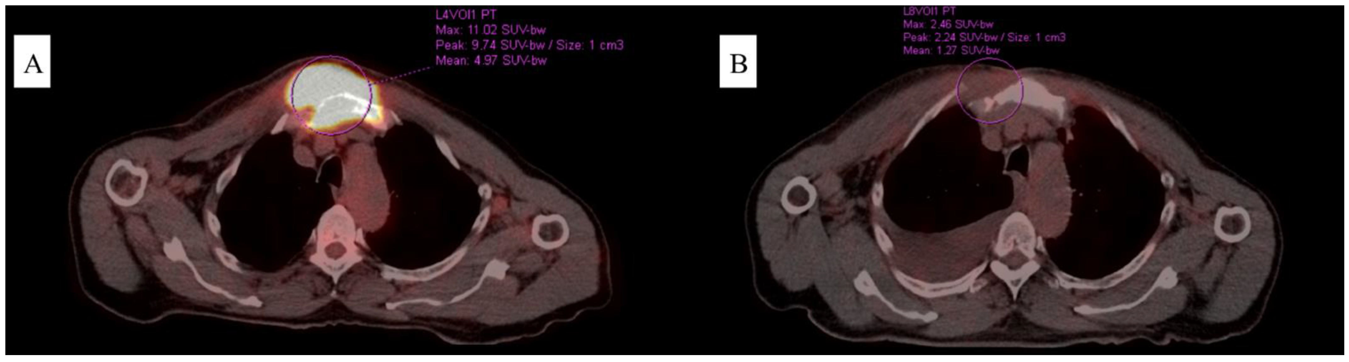
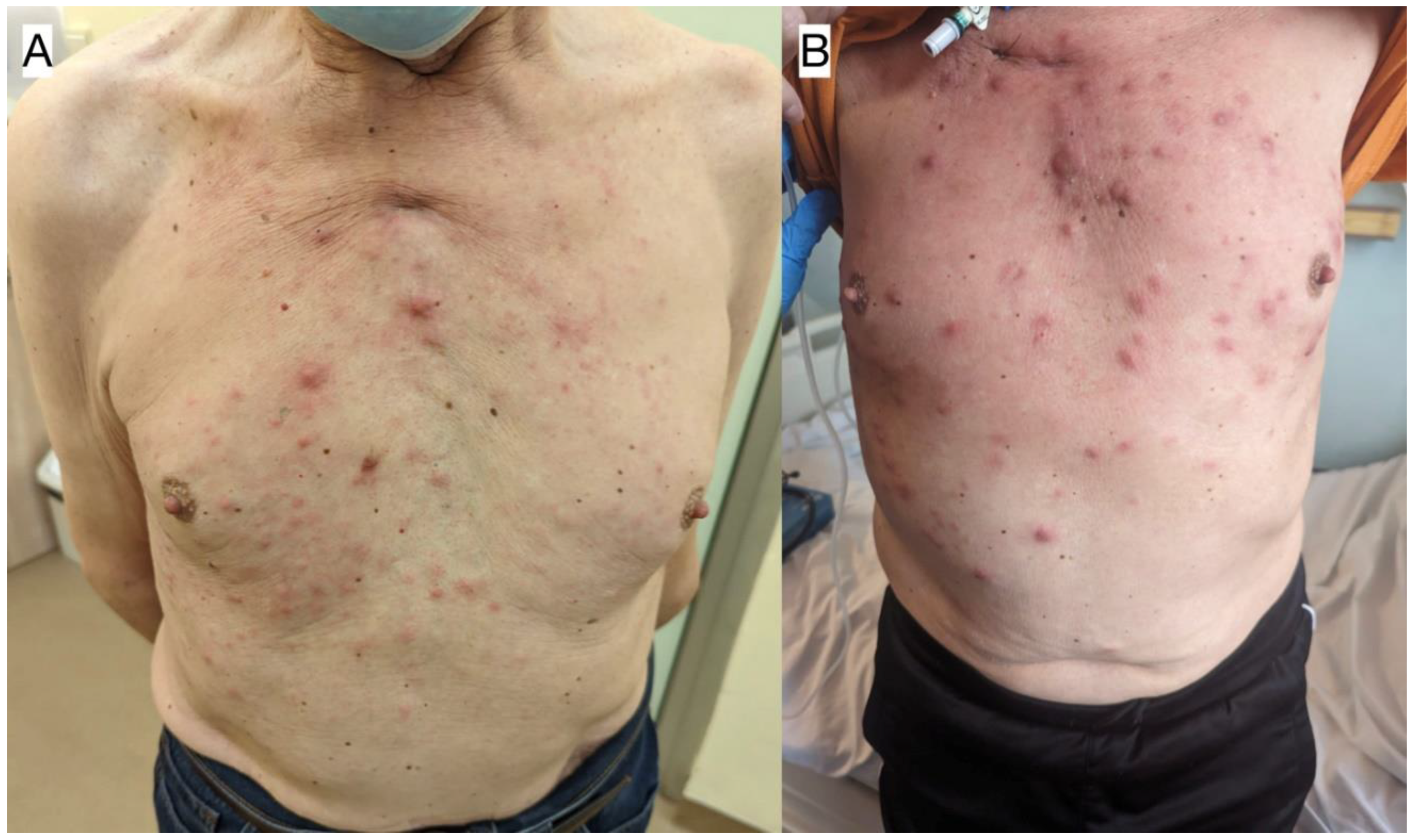
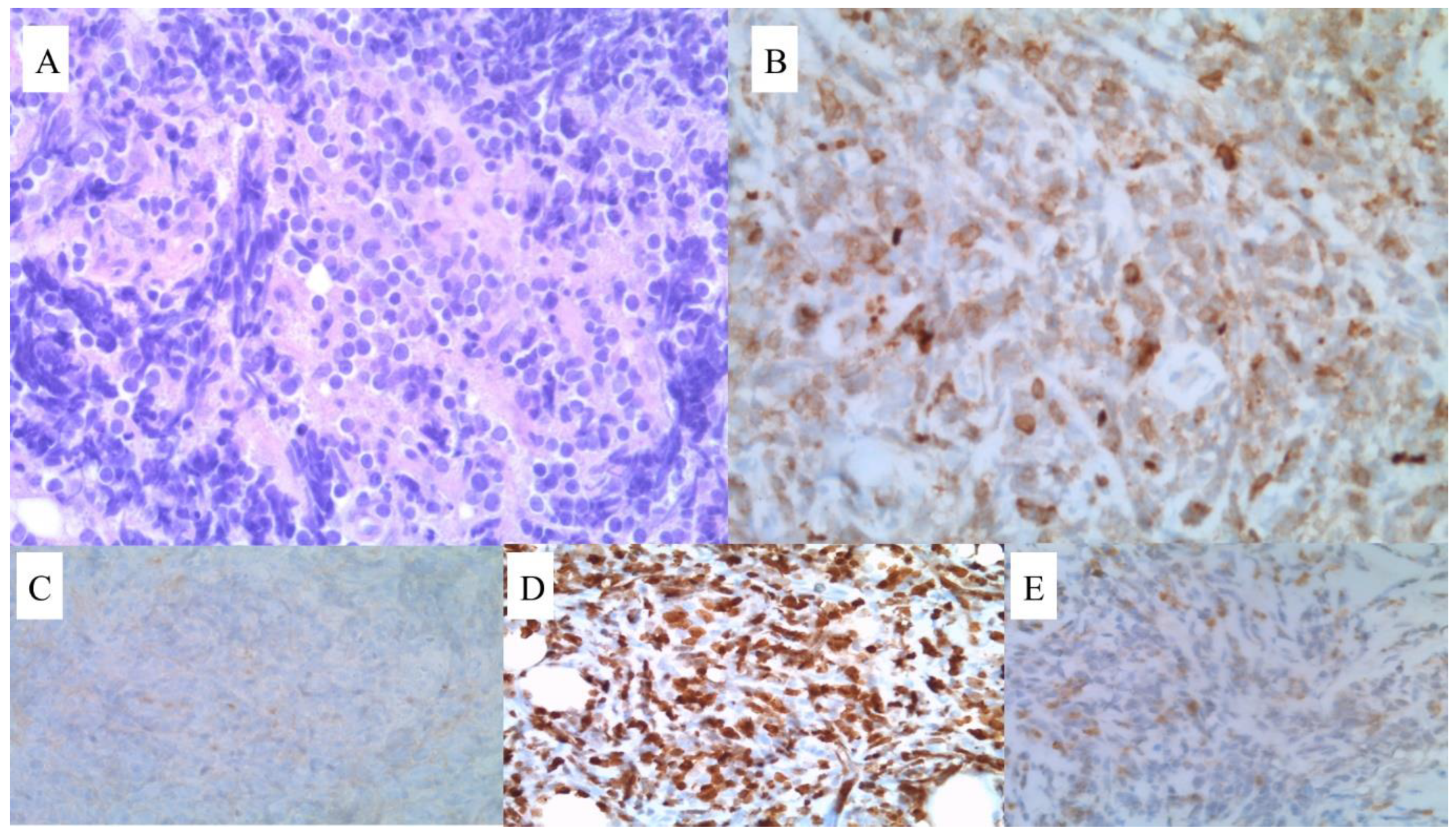
All patients provided informed consent for both the procedures itself and the publication of the resulting data. To gather the relevant information, an extensive literature search was conducted, encompassing publications up until June 2023. Various combinations of keywords, such as myeloid sarcoma, AML, CML, chloroma, and granulocytoma, were utilized. The search was implemented through the relevant scientific databases, including PubMed, UpToDate, and Scopus. As a result, 54 papers were specifically selected for inclusion in our manuscript, as they closely relate to our study. Articles written in the English language were included, while articles written in other languages were excluded.

3. Case 1

A 71-year-old man was admitted to our clinic to assess a growing mass in the area of the sternum, 6 months following their diagnosis of CML treated with imatinib in the standard dose of 400 mg/day (based on a medical interview, the patient had not brought documentation). A thorax computed tomography (CT) scan revealed a pathological mass located in the soft tissues on the right side of the chest. Furthermore, positron emission tomography/computed tomography (PET/CT) confirmed its presence, in addition to disclosing segmental lysis and the involvement of the manubrium of the sternum (Figure 1). Blood testing showed moderate normocytic anemia, lymphopenia, a low reticulocyte count, and high LDH and D-dimer levels (RBC 3.94 × 10⁶/µL, HGB 11.2 g/dL, HCT 34.4%, MCV 87.3 fL, MCH 28.4 pg, LYM 0.58 × 10³/µL, RET 0.22%, LDH 257 U/L, and D-dimer 730 ng/mL). In light of the clinical picture, a sample for pathology assessment was collected (Figure 2). Microscopic examinations revealed fibrous connective tissue covered by an infiltrate of cells with acidophilic cytoplasm and signs of angioinvasion. Immunohistochemical (IH) staining was positive for the following markers: CD45, CD34, CD117, CD15, MPO, Ki-67, and CK Pan, and was negative for CD3, CD20, PAX5, CD138, MUM1, S100, and CD30, respectively. BM biopsy excluded its involvement. Hence, a diagnosis of CML blast crisis in the form of MS was made. At the moment of MS diagnosis, BCR::ABL1 kinase domain sequencing revealed no mutations, which may have resulted in tyrosine kinase inhibitor (TKI) resistance. Upon receiving the results of RQ-PCR testing, which revealed b3a2 (p210) transcript levels of 5.891 (IS) corresponding to the absence of the major molecular response (MMR), the patient was subsequently deemed to be qualified for conformal radiotherapy (24 Gy in 12 fractions) in conjunction with dasatinib 140 mg/day.
After three months of dasatinib therapy, a major response to treatment was noticed both clinically, manifesting as a reduction in tumor volume, and molecularly, as p210 transcript levels dropped below the threshold of the deep molecular response (MR4). Nevertheless, the patient developed skin lesions (Figure 3A) and pleural effusion, which was confirmed with diagnostic imaging. Five months following therapy institution, BCR-ABL1 p210 transcripts reached a level of 1.45 (IS). Further scrutiny revealed the presence of the T315I (c.944C>T) mutation, thus explaining the cause of disease progression and the development of resistance to dasatinib. The patient was then qualified for being treated with ponatinib (45 mg/day), resulting in clinical and laboratory improvements, as one month into the new regimen, p210 transcript levels dropped, yet not below the MMR threshold. Due to a persistent presence of pleural effusion, flow cytometry upon sample collection was performed, showing the presence of blasts that were positive for CD15, CD65, CD13, CD34, CD117, HLA-DR, CD33, CD38, and CD7, and were negative for CD19 and MPO, respectively. Furthermore, 50% of the blast cells demonstrated altered signaling for the ABL1 and BCR genes, of which:
  • Forty-three percent had four fusion signals, one red signal for ABL1 (9q34), and one green for BCR (22q11);
  • Four percent had three fusion signals, one red signal for ABL1 (9q34), and one green for BCR (22q11);
  • Three percent had two fusion signals, one red signal for ABL1 (9q34), and one green for BCR (22q11).
In the context of the cytogenetic testing performed upon diagnosis, these results are proof of clonal evolution and disease progression. Correspondingly, a regimen consisting of DA 3 + 7 (daunorubicin 60 mg/m2 days 1–3, Ara-C 100 mg/m2 days 1–7) and ponatinib 45 mg/day was administered, leading to the patient achieving the MR4 once more with real-time quantitative polymerase chain reaction (RQ-PCR) testing along with a concurrent resolution of dyspnea, which was present at the moment of admission.
Two months later, disease progression was revealed using RQ-PCR, CT, BM biopsy, and the evolution of skin lesions (as displayed in Figure 3B). Histopathological analysis of the skin lesions confirmed the relapse of MS in this patient. The reticular layer of the skin showed a diffuse infiltrate composed of CD34+ blastic cells showing weak expression levels of CD117 and CD15, accompanied with numerous neutrophil granulocytes. Blastic cell IH staining revealed: CD34 (+), CD 117 (+/−), CD15 (+/−), Ki-67 (+++)—reaction in 60–80% of the tumor cells, MPO (−), CD10 (−), CD79a (−), and CD3 (−). BCR::ABL1 kinase domain sequencing ruled out the presence of mutations, including the T315I mutation. Despite a satisfying response having been achieved following the reinduction of chemotherapy following the FLAG-IDA protocol (fludarabine 30 mg/m2, Ara-C 2 g/m2 for 5 days, idarubicin 10 mg/m2 for 3 days, and G-CSF 5 µg/kg days 1–5 and until neutrophil recovery), the patient unfortunately died, having subsequently suffered from neutropenia and respiratory tract infection leading to cardiorespiratory insufficiency. The results of BCR-ABL1 kinase domain sequencing have been presented in Figure 4.

4. Case 2

A 50-year-old male patient arrived at our hospital’s Hematology Department with a yet uncertain diagnosis, owing to an ambiguous result of histopathological analysis of a mediastinal tumor sample. The first and only symptom was reported to be noticed a little over 1 month prior to the admission to our ward, and it consisted of an enlargement of the neck’s circumference. A thorax CT scan was performed by a different clinic, revealing a mass located in the anterior mediastinum compressing the superior vena cava. Subsequently, the patient was referred to our hospital’s Department of Thoracic Surgery with symptoms of superior vena cava syndrome, which were managed with dexamethasone. Then, tumor sampling was performed, whose first analysis resulted being inconclusive, and the patient was subsequently transferred to our department.
Laboratory testing revealed moderate normocytic anemia with elevated LDH and D-dimer levels (RBC 4.24 × 10⁶/µL, HGB 13.4 g/dL, HCT 39.3%, MCV 92.7 fL, MCH 31.6 pg, LDH 277 U/L, and D-dimer 1040 ng/mL, respectively). As for the following diagnostic measures, we opted for PET/CT and BM biopsy (Figure 5). The former confirmed the presence of a mass in the anterior mediastinum with no signs of liver, spleen, BM, or lymph node involvement, while the latter revealed two blast populations, of which the first was positive for Tdt+, CD34, CD33, CD123, CD38, CD7, HLA-DR, and 45dim, and was negative for CD117, while the second one resulted positive for Tdt, CD34, CD33, CD123, CD38, CD7, and 45dim, and was negative for HLA-DR and CD117, respectively. Further scrutiny comprising G-banded karyotyping (GTG) of the BM found that the trisomy of chromosome 21 was present in 45% [9/20] of the metaphase cells. In the remaining 55% [11/20] of cells, a normal male karyotype was identified. Fluorescence in situ hybridization (FISH) revealed a triple signal for the RUNX1 gene (21q22) in 40% [51/127] of the interphase cells, indicating the presence of an extra copy of this gene. The evaluation of the signals retrieved from the metaphase cells confirmed the trisomy of chromosome 21. The presence of mutations, such as NPM1, BCR-ABL, FLT3-ITD, IDH 1/2, and the PML-RARa fusion gene, was excluded. Cerebrospinal fluid examination ruled out central nervous system involvement. A revision of the formerly collected tumor sample proved that the patient suffered from MS, and IH staining confirmed the cooccurrence of the same process both in the BM and in the anterior mediastinum (Figure 6). According to the clinical picture, a diagnosis of MS as the primary manifestation of a coexisting AML was made.
Due to an initial refusal of therapy, the patient was administered treatment following the DAC protocol (daunorubicin 60 mg/m2 days 1–3, cladribin 5 mg/m2 days 1–5, and Ara-C 200 mg/m2 days 1–7) one month after the diagnosis had been made. Despite a reduction in tumor volume and a lowering of blasts’ percentage in the BM, complete remission (CR) was not achieved. Therefore, we opted for a FLAG-IDA reinduction protocol (G-CSF 0.48 mg days 1–7, Ara-C 2 g/m2 days 2–6, fludarabine 30 mg/m2 days 2–6, and idarubicin 8 mg/m2 days 4–6) concomitantly with gemtuzumab ozogamicin (GO) (5 mg i.v. in day 1) resulting in a considerable expression of the CD33 antigen on the surface of the blast cells along with conformal radiotherapy (24 Gy in 12 fractions). This patient tolerated the regimen well, achieved CR, and was eligible for allogeneic peripheral blood stem cell transplantation (allo-PBSCT). Allo-PBSCT was delayed due to a gastrointestinal Salmonella spp. infection, which was resolved upon therapy within a few days. Transplantation from a matched unrelated donor with Bu4Cy2Thymoglobuline conditioning was performed eight months following the diagnosis with an unremarkable post-transplantation period.
Currently, at 8 months after allo-MUD-SCT, and 16 months since the diagnosis was made, the patient remains in CR with minimal residual changes in PET/CT imaging (Figure 3B), chimerism levels ranging 99.54–100%, and no viral reactivations observed.

5. Discussion

MS is frequently associated with CML or AML [1], and occasionally it can be their first manifestation, preceding the diagnosis of an ongoing myeloid malignancy [8]. In a clinical setting, it may be challenging to think of MS owing to its rarity and uncharacteristic clinical appearance. Histological examination and IH staining are two essential modalities, which in the case of MS usually reveal positive results for MPO, CD34, and c-kit. In both of our patients they had a fundamental role in making the diagnosis and ascertaining whether the extramedullary neoplasm corresponded histologically to the process in the BM.
Upon research through the scientific databases, we retrieved a total of 51 cases of MS in patients with either AML or CML considering the English-language literature, which we presented in Table 1 [9,10,11,12,13,14,15,16,17,18,19,20,21,22,23,24,25,26,27,28,29,30,31,32,33,34,35,36,37,38,39,40,41,42,43,44,45,46,47,48,49,50,51,52,53]. We sought to include recent articles that reported their outcomes with adequate clarity and in the light of the rapidly changing AML as well as CML treatment standards (including GO and 3rd generation TKIs). Of the fifty-one cases that were identified, seven of them concerned patients with CML, suggesting that MS occurs more frequently in the course of AML. The male-to-female ratio was 1:0.96. In terms of the time of MS onset, 18 cases were primary, 21 were secondary, and the remaining 12 were concomitant. Interestingly, every MS observed in the included CML cases was secondary, while in AML we noticed a prevalence of primary MS (n = 18) over secondary (n = 16) and cooccurring ones (n = 12). Several of the most commonly affected sites included the breast (n = 4), lymph nodes (n = 4), stomach (n = 3), and the retro-orbital space (n = 3). Bone involvement was observed in a total of six patients, and it included the temporal bone, maxilla, femur, thoracic spine, and twice the cranial base. With respect to therapy, CHTH was by far the most utilized approach (n = 31), in some instances was used in conjunction with surgical treatment (n = 7). It was also the most effective therapeutic approach, achieving CR in 15/31 cases. RT was employed in twelve patients, of whom nine received concomitant CHTH; its effectiveness was scarce, with CR achieved in only 3/12 cases. A less common, yet promising approach is allo-SCT, which was employed in 1/7 cases of MS with CML, and in 9/44 cases of MS and AML. CR was achieved in 5/10 cases including the one with CML, with an average follow-up period of 2.1 years ranging from 6 months to 5 years, respectively. An unusual method termed microtransplantation was instituted by Zhang et al. [22], comprising HLA-mismatched, G-CSF-mobilized, donor peripheral blood stem cell infusions following CHTH, with no graft-vs-host disease prophylaxis [54]. DP was observed in 19/51 cases, while PR was reported in 6/51 cases, respectively. However, 19/51 patients succumbed to their condition, with an average follow-up period of 0.56 year, ranging from 10 days to 3 years, respectively.
Considering the information retrieved from the found articles and our cases, we reached a few conclusions. Based on the available literature, it seems that patients with MS occurring in patients affected by AML are significantly more numerous than those with CML. Despite a claimed prevalence of MS in males over females with a ratio of 1.2:1 [6], we discovered it to be closer to 1:1, at least in the cases concerning coexisting AML or CML. An interesting finding we obtained was that considering the cited articles and our Case 1 patient, MS arose secondarily to CML in every instance, which strongly suggests that in the vast majority of patients with CML, MS will manifest secondarily rather than primarily. Regarding the Case 2 patient, the manifestation of MS briefly preceded the AML diagnosis, which is in line with the available data, where MS most frequently was primary to AML. As for the affected site, our cases present two extremely rare ones, as we found just one previous report of a patient suffering from mediastinal MS, and there have been none implicating MS of the sternum. Finally, it seems that the most frequently administered therapy was CHTH with concomitant RT in some instances along with surgical treatment if the mass results were particularly pronounced and symptomatic. An approach to be considered for the future is allo-SCT. Evidence coming from the analyzed studies and our Case 2 patient, who is currently in CR eight months after transplantation, suggests that allo-SCT can be seen as an effective, potentially curable treatment method. Interestingly, none of the included citations addressing MS-AML mentioned the inclusion of gemtuzumab therapy, which was administered in our Case 2 patient. Our results suggest that the addition of gemtuzumab may be an effective way to achieve and maintain CR in patients suffering from MS-AML. The inclusion of TKIs in MS-CML is reasonable but follows BCR::ABL1 kinase domain sequencing in search of mutations that could potentially rule out some TKI groups due to their inefficacy. Whenever the TKI-based regimen loses its efficaciousness, we have to take into account that a BCR-ABL mutation might have occurred in the course of the disease. Considering the MS-CML reports from Table 1, the T315I mutation was found in just 1/7 cases of MS-CML, and the inclusion of ponatinib therapy led to achieving clinical responses based on imaging. Unfortunately, our experience was different, as despite ponatinib treatment, patient 1 died due to septic complications following systemic chemotherapy. Taking into account both the rarity of MS-CML and the novelty of ponatinib therapy, our Case 1 report may be valuable in the context of opting for the appropriate therapy of MS-CML with the coexisting BCR-ABL1 mutation. It is important to note that MS can progress independently from the achievement of the molecular response, manifesting as a growth of the tumor.
We are aware that due to the very limited evidence available we cannot provide enough proof supporting a particular therapy or diagnostic pathway that could be considered the gold standard. Due to a scarce prevalence of MS, no randomized controlled trials can be conducted. Consequently, case reports are of major value to physicians, providing resourceful data on the clinical course of MS in the context of instituted therapeutic approaches. A particular focus should be put on molecular analysis and in reporting the outcomes of novel therapies, such as gemtuzumab in MS-AML and 3rd generation TKIs in instances of MS-CML with BCR-ABL1 kinase domain mutations.

Author Contributions

U.G. and J.D. provided conceptualization; U.G. and J.D. designed the methodology and performed validation; J.D. provided formal analysis; J.P., J.M. and A.B. were responsible for investigation; J.P., J.M. and A.B. performed data curation; U.G. and M.S. provided writing and original draft preparation; J.D., K.Z. and M.S. was in charge of supervision; U.G. and A.B. were responsible for project administration; and K.Z. provided histopathological expertise. All authors have read and agreed to the published version of the manuscript.

Funding

This research received no external funding.

Institutional Review Board Statement

Ethical review and approval were waived for this study due to local regulations: case reports are not required to be approved by local ethical committee if they describe standard diagnostic and therapeutical procedures. Our cases were diagnosed and treated according to internationally and locally approved standards.

Informed Consent Statement

Informed consent was obtained from all subjects involved in the study. Written informed consent has been obtained from the patients to publish this paper.

Data Availability Statement

Not applicable.

Conflicts of Interest

The authors declare no conflict of interest.

References

  1. Khoury, J.D.; Solary, E.; Abla, O.; Akkari, Y.; Alaggio, R.; Apperley, J.F.; Bejar, R.; Berti, E.; Busque, L.; Chan, J.K.C.; et al. The 5th Edition of the World Health Organization Classification of Haematolymphoid Tumours: Myeloid and Histiocytic/Dendritic Neoplasms. Leukemia 2022, 36, 1703–1719. [Google Scholar] [CrossRef]
  2. Goyal, G.; Bartley, A.C.; Patnaik, M.M.; Litzow, M.R.; Al-Kali, A.; Go, R.S. Clinical Features and Outcomes of Extramedullary Myeloid Sarcoma in the United States: Analysis Using a National Data Set. Blood Cancer J. 2017, 7, e592. [Google Scholar] [CrossRef] [Green Version]
  3. Werstein, B.; Dunlap, J.; Cascio, M.J.; Ohgami, R.S.; Fan, G.; Press, R.; Raess, P.W. Molecular Discordance between Myeloid Sarcomas and Concurrent Bone Marrows Occurs in Actionable Genes and Is Associated with Worse Overall Survival. J. Mol. Diagn. 2020, 22, 338–345. [Google Scholar] [CrossRef]
  4. Greenland, N.Y.; Van Ziffle, J.A.; Liu, Y.C.; Qi, Z.; Prakash, S.; Wang, L. Genomic Analysis in Myeloid Sarcoma and Comparison with Paired Acute Myeloid Leukemia. Hum. Pathol. 2021, 108, 76–83. [Google Scholar] [CrossRef] [PubMed]
  5. Engel, N.W.; Reinert, J.; Borchert, N.M.; Panagiota, V.; Gabdoulline, R.; Thol, F.; Heuser, M.; Fiedler, W. Newly Diagnosed Isolated Myeloid Sarcoma-Paired NGS Panel Analysis of Extramedullary Tumor and Bone Marrow. Ann. Hematol. 2021, 100, 499–503. [Google Scholar] [CrossRef]
  6. Almond, L.M.; Charalampakis, M.; Ford, S.J.; Gourevitch, D.; Desai, A. Myeloid Sarcoma: Presentation, Diagnosis, and Treatment. Clin. Lymphoma Myeloma Leuk. 2017, 17, 263–267. [Google Scholar] [CrossRef]
  7. Yilmaz, A.F.; Saydam, G.; Sahin, F.; Baran, Y. Granulocytic Sarcoma: A Systematic Review. Am. J. Blood Res. 2013, 3, 265. [Google Scholar] [PubMed]
  8. Shallis, R.M.; Gale, R.P.; Lazarus, H.M.; Roberts, K.B.; Xu, M.L.; Seropian, S.E.; Gore, S.D.; Podoltsev, N.A. Myeloid Sarcoma, Chloroma, or Extramedullary Acute Myeloid Leukemia Tumor: A Tale of Misnomers, Controversy and the Unresolved. Blood Rev. 2021, 47, 100773. [Google Scholar] [CrossRef]
  9. Nanote, V.A.; Mahajan, O.A.; Gaidhane, S.A.; Parve, S.D.; Raut, L. Chronic Myeloid Leukemia: Chronic Phase Presenting as Extramedullary Presentation. Cureus 2022, 14, e32691. [Google Scholar] [CrossRef] [PubMed]
  10. Ali, E.A.; Abu-Tineh, M.; Abdelrazek, M.; Petkar, M.; Karzoun, M.; Subahi, E.A.; Szabados, L.; Yassin, M.A. Myeloid Sarcoma Mimicking Dental Abscess in a Patient with Chronic Myeloid Leukemia: Diagnostic and Therapeutic Dilemma. Case Rep. Oncol. 2022, 15, 755–761. [Google Scholar] [CrossRef] [PubMed]
  11. Han, S.; Li, Y.; Niu, T.; Wang, X.; Li, Z.; Ren, X.; Gao, J. Granulocytic Sarcoma Causing Long Spinal Cord Compression: Case Report and Literature Review. J. Spinal Cord Med. 2022, 45, 481–485. [Google Scholar] [CrossRef]
  12. Zhang, Y.; Swoboda, D.M.; Grover, A.; Nodzon, L.; Zhang, L.; Pinilla-Ibarz, J. T315I-Mutated Myeloid Sarcoma. Leuk. Res. Rep. 2019, 12, 100184. [Google Scholar] [CrossRef]
  13. Palejwala, A.H.; O’Connor, K.P.; Shi, H.; Villeneuve, L.; Scordino, T.; Glenn, C.A. Chronic Myeloid Leukemia Manifested as Myeloid Sarcoma: Review of Literature and Case Report. J. Clin. Neurosci. 2019, 64, 269–276. [Google Scholar] [CrossRef]
  14. AI, D.; LIU, W.; LU, G.; PATEL, K.P.; CHEN, Z. Extramedullary Blast Crisis as Initial Presentation in Chronic Myeloid Leukemia with the E1a2 BCR-ABL1 Transcript: A Case Report. Mol. Clin. Oncol. 2015, 3, 1319–1322. [Google Scholar] [CrossRef]
  15. Kwatra, K.S.; Prabhakar, B.R.; Arora, Y. Bilateral Granulocytic Sarcoma (Chloroma) of the Breast in CML in Blast Crisis: A Case Report. Indian J. Pathol. Microbiol. 2004, 47, 66–68. [Google Scholar]
  16. Amiraian, D.; McDonough, M.; Geiger, X. Bilateral Myeloid Sarcoma of the Breast: A Case Report with Radiological and Pathological Correlation. Cureus 2022, 14, e24731. [Google Scholar] [CrossRef] [PubMed]
  17. Mullen, C.; Beverstock, S.; Roddie, H.; Campbell, V.L.; Al-Qsous, W. Myeloid Sarcoma of Uterine Cervix: A Case Report with Review of the Literature. Gynecol. Oncol. Rep. 2022, 39, 100931. [Google Scholar] [CrossRef] [PubMed]
  18. Park, J.H.; Son, Y.; Hyon, J.Y.; Lee, J.Y.; Jeon, H.S. Relapsed Acute Myeloid Leukemia Presenting as Conjunctival Myeloid Sarcoma: A Case Report. BMC Ophthalmol. 2022, 22, 65. [Google Scholar] [CrossRef] [PubMed]
  19. Athukuri, P.; Khan, A.B.; Gadot, R.; Haque, M.; Lee, S.; Gallagher, K.K.; Mims, M.P.; Rivero, G.A.; Barbieri, A.; Patel, A.J.; et al. Myeloid Sarcoma of the Skull Base: A Case Report and Systematic Literature Review. Surg. Neurol. Int. 2022, 13, 220. [Google Scholar] [CrossRef]
  20. Taminishi-Katsuragawa, Y.; Shimura, Y.; Inoue, Y.; Matsumura-Kimoto, Y.; Tsukamoto, T.; Mizutani, S.; Kobayashi, T.; Takeda-Miyata, N.; Nishimura, A.; Takatsuka, K.; et al. Gastric Myeloid Sarcoma Mimicking a Scirrhous Gastric Cancer. Intern. Med. 2022, 61, 1231–1235. [Google Scholar] [CrossRef]
  21. Asawa, P.; Vusqa, U.; Vashistha, K.; Din, M.T.U.; Karna, R.; Chen, F.; Dutton, N.; Samhouri, Y.; Fazal, S. Polyserositis as a Unique Presentation of Isolated Myeloid Sarcoma: A Case Report. Anticancer Res. 2022, 42, 3595–3599. [Google Scholar] [CrossRef]
  22. Zhang, X.; Wang, S.; Wang, F.; Shen, Q.; Jia, Y. Microtransplantation for Myeloid Sarcoma: Two Case Reports. Leuk. Res. Rep. 2022, 17, 100326. [Google Scholar] [CrossRef]
  23. Tuna, R.; Karaman, S.; Oktar, T.; Anak, S.; Doğan, Ö.; Ünüvar, A.; Tuğcu, D.; Bayramoğlu, Z.; Kılıç, S.Ç.; Çelik, A.İ.; et al. Bladder Granulocytic Sarcoma in a Child: Case Report and Literature Review. Turk. J. Pediatr. 2022, 64, 152–159. [Google Scholar] [CrossRef]
  24. Ye, Z.; Jiang, Y. Granulocytic Sarcoma of Cervix: A Rare Case Report. Medicine 2022, 101, E29419. [Google Scholar] [CrossRef] [PubMed]
  25. Shash, H.A.; Khairy, A.M. Successful Treatment of Pediatric Acute Myeloid Leukemia Presenting with Hyperbilirubinemia Secondary to Myeloid Sarcoma: A Case Report. Children 2022, 9, 1699. [Google Scholar] [CrossRef] [PubMed]
  26. Wang, V.; Van Perre, K.; Pu, L.; Liu, Y.; Wang, J.; Choo, E.; Moyers, J.; Cao, H.; Lau, E. Concurrent Epstein Barr Virus-Associated Smooth Muscle Tumor and Myeloid Sarcoma of the Liver and Acute Myeloid Leukemia in a Patient Post Kidney Transplant: A Case Report and Review of the Literature. J. Gastrointest. Oncol. 2022, 13, 3329–3335. [Google Scholar] [CrossRef] [PubMed]
  27. Cross, A.; Chajewski, O.S.; Rutland, C.; Smith, K.; Woodham, P.; Skipper, D.; Lindsey, K.G. Myeloid Sarcoma Diagnosed on Pleural Effusion Cytology: A Case Report and Literature Review. Diagn. Cytopathol. 2021, 49, E316–E319. [Google Scholar] [CrossRef]
  28. Gosavi, A.Y.; Puranik, A.; Agrawal, A.; Purandare, N.C.; Shah, S.; Rangarajan, V.; Thomas, A. Myeloid Sarcoma of the Prostatic Tissue Diagnosed on 18F-FDG PET/CT in Treated Case of Acute Myeloid Leukemia. Indian J. Nucl. Med. 2021, 36, 193. [Google Scholar] [CrossRef]
  29. Huang, C.; Fei, S.; Yao, J.; Chen, P.; Luo, J.; Wang, Y.; Li, J.; Wang, W. Breast Myeloid Sarcoma Presenting as a Palpable Breast Lump after Allogeneic Stem Cell Transplantation for Acute Myelomonocytic Leukemia: A Rare Case Report. World J. Surg. Oncol. 2021, 19, 289. [Google Scholar] [CrossRef] [PubMed]
  30. Wu, K.; Zhang, X.; Zhang, B. Myeloid Sarcoma of the Pancreas: A Case Report and Literature Review. Medicine 2021, 100, E24913. [Google Scholar] [CrossRef] [PubMed]
  31. Thomson, A.; Timm, B.; Nazaretian, S.; Liodakis, P.; Bolton, D. Rare Presentation of Isolated Bilateral Testicular Myeloid Sarcoma: A Case Report. Urol. Case Rep. 2021, 36, 101585. [Google Scholar] [CrossRef]
  32. Liu, R.; Du, J.; Gao, L.; Liu, Y.; Liu, S. Myeloid Sarcoma of the Nasal Cavity in a 15-Month-Old Child: A Case Report. Medicine 2020, 99, e21119. [Google Scholar] [CrossRef] [PubMed]
  33. Hernández, E.M.P.; Vázquez, L.A.Z.; Herevia, A.E.J.; Ugarte, D.H.; Moreno, E.O. Intestinal Chloroma. Cureus 2020, 12, e12080. [Google Scholar] [CrossRef]
  34. Sun, X.; Rong, X.; Nie, H.; Yan, X. Isolated Retro-Orbital Granulocytic Sarcoma Relapse of Acute Myeloid Leukemia after Allogeneic Hematopoietic Stem Cell Transplantation: A Case Report. Eur. J. Ophthalmol. 2020, 32, NP94–NP98. [Google Scholar] [CrossRef] [PubMed]
  35. Slusarenko da Silva, Y.; da Graça Naclério-Homem, M. Myeloid Sarcoma on the Temporal Region before the Onset of the Acute Myeloid Leukemia: An Extremely Rare Case Report. Oral Maxillofac. Surg. 2020, 24, 251–254. [Google Scholar] [CrossRef] [PubMed]
  36. Kumar, M.; Sonia, F.; Hamadani, S.M.; Abbas, S.A. A Rare Case of Acute Cord Compression from Spinal Myeloid Sarcoma: A Complication of Acute Myeloid Leukemia. Cureus 2020, 12, e9502. [Google Scholar] [CrossRef]
  37. Pi, Y.; Wang, B.; Wang, L.; Ren, H. Polyserositis as a Primary Clinical Manifestation of CD7+ Acute Myelogenous Leukemia with Myeloid Sarcoma: A Case Report. Medicine 2020, 99, E23615. [Google Scholar] [CrossRef]
  38. Agarwal, A.; Dadu, T.; Bhalla, V.P.; Malhotra, V. Myeloid Sarcoma of Bile Ducts Presenting as Obstructive Jaundice—A Case Report. Indian J. Pathol. Microbiol. 2019, 62, 602–604. [Google Scholar] [CrossRef]
  39. Marwah, N.; Bhutani, N.; Budhwar, A.; Sen, R. Isolated Myeloid Sarcoma of the Temporal Bone: As the First Clinical Manifestation of Acute Myeloid Leukemia in a Patient of down’s Syndrome. Int. J. Surg. Case Rep. 2019, 58, 77–80. [Google Scholar] [CrossRef]
  40. Almalki, A.M.J.; Alotaibi, F.A.; Jabr, H.F.; Mastan, A.R. Unilateral Proptosis As An Initial Sign Of Acute Myeloid Leukemia In A Child: A Case Report. Int. Med. Case Rep. J. 2019, 12, 319–323. [Google Scholar] [CrossRef] [Green Version]
  41. Abdelnabi, M.H.; Almaghraby, A.; Saleh, Y.; ElSharkawy, E. Cardiac Chloroma or Cardiac Myeloid Sarcoma: Case Report. Echocardiography 2019, 36, 1594–1595. [Google Scholar] [CrossRef] [PubMed]
  42. Bubulac, L.; Bardaş, A.; Popa, D.C.; Didona Vasilache, E.; Ionescu, B.O.; Coriu, D.; Varady, Z.; Dobrea, C.-M. Breast Myeloid Sarcoma after Allogeneic Stem Cell Transplantation for Acute Myelomonocytic Leukemia-Case Report. Rom. J. Morphol. Embryol. 2019, 60, 707–711. [Google Scholar] [PubMed]
  43. Feng, Y.; Zhang, L.; Cheng, N.; Lai, W.; Chen, Y.; Liu, J. A Case of Granulocytic Sarcoma of the Lung in Acute Myeloid Leukemia. Clin. Lab. 2019, 65, 1355–1359. [Google Scholar] [CrossRef] [PubMed]
  44. Nguyen, R.; Sayar, H. Primary Myeloid Sarcoma of the Prostate: A Case Report and Literature Review. Case Rep. Hematol. 2018, 2018, 3604298. [Google Scholar] [CrossRef] [PubMed] [Green Version]
  45. Khaja, M.; Omesh, T.; Niazi, M.; Shaikh, D.; Mudunuru, S.A.; Reyes, O.A.; Malik, S. Co-Occurrence of Myeloid Sarcoma of the Lymph Node and Acute Monocytic Myeloid Leukemia: A Case Report and Literature Review. Case Rep. Oncol. 2018, 11, 791–799. [Google Scholar] [CrossRef]
  46. Siraj, F.; Kaur, M.; Dalal, V.; Khanna, A.; Khan, A.A. Myeloid Sarcoma: A Report of Four Cases at Unusual Sites. Ger. Med. Sci. 2017, 15, Doc03. [Google Scholar] [CrossRef]
  47. Huang, B.; You, P.; Zhu, P.; Du, Z.; Wu, B.; Xu, X.; Chen, Z. Isolated Duodenal Myeloid Sarcoma Associated with the CBFβ/MYH11 Fusion Gene Followed by Acute Myeloid Leukemia Progression: A Case Report and Literature Review. Oncol. Lett. 2014, 8, 1261. [Google Scholar] [CrossRef]
  48. Kini, S.; Amarapurkar, A.; Balasubramanian, M. Small Intestinal Obstruction with Intussusception Due to Acute Myeloid Leukemia: A Case Report. Case Rep. Gastrointest. Med. 2012, 2012, 425358. [Google Scholar] [CrossRef] [Green Version]
  49. Cash, T.; Becton, D.; Mian, A. Cardiac Myeloid Sarcoma: A Case Report and Review of Literature. J. Pediatr. Hematol. Oncol. 2011, 33, e330–e332. [Google Scholar] [CrossRef]
  50. Gadage, V.; Zutshi, G.; Menon, S.; Shet, T.; Gupta, S. Gastric Myeloid Sarcoma—A Report of Two Cases Addressing Diagnostic Issues. Indian J. Pathol. Microbiol. 2011, 54, 832–835. [Google Scholar]
  51. Zhang, X.H.; Zhang, R.; Li, Y. Granulocytic Sarcoma of Abdomen in Acute Myeloid Leukemia Patient with Inv(16) and t(6;17) Abnormal Chromosome: Case Report and Review of Literature. Leuk. Res. 2010, 34, 958–961. [Google Scholar] [CrossRef] [PubMed]
  52. Skeete, D.H.A.; Cesar-Rittenberg, P.; Jong, R.; Murray, S.K.; Colgan, T.J. Myeloid Sarcoma of the Vagina: A Report of 2 Cases. J. Low. Genit. Tract Dis. 2010, 14, 136–141. [Google Scholar] [CrossRef] [PubMed]
  53. Mneimneh, W.S.; Bonte, H.; Bernheim, A.; Martin, A. Myeloid Sarcoma of the Prostate Revealed by Urinary Retention: A Case Report. BMJ Case Rep. 2009, 2009, bcr0620080240. [Google Scholar] [CrossRef] [PubMed] [Green Version]
  54. Guo, M.; Chao, N.J.; Li, J.Y.; Rizzieri, D.A.; Sun, Q.Y.; Mohrbacher, A.; Krakow, E.F.; Sun, W.J.; Shen, X.L.; Zhan, X.R.; et al. HLA-Mismatched Microtransplant in Older Patients Newly Diagnosed with Acute Myeloid Leukemia: Results From the Microtransplantation Interest Group. JAMA Oncol. 2018, 4, 54. [Google Scholar] [CrossRef] [PubMed]
Figure 1. Comparison of the PET/CT scans upon diagnosis (A), and the best result achieved after treatment institution (B) related to Case 1.
Jcm 12 04845 g001
Figure 2. Microscopic examination of the tumor sample. (A) Diffuse proliferation of pleomorphic blasts with an irregular nuclear membrane. HE 400× magnification. (B) Strong CD34 expression. CD34 stain 400× magnification. (C) CD117-negative neoplastic cells. Staining is only visible in the reactive mast cells. (D) Weak expression of MPO in a subset of cells. (E) Ki-67 was positive in 80–90% of cells.
Jcm 12 04845 g002
Figure 3. The evolution of the skin lesions observed in Case 1. (A) Skin lesions after developing resistance to dasatinib. (B) Progression of the skin lesions, shortly before the patient’s death.
Jcm 12 04845 g003
Figure 4. Results of BCR-ABL1 kinase domain sequencing related to Case 1. Samples were collected from the peripheral blood unless otherwise indicated. The blue line represents the MMR threshold, while the green one—MR4. Note: the patient has been administered dasatinib since 17 February 2022 (yellow line), ponatinib since 17 August 2022 (orange line), and CHTH following the DA 3 + 7 protocol between 8–14 October 2022 (red line).
Jcm 12 04845 g004
Figure 5. Comparison of the PET/CT scans upon diagnosis (A), and the best result achieved after treatment institution (B) related to Case 2.
Jcm 12 04845 g005
Figure 6. Microscopic examination of the tumor sample. (A) Diffuse infiltrate composed of monomorphic, medium-sized blasts. Note the strong crush artefact. HE 400× magnification. (B) Most cells weakly express CD79a. CD79a stain 400× magnification. (C) CD117-negative cells. (D) MPO stain was assessed as negative in most cells; however, some nonspecific stains were visible. (E) Ki-67 was positive in 80% of the cells.
Jcm 12 04845 g006
Table 1. Case reports of patients affected by MS with myeloid malignancy.
YearAuthorAgeSexMalignancyOccurrence of MS in Relation to LeukemiaLocationFollow-Up PeriodPatient Status (Dead/Alive)Therapy for MSClinical Outcome of MS
2022Nanote et al. [9]69FCMLSecondaryParotid glandNRAliveCHTHNR
2022Ali et al. [10]26MCML, BPSecondaryMaxillaNRAliveCRTPR
2020Han et al. [11]37MCMLSecondarySpinal canal1 yearAliveSurgical, CHTHCR
2019Zhang et al. [12]21MCML, T315I, BPSecondaryFemurNRAliveCHTH (ponatinib)NR
2019Palejwala et al. [13]50FCML, BPSecondaryFrontal lobe1 monthAliveSurgical, CRTPR
2015Ai et al. [14]23FCML, BPSecondaryLymph nodes4 yearsAliveCHTH, allo-SCT MRDCR
2004Kwatra et al. [15]35FCMLSecondaryBreastN/aN/aN/aN/a
2022Amiraian et al. [16]63FAMLSecondaryBreastNRDeadNone (passed before therapy)DP
2022Mullen et al. [17]26FAMLPrimaryUterine cervix18 monthsAliveCHTH, allo-SCT MRDCR
2022Park et al. [18]50MAMLSecondaryConjuctiva3 yearsAliveCRT, allo-SCTCR
2022Athukuri et al. [19]30MAMLSecondaryCranial base8 monthsAliveSurgical, CHTH PR
2022Taminishi-Katsuragawa et al. [20]66MAMLConcomitantStomachNRAliveCHTHCR
2022Asawa et al. [21]20MAMLPrimaryUpper lobe of the left lung, Mediastinum6 months AliveCHTH, allo-SCT CR
2022Zhang et al. [22]38FAMLPrimaryLymph nodes5.5 yearsAliveCHTH, microtransplantation [54] DP
2022Zhang et al. [22]26FAMLConcomitantCranial base, parapharyngeal space6 yearsAliveCHTH, microtransplantation [54] CR
2022Tuna et al. [23]12FAMLConcomitantBladder5 yearsAliveCRT, surgical, allo-SCT MUD CR
2022Ye et al. [24]45FAMLPrimaryUterine cervix1 yearAliveSurgical, CHTH CR
2022Shash et al. [25]4MAMLPrimaryRetroperitoneum4 yearsAliveCHTH CR
2022Wang et al. [26]23FAMLConcomitantLiverNRDeadCHTH CR
2021Cross et al. [27]45MAMLPrimaryHeart, PancreasNRAliveCHTHPR
2021Gosavi et al. [28]27MAMLSecondaryProstate6 monthsDeadCRT, allo-SCTDP
2021Huang et al. [29]34FAMLSecondaryBreast1 yearDeadCHTH, allo-SCT MMRD DP
2021Wu et al. [30]32FAMLSecondaryPancreasNRAliveCHTHPR
2021Thomson et al. [31]67MAMLPrimaryTesticlesNRAliveCHTH CR
2020Liu et al. [32]15 monthsMAMLPrimaryNasal cavity2 yearsAliveSurgical, CHTH CR
2020Hernández et al. [33]22FAMLConcomitantIntestine12 daysDeadSurgical DP
2020Sun et al. [34]37MAMLSecondaryRetro-orbital space6 monthsDeadRT, allo-SCT DP
2020Slusarenko da Silva et al. [35]29MAMLPrimaryTemporal regionN/aN/aN/a N/a
2020Kumar et al. [36]64FAMLSecondaryThoracic spineNRDeadCRT DP
2020Pi et al. [37]30FAMLConcomitantLymph node4 monthsDeadCHTH DP
2019Agarwal et al. [38]72FAMLPrimaryBile ducts2 monthsDeadSurgical DP
2019Marwah et al. [39]2MAMLPrimaryTemporal boneNRAliveCHTH CR
2019Almalki et al. [40]1MAMLConcomitantRetro-orbital spaceNRAliveCHTH N/a
2019Abdelnabi et al. [41]70MAMLSecondaryLeft cardiac ventricleNRDeadCRTDP
2019Bubulac et al. [42]30FAMLSecondaryBreast3 yearsDeadCRT, allo-SCT MRD DP
2019Feng et al. [43]56MAMLSecondaryLungN/aN/aCHTH CR
2018Nguyen et al. [44]73MAMLPrimaryProstateNRAliveSurgical, CHTH PR
2018Khaja et al. [45]29FAMLConcomitantLymph nodeNRAliveCHTH NR
2017Siraj et al. [46]38MAMLPrimaryNasal cavityNRN/aCHTH N/a
2017Siraj et al. [46]11MAMLPrimaryRetro-orbital space18 monthsAliveCRT CR
2017Siraj et al. [46]49MAMLPrimaryBrain6 monthsDeadSurgical, CHTH DP
2017Siraj et al. [46]52MAMLSecondaryPeritoneumNRNRNR NR
2014Huang et al. [47]58FAMLPrimaryDuodenumNRAliveCHTH CR
2012Kini et al. [48]25MAMLConcomitantGastrointestinal tract10 daysDeadSurgical DP
2011Cash et al. [49]24FAMLSecondaryHeartNRDeadCHTH, allo-SCT DP
2011Gadage et al. [50]35MAMLConcomitantStomach10 monthsDeadCHTH DP
2011Gadage et al. [50]25MAMLPrimaryStomach15 daysDeadSurgical DP
2010Zhang et al. [51]16MAMLConcomitantAbdomenNRAliveCHTH CR
2010Skeete et al. [52]77FAMLPrimaryVagina5 monthsDeadRT DP
2010Skeete et al. [52]36FAMLSecondaryVagina5 monthsDeadRT DP
2009Mneimneh et al. [53]65MAMLConcomitantProstate3 weeksDeadSurgical, CHTH DP
Abbreviations: CHTH—chemotherapy, CRT—chemoradiotherapy, RT—radiotherapy, allo-SCT—allogeneic stem cell transplantation, MUD—matched unrelated donor, MRD—matched related donor, MMRD—mismatched related donor, DP—disease progression, CR—complete remission, PR—partial response, and BP—blast phase upon MS diagnosis, T315I—T315I mutation (in CML), N/a—not available, and NR—not reported.
Disclaimer/Publisher’s Note: The statements, opinions and data contained in all publications are solely those of the individual author(s) and contributor(s) and not of MDPI and/or the editor(s). MDPI and/or the editor(s) disclaim responsibility for any injury to people or property resulting from any ideas, methods, instructions or products referred to in the content.

