Abstract
Background: There are four pairs of paranasal sinuses: maxillary, ethmoidal, frontal, and sphenoidal. It is common to see changes in size and shape throughout life, so understanding the effect of age on sinus volume can help in radiographic studies and in planning dental and surgical procedures in the sinus–nasal region. The aim of the present systematic review was to perform a qualitative synthesis of available studies which assess the volumetric characteristics of the sinuses and their changes according to age. Materials and Methods: The present review followed PRISMA 2020 guidelines. A systematic advanced electronic search was performed in five databases (Medline (via PubMed), Scopus, Embase, Cochrane, and Lilacs) in June–July 2022. Studies that assessed the volumetric changes of paranasal sinuses with age were eligible for inclusion. A qualitative synthesis of the methodology and results of the included studies was performed. Quality assessment was performed using the NIH quality assessment tool. Results: A total of 38 studies were included in the qualitative synthesis. Most authors who studied the maxillary and ethmoidal sinuses concluded that it begins its development from birth until the maximum peak of growth, from which it begins to decrease in volume with age. Results regarding the volumetric changes of the frontal and sphenoidal sinuses are mixed. Conclusions: Based on the results of the studies included in the present review, it can be concluded that the volume of the maxillary and ethmoidal sinus appears to decrease with age. Conclusions on the volumetric changes of the sphenoidal and frontal sinuses require further evidence.
1. Introduction
There are four pairs of paranasal sinuses: maxillary, ethmoidal, frontal, and sphenoidal. They are spaces filled with air and lined with mucous membranes located in the maxillofacial region, centered in the skull, and communicating with the nasal cavity. They begin to develop from the primitive choana between 25 and 28 weeks of gestation. The sinuses, together with the nose, form a fundamental functional unit in the respiratory tract, in conjunction with the tracheal–bronchial tree and the lungs [1,2]. In addition, its presence contributes to the development and conformation of the craniofacial massif throughout human growth. Its size and shape vary from person to person and influence the size and shape of the face [3,4].
These structures are anatomically and physiologically related, forming a system with very specific functions, such as air conditioning, filtration, and warming of inspired air, as well as preparing an immune response to allergens, pollutants, and other particles to protect the lower airways [5]. In humans, the four paranasal sinuses are the most specialized structures for ventilation and are formed by the gradual pneumatization of solid bone tissue, a physiological process in which the sinus increases in volume and fills with air [6,7].
Another function attributed to the paranasal sinuses, specifically the sphenoid, maxillary, and frontal sinuses, is to act as a resonance chamber and transmission of nasal resonance; the state of these cavities (size and shape), especially the maxillary, directly influences the characteristics of the voice [3,8]. In this manner, small cavities reinforce the amplitude of the higher or acute frequencies, and large cavities reinforce the amplitude of the lower or grave frequencies. In addition, the vibration of sound through bone conduction can enhance the sensation of the acoustic resonances of the different sinuses [9,10].
The development and pneumatization of the sinuses continue after birth and undergo changes throughout life. The maxillary sinuses are the first to develop, are the largest in size, and are located in the cheeks. The roof of the sinus is formed by the floor of the orbit. It is a fragile wall that is crossed by the infraorbital nerve in its central part. The floor of the sinus is formed by the alveolar process of the maxilla and the hard palate. Its formation begins in the tenth week of intrauterine development, reaches a volume of approximately 6–8 cm3, and is filled with fluid until birth. In the postnatal period, there are two moments of rapid growth: a first period of transverse growth until the age of three years and a second of vertical growth between seven and twelve years, reaching the nostrils, the nasal-lacrimal duct, and the zygomatic recess. From there on, it continues its development slowly until it reaches its full maturation (volume) between twenty and thirty years of age [7,11].
The growth of the maxillary sinus is also induced by the eruption of the permanent dentition. The apices of the maxillary second premolars and first and second molars are in close relationship with the floor of the maxillary sinus. In some cases, only a thin layer of mucous membrane separates them, so a dental infection can easily spread into the sinus and cause acute sinusitis [5,12].
The sphenoid sinus is immersed in the thickness of the body of the sphenoid bone. It is asymmetrical and variable in shape and size. Its pneumatization begins around two years of age, progresses in the anteroposterior direction until the age of five, and reaches its full development between fifteen and thirty years of age. It is surrounded by neurovascular structures such as the pituitary gland; the internal carotid artery; and the optic, maxillary, and pterygoid nerves. During the pneumatization process, sinus expansion increases contact with these structures, which in many cases, protrude into the sinus [11,13].
The frontal sinuses are a pair of lobulated cavities located in the frontal bone behind the superciliary arches. They are not seen at birth, are the last to begin pneumatization, and are only detectable in radiological images between three and five years of age. Their development continues until approximately twenty years of age. Their shape is highly variable due to differences in pneumatization. It has been suggested that in patients older than sixty years, the frontal sinus has a greater volume due to bone resorption [12,14,15].
The ethmoidal sinus is located in the anterior area of the skull base, above and to the sides of the nasal cavity, and consists of a complex bony labyrinth of thin-walled cells (approximately eight to fifteen), which are divided into anterior and posterior according to their location in the ethmoid and their proximity to the middle turbinate. At birth, some cells are already present. The sinus completes its development around twelve years of age with a marked convexity of its lateral and medial walls. The pneumatization of this sinus is highly variable [5,16,17].
The ethmoidal and maxillary sinuses are pneumatized at birth, while the frontal and sphenoidal sinuses become pneumatized around the second and sixth years of life, respectively [18]. Generally, all pairs of sinuses are asymmetrical, and a correlation of their volume has been described, indicating a harmonic growth of all sinuses with each other. Genetic diseases, environmental conditions, infections, and aging can affect sinus structures. Therefore, it is common to see changes in size and shape throughout life, so understanding the effect of age on sinus volume can help in radiographic studies and in planning dental and surgical procedures in the sinus–nasal region [11,19,20].
However, no efforts have been made to review the available evidence on the volumetric change of the paranasal sinuses with age and reach conclusions in this regard. Therefore, the aim of the present systematic review was to perform a qualitative synthesis of available studies which assess the volumetric characteristics of the sinuses and their changes according to age.
2. Materials and Methods
The present study was carried out following the “Preferred Reporting Items for Systematic Reviews and Meta-Analyses” or PRISMA guidelines [21]. The register for our systematic review is https://doi.org/10.17605/OSF.IO/4U25K. Since this review was not on studies involving human participants, it was not eligible for register in PROSPERO. Instead, we opted for OSF Registries, as performed by previous similar studies.
This systematic review aims to establish current knowledge regarding volumetric changes in the paranasal sinuses (maxillary, sphenoid, ethmoid, and frontal) and possible age-related changes in people with no history of disease or surgery in the oro-rhino-sinus region. For this purpose, we posed the following question: Do the paranasal sinuses change their volume with age?
The question was elaborated following the PICOS framework [21]: population/problem (P): patients of any age without sinus pathology; intervention (I): measurement of sinus volume of any of the sinuses; comparison/control (C): different age groups; outcome (O): volumetric changes of the sinuses with age; study type (S): any. A systematic flow chart of the review methodology is presented in Figure 1.
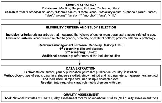
Figure 1.
Systematic flowchart of the review methodology.
2.1. Inclusion/Exclusion Criteria
Original articles that measured the volume of one or more paranasal sinuses and related it to the age of the patients were included in the study, regardless of the study method, and were eligible.
Studies that related the volume to gender, ethnic group, or skeletal pattern of the patient were excluded (unless they complied with the age information) and discarded, as well as those that studied such parameters in patients with sinus pathology.
2.2. Search Strategy
In order to identify the relevant studies for this systematic review, an electronic bibliographic search was carried out in the databases Medline (via PubMed), Scopus, Embase, Cochrane, and Lilacs. The search was conducted during the month of December 2022. The search and study selection process was conducted independently by two researchers (A.I.-G. and J.L.S.). In the event of doubt, a third researcher was consulted (L.F.).
Search terms: the search strategy included 5 Mesh (Medical Subject Heading) terms: “Paranasal sinuses”, “Ethmoid sinus”, “Frontal sinus”, “Maxillary sinus”, and “Sphenoid sinus”; 7 uncontrolled descriptors: “area”, “size”, “volume”, “anatom*”, “morphol*”, “age”, and “child*”. The Boolean operators OR, AND, and NOT were used to join search terms related to the research question.
The following search string was used: (((“frontal sinus” [Title/Abstract] OR “ethmoidal sinus” [Title/Abstract] OR “sphenoidal sinus” [Title/Abstract] OR “maxillary sinus” [Title/Abstract] OR “paranasal sinus” [Title/Abstract]) AND (area [Title/Abstract] OR size [Title/Abstract] OR volume [Title/Abstract] OR morphol* [Title/Abstract] OR anatom* [Title/Abstract])) AND (age [Title/Abstract])) NOT (child*).
2.3. Study Selection
The references yielded from the search strategy were exported to Mendeley Desktop v.1.19.8 (Elsevier Inc.; Amsterdam, The Netherlands). Duplicates were manually discarded using this software. After discarding duplicates, a first screening was performed based on article titles and abstracts of the articles, following the inclusion/exclusion criteria described previously. A second screening of the full text of the remaining studies was then performed to confirm their eligibility. Posteriorly, a screening of the references of the included studies was performed to look for additional potentially eligible studies.
2.4. Data Extraction
In order to perform the bibliometric analysis of the studies included in the review, the following data were collected: author, year of publication, journal of publication, country, and institution to which the authors were linked. For the qualitative synthesis of the methodology used by the included studies, the following variables were recorded: type of study, paranasal sinus/es studied, study method and its parameters, measurement method and tools used, sample size, and sample characteristics. For the results, the data provided by the articles regarding sinus volumetric changes with age were extracted. Statistical data were included for those studies where they were provided.
2.5. Quality Assessment
The quality assessment of the included studies was performed using the National Institutes of Health quality assessment tool for observational studies (NIH quality assessment tool; available at: https://www.nhlbi.nih.gov/health-topics/study-quality-assessment-tools, accessed on 10 January 2023).
3. Results
3.1. Study Selection and Flow Diagram
The study selection process is illustrated in Figure 2. During the search, 1465 articles related to sinus morphometry were found. In the database search, 357 were found in PubMed, 726 in Embase, 276 in Scopus, and 90 in Lilacs. The search in the Cochrane database did not yield any results. Additionally, the screening of the references of the included studies yielded 16 new eligible articles.
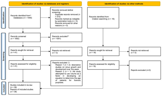
Figure 2.
Systematic flow chart representing the study selection process. Based on the PRISMA 2020 flow diagram. * Only electronic databases were consulted. ** Records were excluded based on title and abstract screening.
A total of 373 duplicates were excluded. From the remaining 1092 records, another 1051 were excluded after reading the title and abstract, as they did not meet the inclusion criteria. The remaining 41 articles were read in their entirety, and 3 additional studies were excluded: two of them because they were descriptive studies of sinus growth and development over the years and a third because it attempted to use volume as a factor in developing an equation to determine the age of patients for forensic purposes. Thus, 38 articles that met the inclusion criteria were included in the systematic review.).
3.2. Study Characteristics
All but two studies [22,23], which used cadaveric dissection techniques, were performed using imaging techniques. The techniques used were X-rays (3 studies), CBCT (8 studies), and tomography (25 studies). Table 1 details the various measurement methods used to determine the dimensions and volume of the paranasal sinuses in each article.

Table 1.
General characteristics of the study methodology.
All articles included in the review were cross-sectional observational studies. Four studies were prospective [20,44,49,50], and the rest were retrospective.
The most studied paranasal sinuses were the maxillary, frontal, and sphenoidal sinuses, either individually or in different combinations between them, which are detailed in Figure 3. Only three studies included the ethmoid sinuses [20,39,45].
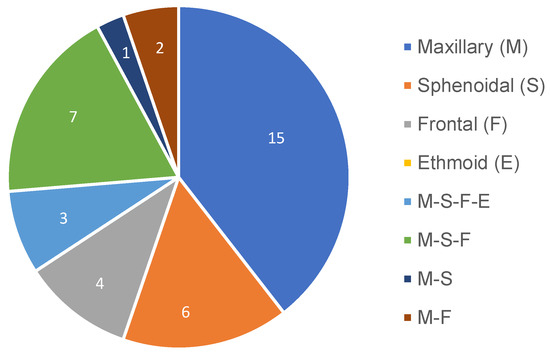
Figure 3.
Pie chart representing the distribution of the studied paranasal sinuses among the included studies.
The ages of the participants included ranged from 0 years to >100 years (Table 1). All studies were performed on living subjects, except those of Uchida et al., 1998 [22]; Takahashi et al., 2010 [40]; Demiralp et al., 2019 [33]; and Andrianakis et al., 2020 [23], which were performed on skulls.
All the authors of the included studies were linked to a university or health institution dedicated to education and research. The breakdown of the studies by year, country, and journal of publication is detailed in Figure 4.
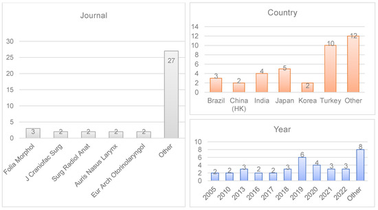
Figure 4.
Bibliometric data of the included studies (year of publication, journal, and country).
3.3. Study Results
3.3.1. Maxillary Sinus
Most authors who studied the maxillary sinus concluded that it begins its development from birth until the maximum peak of growth, from which it begins to decrease in volume with age [12,20,24,25,27,29,30,32,35,37,39,40,43,45,46,48,49,50]. In contrast, Jasso-Ramirez et al., 2022 [11], and de Barros et al., 2022 [28], concluded that maxillary sinus volume increases with age. In six of the studies, no relationship between sinus volume and age was found [6,7,22,31,33,36].
3.3.2. Sphenoidal Sinus
Regarding the sphenoidal sinus, the authors Andrianakis et al., 2020 [23]; Kim et al., 2010 [6]; Demiralp et al., 2019 [33]; Oliveira et al., 2017 [13]; Singh et al., 2021 [38]; Takahashi et al., 2016 [40]; Yilmaz et al., 2020 [12]; and Özer et al., 2018 [47], did not find a linear correlation between sinus volume and age. In contrast, Kumar et al., 2022 [20]; Karakas and Kavakli, 2005 [27]; Cohen et al., 2018 [32]; Emirzeoglu et al., 2017 [39]; Ozdikici, 2018 [45]; Alasmari et al., 2019 [46]; and Yonetsu et al., 2000 [51], found that sinus volume decreases with age. Only Pirinc et al., 2019 [34], and Jasso-Ramirez et al., 2022 [11], reported an increase in sphenoid sinus volume.
3.3.3. Frontal Sinus
A decrease in frontal sinus volume was observed in the studies of Kumar et al., 2022 [20]; Karakas and Kavakli, 2005 [27]; Tatlisumak et al., 2008 [15]; Emirzeoglu et al., 2007 [39]; and Ozdikici, 2018 [45]. In the studies of Jasso-Ramirez et al., 2022 [11]; Demiralp et al., 2019 [33]; Fatu et al., 2006 [42]; and Soman et al., 2016 [44], an increase in sinus area was observed, while Kim et al., 2010 [6]; Sahlstrand-Johnson et al., 2011 [31]; Cohen et al., 2018 [32]; Marino et al., 2020 [36]; Takahashi et al., 2016 [40]; Yilmaz et al., 2020 [12]; and Rubira-Bullen et al., 2010 [41], did not observe sinus volumetric alterations with age.
3.3.4. Ethmoidal Sinus
The three studies that include the ethmoidal sinus in their measurements conclude that the sinus volume decreases with age after reaching its maximum growth peak [20,39,45].
The summary of the results of the articles is summarized in Table 2.
Table 2.
Study results.
3.4. Quality Assessment
The quality analysis of all the articles was carried out using the NIH quality assessment tool. All the authors clearly defined both their study objective and the population (number, characteristics, and eligibility) on which they were carrying out the research (sections 1–3). Except for the studies performed on cadavers, all used similar populations, and their selection was carried out applying the same inclusion and exclusion criteria for all (section 4), but only two of the studies made a sample size calculation (section 5). The authors reported the exposure factors of the population before measuring the results, except for one of them (section 6). All investigators measured the varying degrees of exposure of the study population to the independent variables for a sufficient length of time to observe the results uniformly across the sample (sections 7–9). The participants in the studies were divided into groups according to age brackets so that those in charge of measuring the volumes knew the degree of exposure of each patient (section 11). Most studies took measures to control for confounding factors that could alter or mask the exposure–outcome relationship (section 14). Items 10, 12, and 13 were not considered applicable to the studies included in this literature review (Table 3).
Table 3.
Quality assessment.
4. Discussion
The four pairs of paranasal sinuses (maxillary, sphenoidal, frontal, and ethmoidal) are not only a fundamental part of the upper airway but also contribute to the development of the jaw and face and constitute the largest air cavity of the skull. The radiographic study of the paranasal sinuses is quite frequent because they are usually the site of chronic infectious processes, allergies, hypoplasia, pneumosinus dilatans, or atelectasis [39,45]. For example, sinusitis is one of the most common paranasal diseases, affecting almost 30 million people in the United States and 11% of the European population, making it one of the main causes of antibiotics prescription [41].
Since the paranasal sinuses are basically an air-filled space, one of the most important characteristics for their evaluation is the volume. Having well-defined parameters of the anatomy and normal evolution of the sinus complex is of great help to clinicians and surgeons in its diagnosis and treatment. At present, with increasing life expectancy, knowing the volumetric changes that sinuses undergo with age is an important reference for planning clinical procedures [11,20,52,53]. Accordingly, the aim of this study was to perform a systematic review of the change in volume experienced by the paranasal sinuses throughout life.
In all articles, we found imaging as the method of study. The only exceptions were Uchida et al., 1998 [22], and Andrianakis et al., 2020 [23], who made silicone models of the sphenoid sinuses from cadaver heads, which they then used to measure the volume. Four techniques were used within imaging methods: computed tomography (CT) [6,11,12,13,15,19,20,24,25,26,27,30,31,32,34,36,39,40,43,45,47,48,49,50,51], cone beam computed tomography (CBCT) [7,28,29,33,35,37,38,46], chemically developed radiographs [41,44], and digitized chemically developed radiographs [42].
Most studies used digital methods for image analysis, using specific automatic or semi-automatic study software [6,7,11,12,13,15,19,20,24,25,26,27,28,29,30,31,32,33,34,35,36,37,38,40,42,43,46,47,48,49,50,51]. Other authors made use of non-digital methods on paper media [39,41,44,45].
The Cavalieri Principle is a stereological method of volume measurement. It allows measuring a three-dimensional characteristic, such as volume, from two-dimensional images. These images should be consecutive slices of the organ to be studied, on which a template of points are superimposed to measure the area and, subsequently, the volume [45,54].
This makes it a good technique to be used in radiographic studies performed with CT or CBCT. In addition, the technique has proven to be accurate, fast, reliable, and possible to apply, even without a computer program, since it can be performed on paper prints of the images [20,39]. This was the method used to determine sinus volume by Elamin et al., 2021 [25]; Kumar et al., 2022 [20]; Karakas and Kavakli, 2005 [27]; Emirzeoglu et al., 2007 [39]; Ozdikici, 2018 [45]; and Jasim and Al-Taei, 2013 [50].
Other stereological methods used to calculate sinus volume were the ellipsoid formula [12,24,31,44,48], other formulas [30,46], and the measurement of sinus dimensions [15,41,49].
Another group of studies employed volume measurement tools integrated into the image analysis software [6,7,11,13,19,26,28,29,32,33,34,35,36,37,38,40,42,43,47,51].
Finally, Uchida et al., 1998 [22], and Andrianakis et al., 2020 [23], used the liquid displacement method (based on Archimedes’ Principle) to determine the volume of the sphenoidal sinuses by introducing the silicone models that they had taken out of the skulls included in their study, into a calibrated cylinder filled with water. This is an appropriate method for studying the volume of irregular structures, but it requires that the sample population are cadavers.
The most used study methods were diagnostic imaging, including three-dimensional imaging. This type of radiological technique, which can recognize very slight changes in tissue density, allows for the contour and characteristics of the bony structures and cavities of the skull to be observed in more detail [55].
Their images are accurate and reproducible, they are non-invasive methods, and the development of three-dimensional analysis software facilitates the measurement of the morphometric characteristics of the structures with greater accuracy than the stereological methods used in two-dimensional studies. Although CT was used in a greater number of studies, it has some disadvantages compared to CBCT, such as higher cost, higher radiation dose, and longer exposure time that can cause artifacts and worse image quality due to patient movements [53,56,57].
By far, the most studied sinus of all was the maxillary sinus. It appears in 14 articles individually and, in 13, studied in combination with other sinuses. Possibly this is because, in addition to being the largest paranasal sinus, its location in the maxilla and the close relationship it has with the orbit, nasal cavity, and dental apices make it an anatomical structure of interest for procedures such as sinus endoscopy, maxillofacial surgery, and dental surgery (implants and sinus lift) [19,25,58]. It is also widely studied in the forensic field for the identification of individuals because its structure remains intact, even in cases of death by cremation [7].
In 20 of the articles studying the maxillary sinus, the authors conclude that its volume decreases with age [12,19,20,24,25,26,27,29,30,32,35,37,39,40,43,45,46,48,49,50]. However, in five studies, no relation is found between sinus volume and age [6,7,31,33,36]. This difference may be attributed to the characteristics of the sample population and the ages included in the sample. In the majority of articles where an increase in volume is observed, individuals over 70 years of age are included in the sample, and in those that do not observe changes, only participants up to 65 years of age are included.
The growth of the maxillary sinus remains stable until the sixth decade of life. After the seventh decade, its volume begins to decrease due to a loss of minerals in the bone matrix (osteoporosis) that surrounds the sinus, which shrinks its walls and reduces its volume. Loss of volume has also been reported in the sinuses of totally or partially edentulous persons. Both factors occur in older people [5,25,26]. This may explain why the investigations of Jasso-Ramirez et al., 2022 [11], and de Barros et al., 2022 [28], whose sample consisted of young individuals, obtained an increase in sinus volume.
In contrast, the ethmoidal sinus was the least studied (only three articles), perhaps because the complexity of its anatomy makes it too difficult to perform an accurate and standardized measurement of its anatomy. Moreover, some consider that from an embryological and anatomical point of view, the frontal sinus and ethmoidal cells should be considered as a single structure due to their closeness [32,42].
In the sphenoid sinus study, Jasso-Ramirez et al., 2022 [11], and Pirinc et al., 2019 [34], found that the volume increased. Their study populations were almost entirely young patients under 60 years of age. The outcome of the remaining studies was divided between those that measured a decrease in volume [20,27,32,39,45,46,51] and those that did not find a volume–age correlation [6,12,13,23,33,38,40,47].
When comparing the characteristics of the studies of each group, we see that they differ by the measurement methods; in the volume decrease group, all (except Cohen et al., 2018 [32]) use stereological measurement methods, and in the group that found no relationship, they employ digital methods by using computer software (except Andrianakis et al., 2020 [23]).
Since the sphenoid sinus is an irregularly shaped and difficult-to-access cavity enclosed within the body of the sphenoid and is considered the most variable cavity in the human body, it is possible that non-three-dimensional measurement methods may affect the results. Because of its proximity to vital neurovascular and endocrine structures, the sphenoid sinus is of great clinical relevance, and knowledge of its anatomy and volume is indispensable in planning skull base surgeries [6,11,13,23,32,38].
When analyzing the results obtained in frontal sinus investigations, we see similar results to those of the sphenoid sinus. For Kumar et al., 2022 [20]; Karakas and Kavakli, 2005 [27]; Tatlisumak et al., 2008 [15]; Emirzeoglu et al., 2007 [39]; and Ozdikici, 2018 [45], sinus volume decreases with age. All use stereological measurement methods. In contrast, Kim et al., 2010 [6]; Sahlstrand-Johnson et al., 2011 [31]; Cohen et al., 2018 [32]; Marino et al., 2020 [36]; Takahashi et al., 2016 [40]; Yilmaz et al., 2020 [12], and Rubira-Bullen et al., 2010 [41], found no relationship between sinus volume and age. All used digital study methods, except Rubira-Bullen et al., 2010 [41].
Only Jasso-Ramirez et al., 2022 [11]; Demiralp et al., 2019 [33]; Fatu et al., 2006 [42]; and Soman et al., 2016 [44], detected an increase in sinus volume. It has been described that in elderly patients, the thinning of the bony cortex of the sinus due to osteoporosis can increase sinus volume and thin the cortex of the orbit. Nevertheless, its size is highly variable and can be affected by genetic, environmental, and pathologic factors, craniofacial morphology, and even hormonal levels [42,44]. For example, a recent study reported that cystic fibrosis patients were more prone to present frontal sinus aplasia and hypoplasia [59].
Only three authors made measurements of ethmoid sinus volume: Kumar et al., 2022 [20]; Emirzeoglu et al., 2007 [39]; and Ozdikici, 2018 [45]. All three saw a decrease in sinus volume, used similar study and measurement methods, and populations in the same age ranges.
Despite the heterogeneity of the studies in study methods, measurement, and population, when performing the quality analysis, we observed that all studies followed a similar structure, with a sufficient introduction stating the objectives of the study, appropriate description of the methodology and population used, as well as an explanation of the results and statistical methods. Despite this, only two articles measured the necessary sample size, and because of the type of studies reviewed, neither were the groups randomized nor the investigators blinded.
The methodological heterogeneity in terms of the characteristics of the sample and the parameters used for the measurement of sinus volume made it impossible to perform a quantitative synthesis or meta-analysis. The use of standardized methodology and previously established age groups could be useful in the future to reach statistically significant conclusions [60]. Instead, a qualitative synthesis of the available evidence was presented to provide a global view of the evidence in this regard, with the inherent limitations that this may imply.
The volumetric investigation of the paranasal sinuses is not only useful to know the amount of air that these structures can contain. Studying the sinus volume allows for learning its anatomy, pneumatization, and degenerative and pathological processes. Knowing the normal dimensions and volumes of the sinuses at each stage of life allows for recognizing sinus alterations or deformities and planning endoscopic surgeries at the base of the skull without putting adjacent structures, facial reconstruction surgeries, or dental implants at risk. Knowing normality allows for the recognition of abnormality [11,23,32,58].
To the best of our knowledge, the possible impact of sinus volumetric change on voice resonance, and its implication in presbyphonia, has not been described and may be an interesting topic for future studies.
The substantial heterogeneity of study and measurement methodologies hinders the comparison between studies. Future studies should implement a standardized three-dimensional sinus volume measurement method based on new diagnostic imaging technologies and 3D reconstruction software.
5. Conclusions
Based on the results of the included studies, the ethmoidal and maxillary sinuses experience development and growth from 0 to 20 years, reaching their maximum peak of development. From 20 to 50 years, a discrete decrease in volume may be observed. From 50 to 65 years of age, the decrease in volume is accentuated, which is further accelerated from 65 years onward. Conclusions on the volumetric changes of the sphenoidal and frontal sinuses require further evidence.
These results are based on heterogeneous methodologies and samples with varying characteristics. Therefore, they should be interpreted as an approximation and not as an exact measure.
Author Contributions
Conceptualization, A.I.-G. and L.F.; methodology, A.I.-G. and L.F.; software, J.L.S.; validation, M.M.; formal analysis, A.I.-G.; investigation, A.I.-G.; resources, C.P.-H.; data curation, A.I.-G.; writing—original draft preparation, A.I.-G.; writing—review and editing, J.L.S. and L.F.; visualization, M.M.; supervision, C.P.-H.; project administration, L.F.; funding acquisition, L.F. All authors have read and agreed to the published version of the manuscript.
Funding
This research received no external funding.
Institutional Review Board Statement
Not applicable.
Informed Consent Statement
Not applicable.
Data Availability Statement
The data presented in this study are available on request from the corresponding author.
Conflicts of Interest
The authors declare no conflict of interest.
References
- Onwuchekwa, R.C.; Alazigha, N. Computed tomography anatomy of the paranasal sinuses and anatomical variants of clinical relevants in Nigerian adults. Egypt. J. Ear Nose Throat Allied Sci. 2017, 18, 31–38. [Google Scholar] [CrossRef]
- Whyte, A.; Boeddinghaus, R. Correction to The maxillary sinus: Physiology, development and imaging anatomy. Dentomaxillofacial Radiol. 2019, 48, 20190205c. [Google Scholar] [CrossRef] [PubMed]
- Almenar, A. Morfología de los Senos Maxilares y Sus Relaciones con la Arcada Alveolar Superior en Cráneos Humanos Adultos de la Provincia de Valencia. Ph.D. Thesis, Universitat de València, Valencia, Spain, 1988. [Google Scholar]
- Spence, A.P. Anatomia Humana Básica. 1991. Available online: https://books.google.com/books/about/Anatomia_humana_básica.html?hl=es&id=hheoPgAACAAJ (accessed on 1 December 2022).
- Van Cauwenberge, P.; Sys, L.; De Belder, T.; Watelet, J.-B. Anatomy and physiology of the nose and the paranasal sinuses. Immunol. Allergy Clin. North Am. 2004, 24, 1–17. [Google Scholar] [CrossRef] [PubMed]
- Kim, J.; Song, S.W.; Cho, J.-H.; Chang, K.-H.; Jun, B.C. Comparative study of the pneumatization of the mastoid air cells and paranasal sinuses using three-dimensional reconstruction of computed tomography scans. Surg. Radiol. Anat. 2010, 32, 593–599. [Google Scholar] [CrossRef] [PubMed]
- Gulec, M.; Tassoker, M.; Magat, G.; Lale, B.; Ozcan, S.; Orhan, K. Three-dimensional volumetric analysis of the maxillary sinus: A cone-beam computed tomography study. Folia Morphol. 2020, 79, 557–562. [Google Scholar] [CrossRef] [PubMed]
- Dang, J.; Honda, K. Acoustic characteristics of the human paranasal sinuses derived from transmission characteristic measurement and morphological observation. J. Acoust. Soc. Am. 1996, 100, 3374. [Google Scholar] [CrossRef]
- Méndez, A.M. Fisiología resonancial: Conceptos claves para la fonoaudiología. Areté 2018, 18, 83–92. [Google Scholar] [CrossRef]
- Vampola, T.; Horáček, J.; Radolf, V.; Švec, J.G.; Laukkanen, A.-M. Influence of nasal cavities on voice quality: Computer simulations and experiments. J. Acoust. Soc. Am. 2020, 148, 3218–3231. [Google Scholar] [CrossRef]
- Jasso-Ramirez, N.G.; Elizondo-Omaña, R.E.; Treviño-Gonzalez, J.L.; Quiroga-Garza, A.; Garza-Rico, I.A.; Aguilar-Morales, K.; Elizondo-Riojas, G.; Guzmán-Lopez, S. Morphometric variants of the paranasal sinuses in a Mexican population: Expected changes according to age and gender. Folia Morphol. 2022; Online ahead of print. [Google Scholar] [CrossRef]
- Yilmaz, N.; Mülazimoğlu, S.; Öner, S.; Nacar, E.; Yilmaz, O. Paranasal sinus anatomical differences in elderly patients. Turk. J. Geriatr. 2020, 23, 129–137. [Google Scholar] [CrossRef]
- Oliveira, J.M.M.; Alonso, M.B.C.C.; Tucunduva, M.J.A.P.D.S.E.; Fuziy, A.; Scocate, A.C.R.N.; Costa, A.L.F. Volumetric study of sphenoid sinuses: Anatomical analysis in helical computed tomography. Surg. Radiol. Anat. 2016, 39, 367–374. [Google Scholar] [CrossRef]
- dos Santos, R.M. Desenvolvimento dos Seios Paranasaia: Estudo por Ressonancia Magnetica do Cranio. 2002. Available online: https://repositorio.unifesp.br/handle/11600/18147 (accessed on 15 November 2022).
- Tatlisumak, E.; Ovali, G.Y.; Asirdizer, M.; Aslan, A.; Ozyurt, B.; Bayindir, P.; Tarhan, S. CT study on morphometry of frontal sinus. Clin. Anat. 2008, 21, 287–293. [Google Scholar] [CrossRef] [PubMed]
- Jones, N. The nose and paranasal sinuses physiology and anatomy. Adv. Drug Deliv. Rev. 2001, 51, 5–19. [Google Scholar] [CrossRef] [PubMed]
- De Souza, M.C.Q. Características Espectrais da Nasalidade. Master’s Thesis, Universidade de São Paulo, São Paulo, Brazil, 2003. [Google Scholar] [CrossRef]
- Khandelwal, N.; Gupta Arun, K.; Garg, A. Diagnostic Radiology: Neuroradiology, Including Head and Neck Imaging; Jaypee Brothers Medical Publishers: New Delhi, India, 2010. [Google Scholar]
- Sarilita, E.; Lita, Y.A.; Nugraha, H.G.; Murniati, N.; Yusuf, H.Y. Volumetric growth analysis of maxillary sinus using computed tomography scan segmentation: A pilot study of Indonesian population. Anat. Cell Biol. 2021, 54, 431–435. [Google Scholar] [CrossRef]
- Kumar, S.; Gupta, R.; Kumar, L. Morphometric and Volumetric Measurements of the Paranasal Sinuses among the population of the Bareilly region. Int. J. Health Clin. Res. 2022, 5, 587–590. [Google Scholar]
- Page, M.J.; McKenzie, J.E.; Bossuyt, P.M.; Boutron, I.; Hoffmann, T.C.; Mulrow, C.D.; Shamseer, L.; Tetzlaff, J.M.; Akl, E.A.; Brennan, S.E.; et al. The PRISMA 2020 Statement: An Updated Guideline for Reporting Systematic Reviews. BMJ 2021, 372, n71. [Google Scholar] [CrossRef]
- Uchida, Y.; Goto, M.; Katsuki, T.; Akiyoshi, T. A cadaveric study of maxillary sinus size as an aid in bone grafting of the maxillary sinus floor. J. Oral Maxillofac. Surg. 1998, 56, 1158–1163. [Google Scholar] [CrossRef]
- Andrianakis, A.; Kiss, P.; Wolf, A.; Pilsl, U.; Palackic, A.; Holzmeister, C.; Moser, U.; Tomazic, P.V. Volumetric Investigation of Sphenoid Sinus in an Elderly Population. J. Craniofacial Surg. 2020, 31, 2346–2349. [Google Scholar] [CrossRef]
- Samhitha, G.; Geethanjali, B.S.; Mokhasi, V.; Prakash, R.; Shamkuwar, S.; Kumar, H.K. Measurements of maxillary sinus in correlation to age and gender by computed tomography. Int. J. Anat. Res. 2019, 7, 6732–6739. [Google Scholar] [CrossRef]
- Elamin, A.A.; Acar, T.; Kajoak, S.; Idris, S.A.; Malik, B.A.; Ayad, C.E. Volumetric Measurement of the MaxillarySinuses inNormal Sudanese using Computed Tomography: A Retrospective Study. J. Clin. Diagn. Res. 2021, 15, 14894. [Google Scholar] [CrossRef]
- Jun, B.; Song, S.; Park, C.; Lee, D.; Cho, K.; Cho, J. The analysis of maxillary sinus aeration according to aging process; volume assessment by 3-dimensional reconstruction by high-resolutional CT scanning. Otolaryngol. Neck Surg. 2005, 132, 429–434. [Google Scholar] [CrossRef]
- Karakas, S.; Kavaklıb, A. Morphometric examination of the paranasal sinuses and mastoid air cells using computed tomography. Ann. Saudi Med. 2005, 25, 41–45. [Google Scholar] [CrossRef] [PubMed]
- de Barros, F.; Fernandes, C.M.D.S.; Kuhnen, B.; Filho, J.S.; Gonçalves, M.; Gonçalves, V.; Serra, M.D.C. Three-dimensional analysis of the maxillary sinus according to sex, age, skin color, and nutritional status: A study with live Brazilian subjects using cone-beam computed tomography. Arch. Oral Biol. 2022, 139, 105435. [Google Scholar] [CrossRef] [PubMed]
- Bornstein, M.M.; Ho, J.K.C.; Yeung, A.W.K.; Tanaka, R.; Li, J.Q.; Jacobs, R. A Retrospective Evaluation of Factors Influencing the Volume of Healthy Maxillary Sinuses Based on CBCT Imaging. Int. J. Periodontics Restor. Dent. 2019, 39, 187–193. [Google Scholar] [CrossRef] [PubMed]
- Ariji, Y.; Kuroki, T.; Moriguchi, S.; Ariji, E.; Kanda, S. Age changes in the volume of the human maxillary sinus: A study using computed tomography. Dentomaxillofacial Radiol. 1994, 23, 163–168. [Google Scholar] [CrossRef] [PubMed]
- Sahlstrand-Johnson, P.; Jannert, M.; Strömbeck, A.; Abul-Kasim, K. Computed tomography measurements of different dimensions of maxillary and frontal sinuses. BMC Med. Imaging 2011, 11, 8. [Google Scholar] [CrossRef]
- Cohen, O.; Warman, M.; Fried, M.; Shoffel-Havakuk, H.; Adi, M.; Halperin, D.; Lahav, Y. Volumetric analysis of the maxillary, sphenoid and frontal sinuses: A comparative computerized tomography based study. Auris Nasus Larynx 2018, 45, 96–102. [Google Scholar] [CrossRef]
- Demiralp, K.; Cakmak, S.K.; Aksoy, S.; Bayrak, S.; Orhan, K.; Demir, P. Assessment of paranasal sinus parameters according to ancient skulls’ gender and age by using cone-beam computed tomography. Folia Morphol. 2019, 78, 344–350. [Google Scholar] [CrossRef]
- Pirinc, B.; Fazliogullari, Z.; Guler, I.; Dogan, N.U.; Uysal, I.I.; Karabulut, A.K. Classification and volumetric study of the sphenoid sinus on MDCT images. Eur. Arch. Oto-Rhino-Laryngol. 2019, 276, 2887–2894. [Google Scholar] [CrossRef]
- Belgin, C.A.; Colak, M.; Adiguzel, O.; Akkus, Z.; Orhan, K. Three-dimensional evaluation of maxillary sinus volume in different age and sex groups using CBCT. Eur. Arch. Oto-Rhino-Laryngol. 2019, 276, 1493–1499. [Google Scholar] [CrossRef]
- Marino, M.J.; Riley, C.A.; Wu, E.L.; Weinstein, J.E.; Emerson, N.; McCoul, E.D. Variability of Paranasal Sinus Pneumatization in the Absence of Sinus Disease. Ochsner J. 2020, 20, 170–175. [Google Scholar] [CrossRef]
- Velasco-Torres, M.; Padial-Molina, M.; Ortiz, G.A.; García-Delgado, R.; O’Valle, F.; Catena, A.; Galindo-Moreno, P. Maxillary Sinus Dimensions Decrease as Age and Tooth Loss Increase. Implant. Dent. 2017, 26, 288–295. [Google Scholar] [CrossRef] [PubMed]
- Singh, P.; Hung, K.; Ajmera, D.H.; Yeung, A.W.K.; von Arx, T.; Bornstein, M.M. Morphometric characteristics of the sphenoid sinus and potential influencing factors: A retrospective assessment using cone beam computed tomography (CBCT). Anat. Sci. Int. 2021, 96, 544–555. [Google Scholar] [CrossRef] [PubMed]
- Emirzeoglu, M.; Sahin, B.; Bilgic, S.; Celebi, M.; Uzun, A. Volumetric evaluation of the paranasal sinuses in normal subjects using computer tomography images: A stereological study. Auris Nasus Larynx 2007, 34, 191–195. [Google Scholar] [CrossRef] [PubMed]
- Takahashi, Y.; Watanabe, T.; Iimura, A.; Takahashi, O. A Study of the Maxillary Sinus Volume in Elderly Persons Using Japanese Cadavers. Okajimas Folia Anat. Jpn. 2016, 93, 21–27. [Google Scholar] [CrossRef]
- Rubira-Bullen, I.R.; Rubira, C.; Sarmento, V.A.; Azevedo, R.A. Frontal sinus size on facial plain radiographs. J. Morphol. Sci. 2010, 27, 77–81. [Google Scholar]
- Fatu, C.; Puisoru, M.; Rotaru, M.; Truta, A. Morphometric evaluation of the frontal sinus in relation to age. Ann. Anat. Anat. Anz. 2006, 188, 275–280. [Google Scholar] [CrossRef]
- Iwai, K.; Hashimoto, K.; Kawabe, Y.; Shinoda, K.; Kudo, I. Age-related Morphological Changes of th Maxillary Sinus. Ronen Shika Igaku 1995, 10, 31–41. [Google Scholar] [CrossRef]
- Soman, B.A.; Sujatha, G.; Lingappa, A. Morphometric evaluation of the frontal sinus in relation to age and gender in subjects residing in Davangere, Karnataka. J. Forensic Dent. Sci. 2016, 8, 57. [Google Scholar] [CrossRef]
- Özdikici, M. Volumetric Evaluation of the Paranasal Sinuses with the Cavalieri Method. Anat. Physiol. Biochem. Int. J. 2018, 5, 555657. [Google Scholar]
- Alasmari, D.S.; Mohan, M.P.; Almutairi, A.S.; Satheeshkumar, P.S. Para Nasal Sinuses are Pneumatized in a Synchronized Pattern, a Study Evaluating Volume of the Maxillary and Sphenoid Sinuses using Cone Beam Computed Tomography. Int. J. Contemp. Med. Res. 2019, 6, 587–590. [Google Scholar]
- Özer, C.M.; Atalar, K.D.; Öz, I.I.; Toprak, S.; Barut, M. Sphenoid Sinus in Relation to Age, Gender, and Cephalometric Indices. J. Craniofacial Surg. 2018, 29, 2319–2326. [Google Scholar] [CrossRef] [PubMed]
- Abdulhameed, A.; Zagga, A.D.; Ma’aji, S.M.; Bello, A.; Bello, S.S.; Usman, J.D.; Awwal, M.M.; Tadros, A.A. Three Dimensional Volumetric Analysis of the Maxillary Sinus Using Computed Tomography from Usmanu Danfodiyo University Teaching. Int. J. Health Med. Inf. 2013, 2, 1–9. [Google Scholar]
- Baweja, S.; Dixit, A.; Baweja, S. Study of age related changes of maxillary air sinus from its anteroposterior, transverse and vertical dimensions using computerized tomographic (ct) scan. Int. J. Biomed. Res. 2013, 4, 21–25. [Google Scholar]
- Al-Taei, J.A.; Jasim, H.H. Computed Tomographic Measurement of Maxillary Sinus Volume and Dimension in Correlation to the Age and Gender: Comparative Study among Individuals with Dentate and Edentulous Maxilla. J. Baghdad Coll. Dent. 2013, 25, 87–93. [Google Scholar] [CrossRef]
- Yonetsu, K.; Watanabe, M.; Nakamura, T. Age-Related Expansion and Reduction in Aeration of the Sphenoid Sinus: Volume As-sessment by Helical CT Scanning. AJNR Am. J. Neuroradiol. 2000, 21, 179. [Google Scholar]
- Micó, S.I.; Carceller, M.A.; Aragonés, A.M. Atelectasia crónica maxilar: Causa infrecuente de opacidad radiológica persistente. An. De Pediatría 2005, 63, 169–171. [Google Scholar] [CrossRef]
- Aksoy, S.; Orhan, K. Evaluation of Paranasal Sinus Volumes. Turk. Klin Oral Maxillofac. Radiol. Spec. Top. 2017, 3, 184–188. Available online: https://www.turkiyeklinikleri.com/article/en-paranazal-sinus-hacimlerinin-degerlendirilmesi-79744.html (accessed on 10 November 2022).
- Altunkaynak, B.Z.; Altunkaynak, E.; Unal, D.; Unal, B. A novel application for the cavalieri principle: A stereological and methodo-logical study. Eurasian J. Med. 2009, 41, 99–101. Available online: https://pubmed.ncbi.nlm.nih.gov/25610077/ (accessed on 10 November 2022).
- Bryanskaya, E.O.; Novikova, I.N.; Dremin, V.V.; Gneushev, R.Y.; Bibikova, O.A.; Dunaev, A.V.; Artyushenko, V.G. Optical Diagnostics of the Maxillary Sinuses by Digital Diaphanoscopy Technology. Diagnostics. 2021, 11, 77. [Google Scholar] [CrossRef]
- Koç, A. Are Maxillary and Sphenoid Sinus Volumes Deterministic for Gender and Age Estimation? A Cone Beam Computed Tomography Study. Cumhur. Dent. J. 2020, 23, 348–355. [Google Scholar] [CrossRef]
- Park, I.-H.; Song, J.S.; Choi, H.; Kim, T.H.; Hoon, S.; Lee, S.H.; Lee, H.-M. Volumetric study in the development of paranasal sinuses by CT imaging in Asian: A Pilot study. Int. J. Pediatr. Otorhinolaryngol. 2010, 74, 1347–1350. [Google Scholar] [CrossRef] [PubMed]
- Lee, S.-J.; Yoo, J.-Y.; Yoo, S.-K.; Ha, R.; Lee, D.-H.; Kim, S.-T.; Yi, W.-J. Image-Guided Endoscopic Sinus Surgery with 3D Volumetric Visualization of the Nasal Cavity and Paranasal Sinuses: A Clinical Comparative Study. Appl. Sci. 2021, 11, 3675. [Google Scholar] [CrossRef]
- Chung, J.; Wünnemann, F.; Salomon, J.; Boutin, S.; Frey, D.L.; Albrecht, T.; Joachim, C.; Eichinger, M.; Mall, M.A.; Wielpütz, M.O.; et al. Increased Inflammatory Markers Detected in Nasal Lavage Correlate with Paranasal Sinus Abnormalities at MRI in Adolescent Patients with Cystic Fibrosis. Antioxidants 2021, 10, 1412. [Google Scholar] [CrossRef] [PubMed]
- Page, M.J.; Moher, D.; Bossuyt, P.M.; Boutron, I.; Hoffmann, T.C.; Mulrow, C.D.; Shamseer, L.; Tetzlaff, J.M.; Akl, E.A.; Brennan, S.E.; et al. PRISMA 2020 explanation and elaboration: Updated guidance and exemplars for reporting systematic reviews. BMJ 2021, 372, n160. [Google Scholar] [CrossRef]
Disclaimer/Publisher’s Note: The statements, opinions and data contained in all publications are solely those of the individual author(s) and contributor(s) and not of MDPI and/or the editor(s). MDPI and/or the editor(s) disclaim responsibility for any injury to people or property resulting from any ideas, methods, instructions or products referred to in the content. |
© 2023 by the authors. Licensee MDPI, Basel, Switzerland. This article is an open access article distributed under the terms and conditions of the Creative Commons Attribution (CC BY) license (https://creativecommons.org/licenses/by/4.0/).