Volumetric Changes of the Paranasal Sinuses with Age: A Systematic Review
Abstract
1. Introduction
2. Materials and Methods
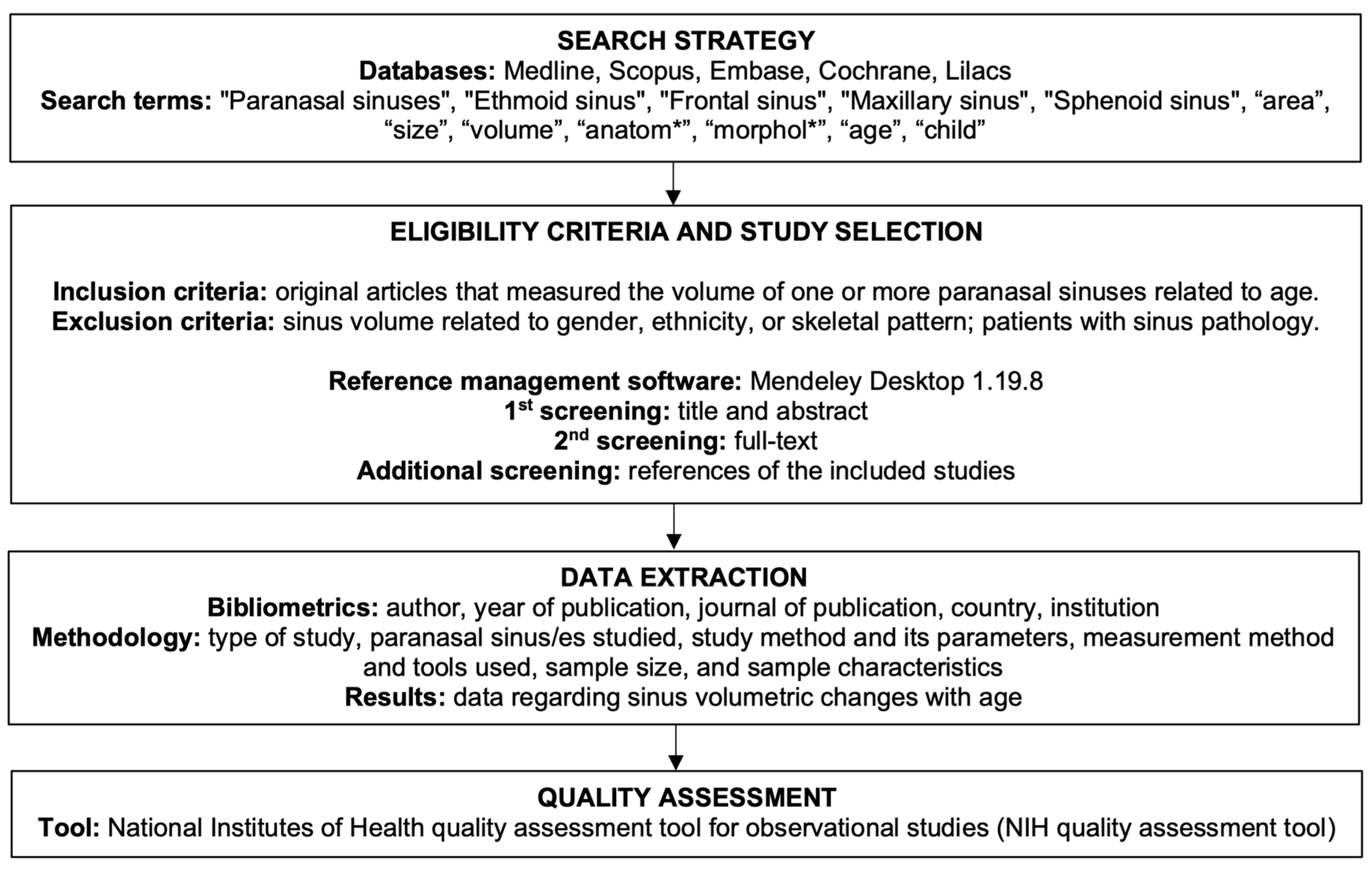
2.1. Inclusion/Exclusion Criteria
2.2. Search Strategy
2.3. Study Selection
2.4. Data Extraction
2.5. Quality Assessment
3. Results
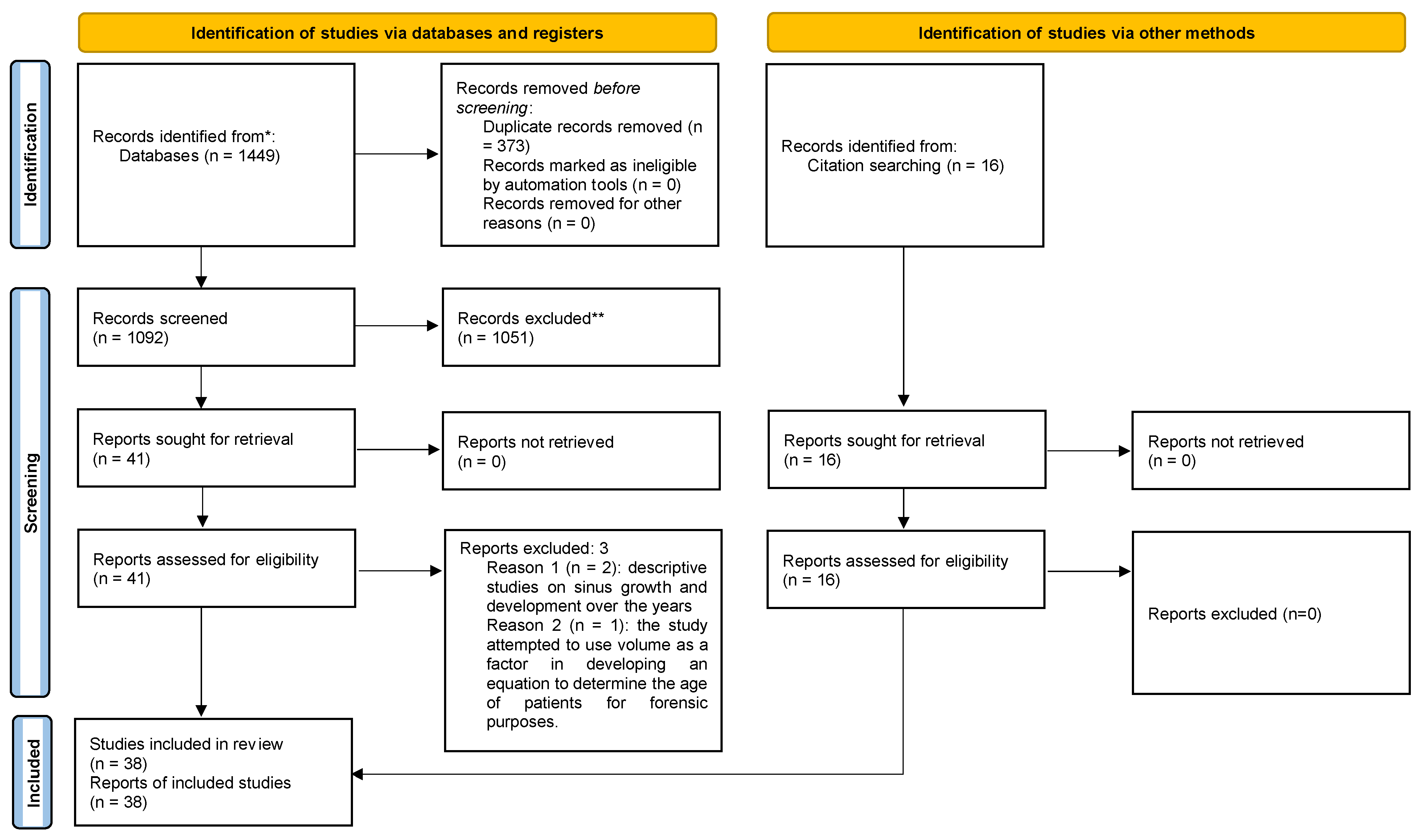
3.1. Study Selection and Flow Diagram
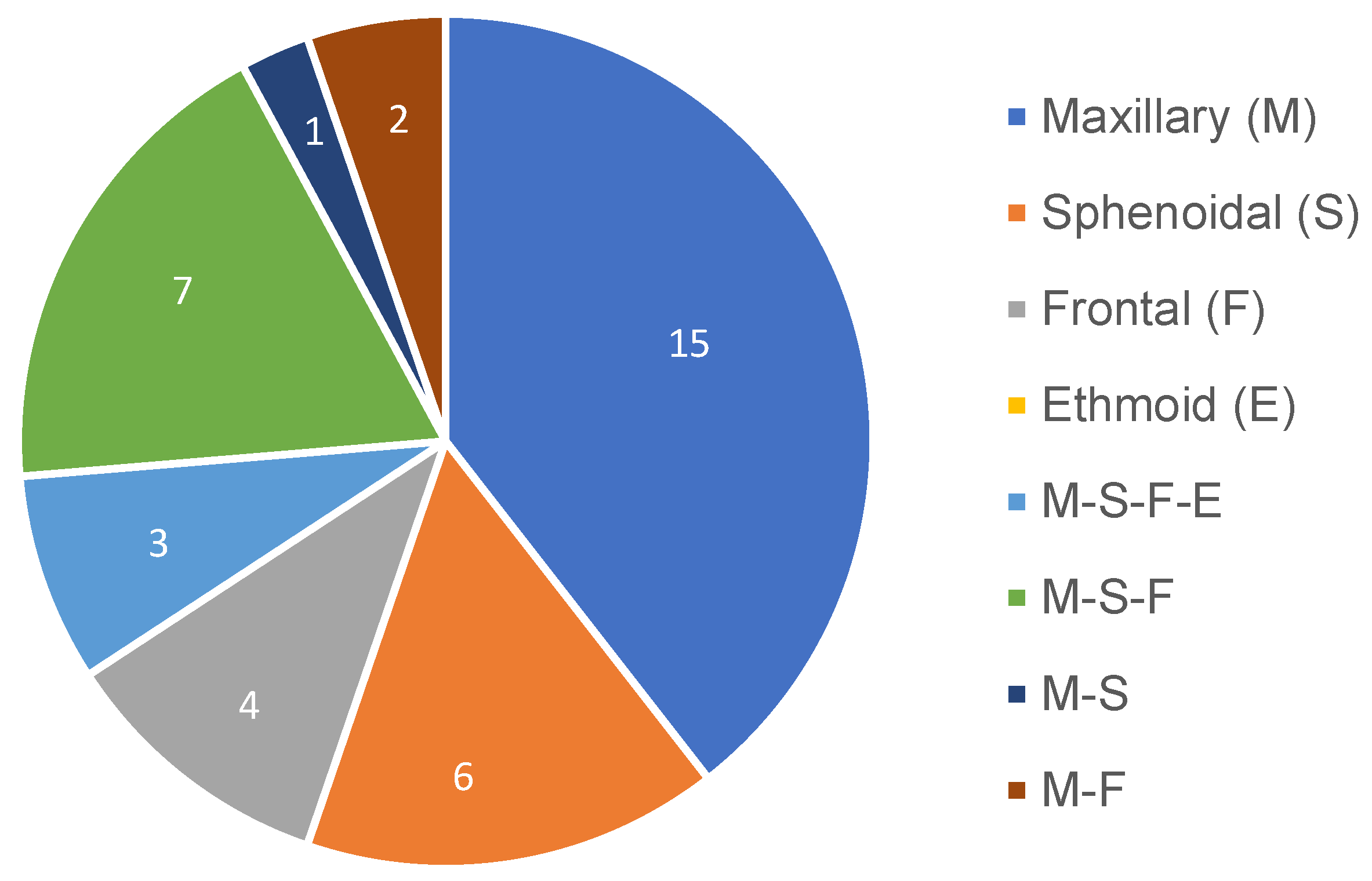
3.2. Study Characteristics
3.3. Study Results
3.3.1. Maxillary Sinus
3.3.2. Sphenoidal Sinus
3.3.3. Frontal Sinus
3.3.4. Ethmoidal Sinus
3.4. Quality Assessment
4. Discussion
5. Conclusions
Author Contributions
Funding
Institutional Review Board Statement
Informed Consent Statement
Data Availability Statement
Conflicts of Interest
References
- Onwuchekwa, R.C.; Alazigha, N. Computed tomography anatomy of the paranasal sinuses and anatomical variants of clinical relevants in Nigerian adults. Egypt. J. Ear Nose Throat Allied Sci. 2017, 18, 31–38. [Google Scholar] [CrossRef]
- Whyte, A.; Boeddinghaus, R. Correction to The maxillary sinus: Physiology, development and imaging anatomy. Dentomaxillofacial Radiol. 2019, 48, 20190205c. [Google Scholar] [CrossRef] [PubMed]
- Almenar, A. Morfología de los Senos Maxilares y Sus Relaciones con la Arcada Alveolar Superior en Cráneos Humanos Adultos de la Provincia de Valencia. Ph.D. Thesis, Universitat de València, Valencia, Spain, 1988. [Google Scholar]
- Spence, A.P. Anatomia Humana Básica. 1991. Available online: https://books.google.com/books/about/Anatomia_humana_básica.html?hl=es&id=hheoPgAACAAJ (accessed on 1 December 2022).
- Van Cauwenberge, P.; Sys, L.; De Belder, T.; Watelet, J.-B. Anatomy and physiology of the nose and the paranasal sinuses. Immunol. Allergy Clin. North Am. 2004, 24, 1–17. [Google Scholar] [CrossRef] [PubMed]
- Kim, J.; Song, S.W.; Cho, J.-H.; Chang, K.-H.; Jun, B.C. Comparative study of the pneumatization of the mastoid air cells and paranasal sinuses using three-dimensional reconstruction of computed tomography scans. Surg. Radiol. Anat. 2010, 32, 593–599. [Google Scholar] [CrossRef] [PubMed]
- Gulec, M.; Tassoker, M.; Magat, G.; Lale, B.; Ozcan, S.; Orhan, K. Three-dimensional volumetric analysis of the maxillary sinus: A cone-beam computed tomography study. Folia Morphol. 2020, 79, 557–562. [Google Scholar] [CrossRef] [PubMed]
- Dang, J.; Honda, K. Acoustic characteristics of the human paranasal sinuses derived from transmission characteristic measurement and morphological observation. J. Acoust. Soc. Am. 1996, 100, 3374. [Google Scholar] [CrossRef]
- Méndez, A.M. Fisiología resonancial: Conceptos claves para la fonoaudiología. Areté 2018, 18, 83–92. [Google Scholar] [CrossRef]
- Vampola, T.; Horáček, J.; Radolf, V.; Švec, J.G.; Laukkanen, A.-M. Influence of nasal cavities on voice quality: Computer simulations and experiments. J. Acoust. Soc. Am. 2020, 148, 3218–3231. [Google Scholar] [CrossRef]
- Jasso-Ramirez, N.G.; Elizondo-Omaña, R.E.; Treviño-Gonzalez, J.L.; Quiroga-Garza, A.; Garza-Rico, I.A.; Aguilar-Morales, K.; Elizondo-Riojas, G.; Guzmán-Lopez, S. Morphometric variants of the paranasal sinuses in a Mexican population: Expected changes according to age and gender. Folia Morphol. 2022; Online ahead of print. [Google Scholar] [CrossRef]
- Yilmaz, N.; Mülazimoğlu, S.; Öner, S.; Nacar, E.; Yilmaz, O. Paranasal sinus anatomical differences in elderly patients. Turk. J. Geriatr. 2020, 23, 129–137. [Google Scholar] [CrossRef]
- Oliveira, J.M.M.; Alonso, M.B.C.C.; Tucunduva, M.J.A.P.D.S.E.; Fuziy, A.; Scocate, A.C.R.N.; Costa, A.L.F. Volumetric study of sphenoid sinuses: Anatomical analysis in helical computed tomography. Surg. Radiol. Anat. 2016, 39, 367–374. [Google Scholar] [CrossRef]
- dos Santos, R.M. Desenvolvimento dos Seios Paranasaia: Estudo por Ressonancia Magnetica do Cranio. 2002. Available online: https://repositorio.unifesp.br/handle/11600/18147 (accessed on 15 November 2022).
- Tatlisumak, E.; Ovali, G.Y.; Asirdizer, M.; Aslan, A.; Ozyurt, B.; Bayindir, P.; Tarhan, S. CT study on morphometry of frontal sinus. Clin. Anat. 2008, 21, 287–293. [Google Scholar] [CrossRef] [PubMed]
- Jones, N. The nose and paranasal sinuses physiology and anatomy. Adv. Drug Deliv. Rev. 2001, 51, 5–19. [Google Scholar] [CrossRef] [PubMed]
- De Souza, M.C.Q. Características Espectrais da Nasalidade. Master’s Thesis, Universidade de São Paulo, São Paulo, Brazil, 2003. [Google Scholar] [CrossRef]
- Khandelwal, N.; Gupta Arun, K.; Garg, A. Diagnostic Radiology: Neuroradiology, Including Head and Neck Imaging; Jaypee Brothers Medical Publishers: New Delhi, India, 2010. [Google Scholar]
- Sarilita, E.; Lita, Y.A.; Nugraha, H.G.; Murniati, N.; Yusuf, H.Y. Volumetric growth analysis of maxillary sinus using computed tomography scan segmentation: A pilot study of Indonesian population. Anat. Cell Biol. 2021, 54, 431–435. [Google Scholar] [CrossRef]
- Kumar, S.; Gupta, R.; Kumar, L. Morphometric and Volumetric Measurements of the Paranasal Sinuses among the population of the Bareilly region. Int. J. Health Clin. Res. 2022, 5, 587–590. [Google Scholar]
- Page, M.J.; McKenzie, J.E.; Bossuyt, P.M.; Boutron, I.; Hoffmann, T.C.; Mulrow, C.D.; Shamseer, L.; Tetzlaff, J.M.; Akl, E.A.; Brennan, S.E.; et al. The PRISMA 2020 Statement: An Updated Guideline for Reporting Systematic Reviews. BMJ 2021, 372, n71. [Google Scholar] [CrossRef]
- Uchida, Y.; Goto, M.; Katsuki, T.; Akiyoshi, T. A cadaveric study of maxillary sinus size as an aid in bone grafting of the maxillary sinus floor. J. Oral Maxillofac. Surg. 1998, 56, 1158–1163. [Google Scholar] [CrossRef]
- Andrianakis, A.; Kiss, P.; Wolf, A.; Pilsl, U.; Palackic, A.; Holzmeister, C.; Moser, U.; Tomazic, P.V. Volumetric Investigation of Sphenoid Sinus in an Elderly Population. J. Craniofacial Surg. 2020, 31, 2346–2349. [Google Scholar] [CrossRef]
- Samhitha, G.; Geethanjali, B.S.; Mokhasi, V.; Prakash, R.; Shamkuwar, S.; Kumar, H.K. Measurements of maxillary sinus in correlation to age and gender by computed tomography. Int. J. Anat. Res. 2019, 7, 6732–6739. [Google Scholar] [CrossRef]
- Elamin, A.A.; Acar, T.; Kajoak, S.; Idris, S.A.; Malik, B.A.; Ayad, C.E. Volumetric Measurement of the MaxillarySinuses inNormal Sudanese using Computed Tomography: A Retrospective Study. J. Clin. Diagn. Res. 2021, 15, 14894. [Google Scholar] [CrossRef]
- Jun, B.; Song, S.; Park, C.; Lee, D.; Cho, K.; Cho, J. The analysis of maxillary sinus aeration according to aging process; volume assessment by 3-dimensional reconstruction by high-resolutional CT scanning. Otolaryngol. Neck Surg. 2005, 132, 429–434. [Google Scholar] [CrossRef]
- Karakas, S.; Kavaklıb, A. Morphometric examination of the paranasal sinuses and mastoid air cells using computed tomography. Ann. Saudi Med. 2005, 25, 41–45. [Google Scholar] [CrossRef] [PubMed]
- de Barros, F.; Fernandes, C.M.D.S.; Kuhnen, B.; Filho, J.S.; Gonçalves, M.; Gonçalves, V.; Serra, M.D.C. Three-dimensional analysis of the maxillary sinus according to sex, age, skin color, and nutritional status: A study with live Brazilian subjects using cone-beam computed tomography. Arch. Oral Biol. 2022, 139, 105435. [Google Scholar] [CrossRef] [PubMed]
- Bornstein, M.M.; Ho, J.K.C.; Yeung, A.W.K.; Tanaka, R.; Li, J.Q.; Jacobs, R. A Retrospective Evaluation of Factors Influencing the Volume of Healthy Maxillary Sinuses Based on CBCT Imaging. Int. J. Periodontics Restor. Dent. 2019, 39, 187–193. [Google Scholar] [CrossRef] [PubMed]
- Ariji, Y.; Kuroki, T.; Moriguchi, S.; Ariji, E.; Kanda, S. Age changes in the volume of the human maxillary sinus: A study using computed tomography. Dentomaxillofacial Radiol. 1994, 23, 163–168. [Google Scholar] [CrossRef] [PubMed]
- Sahlstrand-Johnson, P.; Jannert, M.; Strömbeck, A.; Abul-Kasim, K. Computed tomography measurements of different dimensions of maxillary and frontal sinuses. BMC Med. Imaging 2011, 11, 8. [Google Scholar] [CrossRef]
- Cohen, O.; Warman, M.; Fried, M.; Shoffel-Havakuk, H.; Adi, M.; Halperin, D.; Lahav, Y. Volumetric analysis of the maxillary, sphenoid and frontal sinuses: A comparative computerized tomography based study. Auris Nasus Larynx 2018, 45, 96–102. [Google Scholar] [CrossRef]
- Demiralp, K.; Cakmak, S.K.; Aksoy, S.; Bayrak, S.; Orhan, K.; Demir, P. Assessment of paranasal sinus parameters according to ancient skulls’ gender and age by using cone-beam computed tomography. Folia Morphol. 2019, 78, 344–350. [Google Scholar] [CrossRef]
- Pirinc, B.; Fazliogullari, Z.; Guler, I.; Dogan, N.U.; Uysal, I.I.; Karabulut, A.K. Classification and volumetric study of the sphenoid sinus on MDCT images. Eur. Arch. Oto-Rhino-Laryngol. 2019, 276, 2887–2894. [Google Scholar] [CrossRef]
- Belgin, C.A.; Colak, M.; Adiguzel, O.; Akkus, Z.; Orhan, K. Three-dimensional evaluation of maxillary sinus volume in different age and sex groups using CBCT. Eur. Arch. Oto-Rhino-Laryngol. 2019, 276, 1493–1499. [Google Scholar] [CrossRef]
- Marino, M.J.; Riley, C.A.; Wu, E.L.; Weinstein, J.E.; Emerson, N.; McCoul, E.D. Variability of Paranasal Sinus Pneumatization in the Absence of Sinus Disease. Ochsner J. 2020, 20, 170–175. [Google Scholar] [CrossRef]
- Velasco-Torres, M.; Padial-Molina, M.; Ortiz, G.A.; García-Delgado, R.; O’Valle, F.; Catena, A.; Galindo-Moreno, P. Maxillary Sinus Dimensions Decrease as Age and Tooth Loss Increase. Implant. Dent. 2017, 26, 288–295. [Google Scholar] [CrossRef] [PubMed]
- Singh, P.; Hung, K.; Ajmera, D.H.; Yeung, A.W.K.; von Arx, T.; Bornstein, M.M. Morphometric characteristics of the sphenoid sinus and potential influencing factors: A retrospective assessment using cone beam computed tomography (CBCT). Anat. Sci. Int. 2021, 96, 544–555. [Google Scholar] [CrossRef] [PubMed]
- Emirzeoglu, M.; Sahin, B.; Bilgic, S.; Celebi, M.; Uzun, A. Volumetric evaluation of the paranasal sinuses in normal subjects using computer tomography images: A stereological study. Auris Nasus Larynx 2007, 34, 191–195. [Google Scholar] [CrossRef] [PubMed]
- Takahashi, Y.; Watanabe, T.; Iimura, A.; Takahashi, O. A Study of the Maxillary Sinus Volume in Elderly Persons Using Japanese Cadavers. Okajimas Folia Anat. Jpn. 2016, 93, 21–27. [Google Scholar] [CrossRef]
- Rubira-Bullen, I.R.; Rubira, C.; Sarmento, V.A.; Azevedo, R.A. Frontal sinus size on facial plain radiographs. J. Morphol. Sci. 2010, 27, 77–81. [Google Scholar]
- Fatu, C.; Puisoru, M.; Rotaru, M.; Truta, A. Morphometric evaluation of the frontal sinus in relation to age. Ann. Anat. Anat. Anz. 2006, 188, 275–280. [Google Scholar] [CrossRef]
- Iwai, K.; Hashimoto, K.; Kawabe, Y.; Shinoda, K.; Kudo, I. Age-related Morphological Changes of th Maxillary Sinus. Ronen Shika Igaku 1995, 10, 31–41. [Google Scholar] [CrossRef]
- Soman, B.A.; Sujatha, G.; Lingappa, A. Morphometric evaluation of the frontal sinus in relation to age and gender in subjects residing in Davangere, Karnataka. J. Forensic Dent. Sci. 2016, 8, 57. [Google Scholar] [CrossRef]
- Özdikici, M. Volumetric Evaluation of the Paranasal Sinuses with the Cavalieri Method. Anat. Physiol. Biochem. Int. J. 2018, 5, 555657. [Google Scholar]
- Alasmari, D.S.; Mohan, M.P.; Almutairi, A.S.; Satheeshkumar, P.S. Para Nasal Sinuses are Pneumatized in a Synchronized Pattern, a Study Evaluating Volume of the Maxillary and Sphenoid Sinuses using Cone Beam Computed Tomography. Int. J. Contemp. Med. Res. 2019, 6, 587–590. [Google Scholar]
- Özer, C.M.; Atalar, K.D.; Öz, I.I.; Toprak, S.; Barut, M. Sphenoid Sinus in Relation to Age, Gender, and Cephalometric Indices. J. Craniofacial Surg. 2018, 29, 2319–2326. [Google Scholar] [CrossRef] [PubMed]
- Abdulhameed, A.; Zagga, A.D.; Ma’aji, S.M.; Bello, A.; Bello, S.S.; Usman, J.D.; Awwal, M.M.; Tadros, A.A. Three Dimensional Volumetric Analysis of the Maxillary Sinus Using Computed Tomography from Usmanu Danfodiyo University Teaching. Int. J. Health Med. Inf. 2013, 2, 1–9. [Google Scholar]
- Baweja, S.; Dixit, A.; Baweja, S. Study of age related changes of maxillary air sinus from its anteroposterior, transverse and vertical dimensions using computerized tomographic (ct) scan. Int. J. Biomed. Res. 2013, 4, 21–25. [Google Scholar]
- Al-Taei, J.A.; Jasim, H.H. Computed Tomographic Measurement of Maxillary Sinus Volume and Dimension in Correlation to the Age and Gender: Comparative Study among Individuals with Dentate and Edentulous Maxilla. J. Baghdad Coll. Dent. 2013, 25, 87–93. [Google Scholar] [CrossRef]
- Yonetsu, K.; Watanabe, M.; Nakamura, T. Age-Related Expansion and Reduction in Aeration of the Sphenoid Sinus: Volume As-sessment by Helical CT Scanning. AJNR Am. J. Neuroradiol. 2000, 21, 179. [Google Scholar]
- Micó, S.I.; Carceller, M.A.; Aragonés, A.M. Atelectasia crónica maxilar: Causa infrecuente de opacidad radiológica persistente. An. De Pediatría 2005, 63, 169–171. [Google Scholar] [CrossRef]
- Aksoy, S.; Orhan, K. Evaluation of Paranasal Sinus Volumes. Turk. Klin Oral Maxillofac. Radiol. Spec. Top. 2017, 3, 184–188. Available online: https://www.turkiyeklinikleri.com/article/en-paranazal-sinus-hacimlerinin-degerlendirilmesi-79744.html (accessed on 10 November 2022).
- Altunkaynak, B.Z.; Altunkaynak, E.; Unal, D.; Unal, B. A novel application for the cavalieri principle: A stereological and methodo-logical study. Eurasian J. Med. 2009, 41, 99–101. Available online: https://pubmed.ncbi.nlm.nih.gov/25610077/ (accessed on 10 November 2022).
- Bryanskaya, E.O.; Novikova, I.N.; Dremin, V.V.; Gneushev, R.Y.; Bibikova, O.A.; Dunaev, A.V.; Artyushenko, V.G. Optical Diagnostics of the Maxillary Sinuses by Digital Diaphanoscopy Technology. Diagnostics. 2021, 11, 77. [Google Scholar] [CrossRef]
- Koç, A. Are Maxillary and Sphenoid Sinus Volumes Deterministic for Gender and Age Estimation? A Cone Beam Computed Tomography Study. Cumhur. Dent. J. 2020, 23, 348–355. [Google Scholar] [CrossRef]
- Park, I.-H.; Song, J.S.; Choi, H.; Kim, T.H.; Hoon, S.; Lee, S.H.; Lee, H.-M. Volumetric study in the development of paranasal sinuses by CT imaging in Asian: A Pilot study. Int. J. Pediatr. Otorhinolaryngol. 2010, 74, 1347–1350. [Google Scholar] [CrossRef] [PubMed]
- Lee, S.-J.; Yoo, J.-Y.; Yoo, S.-K.; Ha, R.; Lee, D.-H.; Kim, S.-T.; Yi, W.-J. Image-Guided Endoscopic Sinus Surgery with 3D Volumetric Visualization of the Nasal Cavity and Paranasal Sinuses: A Clinical Comparative Study. Appl. Sci. 2021, 11, 3675. [Google Scholar] [CrossRef]
- Chung, J.; Wünnemann, F.; Salomon, J.; Boutin, S.; Frey, D.L.; Albrecht, T.; Joachim, C.; Eichinger, M.; Mall, M.A.; Wielpütz, M.O.; et al. Increased Inflammatory Markers Detected in Nasal Lavage Correlate with Paranasal Sinus Abnormalities at MRI in Adolescent Patients with Cystic Fibrosis. Antioxidants 2021, 10, 1412. [Google Scholar] [CrossRef] [PubMed]
- Page, M.J.; Moher, D.; Bossuyt, P.M.; Boutron, I.; Hoffmann, T.C.; Mulrow, C.D.; Shamseer, L.; Tetzlaff, J.M.; Akl, E.A.; Brennan, S.E.; et al. PRISMA 2020 explanation and elaboration: Updated guidance and exemplars for reporting systematic reviews. BMJ 2021, 372, n160. [Google Scholar] [CrossRef]


| Author, Year and Country | Studied Sinus | Equipment Used and Parameters | Methods Used to Assess Sinus Volume | Sample Characteristics |
|---|---|---|---|---|
| Andrianakis et al., 2020 [23], Austria | Sphenoidal | Cadaveric dissection. Models of hydrophilic addition silicone sinuses. | The models were immersed in a 0.5 cm3 graduated cylinder filled with water. | 50 skulls (100 sinuses); 65–100 years |
| Samhitha et al., 2019 [24], India | Maxillary | CT scans | Software Radianat (the following formula was used: width × ant-post. × height. × 0.52) | 100 participants; 1–90 years |
| Elamin et al., 2021 [25], Sudán | Maxillary | CT scans (Toshiba Aquilion 64 CT scanner); 120 kVp; 210 mA; 1 s rotation; 64 × 0.625 collimator; 220 mm FOV | Software Image J; Cavalieri Principle: by the number of pixels of each slice, the area was calculated (1–6 mm slices), and the volume was calculated with the formula: Vmax = t × ∑A | 81 participants (46 M 35 W); 17–78 years |
| Kim et al., 2010 [6], Republic of Korea | Frontal; Sphenoidal Maxillary | CT scans; 1 mm sections; 140 kVp; 120 mA | Software Vworks 4.0 (Automatic volume calculation during 3D reconstruction) | 60 participants (46 M 14 W); 18–63 years |
| Jasso-Ramírez et al., 2022 [11], Mexico | Frontal; Sphenoidal; Maxillary | CT scans (Light Speed plus CT, GE medical services); 0.625 collimator; 1.25 mm sections; 50 mA; 120 kVp | Multiplanar reconstruction with Centricity Universal Viewer software | 210 scans (104 M 106 W); 0–20 years |
| Kumar et al., 2022 [20], India | Frontal; Maxillary; Ethmoid; Sphenoidal | CT scans (Bright Speed Elite 16, Wipro, GE) | The tomography slice area was calculated, and by multiplying it by the slice thickness, the volume was calculated. By adding all the partial volumes together, the total volume was calculated according to the Cavalieri Principle | 300 participants (163 M 137 W); 17–65 years |
| Sarilita et al., 2021 [19], Indonesia | Maxillary | CT scans (SOMATOM Definition DS dual source 128, Siemens) | Semi-automatic volume calculation with software ITK-SNAP 3.0 | 194 sinuses; 0–25 years |
| Jun et al., 2005 [26], Republic of Korea | Maxillary | CT scans; 120 kVp; 180 mA; 2.5 mm sections | The volume was measured in 3D reconstruction of the sinus with Vworks software. | 238 sinuses/175 participants (109 M 109 W); 0–80 years |
| Karakas y Kavakli, 2005 [27], Turkey | Maxillary; Sphenoidal; Frontal | CT scan (Prospeed helical CT, GE); 120 kVp; 160 mA | The tomography slice area was calculated, and by multiplying it by the slice thickness, the volume was calculated. By adding all the partial volumes together, the total volume was calculated according to the Cavalieri Principle | 91 participants (47 M 44 W); 5–55 years |
| de Barros et al., 2022 [28], Brazil | Maxillary | CBCT (iCAT); 23 × 17 cm; 40 s; 0.3 mm voxel, 0.5 mm focus | The volume was measured in 3D reconstruction of the sinus with the software DDS-Pro 2.14.2-2022 | 161 participants (72 M 89 W); 6–18 years |
| Bornstein et al., 2019 [29], Hong Kong, China | Maxillary | CBCT (ProMax 3D Mid, Planmeca); 0.2–0.4 mm voxel; 10 × 6–8 × 8–10 × 10–20 × 6–20 × 10–20 × 17 FOV | Romexis v.4.4.0.R software | 87 CBCT/174 sinuses (27 M 60 W); 18–82 years |
| Ariji et al., 1994 [30], Japan | Maxillary | CT scans (SOMATOM DR scanner, Siemens); 125 kVp; 350 mA; 2–4 mm sections | Cosmozone ISA (Nikon) image analysis software. The volume was calculated with the formula: | 115 participants (58 M 57 W): 4–94 years |
| Sahlstrand-Johnson et al., 2011 [31], Sweden | Maxillary; Frontal | CT scans (SOMATOM Sensation 16, Siemens) | Volume was measured twice: 1. Software Leonardo WorkStation (Siemens) in an automatic way; 2. Using the formula: width × ant-post × skull-flow diameter × 0.5 | 60 participants (28 M32 W); 18–65 years |
| Cohen et al., 2018 [32], Israel | Maxillary; Sphenoidal; Frontal | CT scans | Automatic measurement with volume tracing in advanced vessel analysis software (Philips) | 201 participants (201 M 100 W); 25–>65 years |
| Demiralp et al., 2019 [33], Turkey | Maxillary; Frontal; Sphenoidal | CBCT (ProMAx 3D Max, Planmeca) | 3D reconstruction and measurement with Invivo 5.1.2 software (Anatomage Inc.) | 32 skulls (18 M 14 W) 41.4 ± 10.2–39.6 ± 9.2 years |
| Gulec et al., 2020 [7], Turkey | Maxillary | CBCT (3D Accuitomo 170, Morita J); 90 kVp; 5 mA; 17.5 s exp.; 17 × 12 cm area | 3D reconstruction and measurement with MIMICS 21.0 software | 133 participants/266 sinuses (49 M 84 W); 8–51 years |
| Pirinc et al., 2019 [34], Turkey | Sphenoidal | CT scan (SOMATOM Flash, Siemens); 120 kVp; 160 mA; 0.5 s exp.; 64 × 0.625 collimator; 220 mm FOV | Automatic measurement with Snygo Via software (Siemens) | 200 participants (99 M 101 W); 4–84 years |
| Belgin et al., 2019 [35], Turkey | Maxillary | CBCT (i.CAT vision system, Imaging Sciences Intl.); 120 kVp; 5 mA; 8–9 s exp.; 0.3 mm isotropic voxel; 16 × 13 image area | The volume was calculated with the 3D tool of the MIMICS 19.0 software. | 200 participants (86 M 114 W); 18–>65 years |
| Marino et al., 2020 [36], USA | Maxillary; Frontal | CT scans | 3D volumetric analysis and APPS score | 323 scans >13 years |
| Velasco-Torres et al., 2017 [37], Spain | Maxillary | CBCT (i-CAT Next Generation, Imaging Sciences Intl.); 120 kVp; 5 mA; 16 × 8 cm FOV; 10.8 s exp.; 0.3 mm voxel | ViewForum software 3D volume measurement tool (Philips Healthcare) | 394 participants (193 M 201 W); 10–87 years |
| Tatlisumak et al., 2008 [15], Turkey | Frontal | CT scans (Emotion Tomography Machine, Siemens) | Measurement of sinus lengths with a DICOM image viewer | 300 participants (123 M 177 W); 20–83 years |
| Oliveira et al., 2017 [13], Brazil | Sphenoidal | CT scans (SOMATOM AR star, Siemens); 110 kVp; 83 mA | Automatic volume measurement with the ITK/SNAP software | 47 scans (20 M 27 M); 18–86 years |
| Singh et al., 2021 [38], Hong Kong, China | Esf enoidal | CBCT (Promax 3D Mid, Planmeca); 90 kVp 0.4 mm voxel, 9.4 s exp. 20 × 17 cm FOV | Software Romexis v.4.4.0.R (Planmeca) | 148 participants (285 sinuses) (71 M 77 W); 15–85 years |
| Emirzeoglu et al., 2007 [39], Turkey | Frontal; Maxillary; Sphenoidal; Ethmoid | CT scans (Cytec 3000i, GE); 120 kVp; 130 mA; 3 mm sections | Printed images + template to measure densities (square grid test); Cavalieri Principle. The following formula was used: | 77 participants (39 M 38 W); 18–72 years |
| Takahashi et al., 2016 [40], Japan | Maxillary; Frontal; Sphenoidal | CT scan (Asteion super 4, Toshiba); 120 kVp; 225 mA; 1 mm sections | 3D reconstruction of volumes with Osirix v.6.0.2 software (Pixmeo) | 77 skulls (33 M 44 W); <69–>100 years |
| Yilmaz et al., 2020 [12], Turkey | Maxillary; Frontal; Sphenoidal | CT scan (Alexion, Toshiba); 120 kVp; 120 mA; 3 mm sections | Images were analyzed with Osirix MD v.8.0 software (Pixmeo) and volume was measured with the ellipsoidal volume formula: π/6 × transverse diameter × ant-post diameter × cranial-caudal diameter. | 47 participants (26 M 21 W); 65–81 years 47 participants (21 M 26 W); 20–49 years |
| Rubira-Bullen et al., 2010 [41], Brazil | Frontal | Radiographs (Caldwell projection) | Measurement on X-rays (negatoscope) with ruler | 145 participants (116 M 29 W); 17–>51 years |
| Fatu et al., 2006 [42], Romania | Frontal | Digital radiographs | Volume was measured automatically with Imageberger v.4.02 software. | 60 participants; 4–83 years |
| Iwai et al., 1995 [43], Japan | Maxillary | CT scans (TCT-700 S, Toshiba), 2 mm sections | Volume was measured with Area Curve Meter X-PLAN 360 software (Ushikata). | 70 participants (36 M 34 W); 17–87 years |
| Soman et al., 2016 [44], India | Frontal | Cephalogram X-ray; 70–75 kVp; 8 mA | The sinus contour was traced on special paper and measured in cm; the magnification factor (height and width) was subtracted. | 200 participants (100 M 100 W) |
| Özdikici, 2018 [45], Turkey | Frontal; Maxillary; Ethmoid; Sphenoidal | CT scans; 3–5 mm sections | Cavalieri Method. Template ”square grid test” for calculating area and then volume. The following formula was used: | 125 participants (68 M 57 W); 18–75 years |
| Alasmari et al., 2019 [46], Saudi Arabia | Maxillary; Sphenoidal | CBCT (Galileo, Sirona); 12 × 15 × 15 cm3 FOV; 85 kVp; 5–7 mA; 2 s exp. | The volume of the sinuses was calculated with the formula for the volume of the sphere (V1 = 4/3 πr3) and the volume of the pyramid (V2 = 1/3 A × h) | 50 participants; 21–80 years |
| Özer et al., 2018 [47], Turkey | Sphenoidal | CT scans (Activion 16 CT scanner, Toshiba); 120 kVp; 100 mA 2 mm sections, 240 FOV | Osirix (Pixmeo) software and ROI volume program | 144 participants (89 M 55 W); 9–83 years |
| Abdulhameed et al., 2013 [48], Nigeria | Maxillary | CT scans (Neusoft Dual slide helical CT); 15 cm FOV; 120 kVp; 200 mA 5 mm sections | 3D reconstruction with V-works 3.0 software. Volume was calculated with the product of cranial-caudal, ant-post, and transverse diameters and slice thickness. | 130 participants (79 M 51 W); 20–80 years |
| Baweja et al., 2013 [49], India | Maxillary | CT scans (Helicoidal CT/e spiral CT, GE); 125 kVp; 80–160 mA; 5 mm sections | The ant-post, transverse, and vertical dimensions were measured. | 90 participants; 0–>61 years |
| Jasim y Al-Taei, 2013 [50], Irak | Maxillary | CT scans (Aquilion 64, Toshiba) | The volume of the sinus was calculated by sections and then the total with the formula: | 120 participants (60 M 60 W); 40–69 years |
| Yonetsu et al., 2000 [51], Japan | Sphenoidal | CT scans (Helicoidal HiSpeed Advantage SG CT, GE); 1–3 mm collimator; 23 cm FOV; 512 × 512 matrix; 120 kVp; 100 mA | 3D reconstruction of the sinuses | 161 participants (85 M 76 W); 1–80 years |
| Uchida et al., 1998 [22], Japan | Maxillary | Cadaveric dissection Models of hydrophilic addition silicone sinuses | The models were immersed in a graduated cylinder filled with water | 32 skulls (59 sinuses) (20 M (36 sinuses) 12 W (23 sinuses)); 46–94 years |
| Author, Year and Country | Studied Sinus | Results/Obtained Measurements |
|---|---|---|
| Andrianakis et al., 2020 [23], Austria | Sphenoidal | No linear correlation was found between age and sinus volume. Spearman’s correlation analysis showed no significant correlation between the two parameters (p = 0.707). |
| Samhitha et al., 2019 [24], India | Maxillary | Sinus volume does decrease with age. In men, the volume reaches its maximum value between 41 and 50 years, and in women between 51 and 60 years; from that age, it gradually decreases. |
| Elamin et al., 2021 [25], Sudan | Maxillary | A negative correlation was found between sinus volume and age, r = 0.029 (p = 0.9) in the male group and r = 0.313 (p = 0.07) in the female group. Sinus volume decreases with age. This decrease is more pronounced in women. |
| Kim et al., 2010 [6], Republic of Korea | Frontal; Sphenoidal Maxillary | No changes in sinus volume with age were found. |
| Jasso-Ramírez et al., 2022 [11], Mexico | Frontal Sphenoidal Maxillary | Statistically significant differences were found between the different age groups. The volume of sinuses increases with age and reaches its maximum at 15 years of age. |
| Kumar et al., 2022 [20], India | Frontal Maxillary Ethmoid Sphenoidal | Sinus volume has an inverse correlation with age. From the age group 17–26 years, the volume of the sinuses gradually decreases as the patient’s age increases. Considering the average volume values obtained for the right-sided sinuses in the extreme age groups, they summarized: Frontal: 17–26 years (15.11 + 4.95 cm3) 47–55 years (10.67 + 4.39 cm3) p = 0.826. Maxillary: 17–26 years (31.97 + 8.97 cm3) 47–55 years (21.14 + 8.74 cm3) p = 0.047 Ethmoid: 17–26 years (9.68 + 2.62 cm3) 47–55 years (6.00 + 3.02 cm3) p = 0.377 Sphenoidal: 17–26 years (9.39 + 5.53 cm3) 47–55 years (6.33 + 3.14 cm3) p = 0.052 |
| Sarilita et al., 2021 [19], Indonesia | Maxillary | The average sinus volume increases from the first year of age until the 16–20 years group, after which it begins to decrease. Group 0–5 years (1361.12 mm3); group 16–20 years (13,278.73 mm3); group 20–25 years (12,325.21 mm3). |
| Jun et al., 2005 [26], Republic of Korea | Maxillary | Sinus development continues until the 30s in men (maximum volume 24.043 mm3) and 20s in women (maximum volume 15,859.5 mm3). From that age, the volume decreases. |
| Karakas y Kavakli, 2005 [27], Turkey | Maxillary Sphenoidal Frontal | The volume of sinuses increases, in men, until the age group 21–25 years (maxillary—31.97 ± 8.97 cm3; frontal—8.83 ± 4.46 cm3; sphenoidal—9.68 ± 2.62 cm3); in women, it increases until the age group 16–20 years (maxillary—21.81 ± 7.83 cm3; frontal—3.51 ± 3.11 cm3; sphenoidal—8.71 ± 2.44 cm3). After these age groups, the volume decreases. |
| de Barros et al., 2022 [28], Brazil | Maxillary | Sinus size increases with age. The mean values of sinus volume were lower in the group aged 6–11 years (8.560,61 mm3) than in those aged 12–17 years (10,678.83 mm3) and >18 years (12,329.65 mm3). |
| Bornstein et al., 2019 [29], Hong Kong, China | Maxillary | Subjects in the 18–24.3 years group had a larger sinus volume (17.16 cm3) than those in the 24.4–82 years group (14.72 cm3). Sinus volume decreases with age. |
| Ariji et al., 1994 [30], Japan | Maxillary | Sinus volume increased until the age of 20 years, with a correlation coefficient of 0.72. After that, it decreased with a correlation coefficient of −0.43. |
| Sahlstrand-Johnson et al., 2011 [31], Sweden | Maxillary Frontal | No correlation was found between sinus volume and age. |
| Cohen et al., 2018 [32], Israel | Maxillary Sphenoidal Frontal | Maxillary and Sphenoidal sinus volumes decrease with age. This is evidenced by a negative Pearson correlation coefficient with age (−0.34 and −0.24, respectively). No volume changes with age were found for the frontal sinus. |
| Demiralp et al., 2019 [33], Turkey | Maxillary Frontal Sphenoidal | Frontal sinus volume increases statistically with age. Sphenoidal and maxillary sinuses are not affected. |
| Gulec et al., 2020 [7], Turkey | Maxillary | No correlation was found between sinus volume and age. |
| Pirinc et al., 2019 [34], Turkey | Sphenoidal | Sinus volume increases until the age of nine years. At ten years, it reaches its size, which remains stable throughout life. |
| Belgin et al., 2019 [35], Turkey | Maxillary | Sinus volume decreased with age. It was significantly larger in the 18–24 years group (33.59 cm3) than in the >55 years group (25.23 cm3). |
| Marino et al., 2020 [36], USA | Maxillary Frontal | No correlation was found between breast volume and age. |
| Velasco-Torres et al., 2017 [37], Spain | Maxillary | Sinus size does decrease with age (rho = −0.249 and −0.186, and p < 0.001, right and left, respectively). |
| Tatlisumak et al., 2008 [15], Turkey | Frontal | Sinus size does decrease with age. Especially after 60 years of age. |
| Oliveira et al., 2017 [13], Brazil | Sphenoidal | According to Spearman’s correlation analysis, no linear correlation was found between breast volume and age. |
| Singh et al., 2021 [38], Hong Kong, China | Sphenoidal | No relationship was found between breast volume and age. |
| Emirzeoglu et al., 2007 [39], Turkey | Frontal Maxillary Sphenoidal Ethmoid | A negative correlation was found between the total volume of the 4 paranasal sinuses and age (r = −0.238; p < 0.05). This means that the total volume of the sinuses tends to decrease with age. |
| Takahashi et al., 2016 [40], Japan | Maxillary Frontal Sphenoidal | The maxillary sinus decreases with age after 90 years of age. No relationship between volume and age was found for the frontal and sphenoidal sinuses. |
| Yilmaz et al., 2020 [12], Turkey | Maxillary Frontal Sphenoidal | Maxillary sinus volume decreased significantly in the elderly participants (18.31 mm3 right/18.45 mm3 left) compared to the young participants’ group (22.2 mm3 right/23.11 mm3 left). No significant differences were observed for frontal and sphenoidal sinuses. |
| Rubira-Bullen et al., 2010 [41], Brazil | Frontal | There are no significant differences in sinus size between the different age groups and no change with age. |
| Fatu et al., 2006 [42], Romania | Frontal | The sinus begins to develop from the age of 4 years, reaches its adult size at about 16 years, and remains stable until about 60 years of age, when it increases in size, possibly due to bone resorption. |
| Iwai et al., 1995 [43], Japan | Maxillary | The sinus area increases and reaches its maximum growth peak between the third and fifth decade of life. After the age of 50 years, it begins to decrease. |
| Soman et al., 2016 [44], India | Frontal | The frontal sinus area increases with age. In men, this value decreases after 45 years, while in women, it continues to increase with age. |
| Özdikici, 2018 [45], Turkey | Frontal Maxillary Ethmoid Sphenoidal | The volume of the sinuses is highest at 20 years of age and then gradually decreases. There is a negative correlation between age and the total volume of the four paranasal sinuses (r = −0.240; p < 0.05). |
| Alasmari et al., 2019 [46], Saudi Arabia | Maxillary Sphenoidal | Sinus volume increases with age (21–30 years: 10.0 cm3 Maxillary/7.9 cm3 Sphenoidal), and there is a uniform pattern of growth until 51–60 years (16.1 cm3 Maxillary/9.0 cm3 Sphenoidal). Thereafter the volume decreases as aging progresses (71–80 years: 2.7 cm3 Maxillary/1.1 cm3 Sphenoidal). |
| Özer et al., 2018 [47], Turkey | Sphenoidal | There is no linear correlation between sinus volume and age. |
| Abdulhameed et al., 2013 [48], Nigeria | Maxillary | Maxillary sinus volume decreases after the age of 20 years. Although there is no uniform pattern of volume decrease with age, a decrease was found on both the right (from 14.15 cm3 to 13.51 cm3) and left side (from 14.33 cm3 to 9.61 cm3) when comparing the extreme age groups (20–29 years and 80–89 years, respectively). |
| Baweja et al., 2013 [49], India | Maxillary | Sinus size and volume increase until the second decade of life and thereafter decrease. Up to the age of 20 years, the changes are anteroposterior, and in adults, the greatest decrease is in the vertical direction. |
| Jasim y Al-Taei, 2013 [50], Irak | Maxillary | Sinus volume decreases with age. |
| Yonetsu et al., 2000 [51], Japan | Sphenoidal | The volume of the sinus increases until the third decade of life, after which it decreases. The mean maximum volume was 8.2 + 0.5 cm3 and decreased to 71% in the seventh decade. |
| Uchida et al., 1998 [22], Japan | Maxillary | No statistically significant differences were found in breast volume measurements between the different age groups. |
| Author, Year and Country | Items | |||||||||||||
|---|---|---|---|---|---|---|---|---|---|---|---|---|---|---|
| 1 | 2 | 3 | 4 | 5 | 6 | 7 | 8 | 9 | 10 | 11 | 12 | 13 | 14 | |
| Andrianakis et al., 2020 [23], Austria | Y | Y | Y | N | N | Y | Y | Y | Y | NA | Y | NA | NA | Y |
| Samhitha et al., 2019 [24], India | Y | Y | Y | Y | N | Y | Y | Y | Y | NA | Y | NA | NA | N |
| Elamin et al., 2021 [25], Sudán | Y | Y | Y | Y | N | Y | Y | Y | Y | NA | Y | NA | NA | Y |
| Kim et al., 2010 [6], Republic of Korea | Y | Y | Y | Y | N | Y | Y | Y | Y | NA | Y | NA | NA | N |
| Jasso-Ramírez et al., 2022 [11], México | Y | Y | Y | Y | N | Y | Y | Y | Y | NA | Y | NA | NA | Y |
| Kumar et al., 2022 [20], India | Y | Y | Y | Y | N | Y | Y | Y | Y | NA | Y | NA | NA | Y |
| Sarilita et al., 2021 [19], Indonesia | Y | Y | Y | Y | N | Y | Y | Y | Y | NA | Y | NA | NA | Y |
| Jun et al., 2005 [26], Republic of Korea | Y | Y | Y | Y | N | Y | Y | Y | Y | NA | Y | NA | NA | Y |
| Karakas y Kavakli, 2005 [27], Turquía | Y | Y | Y | Y | N | Y | Y | Y | Y | NA | Y | NA | NA | Y |
| de Barros et al., 2022 [28], Brasil | Y | Y | Y | Y | N | Y | Y | Y | Y | NA | Y | NA | NA | Y |
| Bornstein et al., 2019 [29], Hong Kong, China | Y | Y | Y | Y | N | Y | Y | Y | Y | NA | Y | NA | NA | Y |
| Ariji et al., 1994 [30], Japón | Y | Y | Y | Y | N | Y | Y | Y | Y | NA | Y | NA | NA | Y |
| Sahlstrand-Johnson et al., 2011 [31], Suecia | Y | Y | Y | Y | N | Y | Y | Y | Y | NA | Y | NA | NA | Y |
| Cohen et al., 2018 [32], Israel | Y | Y | Y | Y | N | Y | Y | Y | Y | NA | Y | NA | NA | Y |
| Demiralp et al., 2019 [33], Turquía | Y | Y | Y | N | N | Y | Y | Y | Y | NA | Y | NA | NA | Y |
| Gulec et al., 2020 [7], Turquía | Y | Y | Y | Y | Y | N | Y | Y | Y | NA | Y | Y | NA | Y |
| Pirinc et al., 2019 [34], Turquía | Y | Y | Y | Y | N | Y | Y | Y | Y | NA | Y | NA | NA | Y |
| Belgin et al., 2019 [35], Turquía | Y | Y | Y | Y | N | Y | Y | Y | Y | NA | Y | NA | NA | Y |
| Marino et al., 2020 [36], E.E.U.U. | Y | Y | Y | Y | N | Y | Y | Y | Y | NA | Y | NA | NA | Y |
| Velasco-Torres et al., 2017 [37], España | Y | Y | Y | Y | N | Y | Y | Y | Y | NA | Y | NA | NA | Y |
| Tatlisumak et al., 2008 [15], Turquía | Y | Y | Y | Y | N | Y | Y | Y | Y | NA | Y | NA | NA | Y |
| Oliveira et al., 2017 [13], Brasil | Y | Y | Y | Y | N | Y | Y | Y | Y | NA | Y | NA | NA | Y |
| Singh et al., 2021 [38], Hong Kong, China | Y | Y | Y | Y | Y | Y | Y | Y | Y | NA | Y | NA | NA | Y |
| Emirzeoglu et al., 2007 [39], Turquía | Y | Y | Y | Y | N | Y | Y | Y | Y | NA | Y | NA | NA | Y |
| Takahashi et al., 2016 [40], Japón | Y | Y | Y | N | N | Y | Y | Y | Y | NA | Y | NA | NA | Y |
| Yilmaz et al., 2020 [12], Turquía | Y | Y | Y | Y | N | Y | Y | Y | Y | NA | Y | NA | NA | Y |
| Rubira-Bullen et al., 2010 [41], Brasil | Y | Y | Y | Y | N | Y | Y | Y | Y | NA | Y | NA | NA | N |
| Fatu et al., 2006 [42], Rumanía | Y | Y | Y | Y | N | Y | Y | Y | Y | NA | Y | NA | NA | N |
| Iwai et al., 1995 [43], Japón | Y | Y | Y | Y | N | Y | Y | Y | Y | NA | Y | NA | NA | N |
| Soman et al., 2016 [44], India | Y | Y | Y | Y | N | Y | Y | Y | Y | NA | Y | NA | NA | Y |
| Özdikici 2018 [45], Turquía | Y | Y | Y | Y | N | Y | Y | Y | Y | NA | Y | NA | NA | Y |
| Alasmari et al., 2019 [46], Arabia Saudí | Y | Y | Y | Y | N | Y | Y | Y | Y | NA | Y | NA | NA | N |
| Özer et al., 2018 [47], Turquía | Y | Y | Y | Y | N | Y | Y | Y | Y | NA | Y | NA | NA | Y |
| Abdulhameed et al., 2013 [48], (Nigeria) | Y | Y | Y | Y | N | Y | Y | Y | Y | NA | Y | NA | NA | Y |
| Baweja et al., 2013 [49], India | Y | Y | Y | Y | N | Y | Y | Y | Y | NA | Y | NA | NA | N |
| Jasim y Al-Taei, 2013 [50], Irak | Y | Y | Y | Y | N | Y | Y | Y | Y | NA | Y | NA | NA | N |
| Yonetsu et al., 2000 [51], Japón | Y | Y | Y | Y | N | Y | Y | Y | Y | NA | Y | NA | NA | Y |
| Uchida et al., 1998 [22], Japón | Y | Y | Y | N | N | Y | Y | Y | Y | NA | Y | NA | NA | Y |
Disclaimer/Publisher’s Note: The statements, opinions and data contained in all publications are solely those of the individual author(s) and contributor(s) and not of MDPI and/or the editor(s). MDPI and/or the editor(s) disclaim responsibility for any injury to people or property resulting from any ideas, methods, instructions or products referred to in the content. |
© 2023 by the authors. Licensee MDPI, Basel, Switzerland. This article is an open access article distributed under the terms and conditions of the Creative Commons Attribution (CC BY) license (https://creativecommons.org/licenses/by/4.0/).
Share and Cite
Iturralde-Garrote, A.; Sanz, J.L.; Forner, L.; Melo, M.; Puig-Herreros, C. Volumetric Changes of the Paranasal Sinuses with Age: A Systematic Review. J. Clin. Med. 2023, 12, 3355. https://doi.org/10.3390/jcm12103355
Iturralde-Garrote A, Sanz JL, Forner L, Melo M, Puig-Herreros C. Volumetric Changes of the Paranasal Sinuses with Age: A Systematic Review. Journal of Clinical Medicine. 2023; 12(10):3355. https://doi.org/10.3390/jcm12103355
Chicago/Turabian StyleIturralde-Garrote, Amaya, José Luis Sanz, Leopoldo Forner, María Melo, and Clara Puig-Herreros. 2023. "Volumetric Changes of the Paranasal Sinuses with Age: A Systematic Review" Journal of Clinical Medicine 12, no. 10: 3355. https://doi.org/10.3390/jcm12103355
APA StyleIturralde-Garrote, A., Sanz, J. L., Forner, L., Melo, M., & Puig-Herreros, C. (2023). Volumetric Changes of the Paranasal Sinuses with Age: A Systematic Review. Journal of Clinical Medicine, 12(10), 3355. https://doi.org/10.3390/jcm12103355


