
Protein Biomarkers in Glaucoma: A Review


Abstract
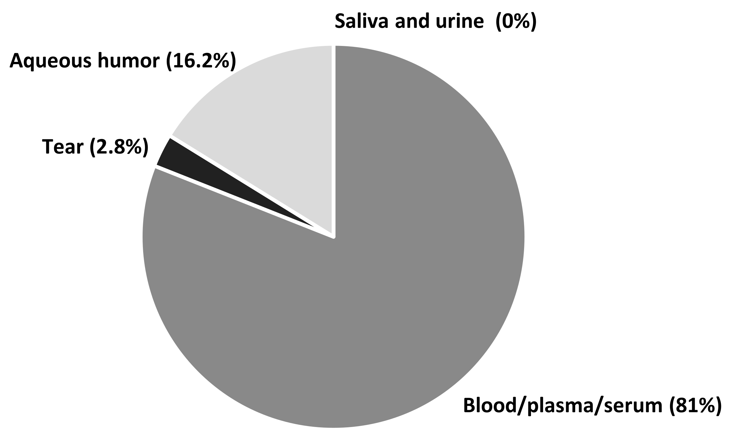
:1. Introduction
2. Proteins
3. Peptides and Amino Acids
4. Autoantibodies and Antibodies
5. Cytokines and Growth Factors
6. Hormones and Enzymes
7. Uric Acid: An Important Biomarker Combined with Protein Metabolism
8. Detection Methods
9. Conclusions
Author Contributions
Funding
Institutional Review Board Statement
Informed Consent Statement
Conflicts of Interest
References
- Tuulonen, A.; Airaksinen, P.J.; Erola, E.; Forsman, E.; Friberg, K.; Kaila, M.; Klemetti, A.; Mäkelä, M.; Oskala, P.; Puska, P.; et al. The Finnish evidence-based guideline for open angle glaucoma. Acta Ophthalmol. Scand. 2003, 81, 3–18. [Google Scholar] [CrossRef]
- Shon, K.; Wollstein, G.; Schuman, J.S.; Sung, K.R. Prediction of glaucomatous field progression: Pointwise analysis. Curr. Eye Res. 2014, 39, 705–710. [Google Scholar] [CrossRef] [PubMed] [Green Version]
- Weinreb, R.N.; Khaw, P.T. Primary open-angle glaucoma. Lancet 2004, 363, 1711–1720. [Google Scholar] [CrossRef]
- Rudnicka, A.R.; Mt-Isa, S.; Owen, C.G.; Cook, D.G.; Ashby, D. Variations in primary open-angle glaucoma prevalence by age, gender, and race: A Bayesian meta-analysis. Invest Ophthalmol. Vis. Sci. 2006, 47, 4254–4261. [Google Scholar] [CrossRef] [Green Version]
- EGS Foundation. European Glaucoma Society Terminology and Guidelines for Glaucoma, 4th Edition—Part 1. Supported by the EGS Foundation. Br. J. Ophthalmol. 2017, 101, 1–72. [Google Scholar] [CrossRef] [Green Version]
- Nakabayashi, M. Review of the ischemia hypothesis for ocular hypertension other than congenital glaucoma and closed-angle glaucoma. Ophthalmologica 2004, 218, 344–349. [Google Scholar] [CrossRef] [PubMed]
- Tezel, G. The immune response in glaucoma: A perspective on the roles of oxidative stress. Exp. Eye Res. 2011, 93, 178–186. [Google Scholar] [CrossRef] [Green Version]
- Shazly, T.A.; Aljajeh, M.; Latina, M.A. Autoimmune basis of glaucoma. Semin. Ophthalmol. 2011, 26, 278–281. [Google Scholar] [CrossRef]
- Tezel, G. A proteomics view of the molecular mechanisms and biomarkers of glaucomatous neurodegeneration. Prog. Retin. Eye Res. 2013, 35, 18–43. [Google Scholar] [CrossRef] [Green Version]
- Soto, I.; Howell, G.R. The complex role of neuroinflammation in glaucoma. Cold Spring Harb. Perspect. Med. 2014, 4, 1–15. [Google Scholar] [CrossRef]
- Chidlow, G.; Wood, J.P.M.; Casson, R.J. Investigations into hypoxia and oxidative stress at the optic nerve head in a rat model of glaucoma. Front. Neurosci. 2017, 11, 1–22. [Google Scholar] [CrossRef] [PubMed]
- Quigley, H.A.; Broman, A.T. The number of people with glaucoma worldwide in 2010 and 2020. Br. J. Ophthalmol. 2006, 90, 262–267. [Google Scholar] [CrossRef] [PubMed] [Green Version]
- Tezel, G.; Yang, X.; Luo, C.; Kain, A.D.; Powell, D.W.; Kuehn, M.H.; Kaplan, K.J. Oxidative stress and the regulation of complement activation in human glaucoma. Invest. Ophthalmol. Vis. Sci. 2010, 51, 5071–5082. [Google Scholar] [CrossRef]
- Yanagi, M.; Kawasaki, R.; Wang, J.J.; Wong, T.Y.; Crowston, J.; Kiuchi, Y. Vascular risk factors in glaucoma: A review. Clin. Exp. Ophthalmol. 2011, 39, 252–258. [Google Scholar] [CrossRef] [PubMed]
- Bell, K.; Gramlich, O.W.; Von Thun Und Hohenstein-Blaul, N.; Beck, S.; Funke, S.; Wilding, C.; Pfeiffer, N.; Grus, F.H. Does autoimmunity play a part in the pathogenesis of glaucoma? Prog. Retin. Eye Res. 2013, 36, 199–216. [Google Scholar] [CrossRef]
- Grzybowski, A.; Och, M.; Kanclerz, P.; Leffler, C.; De Moraes, C.G. Primary open angle glaucoma and vascular risk factors: A review of population based studies from 1990 to 2019. J. Clin. Med. 2020, 9, 761. [Google Scholar] [CrossRef] [Green Version]
- McMonnies, C.W. Glaucoma history and risk factors. J. Optom. 2017, 10, 71–78. [Google Scholar] [CrossRef] [Green Version]
- Hohenstein-Blaul, N.V.T.U.; Kunst, S.; Pfeiffer, N.; Grus, F.H. Biomarkers for glaucoma: From the lab to the clinic. Eye 2017, 31, 225–231. [Google Scholar] [CrossRef] [PubMed] [Green Version]
- Hondur, G.; Göktas, E.; Yang, X.; Al-Aswad, L.; Auran, J.D.; Blumberg, D.M.; Cioffi, G.A.; Liebmann, J.M.; Suh, L.H.; Trief, D.; et al. Oxidative stress-related molecular biomarker candidates for glaucoma. Investig. Ophthalmol. Vis. Sci. 2017, 58, 4078–4088. [Google Scholar] [CrossRef] [Green Version]
- Beykin, G.; Goldberg, J.L. Molecular biomarkers for glaucoma. Curr. Ophthalmol. Rep. 2019, 7, 171–176. [Google Scholar] [CrossRef] [Green Version]
- Biomarkers Definitions Working Group. Biomarkers and surrogate endpoints: Preferred definitions and conceptual framework. Clin. Pharmacol. Ther. 2001, 69, 89–95. [Google Scholar] [CrossRef]
- Strimbu, K.; Tavel, J.A. What are biomarkers? Curr. Opin. HIV AIDS 2010, 5, 463–466. [Google Scholar] [CrossRef]
- Tezel, G.; Thornton, I.L.; Tong, M.G.; Luo, C.; Yang, X.; Cai, J.; Powell, D.W.; Soltau, J.B.; Liebmann, J.M.; Ritch, R. Immunoproteomic analysis of potential serum biomarker candidates in human glaucoma. Invest. Ophthalmol. Vis. Sci. 2012, 53, 8222–8231. [Google Scholar] [CrossRef] [PubMed] [Green Version]
- Damodaran, S. Amino Acids, Peptides, and Proteins. In Fennema’s Food Chemistry, 4th ed.; Damodaran, S., Parkin, K.L., Eds.; CRC Press/Taylor & Francis: Boca Raton, FL, USA, 2008; pp. 425–439. [Google Scholar]
- Lin, Z.; Huang, S.; Yu, H.; Sun, J.; Huang, P.; Zhong, Y. Analysis of plasma hydrogen sulfide, homocysteine, and L-cysteine in open-angle flaucoma patients. J. Ocul. Pharmacol. Ther. 2020, 36, 649–657. [Google Scholar] [CrossRef]
- Lee, W.J.; Na, K.I.; Kim, Y.K.; Jeoung, J.W.; Park, K.H. Diagnostic ability of wide-field retinal nerve fiber layer maps using swept-source optical coherence tomography for detection of preperimetric and early perimetric glaucoma. J. Glaucoma 2017, 26, 577–585. [Google Scholar] [CrossRef]
- Leibovitzh, H.; Cohen, E.; Levi, A.; Kramer, M.; Shochat, T.; Goldberg, E.; Krause, I. Relationship between homocysteine and intraocular pressure in men and women: A population-based study. Medicine 2016, 95, e4858. [Google Scholar] [CrossRef] [PubMed]
- López-Riquelme, N.; Villalba, C.; Tormo, C.; Belmonte, A.; Fernandez, C.; Torralba, G.; Hernández, F. Endothelin-1 levels and biomarkers of oxidative stress in glaucoma patients. Int. Ophthalmol. 2015, 35, 527–532. [Google Scholar] [CrossRef] [PubMed]
- Oddone, F.; Roberti, G.; Micera, A.; Busanello, A.; Bonini, S.; Quaranta, L.; Agnifili, L.; Manni, G. Exploring serum levels of brain derived neurotrophic factor and nerve frowth factor across glaucoma stages. PLoS ONE 2017, 12, e0168565. [Google Scholar] [CrossRef] [Green Version]
- Ghaffariyeh, A.; Honarpisheh, N.; Heidari, M.H.; Puyan, S.; Abasov, F. Brain-derived neurotrophic factor as a biomarker in primary open-angle glaucoma. Optom. Vis. Sci. 2011, 88, 80–85. [Google Scholar] [CrossRef]
- Ghaffariyeh, A.; Honarpisheh, N.; Shakiba, Y.; Puyan, S.; Chamacham, T.; Zahedi, F.; Zarrineghbal, M. Brain-derived neurotrophic factor in patients with normal-tension glaucoma. Optometry 2009, 80, 635–638. [Google Scholar] [CrossRef]
- Baumane, K.; Ranka, R.; Laganovska, G. Association of NT-proANP level in plasma and humor aqueous with primary open-angle glaucoma. Curr. Eye Res. 2017, 42, 233–236. [Google Scholar] [CrossRef] [PubMed]
- Javadiyan, S.; Burdon, K.P.; Whiting, M.J.; Abhary, S.; Straga, T.; Hewitt, A.W.; Mills, R.A.; Craig, J.E. Elevation of serum asymmetrical and symmetrical dimethylarginine in patients with advanced glaucoma. Investig. Ophthalmol. Vis. Sci. 2012, 53, 1923–1927. [Google Scholar] [CrossRef] [PubMed]
- Gye, H.J.; Kim, J.M.; Yoo, C.; Shim, S.H.; Won, Y.S.; Sung, K.C.; Lee, M.Y.; Park, K.H. Relationship between high serum ferritin level and glaucoma in a South Korean population: The Kangbuk Samsung health study. Br. J. Ophthalmol. 2016, 100, 1703–1707. [Google Scholar] [CrossRef] [PubMed]
- Lin, S.C.; Wang, S.Y.; Yoo, C.; Singh, K.; Lin, S.C. Association between serum ferritin and glaucoma in the South Korean population. JAMA Ophthalmol. 2014, 132, 1414–1420. [Google Scholar] [CrossRef] [Green Version]
- González-Iglesias, H.; Álvarez, L.; García, M.; Escribano, J.; Rodríguez-Calvo, P.P.; Fernández-Vega, L.; Coca-Prados, M. Comparative proteomic study in serum of patients with primary open-angle glaucoma and pseudoexfoliation glaucoma. J. Proteom. 2014, 98, 65–78. [Google Scholar] [CrossRef]
- Wang, J.; Fu, M.; Liu, K.; Wang, N.; Zhang, Z.; Zhou, M.; Xu, X. Matricellular proteins play a potential role in acute primary angle closure. Curr. Eye Res. 2018, 43, 771–777. [Google Scholar] [CrossRef]
- Farkas, R.H.; Chowers, I.; Hackam, A.S.; Kageyama, M.; Nickells, R.W.; Otteson, D.C.; Duh, E.J.; Wang, C.; Valenta, D.F.; Gunatilaka, T.L.; et al. Increased expression of iron-regulating genes in monkey and human glaucoma. Invest. Ophthalmol. Vis. Sci. 2004, 45, 1410–1417. [Google Scholar] [CrossRef] [Green Version]
- Wiesmann, C.; de Vos, A.M. Nerve growth factor: Structure and function. Cell Mol Life Sci. 2001, 58, 748–759. [Google Scholar] [CrossRef]
- Binder, D.K.; Scharfman, H.E. Brain-derived neurotrophic factor. Growth Factors 2004, 22, 123–131. [Google Scholar] [CrossRef] [Green Version]
- Skaper, S.D. The neurotrophin family of neurotrophic factors: An overview. Methods Mol. Biol. 2012, 846, 1–12. [Google Scholar]
- Wu, G. Amino Acids: Biochemistry and Nutrition, 1st ed.; CRC Press: Boca Raton, FL, USA, 2013. [Google Scholar]
- Blom, H.J.; Smulders, Y. Overview of homocysteine and folate metabolism. With special references to cardiovascular disease and neural tube defects. J. Inherit. Metab. Dis. 2011, 34, 75–81. [Google Scholar] [CrossRef] [Green Version]
- Ye, S.; Chang, Y.; Kim, C.W.; Kwon, M.J.; Choi, Y.; Ahn, J.; Kim, J.M.; Kim, H.S.; Shin, H.; Ryu, S. Intraocular pressure and coronary artery calcification in asymptomatic men and women. Br. J. Ophthalmol. 2015, 99, 932–936. [Google Scholar] [CrossRef] [PubMed]
- Stow, L.R.; Jacobs, M.E.; Wingo, C.S.; Cain, B.D. Endothelin-1 gene regulation. FASEB J. 2011, 25, 16–28. [Google Scholar] [CrossRef] [PubMed] [Green Version]
- Ludwig, R.J.; Vanhoorelbeke, K.; Leypoldt, F.; Kaya, Z.; Bieber, K.; McLachlan, S.M.; Komorowski, L.; Luo, J.; Cabral-Marques, O.; Hammers, C.M.; et al. Mechanisms of autoantibody-induced pathology. Front. Immunol. 2017, 8, 603. [Google Scholar] [CrossRef] [PubMed] [Green Version]
- Grus, F.H.; Joachim, S.C.; Bruns, K.; Lackner, K.J.; Pfeiffer, N.; Wax, M.B. Serum autoantibodies to alpha-fodrin are present in glaucoma patients from Germany and the United States. Invest. Ophthalmol. Vis. Sci. 2006, 47, 968–976. [Google Scholar] [CrossRef] [Green Version]
- Gramlich, O.W.; Bell, K.; von Thun Und Hohenstein-Blaul, N.; Wilding, C.; Beck, S.; Pfeiffer, N.; Grus, F.H. Autoimmune biomarkers in glaucoma patients. Curr. Opin. Pharmacol. 2013, 13, 90–97. [Google Scholar] [CrossRef]
- Beutgen, V.M.; Perumal, N.; Pfeiffer, N.; Grus, F.H. Autoantibody biomarker discovery in primary open angle glaucoma using serological proteome analysis (SERPA). Front. Immunol. 2019, 10, 381. [Google Scholar] [CrossRef]
- Joachim, S.C.; Pfeiffer, N.; Grus, F.H. Autoantibodies in patients with glaucoma: A comparison of IgG serum antibodies against retinal, optic nerve, and optic nerve head antigens. Graefes Arch. Clin. Exp. Ophthalmol. 2005, 243, 817–823. [Google Scholar] [CrossRef]
- Joachim, S.C.; Wuenschig, D.; Pfeiffer, N.; Grus, F.H. IgG antibody patterns in aqueous humor of patients with primary open angle glaucoma and pseudoexfoliation glaucoma. Mol. Vis. 2007, 13, 1573–1579. [Google Scholar]
- Joachim, S.C.; Bruns, K.; Lackner, K.J.; Pfeiffer, N.; Grus, F.H. Antibodies to alpha B-crystallin, vimentin, and heat shock protein 70 in aqueous humor of patients with normal tension glaucoma and IgG antibody patterns against retinal antigen in aqueous humor. Curr. Eye Res. 2007, 32, 501–509. [Google Scholar] [CrossRef]
- Schmelter, C.; Perumal, N.; Funke, S.; Bell, K.; Pfeiffer, N.; Grus, F.H. Peptides of the variable IgG domain as potential biomarker candidates in primary open-angle glaucoma (POAG). Hum. Mol. Genet. 2017, 26, 4451–4464. [Google Scholar] [CrossRef]
- Li, S.; Zhang, H.; Shao, M.; Li, Y.; Song, Y.; Sun, X.; Cao, W. Association between 17-β-estradiol and interleukin-8 and visual field progression in postmenopausal omen with primary angle closure glaucoma. Am. J. Ophthalmol. 2020, 217, 55–67. [Google Scholar] [CrossRef] [PubMed]
- Gupta, D.; Wen, J.C.; Huebner, J.L.; Stinnett, S.; Kraus, V.B.; Tseng, H.C.; Walsh, M. Cytokine biomarkers in tear film for primary open-angle glaucoma. Clin. Ophthalmol. 2017, 11, 411–416. [Google Scholar] [CrossRef] [PubMed] [Green Version]
- Paschalis, E.I.; Taniguchi, E.V.; Chodosh, J.; Pasquale, L.R.; Colby, K.; Dohlman, C.H.; Shen, L.Q. Blood levels of tumor necrosis factor alpha and its type 2 receptor are elevated in patients with Boston Type I Keratoprosthesis. Curr. Eye Res. 2019, 44, 599–606. [Google Scholar] [CrossRef] [PubMed]
- Kondkar, A.A.; Sultan, T.; Almobarak, F.A.; Kalantan, H.; Al-Obeidan, S.A.; Abu-Amero, K.K. Association of increased levels of plasma tumor necrosis factor alpha with primary open-angle glaucoma. Clin. Ophthalmol. 2018, 12, 701–706. [Google Scholar] [CrossRef] [Green Version]
- Kondkar, A.A.; Azad, T.A.; Almobarak, F.A.; Kalantan, H.; Al-Obeidan, S.A.; Abu-Amero, K.K. Elevated levels of plasma tumor necrosis factor alpha in patients with pseudoexfoliation glaucoma. Clin. Ophthalmol. 2018, 12, 153–159. [Google Scholar] [CrossRef] [Green Version]
- Mohammadi, A.; Amooeian, V.G.; Rashidi, E. Dysfunction in brain-derived neurotrophic factor signaling pathway and susceptibility to schizophrenia, Parkinson’s and Alzheimer’s diseases. Curr. Gene Ther. 2018, 18, 45–63. [Google Scholar] [CrossRef]
- Arvat, E.; Broglio, F.; Ghigo, E. Insulin-like growth factor I: Implications in aging. Drugs Aging 2000, 16, 29–40. [Google Scholar] [CrossRef]
- Puche, J.E.; Castilla-Cortázar, I. Human conditions of insulin-like growth factor-I (IGF-I) deficiency. J. Transl. Med. 2012, 10, 224. [Google Scholar] [CrossRef] [Green Version]
- Ferreira, S.T. Brain insulin, insulin-like growth factor 1 and glucagon-like peptide 1 signalling in Alzheimer’s disease. J. Neuroendocrinol. 2021, 33, e12959. [Google Scholar] [CrossRef]
- Dogan, A.S.; Kabatas, N.; Erden, G.; Celikay, O.; Arzuhal, A.E.; Gurdal, C. Serum insulin-like growth factor-1 levels in patients with pseudoexfoliation syndrome and glaucoma. Int. Ophthalmol. 2017, 37, 371–375. [Google Scholar] [CrossRef] [PubMed]
- Schlötzer-Schrehardt, U.; Naumann, G.O. Ocular and systemic pseudoexfoliation syndrome. Am. J. Ophthalmol. 2006, 141, 921–937. [Google Scholar] [CrossRef]
- Ekström, C.; Kilander, L. Pseudoexfoliation and Alzheimer’s disease: A population-based 30-year follow-up study. Acta Ophthalmol. 2014, 92, 355–358. [Google Scholar] [CrossRef] [PubMed]
- Guo, T.; Guo, L.; Fan, Y.; Fang, L.; Wei, J.; Tan, Y.; Chen, Y.; Fan, X. Aqueous humor levels of TGFβ2 and SFRP1 in different types of glaucoma. BMC Ophthalmol. 2019, 19, 170. [Google Scholar] [CrossRef] [PubMed] [Green Version]
- Newman-Casey, P.A.; Talwar, N.; Nan, B.; Musch, D.C.; Pasquale, L.R.; Stein, J.D. The potential association between postmenopausal hormone use and primary open-angle glaucoma. JAMA Ophthalmol. 2014, 132, 298–303. [Google Scholar] [CrossRef] [PubMed]
- Lee, S.S.; Sanfilippo, P.G.; Yazar, S.; Pennell, C.E.; Hewitt, A.W.; Wang, C.A.; Martin, W.N.; Mackey, D.A. Do levels of stress markers influence the retinal nerve fiber layer thickness in young adults? J. Glaucoma 2020, 29, 587–592. [Google Scholar] [CrossRef]
- Canizales, L.; Rodriguez, L.; Rivera, C.; Martinez, A.; Mendez, F.; Castillo, A. Low-level expression of SOD1 in peripheral blood samples of patients diagnosed with primary open-angle glaucoma. Biomark. Med. 2016, 10, 1218–1223. [Google Scholar] [CrossRef]
- Li, S.; Shao, M.; Li, Y.; Li, X.; Wan, Y.; Sun, X.; Cao, W. Relationship between oxidative stress biomarkers and visual field progression in patients with primary angle closure glaucoma. Oxid. Med. Cell Longev. 2020, 2020, 2701539. [Google Scholar] [CrossRef]
- Glantzounis, G.K.; Tsimoyiannis, E.C.; Kappas, A.M.; Galaris, D.A. Uric acid and oxidative stress. Curr. Pharm. Des. 2005, 11, 4145–4151. [Google Scholar] [CrossRef]
- Li, S.; Shao, M.; Li, D.; Tang, B.; Cao, W.; Sun, X. Association of serum uric acid levels with primary open-angle glaucoma: A 5-year case-control study. Acta Ophthalmol. 2019, 97, 356–363. [Google Scholar] [CrossRef]
- Li, S.; Shao, M.; Tang, B.; Zhang, A.; Cao, W.; Sun, X. The association between serum uric acid and glaucoma severity in primary angle closure glaucoma: A retrospective case-control study. Oncotarget 2017, 8, 2816–2824. [Google Scholar] [CrossRef] [Green Version]
- Good, D.M.; Thongboonkerd, V.; Novak, J.; Bascands, J.L.; Schanstra, J.P.; Coon, J.J.; Dominiczak, A.; Mischak, H. Body fluid proteomics for biomarker discovery: Lessons from the past hold the key to success in the future. J. Proteom. Res. 2007, 6, 4549–4555. [Google Scholar] [CrossRef] [PubMed]
- Thongboonkerd, V. (Ed.) Proteomics of Human Body Fluids. Principles, Methods, and Applications, 1st ed.; Humana Press: Totowa, NJ, USA, 2007. [Google Scholar]
- Zhou, L.; Beuerman, R.W.; Foo, Y.; Liu, S.; Ang, L.P.; Tan, D.T. Characterisation of human tear proteins using high-resolution mass spectrometry. Ann. Acad. Med. Singap. 2006, 35, 400–407. [Google Scholar] [PubMed]
- Zhou, L.; Zhao, S.Z.; Koh, S.K.; Chen, L.; Vaz, C.; Tanavde, V.; Li, X.R.; Beuerman, R.W. In-depth analysis of the human tear proteome. J. Proteom. 2012, 75, 3877–3885. [Google Scholar] [CrossRef]
- Pan, C.W.; Ke, C.; Chen, Q.; Tao, Y.J.; Zha, X.; Zhang, Y.P.; Zhong, H. Differential metabolic markers associated with primary open-angle glaucoma and cataract in human aqueous humor. BMC Ophthalmol. 2020, 20, 183. [Google Scholar] [CrossRef] [PubMed]
- Celis, J.E.; Gromov, P. 2D protein electrophoresis: Can it be perfected? Curr. Opin. Biotechnol. 1999, 10, 16–21. [Google Scholar] [CrossRef]
- Boehm, N.; Wolters, D.; Thiel, U.; Lossbrand, U.; Wiegel, N.; Pfeiffer, N.; Grus, F.H. New insights into autoantibody profiles from immune privileged sites in the eye: A glaucoma study. Brain Behav. Immun. 2012, 26, 96–102. [Google Scholar] [CrossRef] [PubMed]
- Hosseini, S.; Vázquez-Villegas, P.; Rito-Palomares, M.; Martinez-Chapa, S.O. Advantages, disadvantages and modifications of conventional ELISA. In Enzyme-Linked Immunosorbent Assay (ELISA), 1st ed.; Hosseini, S., Vázquez-Villegas, P., Rito-Palomares, M., Martinez-Chapa, S.O., Eds.; Springer: Singapore, 2018; pp. 67–115. [Google Scholar]
- Aretz, I.; Meierhofer, D. Advantages and pitfalls of mass spectrometry based metabolome profiling in systems biology. Int. J. Mol. Sci. 2016, 17, 632. [Google Scholar] [CrossRef] [Green Version]


| Biomarker | Patient Diagnosis | Number of Patients (n)/Biological Material | Results | References |
|---|---|---|---|---|
| Homocysteine (Hcy) | Primary open-angle glaucoma (POAG), normal tension glaucoma (NTG), ocular hypertension (OHT) | n = 44 POAG, n = 20 NTG, n = 52 OHT, n = 78 controls/plasma | Increased levels of Hcy may be associated with glaucoma, especially in patients with POAG. Hcy levels in patients with POAG and NTG groups were significantly higher (p = 0.007–0.043). | [25] |
| Hcy | Glaucomatous retinal nerve fiber layer (RNFL) defect | n = 78,049/plasma | The mean Hcy level in the male group with RNFL defects (11.05 ± 3.80 µmol/L) was higher than those without RNFL defects (10.81 ± 4.12 µmol/L (p = 0.000, χ2 test). | [26] |
| Hcy | Glaucoma | n = 11,850/a retrospective cross-sectional analysis of a database | Mean Hcy levels in subjects with normal intraocular pressure (IOP) of ≤21 mmHg was 11.7 ± 5.5 μmol/L and 12.09 ± 3.43 μmol/L in those with elevated IOP (p = 0.4, 95% CI 1.1–1.8). Mean Hcy levels in subjects with glaucoma were 11.2 ± 3.5 μmol/L compared to 11.7 ± 5.5 μmol/L in subjects without glaucoma and with normal IOP ≤ 21 mmHg (p = 0.4, 95% CI 1.2–2.1). | [27] |
| Hcy | POAG, NTG | n = 48 POAG, n = 15 NTG, n = 75 control/serum | Hcy levels were significantly higher (p = 0.002) in the POAG group compared to the NTG and control groups. | [28] |
| L-cysteine (Cys) | POAG, NTG, OHT | n = 44 POAG, n = 20 NTG, n = 52 OHT, n = 78 controls/plasma | Increased levels of Cys may be associated with glaucoma, especially in patients with POAG. The Cys levels in POAG and NTG groups were significantly higher (p = 0.007–0.043). | [25] |
| Endothelin-1 (ET-1) | POAG, NTG | n = 48 POAG, n = 15 NTG, n = 75 control/serum | The ET-1 levels were significantly higher (p = 0.002) in the POAG group compared to the NTG and control groups. | [28] |
| Brain-derived neurotrophic factor (BDNF) | POAG | n = 45 POAG, n = 15 control/serum | Serum levels of BDNF in glaucoma patients were significantly lower than those measured in healthy control patient (261.2 ± 75.0 pg/mL vs. 313.6 ± 79.6 pg/mL, p = 0.03). Subgroup analysis showed that serum levels of BDNF patients were significantly lower in early (253.8 ± 40.7 pg/mL, p = 0.019) and moderate glaucoma (231.3 ± 54.3 pg/mL, p = 0.04) but not in advanced glaucoma (296.2 ± 103.1 pg/mL, p = 0.06) compared to healthy control subjects. | [29] |
| Nerve growth factor (NGF) | POAG | n = 45 POAG, n = 15 control/serum | Serum levels of NGF in glaucoma patients were significantly lower than those measured in healthy control subject (4.1 ± 1 pg/mL vs. 5.5 ± 1.2 pg/mL, p = 0.01). Subgroup analysis showed that serum levels of NGF were significantly lower in early (3.5 ± 0.9 pg/mL, p = 0.0008) and moderate glaucoma (3.8 ± 0.7 pg/mL p < 0.0001) but not in advanced glaucoma (5.0 ± 0.7 pg/mL, p = 0.32) compared to healthy control subjects. | [29] |
| BDNF | NTG | n = 20 NTG, n = 20 control/tear | The mean level of BDNF detected in the tears of the normal subjects was 77.09 ± 4.84 ng/mL, and the BDNF level in the tears of case group subjects was 24.33 ± 1.48 ng/mL (p < 0.001). | [30] |
| BDNF | POAG | n = 25 POAG, n = 25 control/serum | Mean BDNF level in the serum was 27.16 ± 5.53 ng/mL in the control subjects and 18.42 ± 4.05 ng/mL in the subjects with early-stage glaucoma; there were no significant differences in serum BDNF levels according to the subjects’ age, gender, duration of glaucoma, mean IOP, or blood pressure (p > 0.05). | [31] |
| N-terminal fragment of the proatrial natriuretic peptide (NT-proANP, 1-98) | Glaucoma and cataract | n = 58 POAG, n = 32 cataract (control)/plasma and aqueous humor | The plasma NT-proANP concentration was significantly increased in patients with POAG compared to those in the control group (7.00 vs. 4.65 nmol/L, p = 0.0054). The NT-proANP concentration in the aqueous humor was significantly higher in the POAG patients (0.47 vs. 0.09 nmol/L, p = 0.0112). There was no correlation between the NT-proANP values in the aqueous humor and the plasma of the POAG patients or between the NT-proANP values in the aqueous humor and IOP. | [32] |
| Asymmetric dimethylarginine (ADMA), a dimethylated isomeric derivative of the amino acid l-arginine | Glaucoma | n = 211 glaucoma, n = 295 control/serum | A significant increase in serum ADMA concentration was detected in advanced glaucoma cases compared with control cases (p ≤ 0.0001). | [33] |
| Symmetric dimethylarginine (SDMA), a dimethylated isomeric derivative of the amino acid l-arginine | Glaucoma | n = 211 glaucoma, n = 295 control/serum | A significant increase in serum SDMA concentration was detected in advanced glaucoma cases compared with control cases (p ≤ 0.0001). | [33] |
| Ferritin | Glaucoma | n = 164,029/serum | The mean serum ferritin level was 56.98 ng/mL in women and 223.82 ng/mL in men. | [34] |
| Ferritin | Glaucoma | n = 17,476/serum | Participants whose serum ferritin level was greater than 61 ng/mL had significantly higher odds of a glaucoma diagnosis when compared with those with a level less than 31 ng/mL, after adjustment for potential confounders (ferritin levels of 31−61 ng/mL: odds ratio [OR], 1.17; 95% CI, 0.84−1.62; ferritin levels of 62−112 ng/mL: OR, 1.60; 95% CI, 1.16−2.20; and ferritin levels of 113−3018 ng/mL: OR, 1.89; 95% CI, 1.32−2.72). | [35] |
| Panel of 17 most differentially altered proteins | POAG, PEXG | n = 73(POAG), n = 59 (PEXG), n = 70 healthy controls/serum | Seventeen most differentially altered proteins identified in this analysis confirmed that they were also overexpressed in the intact serum of newly recruited glaucoma patients. | [36] |
| Main matricellular proteins (SPARC, thrombospondin-2, and osteopontin) | Acute primary angle closure (APAC), non-glaucomatous cataract | n = 29 APAC, n = 12 previous APAC, n= 22 cataract/aqueous humor | The levels of SPARC, thrombospondin-2, and osteopontin were significantly elevated in the APAC group as compared to the cataract group (p < 0.001, p < 0.001, and p = 0.009, respectively). | [37] |
| Biomarker | Patient Diagnosis | Number of Patients (n)/Biological Material | Results | References |
|---|---|---|---|---|
| IgG | Primary open-angle glaucoma (POAG) | n = 111 POAG, n = 49 controls/serum | A number of serum proteins were identified and serve as candidate biomarkers of glaucoma. It is unclear whether the IgG-bound serum proteins identified in this study reflect disease-causing antigens. | [23] |
| IgG (heat shock protein 27, α-enolase, actin, and GAPDH) | POAG, pseudoexfoliation glaucoma (PEX) | n = 15 POAG, n = 14 PEX, n = 15 controls/aqueous humor | Significant differences were found between the IgG antibody profiles of the glaucoma groups (PEX and POAG) and the control subjects. | [51] |
| IgG | POAG, normal-tension glaucoma (NTG) | n = 19 POAG, n = 17 NTG, n = 30 controls/serum | All patients showed a complex repertoire of IgG antibodies against retinal, optic nerve, and optic nerve head antigens. The POAG group had the most significant difference against retinal antigens (p = 0.0021) compared with other antigens. In the NTG group, the highest reactivity appeared against the optic nerve head (p = 0.00053) and the optic nerve (p = 0.0025). | [50] |
| Autoantibody, IgG | POAG | n = 13 POAG, n = 15 controls/serum | In total, 75 peptides of the variable IgG domain showed significant glaucoma-related level changes (p < 0.05; log2 fold change ≥ 0.5): 6 peptides were highly abundant in POAG sera, whereas 69 peptides were minimally abundant in comparison to the control group. | [53] |
| Antibodies against trabecular meshwork | POAG | n = 60 POAG, n = 40 controls/serum | VDAC2, PGAM1, and CALD1, new autoantibodies, have been identified in association with POAG. | [49] |
| IgG antibodies against retinal antigens | NTG | n = 21 NTG, n = 21 controls/aqueous humor | α B-crystallin, vimentin, and heat-shock protein were analyzed as the antigen bands. | [52] |
| Biomarker | Patient Diagnosis | Number of Patients (n)/Biological Material | Results | References |
|---|---|---|---|---|
| Interleukin-8 (IL-8) and 17-β-estradiol (E2) | Primary angle closure glaucoma (PACG) and processes related to menopause | n = 200 postmenopausal women with PACG, n = 151 healthy postmenopausal women (control)/serum | Decreased E2 (p = 0.007) and increased IL-8 (p < 0.001) levels were risk factors in postmenopausal women with PACG. A significant negative correlation was observed between IL-8 levels and E2 (p = 0.02). | [25] |
| TNF-α | Primary open-angle glaucoma (POAG) | n = 51 POAG patients n = 88 controls/plasma | The mean TNF-α level was significantly more increased in POAG cases than in control cases (p = 0.003). Logistic regression analysis showed that the risk of POAG was most significantly affected by TNF-α level (not by age and sex). | [57] |
| TNF-α | Pseudoexfoliation glaucoma (PEG) | n = 49 PEG patients n = 88 controls (non-glaucomatous patients)/plasma | The mean TNF-α concentration was significantly higher in PEG patients than in the control subjects (p = 0.000). A positive and significant correlation was observed between TNF-α concentration and cup/disc ratio, providing an important clinical index for PEG. | [58] |
| TNF-α, TNF receptors 1 (TNFR1) and 2 (TNFR2) | Boston keratoprosthesis patients with glaucoma (KPro G), Boston keratoprosthesis patients without glaucoma (KPro NoG), primary angle-closure glaucoma patients without Boston keratoprosthesis (PACG) | n = 19 KPro patients with glaucoma (KPro G), n = 12 KPro patients without glaucoma (KPro NoG), n = 13 patients with primary angle-closure glaucoma without KPro (PACG), n = 21 patients with narrow-angle without glaucoma or KPro (NA)/plasma | KPro G and KPro NoG patients had higher blood plasma levels of TNF-α (p = 0.006, and p = 0.04, respectively) compared to NA patients. KPro G patients had higher blood plasma levels of TNFR2 than NA patients (p = 0.048). KPro status was positively associated with TNF-α levels (p = 0.002) and TNFR2 levels (p = 0.035) after adjusting for age, gender, BMI, glaucoma status, and erythrocyte sedimentation rate (ESR). | [56] |
| Proinflammatory cytokines (IFNγ, IL-10, IL-12p70, IL-13, IL-1β, IL-2, IL-4, IL-6, IL-8, and TNFα) | POAG | n = 10 patients with early POAG, n = 9 control/human tear samples | Mean concentrations of tear-film cytokines were lower in the POAG group for 8 of 10 cytokines tested (for IFNγ, IL-12p70, IL-13, IL-1β, IL-2, IL-4, IL-6, IL-8). IL-12p70 was significantly lower in the tear films of patients with newly diagnosed POAG compared to control subjects (p = 0.035). | [55] |
| Brain-derived neurotrophic factor (BDNF), nerve growth factor (NGF) | POAG with a wide spectrum of disease severity | n = 45 patients affected by glaucoma at different stages, n = 15 control (age-matched)/serum | Serum levels of BDNF in glaucoma patients were significantly lower compared to healthy control subjects (p = 0.03). Additionally, serum levels of BDNF were significantly lower in early (p = 0.019) and moderate (p = 0.04) glaucoma but not in advanced glaucoma (p = 0.06) comparied to control subjects. Serum levels of NGF in glaucoma patients were significantly lower compared to control subjects (p = 0.01). Additonally, serum levels of NGF were significantly lower in early (p = 0.0008) and moderate glaucoma (p < 0.0001) but not in advanced glaucoma (p = 0.32) compared to healthy control subjects. | [29] |
| Insulin-like growth factor-1 (IGF-1) | Pseudoexfoliation (PEX) with or without glaucoma | n = 110 participants (age 65 years or older) who were divided into groups: 1. patients with PEX syndrome (n = 35), 2. patients with PEX glaucoma (n = 34), 3. participants without PEX or glaucoma (n = 41)/serum | Statistically significant differences between the groups in terms of IGF-1 concentration were not observed (p = 0.276). IGF-1 levels in circulation did not differ in the presence of PEX syndrome with or without glaucoma. | [63] |
| Transforming growth factor-β2 (TGFβ2), secreted frizzled-related protein-1 (SFRP1) | Different types of glaucoma | n = 105 patients divided into five groups: cataract (control), POAG, chronic angle-closure glaucoma (CACG), primary angle-closure suspects (PACS), and acute angle-closure glaucoma (AACG)/aqueous humor | The concentration of TGFβ2 in POAG patients (but not CACG, PACS, or AACG patients) was significantly higher compared to control subjectsDifferences in TGFβ2 concentration within AACG patients were observed after consideration of IOP (high and normal). The concentration of SFRP1 was not significantly different among the groups, but a statistically significant negative correlation between SFRP1 and IOP existed in the POAG group. | [66] |
| Hormone | Patient Diagnosis | Number of Patients (n)/Material | Results | References |
|---|---|---|---|---|
| Adrenocorticotropic hormone (ACTH) | RNFL thickness | n = 863/serum | Higher levels of ACTH were associated with thinner RNFL globally (p = 0.009). Lower adrenal sensitivity was associated with thinner RNFL inferotemporally (p < 0.001) and temporally (p = 0.037). | [68] |
| Cortisol | RNFL thickness | n = 863/serum | RNFL thickness was not associated with plasma cortisol levels. | [68] |
| Postmenopausal hormones | Primary open-angle glaucoma (POAG) | 152,163/statistical analysis. Postmenopausal hormone medications containing estrogen only, estrogen + progesterone, and estrogen + androgen, as captured from outpatient pharmacy claims over a 4-year period | Of 152,163 eligible enrollees, 2925 (1.9%) developed POAG. After adjustment for confounding factors, each additional month of use of PMH containing estrogen only was associated with a 0.4% reduced risk for POAG (HR, 0.996 (95% CI, 0.993–0.999); p = 0.02). The risk for POAG did not differ with each additional month of use of estrogen + progesterone (HR, 0.994 (95% CI, 0.987–1.001); p = 0.08) or estrogen + androgen (HR, 0.999 (95% CI, 0.988–1.011); p = 0.89). | [67] |
| Superoxide dismutase (SOD), total antioxidant status (TAS), hydrogen peroxide (H2O2), malondialdehyde (MDA), glutathione peroxidase, glutathione reductase | Primary angle-closure glaucoma (PACG). | n = 94 PACG, n = 89 controls/serum | Serum levels of TAS, SOD, MDA, and H2O2 were independent risk/protective factors for PACG. | [70] |
| Superoxide dismutase 1 (SOD1) | POAG | n = 15 POAG, n = 11 controls/peripheral blood | The mRNA expression level of SOD1 showed significant downregulation in patients diagnosed with POAG. | [69] |
| Patient Diagnosis | Number of Patients (n)/Biological Material | Results | References |
|---|---|---|---|
| Primary open-angle glaucoma (POAG) | n = 163 POAG, n = 103 controls/serum | The level of serum UA in the POAG group (0.321 ± 0.084 mmol/L) was approximately 12.77% lower (p < 0.001) than that of the control group (0.362 ± 0.053 mmol/L). The UA/creatinine (Cr) ratio was approximately 14.99% lower (p < 0.001) in patients with POAG (4.47 ± 1.15) compared with the control group (5.14 ± 1.05). | [72] |
| Primary angle closure glaucoma (PACG) | n = 886 PACG, n = 994 control/serum | The levels of UA were significantly lower (p = 0.025) in PACG patients (0.286 ± 0.082 mmol/L) compared with control subjects (0.295 ± 0.085 mmol/L). The mean serum UA levels were lowest in the severe group (0.281 ± 0.074 mmol/L) followed by moderate (0.282 ± 0.080 mmol/L) and mild (0.297 ± 0.090 mmol/L), with significant differences among the three groups (p = 0.032). | [73] |
Publisher’s Note: MDPI stays neutral with regard to jurisdictional claims in published maps and institutional affiliations. |
© 2021 by the authors. Licensee MDPI, Basel, Switzerland. This article is an open access article distributed under the terms and conditions of the Creative Commons Attribution (CC BY) license (https://creativecommons.org/licenses/by/4.0/).
Share and Cite
Fiedorowicz, E.; Cieślińska, A.; Kuklo, P.; Grzybowski, A. Protein Biomarkers in Glaucoma: A Review. J. Clin. Med. 2021, 10, 5388. https://doi.org/10.3390/jcm10225388
Fiedorowicz E, Cieślińska A, Kuklo P, Grzybowski A. Protein Biomarkers in Glaucoma: A Review. Journal of Clinical Medicine. 2021; 10(22):5388. https://doi.org/10.3390/jcm10225388
Chicago/Turabian StyleFiedorowicz, Ewa, Anna Cieślińska, Patrycja Kuklo, and Andrzej Grzybowski. 2021. "Protein Biomarkers in Glaucoma: A Review" Journal of Clinical Medicine 10, no. 22: 5388. https://doi.org/10.3390/jcm10225388
APA StyleFiedorowicz, E., Cieślińska, A., Kuklo, P., & Grzybowski, A. (2021). Protein Biomarkers in Glaucoma: A Review. Journal of Clinical Medicine, 10(22), 5388. https://doi.org/10.3390/jcm10225388