
Prediction of Late-Onset Small for Gestational Age and Fetal Growth Restriction by Fetal Biometry at 35 Weeks and Impact of Ultrasound–Delivery Interval: Comparison of Six Fetal Growth Standards

 ,
,  , , , , and
Abstract
:1. Introduction
2. Materials and Methods
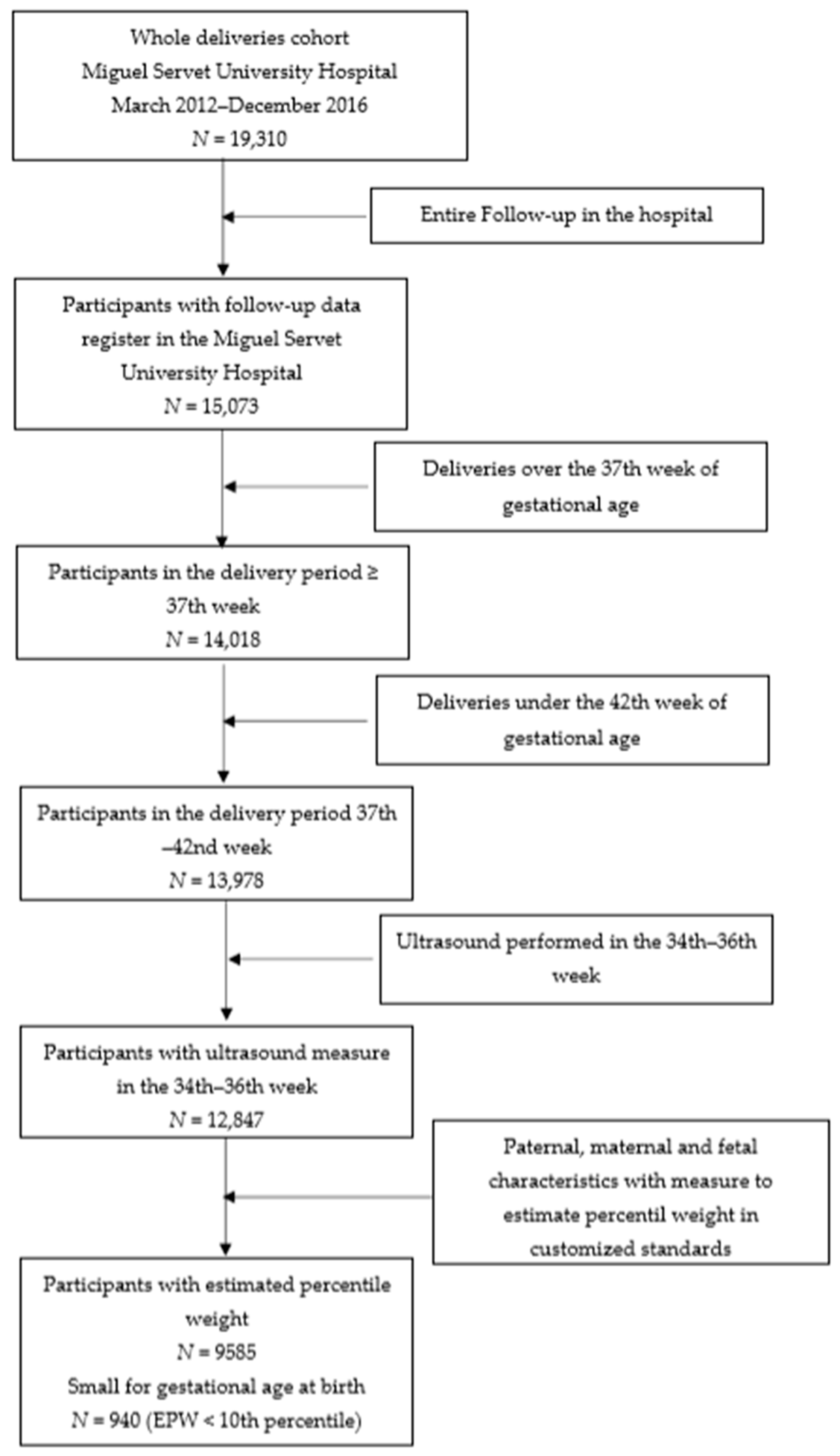
2.1. Study Design
2.2. Estimated Percentile Weight
2.3. Statistical Analysis
3. Results
3.1. Descriptive Results
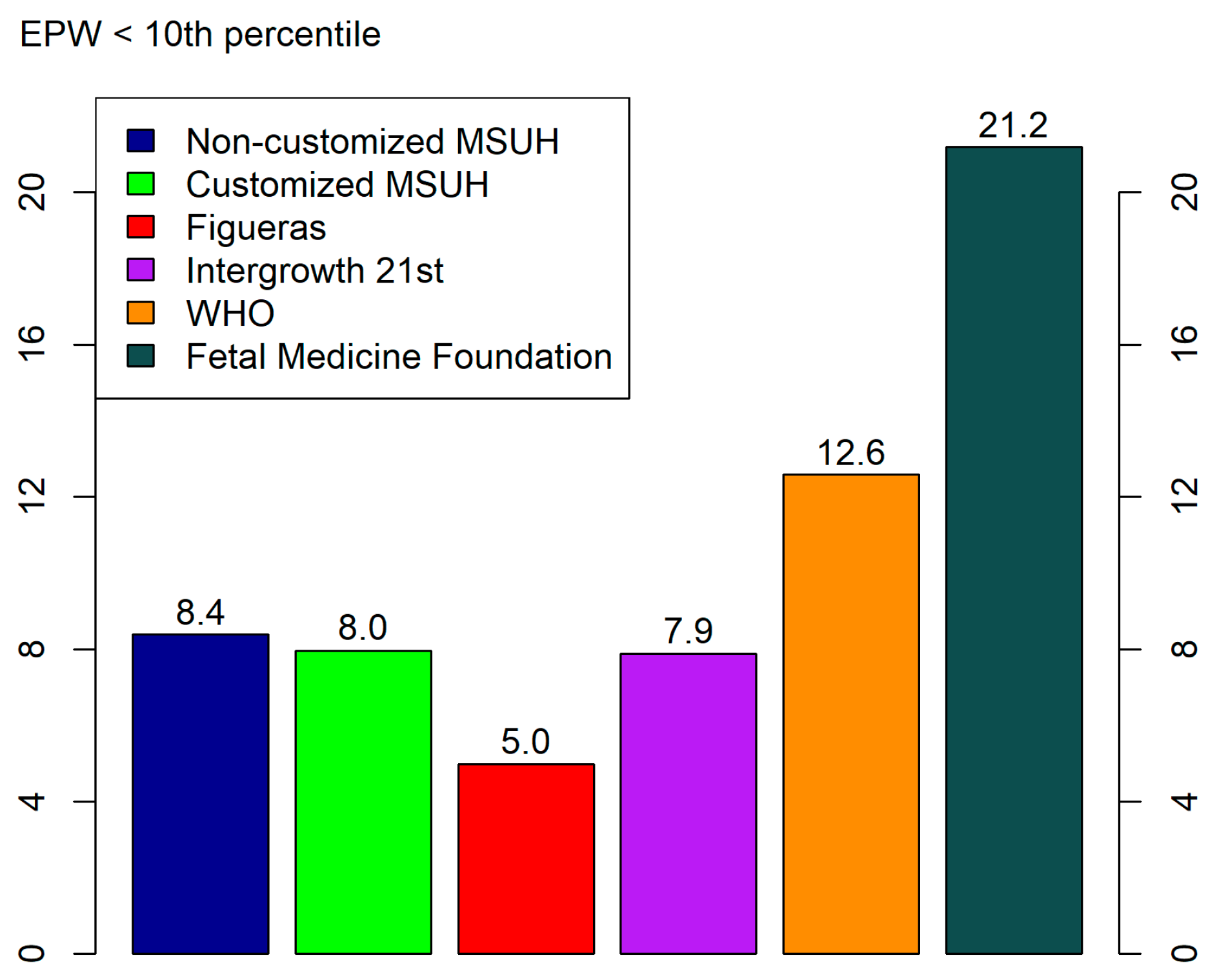
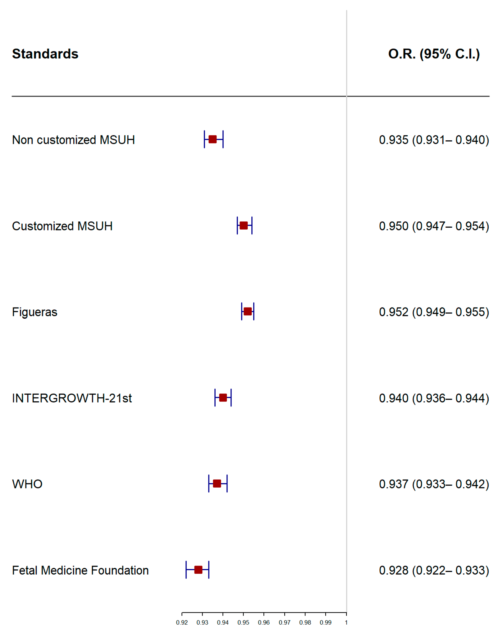
3.2. Comparison of Standards
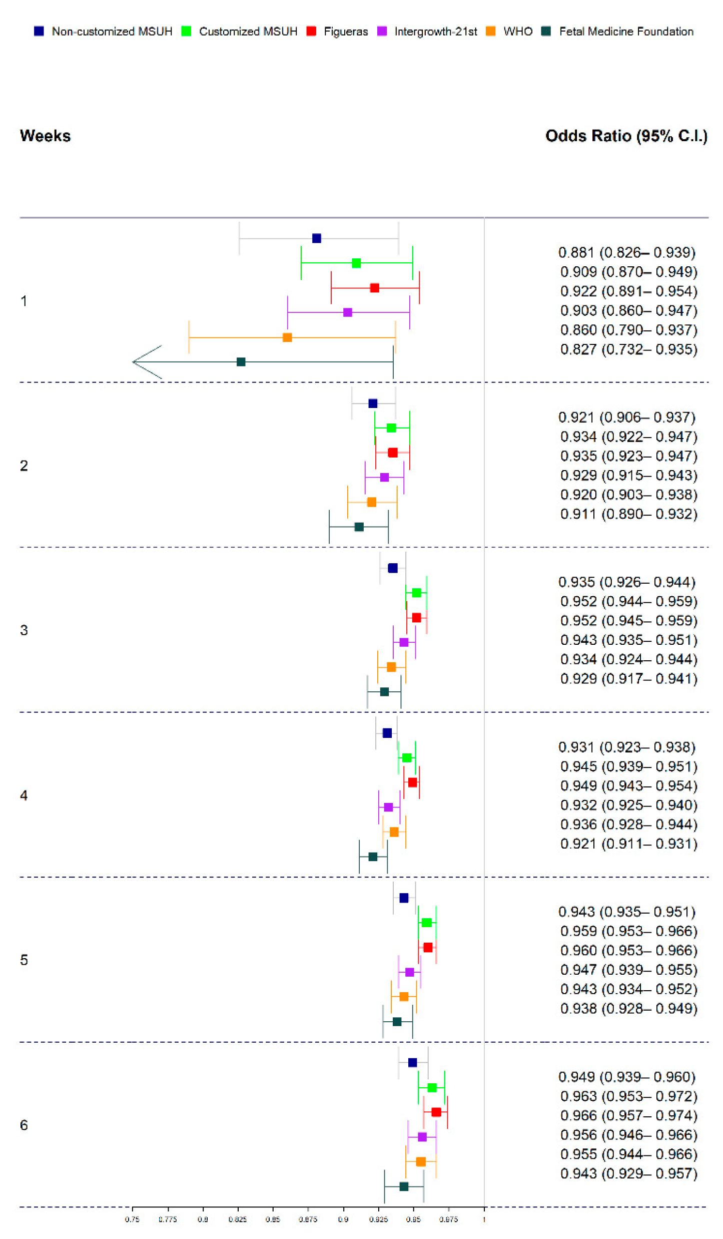
3.3. Ultrasound-Delivery Interval: Comparison of Standards
4. Discussion
4.1. Principal Findings
4.2. Prediction by Fetal Biometry and Ultrasound–Delivery Interval
4.3. Prediction by Fetal Biometry and Ultrasound–Delivery Interval: Comparison of Standards
4.4. Clinical and Research Implications
4.5. Strengths and Limitations of the Study
5. Conclusions
Author Contributions
Funding
Institutional Review Board Statement
Informed Consent Statement
Data Availability Statement
Acknowledgments
Conflicts of Interest
References
- Jelks, A.; Cifuentes, R.; Ross, M.G. Clinician bias in fundal height measurement. Obstet. Gynecol. 2007, 110, 892–899. [Google Scholar] [CrossRef]
- Chauhan, S.P.; Magann, E.F. Screening for fetal growth restriction. Clin. Obstet. Gynecol. 2006, 49, 284–294. [Google Scholar] [CrossRef] [PubMed]
- Conde-Agudelo, A.; Papageorghiou, A.T.; Kennedy, S.H.; Villar, J. Novel biomarkers for predicting intrauterine growth restriction: A systematic review and meta-analysis. BJOG 2013, 120, 681–694. [Google Scholar] [CrossRef] [PubMed] [Green Version]
- Triunfo, S.; Crispi, F.; Gratacos, E.; Figueras, F. Prediction of delivery of small-for-gestational age neonates and adverse perinatal outcome by fetoplacental Doppler at 37 weeks' gestation. Ultrasound Obstet. Gynecol. 2017, 49, 364–371. [Google Scholar] [CrossRef] [Green Version]
- McCowan, L.M.; Figueras, F.; Anderson, N.H. Evidence-based national guidelines for the management of suspected fetal growth restriction: Comparison, consensus, and controversy. Am. J. Obstet. Gynecol. 2018, 218, S855–S868. [Google Scholar] [CrossRef] [Green Version]
- Gardosi, J.; Giddings, S.; Buller, S.; Southam, M.; Williams, M. Preventing stillbirths through improved antenatal recognition of pregnancies at risk due to fetal growth restriction. Public Health 2014, 128, 698–702. [Google Scholar] [CrossRef] [PubMed]
- Gardosi, J.; Madurasinghe, V.; Williams, M.; Malik, A.; Francis, A. Maternal and fetal risk factors for stillbirth: Population based study. BMJ 2013, 346, f108. [Google Scholar] [CrossRef] [Green Version]
- Smith, N.A.; Bukowski, R.; Thomas, A.M.; Cantonwine, D.; Zera, C.; Robinson, J.N. Identification of pathologically small fetuses using customized, ultrasound and population-based growth norms. Ultrasound Obstet. Gynecol. 2014, 44, 595–599. [Google Scholar] [CrossRef]
- Jarvis, S.; Glinianaia, S.V.; Torrioli, M.G.; Platt, M.J.; Miceli, M.; Jouk, P.S.; Johnson, A.; Hutton, J.; Hemming, K.; Hagberg, G.; et al. Surveillance of Cerebral Palsy in Europe (SCPE) collaboration of European Cerebral Palsy Registers. Cerebral palsy and intrauterine growth in single births: European collaborative study. Lancet 2003, 362, 1106–1111. [Google Scholar] [CrossRef]
- Kady, S.; Gardosi, J. Perinatal mortality and fetal growth restriction. Best Pract. Res. Clin. Obstet. Gynaecol. 2004, 18, 397–410. [Google Scholar] [CrossRef]
- Vasak, B.; Koenen, S.V.; Koster, M.P.; Hukkelhoven, C.W.; Franx, A.; Hanson, M.A.; Visser, G.H. Human fetal growth is constrained below optimal for perinatal survival. Ultrasound Obstet. Gynecol. 2015, 45, 162–167. [Google Scholar] [CrossRef] [PubMed]
- Lindqvist, P.G.; Molin, J. Does antenatal identification of small-for-gestational age fetuses significantly improve their outcome? Ultrasound Obstet. Gynecol. 2005, 25, 258–264. [Google Scholar] [CrossRef]
- Sovio, U.; Smith, G.C.S. The effect of customization and use of a fetal growth standard on the association between birthweight percentile and adverse perinatal outcome. Am. J. Obstet. Gynecol. 2018, 218, S738–S744. [Google Scholar] [CrossRef] [PubMed] [Green Version]
- Caradeux, J.; Martinez-Portilla, R.J.; Peguero, A.; Sotiriadis, A.; Figueras, F. Diagnostic performance of third-trimester ultrasound for the prediction of late-onset fetal growth restriction: A systematic review and meta-analysis. Am. J. Obstet. Gynecol. 2019, 220, 449–459. [Google Scholar] [CrossRef]
- Figueras, F.; Meler, E.; Iraola, A.; Eixarch, E.; Coll, O.; Figueras, J.; Francis, A.; Gratacos, E.; Gardosi, J. Customized birthweight standards for a Spanish population. Eur. J. Obstet. Gynecol. Reprod. Biol. 2008, 136, 20–24. [Google Scholar] [CrossRef]
- Saviron-Cornudella, R.; Esteban, L.M.; Lerma, D.; Cotaina, L.; Borque, Á.; Sanz, G.; Castán, S. Comparison of fetal weight distribution improved by paternal height by Spanish standard versus Intergrowth 21st standard. J. Perinat. Med. 2018, 46, 750–759. [Google Scholar] [CrossRef] [PubMed] [Green Version]
- Kiserud, T.; Piaggio, G.; Carroli, G.; Widmer, M.; Carvalho, J.; Neerup Jensen, L.; Giordano, D.; Cecatti, J.G.; Abdel Aleem, H.; Talegawkar, S.A.; et al. The World Health Organization Fetal Growth Charts: A Multinational Longitudinal Study of Ultrasound Biometric Measurements and Estimated Fetal Weight. PLoS Med. 2017, 14, e1002220. [Google Scholar]
- Hadlock, F.P.; Harrist, R.B.; Martinez-Poyer, J. In utero analysis of fetal growth: A sonographic weight standard. Radiology 1991, 181, 129–133. [Google Scholar] [CrossRef] [PubMed]
- Hadlock, F.P.; Harrist, R.B.; Sharman, R.S.; Deter, R.L.; Park, S.K. Estimation of fetal weight with the use of head, body, and femur measurements: A prospective study. Am. J. Obstet. Gynecol. 1985, 151, 333–337. [Google Scholar] [CrossRef]
- Stirnemann, J.; Villar, J.; Salomon, L.J.; Ohuma, E.; Ruyan, P.; Altman, D.G.; Nosten, F.; Craik, R.; Munim, S.; Cheikh Ismail, L.; et al. International estimated fetal weight standards of the INTERGROWTH-21st project. Ultrasound Obstet. Gynecol. 2017, 49, 478–486. [Google Scholar] [CrossRef] [PubMed] [Green Version]
- Villar, J.; Cheikh Ismail, L.; Victora, C.G.; Ohuma, E.O.; Bertino, E.; Altman, D.G.; Lambert, A.; Papageorghiou, A.T.; Carvalho, M.; Jaffer, Y.A.; et al. International standards for newborn weight, length, and head circumference by gestational age and sex: The newborn cross-sectional study of the INTERGROWTH-21st project. Lancet 2014, 384, 857–868. [Google Scholar] [CrossRef]
- Nicolaides, K.H.; Wright, D.; Syngelaki, A.; Wright, A.; Akolekar, R. Fetal Medicine Foundation fetal and neonatal population weight charts. Ultrasound Obstet. Gynecol. 2018, 52, 44–51. [Google Scholar] [CrossRef] [PubMed] [Green Version]
- Gardosi, J.; Chang, A.; Kalyan, B.; Sahota, D.; Symonds, E.M. Customised antenatal growth charts. Lancet 1992, 339, 283–287. [Google Scholar] [CrossRef]
- Gardosi, J.; Francis, A. Controlled trial of fundal height measurement plotted on customized antenatal growth charts. Br. J. Obstet. Gynaecol. 1999, 106, 309–317. [Google Scholar] [CrossRef] [PubMed]
- The Royal College of Obstetricians and Gynaecologists. Small-for-Gestational-Age Fetus, Investigation and Management (Green-top Guideline No. 31), 2nd ed.; The Royal College of Obstetricians and Gynaecologists: London, UK, 2013. [Google Scholar]
- Savirón-Cornudella, R.; Esteban, L.M.; Tajada-Duaso, M.; Castán-Mateo, S.; Dieste-Pérez, P.; Cotaina-Gracia, L.; Lerma-Puertas, D.; Sanz, G.; Perez-Lopez, F.R. Detection of Adverse Perinatal Outcomes at Term Delivery Using Ultrasound Estimated Percentile Weight at 35 Weeks of Gestation: Comparison of Five Fetal Growth Standards. Fetal Diagn. Ther. 2020, 47, 104–114. [Google Scholar] [CrossRef]
- Kabiri, D.; Romero, R.; Gudicha, D.W.; Hernandez-Andrade, E.; Pacora, P.; Benshalom-Tirosh, N.; Tirosh, D.; Yeo, L.; Erez, O.; Hassan, S.S.; et al. Prediction of adverse perinatal outcomes by fetal biometry: A comparison of customized and population-based standards. Ultrasound Obstet. Gynecol. 2020, 55, 177–188. [Google Scholar] [CrossRef]
- Hutcheon, J.A.; Zhang, X.; Platt, R.W.; Cnattingius, S.; Kramer, M.S. The case against customised birthweight standards. Pediatric Perinat. Epidemiol. 2011, 25, 11–16. [Google Scholar] [CrossRef]
- Ohuma, E.O.; Altman, D.G. Statistical methodology for constructing gestational age-related charts using cross-sectional and longitudinal data: THE INTERGROWTH-21st Project as a case study. Stat. Med. 2019, 38, 3507–3526. [Google Scholar] [CrossRef] [Green Version]
- Committee on Obstetric Practice and American Institute of Ultrasound in Medicine and Society for Maternal-Fetal Medicine. Committee Opinion No 700: Methods for Estimating the Due Date. Obstet. Gynecol. 2017, 129, e150–e154. [Google Scholar] [CrossRef]
- Carrascosa, A.; Fernández, J.M.; Ferrández, A.; López-Siguero, J.P.; López, D.; Sánchez, E.; Colaborador, G. Estudios españoles de crecimiento 2010. Rev. Esp. Endocrinol. Pediatric 2011, 2, 59–62. [Google Scholar]
- Hanley, J.A.; McNeil, B.J. The meaning and use of the area under a receiver operating characteristic (ROC) curve. Radiology 1982, 143, 29–36. [Google Scholar] [CrossRef] [PubMed] [Green Version]
- R Core Team. R: A Languaje and Environment for Statistical Computing; R Foundation for Statistical Computing: Vienna, Austria, 2014; Available online: http://www.R-project.org (accessed on 1 June 2021).
- Savirón-Cornudella, R.; Esteban, L.M.; Dieste-Pérez, P.; Pérez-López, F.R.; Castán-Larraz, B.; Sanz, G.; Tajada-Duaso, M. Prediction of Large for Gestational Age by Ultrasound at 35 Weeks and Impact of Ultrasound-Delivery Interval: Comparison of 6 Standards. Fetal Diagn. Ther. 2021, 48, 15–23. [Google Scholar] [CrossRef]
- National Collaborating Centre for Women’s and Children’s Health (NCC-WCH) on behalf of the National Institute of Health and Care Excellence (NICE). Antenatal Care (NICE Clinical Guideline 62); National Institute of Health and Care Excellence: London, UK, 2008. [Google Scholar]
- American College of Obstetricians and Gynecologists. ACOG Practice Bulletin No. 101: Ultrasonography in pregnancy. Obstet. Gynecol. 2009, 113, 451–461. [Google Scholar] [CrossRef]
- Sovio, U.; White, I.R.; Dacey, A.; Pasupathy, D.; Smith, G.C. Screening for fetal growth restriction with universal third trimester ultrasonography in nulliparous women in the Pregnancy Outcome Prediction (POP) study: A prospective cohort study. Lancet 2015, 386, 2089–2097. [Google Scholar] [CrossRef] [Green Version]
- Miranda, J.; Rodriguez-Lopez, M.; Triunfo, S.; Sairanen, M.; Kouru, H.; Parra-Saavedra, M.; Crovetto, F.; Figueras, F.; Crispi, F.; Gratacós, E. Prediction of fetal growth restriction using estimated fetal weight vs a combined screening model in the third trimester. Ultrasound Obstet. Gynecol. 2017, 50, 603–611. [Google Scholar] [CrossRef] [Green Version]
- Fadigas, C.; Saiid, Y.; Gonzalez, R.; Poon, L.C.; Nicolaides, K.H. Prediction of small for gestational age neonates: Screening by fetal biometry at 35–37 weeks. Ultrasound Obstet. Gynecol. 2015, 45, 559–565. [Google Scholar] [CrossRef] [PubMed]
- Dudley, N.J. A systematic review of the ultrasound estimation of fetal weight. Ultrasound Obstet. Gynecol. 2005, 25, 80–89. [Google Scholar] [CrossRef]
- Degani, S. Fetal biometry: Clinical, pathological, and technical considerations. Obstet. Gynecol. Surv. 2001, 56, 159–167. [Google Scholar] [CrossRef]
- Proctor, L.K.; Rushworth, V.; Shah, P.S.; Keunen, J.; Windrim, R.; Ryan, G.; Kingdom, J. Incorporation of femur length leads to underestimation of fetal weight in asymmetric preterm growth restriction. Ultrasound Obstet. Gynecol. 2010, 35, 442–448. [Google Scholar] [CrossRef]
- Souka, A.P.; Papastefanou, I.; Pilalis, A.; Michalitsi, V.; Panagopoulos, P.; Kassanos, D. Performance of the ultrasound examination in the early and late third trimester for the prediction of birth weight deviations. Prenat. Diagn. 2013, 33, 915–920. [Google Scholar] [CrossRef]
- Roma, E.; Arnau, A.; Berdala, R.; Bergos, C.; Montesinos, J.; Figueras, F. Ultrasound screening for fetal growth restriction at 36 vs 32 weeks' gestation: A randomized trial (ROUTE). Ultrasound Obstet. Gynecol. 2015, 46, 391–397. [Google Scholar] [CrossRef] [Green Version]
- Blue, N.R.; Beddow, M.E.; Savabi, M.; Katukuri, V.R.; Mozurkewich, E.L.; Chao, C.R. A Comparison of Methods for the Diagnosis of Fetal Growth Restriction Between the Royal College of Obstetricians and Gynaecologists and the American College of Obstetricians and Gynecologists. Obstet. Gynecol. 2018, 131, 835–841. [Google Scholar] [CrossRef] [PubMed]
- Blue, N.R.; Savabi, M.; Beddow, M.E.; Katukuri, V.R.; Fritts, C.M.; Izquierdo, L.A.; Chao, C.R. The Hadlock Method Is Superior to Newer Methods for the Prediction of the Birth Weight Percentile. J. Ultrasound Med. 2019, 38, 587–596. [Google Scholar] [CrossRef]
- Odibo, A.O.; Nwabuobi, C.; Odibo, L.; Leavitt, K.; Obican, S.; Tuuli, M.G. Customized fetal growth standard compared with the INTERGROWTH-21st century standard at predicting small-for-gestational-age neonates. Acta Obstet. Gynecol. Scand. 2018, 97, 1381–1387. [Google Scholar] [CrossRef] [PubMed] [Green Version]
- Nwabuobi, C.; Odibo, L.; Camisasca-Lopina, H.; Leavitt, K.; Tuuli, M.; Odibo, A.O. Comparing INTERGROWTH-21st Century and Hadlock growth standards to predict small for gestational age and short-term neonatal outcomes. J. Matern. Fetal Neonatal Med. 2019, 6, 1–7. [Google Scholar] [CrossRef]
- Reboul, Q.; Delabaere, A.; Luo, Z.C.; Nuyt, A.M.; Wu, Y.; Chauleur, C.; Fraser, W.; Audibert, F. Prediction of small-for-gestational-age neonate by third-trimester fetal biometry and impact of ultrasound-delivery interval. Ultrasound Obstet. Gynecol. 2017, 49, 372–378. [Google Scholar] [CrossRef] [Green Version]
- Lappen, J.R.; Myers, S.A. The systematic error in the estimation of fetal weight and the underestimation of fetal growth restriction. Am. J. Obstet. Gynecol. 2017, 216, 477–483. [Google Scholar] [CrossRef] [PubMed]


| Clinical Characteristics | Pregnancies (n = 9585) |
|---|---|
| Parental characteristics | |
| Maternal age (years) | 33.3 (30.1–36.1) |
| Maternal body mass index (kg/m2) | 23.2 (21.1–26.2) |
| Maternal height (cm) | 163 (159–168) |
| Paternal height (cm) | 176 (172–181) |
| Parity | |
| 0 | 5077 (53.0%) |
| 1 | 3724 (38.9%) |
| ≥ 2 | 784 (8.1%) |
| Maternal ethnicity | |
| Caucasian | 9243 (96.4%) |
| Asian | 110 (1.1%) |
| African | 232 (2.4%) |
| Maternal smoking habits | |
| Yes | 1546 (16.1%) |
| No | 8039 (83.9%) |
| Ultrasound parameters at 35 (34–36) weeks | |
| Gestational age (weeks) at ultrasound | 35.1 (35.0–35.3) |
| Estimated fetal weight (grams) by Hadlock | 2495 (2314–2697) |
| Estimated fetal weight (grams) by Stirnemann | 2421 (2209–2648) |
| Percentile by standard | P50 (P10–P90) |
| Non-customized MSUH | 52.6 (11.9–93.3) |
| Customized MSUH | 52.9 (12.2–92.9) |
| Figueras | 59.3 (18.1–93.5) |
| INTERGROWTH-21st | 51.9 (12.7–89.8) |
| WHO | 43.1 (7.5–74.9) |
| Fetal Medicine Foundation | 37.6 (2.7–89.9) |
| Pregnancy and perinatal outcomes | |
| Gestational age at delivery | 40.0 (39.1–40.7) |
| Newborn gender | |
| Female | 4652 (48.5%) |
| Male | 4933 (51.5%) |
| Birth weight | 3310 (3030–3590) |
| Small for gestational age (<10th percentile) | 902 (9.4%) |
| 5-min Apgar score < 7 | 42 (0.4%) |
| Instrumental delivery for NRFS | 161 (1.7%) |
| Cesarean delivery for NRFS | 265 (2.8%) |
| Arterial cord blood pH < 7.10 | 254 (2.6%) |
| Stillbirth | 19 (0.2%) |
| Any adverse perinatal outcome * | 645 (6.7%) |
| 5-Min Apgar Score < 7 | Instrumental Delivery for NRFS | Cesarean Delivery for NRFS | Arterial Cord Blood pH < 7.10 | Stillbirth | Any APO | |
|---|---|---|---|---|---|---|
| Total cohort | 42 | 161 | 265 | 254 | 19 | 645 |
| SGA | 12 (28.6%) | 32 (19.9%) | 71 (26.8%) | 45 (17.7%) | 5 (26.3%) | 139 (21.6%) |
| EPW < 10 | ||||||
| Non-customized MSUH | 8 (19.0%) | 15 (9.3%) | 43 (16.2%) | 26 (10.2%) | 5 (26.3%) | 76 (11.8%) |
| Customized MSUH | 6 (14.3%) | 9 (5.6%) | 38 (14.3%) | 22 (8.7%) | 5 (26.3%) | 62 (9.6%) |
| Figueras | 5 (11.9%) | 8 (5.0%) | 32 (12.1%) | 18 (7.1%) | 4 (21.1%) | 51 (7.9%) |
| INTERGROWTH-21st | 7 (16.7%) | 11 (6.8%) | 39 (14.7%) | 26 (10.2%) | 5 (26.3%) | 69 (10.7%) |
| WHO | 12 (28.6%) | 24 (14.9%) | 57 (21.5%) | 42 (16.5%) | 7 (36.8%) | 112 (17.4%) |
| FMF | 17 (40.5%) | 37 (23.0%) | 89 (33.6%) | 62 (24.4%) | 10 (52.6%) | 174 (27.0%) |
| Prediction of Small for Gestational Age by Standard | Area under the Curve (95% C.I.) | Sensitivity (95% C.I.) and Threshold Percentile Points * | |||
|---|---|---|---|---|---|
| FPR 5% | FPR 10% | FPR 15% | FPR 20% | ||
| Small for gestational age | |||||
| Non-customized MSUH | 0.87 (0.85–0.88) | 42.6 (39.4–45.9) (Thr: 10.3) | 60.4 (57.1–63.6) (Thr: 17.3) | 70.5 (67.4–73.4) (Thr: 23.3) | 78.2 (75.3–80.8) (Thr: 28.5) |
| Customized MSUH | 0.82 (0.80–0.83) | 35.5 (32.3–38.6) (Thr: 9.9) | 51.1 (47.8–54.4) (Thr: 16.3) | 60.9 (57.6–64.1) (Thr: 22.7) | 67.6 (64.4–70.6) (Thr: 28.1) |
| Figueras | 0.82 (0.80–0.83) | 35.4 (32.3–38.6) (Thr: 14.5) | 48.9 (45.6–52.2) (Thr: 22.9) | 60.8 (57.5–64.0) (Thr: 29.5) | 66.4 (63.2–69.5) (Thr: 34.6) |
| INTERGROWTH-21st | 0.85 (0.84–0.86) | 37.8 (34.6–41.1) (Thr: 10.2) | 56.3 (53.0–59.6) (Thr: 17.3) | 66.7 (63.5–69.8) (Thr: 22.9) | 73.7 (70.7–76.5) (Thr: 28.3) |
| WHO | 0.84 (0.83–0.85) | 38.6 (35.4–41.9) (Thr: 6.2) | 56.1 (52.8–59.4) (Thr: 11.1) | 61.0 (57.7–64.2) (Thr: 14.6) | 70.6 (67.5–73.5) (Thr: 19.1) |
| Fetal Medicine Foundation | 0.87 (0.85–0.88) | 42.4 (39.2–45.7) (Thr: 1.9) | 60.8 (57.5–64.0) (Thr: 5.3) | 70.7 (67.6–73.6) (Thr: 9.3) | 76.3 (73.4–79.0) (Thr: 13.1) |
| Customized MSUH | Figueras | INTERGROWTH-21st | WHO | Fetal Medicine Foundation | |||||||||||
|---|---|---|---|---|---|---|---|---|---|---|---|---|---|---|---|
| AUC | Sens | APOs | AUC | Sens | APOs | AUC | Sens | APOs | AUC | Sens | APOs | AUC | Sens | APOs | |
| NC MSUH | <0.001 | <0.001 | 0.242 | <0.001 | <0.001 | 0.025 | <0.001 | 0.086 | 0.597 | <0.001 | 0.071 | 0.006 | 0.169 | 0.900 | <0.001 |
| C MSUH | 0.053 | 0.375 | 0.325 | <0.001 | 0.030 | 0.580 | <0.001 | 0.037 | <0.001 | <0.001 | <0.001 | <0.001 | |||
| Figueras | <0.001 | 0.002 | 0.103 | <0.001 | 0.003 | <0.001 | <0.001 | <0.001 | <0.001 | ||||||
| IG-21st | 0.094 | 0.970 | <0.001 | <0.001 | 0.058 | <0.001 | |||||||||
| WHO | <0.001 | 0.048 | <0.001 | ||||||||||||
| Prediction of Small for Gestational by Standard and Ultrasound–Delivery Interval | N | Area under the Curve (95% C.I.) | Sensitivity | |||
|---|---|---|---|---|---|---|
| FPR 5% | FPR 10% | FPR 15% | FPR 20% | |||
| Non-customized MSUH | ||||||
| 1 week (8–14 days) | 156 | 0.94 (0.90–0.98) | 58.3 (36.9–77.2) | 75.0 (52.9–89.4) | 92.0 (71.9–98.7) | 96.0 (77.1–99.8) |
| 2 weeks (15–21 days) | 767 | 0.91 (0.88–0.94) | 63.6 (53.3–72.9) | 74.7 (64.8–82.7) | 85.9 (77.1–91.8) | 88.9 (80.6–94.1) |
| 3 weeks (22–28 days) | 1725 | 0.87 (0.84–0.90) | 46.3 (39.1–53.7) | 61.1 (53.7–68.0) | 68.9 (61.7–75.3) | 77.4 (70.7–83.0) |
| 4 weeks (29–35 days) | 2965 | 0.88 (0.86–0.90) | 48.4 (42.5–54.3) | 65.7 (59.9–71.1) | 75.4 (69.9–80.2) | 81.0 (75.9–85.3) |
| 5 weeks (36–42 days) | 2596 | 0.84 (0.81–0.87) | 32.5 (26.1–39.6) | 52.1 (44.8–59.3) | 65.5 (58.3–72.1) | 71.6 (64.6–77.7) |
| 6 weeks (43–49 days) | 1276 | 0.81 (0.77–0.85) | 26.0 (17.8–36.1) | 41.7 (31.9–52.2) | 53.1 (42.7–63.3) | 65.6 (55.1–74.8) |
| Customized MSUH | ||||||
| 1 week (8–14 days) | 156 | 0.92 (0.86–0.98) | 58.3 (36.9–77.2) | 79.2 (57.3–92.1) | 83.3 (61.8–94.5) | 91.7 (71.6–98.6) |
| 2 weeks (15–21 days) | 767 | 0.89 (0.85–0.92) | 56.6 (46.3–66.4) | 68.7 (58.5–77.4) | 77.8 (68.1–85.3) | 79.8 (70.3–86.9) |
| 3 weeks (22–28 days) | 1725 | 0.82 (0.79–0.85) | 35.3 (28.6–42.6) | 51.6 (44.3–58.9) | 58.4 (51.0–65.4) | 68.4 (61.2–74.8) |
| 4 weeks (29–35 days) | 2965 | 0.84 (0.82–0.86) | 40.1 (34.4–46.0) | 55.7 (49.8–61.5) | 66.8 (61.0–72.1) | 73.0 (67.4–78.0) |
| 5 weeks (36–42 days) | 2596 | 0.78 (0.74–0.81) | 25.3 (19.5–32.1) | 40.7 (33.8–48.0) | 52.6 (45.3–59.8) | 58.8 (51.5–65.7) |
| 6 weeks (43–49 days) | 1276 | 0.76 (0.71–0.80) | 21.9 (14.4–31.7) | 39.6 (29.9–50.1) | 47.9 (37.7–58.3) | 55.2 (44.7–65.2) |
| Figueras | ||||||
| 1 week (8–14 days) | 156 | 0.91 (0.85–0.96) | 54.2 (33.3–73.9) | 66.7 (44.7–83.6) | 79.2 (57.3–92.1) | 87.5 (66.5–96.7) |
| 2 weeks (15–21 days) | 767 | 0.89 (0.85–0.92) | 56.6 (46.3–66.4) | 65.7 (55.4–74.8) | 79.8 (70.3–86.9) | 83.8 (74.7–90.2) |
| 3 weeks (22–28 days) | 1725 | 0.82 (0.79–0.85) | 37.4 (30.6–44.7) | 48.9 (41.6–56.2) | 61.6 (54.3–68.5) | 65.8 (58.5–72.4) |
| 4 weeks (29–35 days) | 2965 | 0.83 (0.81–0.86) | 37.0 (31.5–42.9) | 51.2 (45.3–57.1) | 63.0 (57.1–68.5) | 72.3 (66.7–77.3) |
| 5 weeks (36–42 days) | 2596 | 0.77 (0.74–0.81) | 26.8 (20.8–33.7) | 42.3 (35.3–49.6) | 50.5 (43.3–57.7) | 57.7 (50.6–64.7) |
| 6 weeks (43–49 days) | 1276 | 0.74 (0.70–0.79) | 20.8 (13.5–30.5) | 35.4 (26.1–45.9) | 42.7 (32.8–53.2) | 51.0 (40.7–61.3) |
| INTERGROWTH–21st | ||||||
| 1 week (8–14 days) | 156 | 0.92 (0.87–0.96) | 37.5 (19.6–59.2) | 66.7 (44.7–83.6) | 79.2 (57.3–92.1) | 87.5 (66.5–96.7) |
| 2 weeks (15–21 days) | 767 | 0.89 (0.86–0.92) | 54.5 (44.2–64.5) | 70.7 (60.6–79.2) | 76.8 (67.0–84.4) | 81.8 (72.5–88.6) |
| 3 weeks (22–28 days) | 1725 | 0.84 (0.81–0.87) | 38.4 (31.5–45.7) | 55.8 (48.4–62.9) | 63.7 (56.4–70.5) | 72.6 (65.6–78.7) |
| 4 weeks (29–35 days) | 2965 | 0.87 (0.85–0–89) | 41.5 (35.8–47.4) | 62.6 (56.7–68.1) | 73.3 (67.7–78.2) | 79.9 (74.7–84.3) |
| 5 weeks (36–42 days) | 2596 | 0.82 (0.79–0.85) | 32.5 (26.1–39.6) | 49.0 (41.8–56.2) | 61.3 (54.0–68.1) | 68.0 (60.9–74.4) |
| 6 weeks (43–49 days) | 1276 | 0.79 (0.74–0.83) | 17.7 (10.9–27.1) | 34.4 (25.2–44.9) | 51.0 (40.7–61.3) | 56.2 (45.7–66.2) |
| WHO | ||||||
| 1 week (8–14 days) | 156 | 0.92 (0.87–0.97) | 42.5 (23.5–63.8) | 83.3 (61.8–94.5) | 95.8 (76.8–99.8) | 95.8 (76.8–99.8) |
| 2 weeks (15–21 days) | 767 | 0.89 (0.86–0.93) | 59.6 (49.2–69.2) | 71.7 (61.6–80.1) | 71.7 (61.6–80.1) | 78.8 (69.2–86.1) |
| 3 weeks (22–28 days) | 1725 | 0.86 (0.83–0.89) | 44.7 (37.6–52.1) | 60.5 (53.1–67.4) | 66.3 (59.0–72.9) | 74.2 (67.2–80.1) |
| 4 weeks (29–35 days) | 2965 | 0.85 (0.82–0.87) | 35.6 (30.1–41.5) | 57.1 (51.2–62.8) | 63.3 (57.4–68.8) | 74.4 (68.9–79.2) |
| 5 weeks (36–42 days) | 2596 | 0.82 (0.79–0.84) | 30.4 (24.1–37.5) | 47.9 (40.7–55.2) | 52.6 (45.3–59.8) | 61.3 (54.0–68.1) |
| 6 weeks (43–49 days) | 1276 | 0.77 (0.72–0.82) | 28.1 (19.6–38.3) | 39.6 (29.9–50.1) | 44.8 (34.7–55.3) | 57.3 (46.8–67.2) |
| Fetal Medicine Foundation | ||||||
| 1 week (8–14 days) | 156 | 0.94 (0.89–0.98) | 58.3 (36.9–77.2) | 75.0 (52.9–89.4) | 91.7 (71.6–98.6) | 95.8 (76.8–99.8) |
| 2 weeks (15–21 days) | 767 | 0.91 (0.88–0.94) | 59.6 (49.2–69.2) | 74.7 (64.8–82.7) | 84.8 (75.9–91.0) | 87.9 (79.4–93.3) |
| 3 weeks (22–28 days) | 1725 | 0.87 (0.84–0.90) | 44.7 (37.6–52.1) | 61.1 (53.7–68.0) | 70.0 (62.9–76.3) | 77.9 (71.2–83.4) |
| 4 weeks (29–35 days) | 2965 | 0.88 (0.86–0.90) | 45.3 (39.5–51.2) | 66.8 (61.0–72.1) | 75.1 (69.6–79.9) | 79.9 (74.7–84.3) |
| 5 weeks (36–42 days) | 2596 | 0.84 (0.81–0.86) | 33.5 (27.0–40.7) | 52.6 (45.3–59.8) | 64.4 (57.2–71.0) | 71.1 (64.1–77.3) |
| 6 weeks (43–49 days) | 1276 | 0.81 (0.77–0.85) | 28.1 (19.6–38.3) | 42.7 (32.8–53.2) | 55.2 (44.7–65.2) | 63.5 (53.0–72.9) |
Publisher’s Note: MDPI stays neutral with regard to jurisdictional claims in published maps and institutional affiliations. |
© 2021 by the authors. Licensee MDPI, Basel, Switzerland. This article is an open access article distributed under the terms and conditions of the Creative Commons Attribution (CC BY) license (https://creativecommons.org/licenses/by/4.0/).
Share and Cite
Savirón-Cornudella, R.; Esteban, L.M.; Aznar-Gimeno, R.; Dieste-Pérez, P.; Pérez-López, F.R.; Campillos, J.M.; Castán-Larraz, B.; Sanz, G.; Tajada-Duaso, M. Prediction of Late-Onset Small for Gestational Age and Fetal Growth Restriction by Fetal Biometry at 35 Weeks and Impact of Ultrasound–Delivery Interval: Comparison of Six Fetal Growth Standards. J. Clin. Med. 2021, 10, 2984. https://doi.org/10.3390/jcm10132984
Savirón-Cornudella R, Esteban LM, Aznar-Gimeno R, Dieste-Pérez P, Pérez-López FR, Campillos JM, Castán-Larraz B, Sanz G, Tajada-Duaso M. Prediction of Late-Onset Small for Gestational Age and Fetal Growth Restriction by Fetal Biometry at 35 Weeks and Impact of Ultrasound–Delivery Interval: Comparison of Six Fetal Growth Standards. Journal of Clinical Medicine. 2021; 10(13):2984. https://doi.org/10.3390/jcm10132984
Chicago/Turabian StyleSavirón-Cornudella, Ricardo, Luis Mariano Esteban, Rocío Aznar-Gimeno, Peña Dieste-Pérez, Faustino R. Pérez-López, Jose Manuel Campillos, Berta Castán-Larraz, Gerardo Sanz, and Mauricio Tajada-Duaso. 2021. "Prediction of Late-Onset Small for Gestational Age and Fetal Growth Restriction by Fetal Biometry at 35 Weeks and Impact of Ultrasound–Delivery Interval: Comparison of Six Fetal Growth Standards" Journal of Clinical Medicine 10, no. 13: 2984. https://doi.org/10.3390/jcm10132984
APA StyleSavirón-Cornudella, R., Esteban, L. M., Aznar-Gimeno, R., Dieste-Pérez, P., Pérez-López, F. R., Campillos, J. M., Castán-Larraz, B., Sanz, G., & Tajada-Duaso, M. (2021). Prediction of Late-Onset Small for Gestational Age and Fetal Growth Restriction by Fetal Biometry at 35 Weeks and Impact of Ultrasound–Delivery Interval: Comparison of Six Fetal Growth Standards. Journal of Clinical Medicine, 10(13), 2984. https://doi.org/10.3390/jcm10132984