Review

The Role and Mechanisms of Selenium Supplementation on Fatty Liver-Associated Disorder

School of Chinese Medicine, Li Ka Shing Faculty of Medicine, The University of Hong Kong, Hong Kong 999077, China
*
Author to whom correspondence should be addressed.
Antioxidants 2022, 11(5), 922; https://doi.org/10.3390/antiox11050922
Submission received: 31 March 2022 / Revised: 28 April 2022 / Accepted: 5 May 2022 / Published: 8 May 2022

Abstract

:
Non-alcoholic fatty liver disease (NAFLD) is the most frequent chronic liver disease without effective therapy. Selenium, as an essential trace element for humans, is notable for its antioxidant properties. The previous study shows that selenium levels in NAFLD patients are lower than normal ones. Selenium supplementation can effectively alleviate metabolic disorders by relieving anti-oxidative stress and anti-inflammatory regulation. However, the correlation between selenium and NAFLD has not been fully clarified. Herein, we review the current studies on selenium in regulating the different stages of NAFLD and summarize relevant clinical trials to highlight the potential roles of selenium in NAFLD treatment.

Graphical Abstract

1. Introduction

Selenium is an essential trace element in human nutrition, named after the Greek word “Selene”, which was initially characterized as a by-product of sulphuric acid manufacturing in 1817 [1]. The earliest evidence of selenium’s biological activity was identified in microbes in 1954 [2]. The endogenous level of selenium in plants varies from place to place and among different populations due to the selenium content in the soil being changed in different geographical regions. Selenium is a trace element that is vital for human health. Poor selenium status leads to leukonychia, leukoderma, poor wound healing, and gestational disorders [3,4]. Selenium is also found in the body’s natural defense system against infection and oxidative stress [3]. An excessive amount of selenium, on the other hand, can be harmful to people. For adult men and women over 19, the Recommended Dietary Allowance (RDA) is 55 μg per day, and for all individuals, including pregnant or breastfeeding women, the Tolerable Upper Intake Level (UL) for selenium is 400 μg per day [5]. Dietary selenium is obtained chiefly from the following food groups: meat and fish, eggs and dairy products, and bread and cereals [6]. For clinical application, selenium can be used to treat erosive oral lichen planus [7] and relieve side effects in cancer patients after chemotherapy and radiotherapy [8]. In addition, selenium nanoparticles can be used as cancer-targeted drugs [9]. Selenium is commonly found in multivitamin/multimineral supplements and as a stand-alone supplement, most frequently in the form of selenomethionine or selenium-enriched yeast (produced in a high-selenium culture) or as sodium selenite or sodium selenate. Compared to selenite, the human body can absorb more from selenomethionine, reaching 90% [10]. However, the difference between various selenium types in their bioavailability has not been fully investigated. In one of the studies, ten groups of selenium-deficient subjects were randomly assigned to receive a placebo, 200 or 600 μg/day of selenium in the mentioned supplementary form, such as selenomethionine, sodium selenite, or high-selenium yeast. Based on urine excretion, selenomethionine had the highest bioavailability, while selenite had the lowest [11].
Non-alcoholic fatty liver disease (NAFLD) is the most frequent obesity-related metabolic syndrome in the liver, defined by the excessive accumulation of hepatic lipids in the absence of alcohol abuse [12]. Obesity and type 2 diabetes are both known to increase the risk of NAFLD. Other risks include being overweight, having metabolic syndrome with the following medical conditions: abdominal obesity, high blood pressure, high blood sugar, high serum triglycerides, and low serum HDL cholesterol, and having a high-fructose diet. With a 15–40% high incidence in the general population, it has become the most common chronic liver disease [13]. By 2030, the global prevalence is expected to climb by 21% [14]. Furthermore, fatty liver can progress to more advanced hepatic metabolic disorders such as non-alcoholic steatohepatitis, fibrosis, cirrhosis, and hepatocellular carcinoma [15]. Indication of lifestyle changes, including diet and physical activity, is the most effective strategy to treat NAFLD, either non-alcoholic fatty liver (NAFL) or non-alcoholic steatohepatitis (NASH). For pharmacological treatments, vitamin E or pioglitazone can only be used in nondiabetic patients with biopsy-proven NASH [16].
NAFLD is a multifactorial metabolic disorder for which there are currently no Food and Drug Administration (FDA)-approved therapies. Drugs for the therapy of NAFLD include antioxidants, insulin sensitizers, hypoglycemic, and lipid-lowering agents, all of which are used to alleviate the symptoms. Recent studies have discovered that selenium levels in patients with NAFLD are relatively lower than those in healthy individuals. Such selenium reduction is frequently observed in patients with advanced hepatic metabolic diseases such as hepatitis, cirrhosis, and liver cancer [17]. However, the mechanism of selenium has not been entirely explained. In this review, the significance of selenium in the pathological progression of NAFLD and the underlying regulatory mechanism are the focuses of this review, and we provide a novel insight into the implication of selenium in NAFLD treatment.

2. Regulatory Mechanisms of Selenium

The pathogenesis of NAFLD is extremely complex and has not yet been fully understood. What is generally recognized by the medical community is the hypothesis of “two-hit”, jointly proposed by Day and James in 1998 [18], which has become the main theory to clarify the mechanism of the disease. According to this hypothesis, lipid accumulation in the liver was caused by insulin resistance (IR), forming the “first hit”. On this basis, the damage to liver cells was further accelerated and aggravated through inflammatory cytokines, mitochondrial dysfunction, and oxidative stress. It makes up the “second hits” and eventually leads to steatohepatitis or fibrosis. These factors affecting NAFLD etiology are neither mutually exclusive nor independent. Rather than that, it coordinates and cooperatively promotes the advancement of NAFLD. For instance, obesity is related to elevated levels of inflammatory markers and oxidative stress, as well as an exacerbated inflammatory response. Cytokines can either cause direct harm to the liver or impair its function indirectly through their effects on oxidative stress and inflammatory responses. Relative pathophysiological mechanisms that cause oxidative stress and inflammation in NAFLD are shown in Figure 1.

2.1. The Effect of Selenium on Insulin Resistance

Insulin plays a critical role in glucose homeostasis by balancing the synthesis of glucose by the liver and glucose absorption by muscle and adipose tissue. Insulin resistance impairs the antilipolytic action of adipose tissue, resulting in increased fatty acid release [19]. Insulin resistance is associated with elevated insulin levels, which stimulate hepatic triglyceride production in the setting of increased lipolysis and/or fat intake [20]. The changes in selenium status seem to play a key role in the development of insulin resistance [21]. After 4 weeks of treatment, sodium selenite was found could increase both mRNA and protein levels of the glucagon-like peptide-1 receptor (GLP-1R), insulin receptor substrate-1 (IRS-1), and preproinsulin mRNA levels in the liver of diabetic rats [22]. GLP-1 tends to increase glycogen synthesis, hence decreasing hepatic glucose production [23], and it has also been found to affect insulin synthesis and secretion, as well as proliferation, differentiation, and apoptosis of pancreatic β cells [24]. What is more, the number of endocrine islets increased after selenium treatment, and the cellularity and viability of pancreatic tissue were restored [25], further suggesting that selenium may have a role in the physiological effects of incretin hormones. In addition, a study found that in diabetic rats given sodium selenite intraperitoneally for 14 days, glucose absorption by peripheral organs and adipocytes increased [26]. This was most likely due to selenium’s impact on the insulin receptor, which enhanced insulin sensitivity and alleviated hyperglycemia by allowing for the regulation of hepatic glucose production.

2.2. The Regulation of Selenoproteins as Endogenous Antioxidant Defense

Living organisms produce reactive oxygen species (ROS) on account of regular cellular metabolism and extrinsic effects. ROS are extremely reactive chemicals that are able to damage and change the functioning of cell components such as carbohydrates, nucleic acids, lipids, and proteins. Oxidative stress (OS) refers to a shift in the equilibrium of oxidants and antioxidants in favor of oxidants. The redox state regulation is crucial for cell survival, activation, proliferation, and organ function [27]. NAFLD is strongly associated with the presence of oxidative stress. In NAFLD patients, elevated OS has been widely reported [28]. When oxidative stress is elevated, as occurs in NAFLD, lipid peroxidation would subsequently arise and lead to lipid hydroperoxide production. Those lipid hydroperoxides, together with increased cytokines produced by Kupffer cells and hepatocytes, can cause direct damage to liver cell membranes, enhance intrahepatic inflammation, and induce fibrosis in the liver. Changes in intracellular redox status and oxidative changes in proteins are the two main mechanisms of action in current ROS signaling theories. Thiol redox systems, primarily glutathione and thioredoxin, mitigate intracellular oxidative stress by decreasing H2O2 and lipid hydroperoxides in the first mechanism [29].
Selenium, in the form of selenoproteins, serves crucial biological activities in organisms. From the human genome, 25 selenoproteins have been discovered thus far [30], including glutathione peroxidase (GPxs), and thioredoxin reductase (TrxRs), as well as several uncharacterized selenoproteins. The relevance of selenium is highlighted by its involvement as a component of several important antioxidants, as well as its unique redox properties and utilization in antioxidant enzymes such as GPXs and TrxRs [31]. GPxs belong to the family of antioxidant enzymes. Cytosolic GPx, gastrointestinal-specific GPx, plasma GPx, and phospholipid hydroperoxide GPx, representing GPx1 to 4, respectively, are the four main GPxs. Their primary function is to neutralize intracellular and extracellular hydrogen peroxide and organic peroxides, affecting signaling and preventing oxidative damage. Because the enzymatic activity is proportional to selenium intake, selenium deficiency is strongly associated with the oxidation of the body. GPx1–3 are responsible for the reduction of hydrogen peroxide and organic hydroperoxides, whereas GPx4 is responsible for the reduction of phospholipid hydroperoxides and cholesterol hydroperoxides directly [6]. The increased vulnerability of animals that lacked GPx1 and GPx2 to oxidative stress indicates that GPx1 and 2 have well-defined antioxidant activities [32]. The transgenic mice with GPx1 deficiency or overexpression showed additional regulatory functions of GPx1 in reactive oxygen and nitrogen species and insulin secretion and insulin resistance [33]. TrxRs are pyridine nucleotide sulfide oxidoreductases belonging to the flavoprotein family. TrxRs regulate cellular proliferation, survival, and apoptosis by regulating thioredoxin activity and redox status. They also play an important part in oxidative stress’s biological response via the efficient removal of various ROS [34]. The thioredoxin (Trx) system functions in numerous physiological signaling cascades by regulating the activity of transcription factors with crucial cysteines in their DNA-binding domains, such as nuclear factor kappa B (NF-κB), activator protein-1 (AP-1), p53, and the glucocorticoid receptor [35]. The redox regulation by thioredoxin reductase (TXNRD) systems could affect the biological response to oxidative stress, cell proliferation, as well as inflammatory modulation [36].

2.3. The Modulation of Selenium on Inflammation

Selenium and selenoproteins are crucial for establishing or enhancing immunity. They can also play a role in immunological modulation, which is important for avoiding overreactions that can lead to autoimmunity or chronic inflammation. In addition, inflammation is also widely recognized as a key factor in the development and progression of NAFLD and liver damage. Studies showed that supplementing selenium to selenium-deficient macrophages could reduce the expression of two pro-inflammatory genes, cyclooxygenase-2 (COX-2) and tumor necrosis factor-alpha (TNF-α), which were induced by lipopolysaccharide (LPS) [37]. Vunta et al. found that selenium supplementation increases the production of 15-deoxy-Delta12,14-prostaglandin J2 (15d-PGJ2), which is the endogenous inhibitor of IkappaB-kinase beta (IKKbeta) activity, as an adaptive response to protect cells from pro-inflammatory gene expression triggered by oxidative stress [38]. Therefore, selenium contributes to the anti-inflammatory function of immune cells by modulating the expression of pro-inflammatory genes. In vivo, selenium can modulate glutathione peroxidase activity. Selenium can also suppress NF-κB activation and up-regulate the half-life of I kappa B alpha [39]. Selenoprotein S (SEPS1) is also an important selenoprotein, localizing to the endoplasmic reticulum and acting as an anti-inflammatory factor. In the pancreas and blood vessels, it provides antioxidant protection and has anti-ER stress actions, but in the liver, adipose tissue, and skeletal muscle, it increases the onset and development of insulin resistance [40]. SEPS1 displays regulatory functions in stress response and inflammation control. In macrophage cells, the suppression of SEPS1 by short interfering RNA increased the release of IL-6 and TNF-α, which reveals a direct mechanistic relationship between SEPS1 and the production of inflammatory cytokines and implies that SEPS1 plays a role in mediating inflammation [39].

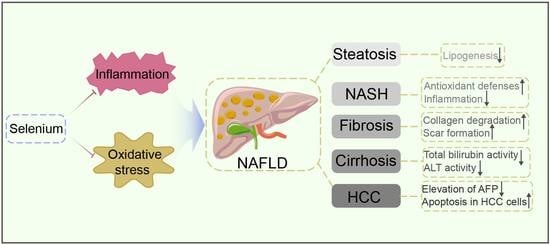
3. The Role of Selenium on the Progression of NAFLD

3.1. Selenium and Lipid Metabolism in the Liver

For circulating lipids, the liver serves as a metabolic hub. The net retention of lipids within hepatocytes, usually in the form of triglycerides, is the first step in the initiation of fatty liver [41]. The major pathways of lipid metabolic abnormalities are related to the changes in lipid uptake, lipogenesis, lipolysis, lipophagy, and expression of hepatic lipoproteins. Speckmann B et al. showed that selenium supplementation increased the hepatic mRNA expression of PPARα in the mice [42], which can mediate the prevention of triglyceride accumulation [43]. For patients with gestational diabetes mellitus, taking selenium yeast 200 μg per day as selenium supplements for 6 weeks upregulated peroxisome proliferator-activated receptor gamma (PPARγ) and glucose transporter 1 (GLUT-1), which inhibited lipogenesis [44]. Moreover, selenium deficiency could decrease the expression of intracellular antioxidant enzymes, including Superoxide dismutase-1 (SOD1) [45], which results in the development of fatty liver [46]. Furthermore, supplementing with selenium and magnesium inhibits the mRNA expression level of hepatic lipogenesis genes liver X receptor alpha (LXRα), SREBP-1c, and FASN (fatty acid synthase) to reduce the hepatic total cholesterol (TC) and attenuate liver steatosis in the rats fed by high-fat diet [47]. Moreover, a study found zinc and selenium co-supplementation could significantly reduce the level of serum triglyceride and TC. The fat accumulation was considerably reduced in high-fat diet-fed rats [48].

3.2. Selenium and Inflammatory Response in Liver

The management of inflammatory processes in NAFLD may prevent the development of NASH to liver fibrosis [49]. It has been proven that selenium deficiency could initiate inflammation by activating the NF-κB pathway through multiple mechanisms at an organ level [50]. Selenium supplementation can reduce pro-inflammatory cytokines and the expression of inflammation-related proteins in the liver, such as TLR4, NF-κB, JNK, and p38, as well as upregulating heme oxygenase-1 (HO-1) to reduce the inflammatory response. Al-Dossari et al. found that the selenium treatment (0.1 mg/kg/d) mitigated lipopolysaccharide (LPS)/Diclofenac (DCL)-induced injury in the liver through suppressing the LPS-induced TLR4 signaling pathway and boosted antioxidant defenses to reduce oxidative stress in rats [51]. In the mercury chloride (HgCl2)-induced hepatotoxicity animals, selenium alleviated the inflammation by blocking the NF-κB/NLR family pyrin domain containing 3 (NLRP3) inflammasome signaling pathway. Moreover, selenium prevented HgCl2-induced liver lipid accumulation and dyslipidemia by regulating gene expression related to lipid and glucose metabolism [52].

3.3. Selenium and Hepatic Fibrosis Formation

Liver fibrosis is common in the majority of types of chronic liver diseases, and its obvious characteristic is a condition in which extracellular matrix proteins, such as collagen, build up excessively and generate fibrous scars. According to a study, serum selenium levels are inversely related to the risk of advanced liver fibrosis, especially in older individuals, Caucasians, and females. Increased selenium levels reduced all-cause mortality [53]. In the N-nitrosodimethylamine (NDMA)-driven hepatic fibrosis rat model, reduced selenium and glutathione peroxidase can be observed to contribute to the weakening of cellular antioxidant defense to aggravate the progression of hepatic fibrosis [54]. In carbon tetrachloride (CCl4-induced liver fibrosis, selenium supplementation in the form of sodium selenite in the drinking water boosted selenium-dependent glutathione peroxidase activity and reduced malondialdehyde in liver tissues. Selenium can lower the number of collagen-producing stellate cells and increase collagen degradation to inhibit fibrosis [55]. Zhang et al. observed that compared to conventional green tea, selenium-enriched green tea products had a more pronounced improvement in liver extracellular matrix deposition, scar formation, and peripheral 5-hydroxytryptamine signals [56]. Selenium-enriched probiotics contain 10 g of selenium per milliliter, of which more than 90% is organic Se significantly reduced the fibrosis-related gene expression of α-smooth muscle actin, collagen, transforming growth factor-beta (TGF-β1), and TIMP-1, which involved the apoptosis of stimulated hepatic stellate cells [57]. Other studies show that selenium-glutathione-enriched probiotics, which contained 38.4 μg/g of organic selenium and 34.1 mg/g of intracellular glutathione, attenuated liver fibrosis by increasing the hepatic silent information regulator 1 (SIRT1) and MAPK pathway and involved the down-regulation of oxidative stress, endoplasmic reticulum stress, and inflammation in CCl4-treated rats [58]. Vitamin E and selenium in combination could have a synergistic effect on inhibiting CCl4-induced hepatic injury. They have been shown to limit hepatic stellate cell growth and increase apoptosis in activated hepatic stellate cells (HSCs) during the acute injury phase. Vitamin E and selenium can also significantly reduce the severity of hepatic fibrosis and facilitate the healing process [59].

3.4. Selenium and Liver Cirrhosis Development

Studies have long found that the plasma levels of selenium in patients with cirrhosis were lower than physical plasma concentrations and that they decrease in proportion to the severity of the cirrhotic condition [60]. In rats with carbon tetrachloride (CCl4)/ethanol-induced cirrhosis, the co-treatment of selenium and vitamin E significantly decreases the amount of hepatic fibrosis [61]. The study of thioacetamide-induced liver cirrhosis showed that sodium selenite supplementation (1 mg/kg b.w, i.p. for 12 weeks) reduced total bilirubin and ALT activity, as well as restored the antioxidant enzymes (SOD and GSH), MDA, and catalase activity [62]. However, selenium supplementation could not restore hepatic GPx and selenium to normal levels in selenium-deficient thioacetamide-induced cirrhosis mice [63].

3.5. Selenium and Hepatocellular Carcinoma

Hepatocellular carcinoma (HCC) is the most frequent type of primary liver cancer in adults, and it is now the leading cause of mortality in cirrhotic patients [64]. A meta-analysis shows the increased concentration of serum selenium could be related to a lower risk of HCC [65,66]. In CCl4-induced liver cancer, selenium was observed to decrease the elevation of alpha-fetoprotein (AFP) and TNF-α compared with control mice. The serum AFP is considered a tumor marker for liver cancer. Studies found that at the early stage of HCC, the low selenium levels caused the accumulation of lipid peroxides, which sped up the development of the disease by enhancing AP-1 activation and resulting in increased production of VEGF and IL-8. Therefore, selenium supplantation in the form of selenite could be investigated as chemoprevention or supplementary therapy for early HCC patients with low selenium levels [67]. Furthermore, pleomorphic adenoma gene like-2 (PLAGL2) overexpression has been linked to tumorigenesis. Yang et al. found that selenium sulfide (SeS2) suppressed C-MET/STAT3, AKT/mTOR, and MAPK signaling and prompted Bcl-2/Cyto C/Caspase-mediated intrinsic mitochondrial apoptosis in HCC cells in a PLAGL2-dependent manner by triggering Bcl-2/Cyto C/Caspase-mediated intrinsic mitochondrial apoptosis in a PLAGL2-dependent manner [68]. Thus, selenium shows a protective role against liver cancer [69]. The role of selenium in animal models has been summarized in Table 1.

4. Discussion and Conclusions

In this paper, the role of selenium in the development of NAFLD has been summarized. According to most of the evidence from experimental trials, selenium appears to have a beneficial effect on the improvement of hepatic steatosis, NASH, and fibrosis. At first, selenium can promote the upregulation of lipid oxidation enzymes as well as the downregulation of de novo lipogenesis enzymes, which may be used to combat steatosis [70]. Moreover, the antioxidant impact of selenium on the liver is at least in part through enhancing the activity of GPx, whose decrease leads to hepatic inflammation and liver fibrosis [71,72]. Selenium potentially suppresses the metalloproteinases, TNF-α, IL-6, TGF-β1, and other cytokines and growth factors involved in NAFLD etiology, which might help to reduce hepatic inflammation and fibrosis [71,73,74,75]. It has been postulated that Se supplementation in chronic inflammation recovers depleted hepatic Se levels and, by boosting selenoprotein biosynthesis, restores circulating Se levels, suppressing C-reactive protein production and finally attenuating the inflammatory response. However, most of the models are induced by CCl4 for manifesting hepatic inflammation and fibrosis; technically, it represents the toxic influence on the liver instead of the non-alcoholic fatty liver. Thus, the majority of animal research has been conducted on models that do not fully resemble human NAFLD. A randomized clinical trial revealed the relationship between selenium supplementation and insulin resistance. However, compared to the placebo group, the selenium group (200 µg/day) did not have a significant adverse effect on β-cell function or insulin sensitivity [76]. Since the in vitro and in vivo effects of selenium on NAFLD progression have been well outlined, the clinical trials of selenium warrant further estimation. In Korea, selenium levels were significantly lower in hepatitis and hepatoma patients compared to healthy controls, indicating a similar correlation between liver disease progression and decreases in Se levels, except for liver cirrhosis. Furthermore, the findings of significantly reduced Se levels in Korean hepatoma patients were validated in this investigation [77]. However, in China, higher plasma selenium levels were linked to a higher frequency of NAFLD and higher levels of fasting plasma glucose, post-loading plasma glucose, triglycerides, ALT, and AST. By triggering inflammation and infiltrating the liver, selenium exposure exacerbated liver damage [78].
Although selenium benefits the organism to some extent, an overdose of selenium can lead to severe effects. It is vital to be aware of its toxicity, most often attributable to excessive daily food and water intake. Selenium may take directly toxic effects by the modulation of ROS-related oxidative stress [79,80]. An outbreak of acute selenium poisoning in America was investigated; a high concentration of selenium in a liquid nutritional supplement containing 200 times (200 μg of selenium per fluid ounce) was found as the outbreak’s source. Diarrhea, weariness, hair loss, joint discomfort, nail discoloration or brittleness, and nausea are the most reported symptoms of its side effects. Fingernail discoloration and loss, weariness, and hair loss were among the signs that lasted 90 days or longer [81]. After ingesting 10 g of sodium selenite, a 75-years-old man was reported to suffer a cardiac arrest and die 6 h after ingestion [82]. Because of the limited range between therapeutic and dangerous amounts of selenium and because its effect depends on the form, dose, and type of therapy used, selecting the most effective supplement is a difficult task and needs further study. Additionally, though the supplement of selenium exists in different forms such as selenomethionine, selenium-enriched yeast, or sodium selenite or sodium selenate, there has been no investigation of how different kinds of selenium supplementation influence molecular signaling in vivo or how they affect treatment outcomes. Due to the restricted treatment window, the changes in effect and intake of selenium caused by the difference in form require further exploration in order to provide a more rigorous experimental basis for future therapeutic uses.

5. Challenges and Outlooks

In recent years, selenium has been promoted as a beneficial supplement for humans. A prudent selenium intake should be safe and useful for both healthy people and those suffering from metabolic diseases. It is expected that more clinically relevant research will be conducted in the future as attempts to understand the role of selenium in metabolic diseases continues to be made. Since the non-alcoholic fatty liver is a chronic disease, long-term observation is required in future clinical monitoring. In addition, the in vitro models of liver cirrhosis, liver fibrosis, and hepatocellular carcinoma in the late stage of NAFLD progression need to be further standardized so that the regulation of selenium on the overall progression of NAFLD can be precisely and fully evaluated rather than specific phenomena.
In conclusion, we conducted a thorough study of the involvement of selenium in a variety of non-alcoholic fatty acid metabolism pathways. It takes effect on the development of NAFLD by acting as an antioxidant and controlling the inflammatory response. Our study demonstrates the therapeutic potential of selenium supplementation in the treatment of metabolic disorders.

Author Contributions

Conceptualization, N.W. and Y.F.; writing—original draft preparation, L.X. and Y.L.; writing—review and editing, L.X., Y.L. and N.W.; supervision, N.W. and Y.F.; project administration, Y.F.; funding acquisition, Y.F. All authors have read and agreed to the published version of the manuscript.

Funding

This research was partially supported by the Research Council of the University of Hong Kong (project codes: 104004092 and 104004460), the Wong’s donation (project code: 200006276), a donation from the Gaia Family Trust of New Zealand (project code: 200007008), the Research Grants Committee (RGC) of Hong Kong, HKSAR (Project Codes: 740608, 766211, 17152116 and 17121419), the Health and Medical Research Fund (Project code: 15162961 and 16172751), the Enhanced new staff start-up fund (Project code: 204610519) and the Pre-emptive retention fund (Project code: 202007002).

Conflicts of Interest

The authors declare no conflict of interest.

References

  1. Wang, N.; Tan, H.Y.; Li, S.; Xu, Y.; Guo, W.; Feng, Y. Supplementation of Micronutrient Selenium in Metabolic Diseases: Its Role as an Antioxidant. Oxid. Med. Cell. Longev. 2017, 2017, 7478523. [Google Scholar] [CrossRef] [PubMed]
  2. Pinsent, J. The need for selenite and molybdate in the formation of formic dehydrogenase by members of the Coli-aerogenes group of bacteria. Biochem. J. 1954, 57, 10–16. [Google Scholar] [CrossRef] [PubMed] [Green Version]
  3. Hogan, C.; Perkins, A.V. Selenoproteins in the Human Placenta: How Essential Is Selenium to a Healthy Start to Life? Nutrients 2022, 14, 628. [Google Scholar] [CrossRef] [PubMed]
  4. Nosewicz, J.; Spaccarelli, N.; Roberts, K.M.; Hart, P.A.; Kaffenberger, J.A.; Trinidad, J.C.; Kaffenberger, B.H. The epidemiology, impact, and diagnosis of micronutrient nutritional dermatoses part 1: Zinc, selenium, copper, vitamin A., and vitamin C. J. Am. Acad. Dermatol. 2022, 86, 267–278. [Google Scholar] [CrossRef]
  5. Tóth, R.J.; Csapó, J. The role of selenium in nutrition—A review. Acta Univ. Sapientiae Aliment. 2018, 11, 128–144. [Google Scholar] [CrossRef] [Green Version]
  6. Fairweather-Tait, S.J.; Bao, Y.; Broadley, M.R.; Collings, R.; Ford, D.; Hesketh, J.E.; Hurst, R. Selenium in human health and disease. Antioxid. Redox Signal. 2011, 14, 1337–1383. [Google Scholar] [CrossRef]
  7. Qataya, P.O.; Elsayed, N.M.; Elguindy, N.M.; Ahmed Hafiz, M.; Samy, W.M. Selenium: A sole treatment for erosive oral lichen planus (Randomized controlled clinical trial). Oral Dis. 2020, 26, 789–804. [Google Scholar] [CrossRef]
  8. Dennert, G.; Horneber, M. Selenium for alleviating the side effects of chemotherapy, radiotherapy and surgery in cancer patients. Cochrane Database Syst Rev. 2006, CD005037. [Google Scholar] [CrossRef]
  9. Liu, T.; Zeng, L.; Jiang, W.; Fu, Y.; Zheng, W.; Chen, T. Rational design of cancer-targeted selenium nanoparticles to antagonize multidrug resistance in cancer cells. Nanomedicine 2015, 11, 947–958. [Google Scholar] [CrossRef]
  10. Fairweather-Tait, S.J.; Collings, R.; Hurst, R. Selenium bioavailability: Current knowledge and future research requirements. Am. J. Clin. Nutr. 2010, 91, 1484S–1491S. [Google Scholar] [CrossRef] [Green Version]
  11. Burk, R.F.; Norsworthy, B.K.; Hill, K.E.; Motley, A.K.; Byrne, D.W. Effects of chemical form of selenium on plasma biomarkers in a high-dose human supplementation trial. Cancer Epidemiol. Biomark. Prev. 2006, 15, 804–810. [Google Scholar] [CrossRef] [PubMed] [Green Version]
  12. Masuoka, H.C.; Chalasani, N. Nonalcoholic fatty liver disease: An emerging threat to obese and diabetic individuals. Ann. N. Y. Acad. Sci. 2013, 1281, 106–122. [Google Scholar] [CrossRef] [PubMed]
  13. Kwok, R.; Choi, K.C.; Wong, G.L.; Zhang, Y.; Chan, H.L.; Luk, A.O.; Shu, S.S.; Chan, A.W.; Yeung, M.W.; Chan, J.C.; et al. Screening diabetic patients for non-alcoholic fatty liver disease with controlled attenuation parameter and liver stiffness measurements: A prospective cohort study. Gut 2016, 65, 1359–1368. [Google Scholar] [CrossRef] [PubMed]
  14. Estes, C.; Razavi, H.; Loomba, R.; Younossi, Z.; Sanyal, A.J. Modeling the epidemic of nonalcoholic fatty liver disease demonstrates an exponential increase in burden of disease. Hepatology 2018, 67, 123–133. [Google Scholar] [CrossRef] [PubMed]
  15. Yang, Y.; Cai, J.; Yang, X.; Wang, K.; Sun, K.; Yang, Z.; Zhang, L.; Yang, L.; Gu, C.; Huang, X.; et al. Dysregulated m6A Modification Promotes Lipogenesis and Development of Non-alcoholic Fatty Liver Disease and Hepatocellular Carcinoma. Mol. Ther. 2022. [Google Scholar] [CrossRef] [PubMed]
  16. Arab, J.P.; Candia, R.; Zapata, R.; Munoz, C.; Arancibia, J.P.; Poniachik, J.; Soza, A.; Fuster, F.; Brahm, J.; Sanhueza, E.; et al. Management of nonalcoholic fatty liver disease: An evidence-based clinical practice review. World J. Gastroenterol. 2014, 20, 12182–12201. [Google Scholar] [CrossRef] [PubMed]
  17. Thuluvath, P.J.; Triger, D.R. Selenium in chronic liver disease. J. Hepatol. 1992, 14, 176–182. [Google Scholar] [CrossRef]
  18. Day, C.P.; James, O.F. Steatohepatitis: A tale of two “hits”? Gastroenterology 1998, 114, 842–845. [Google Scholar] [CrossRef]
  19. Arner, P. Insulin resistance in type 2 diabetes: Role of fatty acids. Diabetes Metab. Res. Rev. 2002, 18 (Suppl. S2), S5–S9. [Google Scholar] [CrossRef]
  20. Gastaldelli, A.; Cusi, K.; Pettiti, M.; Hardies, J.; Miyazaki, Y.; Berria, R.; Buzzigoli, E.; Sironi, A.M.; Cersosimo, E.; Ferrannini, E.; et al. Relationship between hepatic/visceral fat and hepatic insulin resistance in nondiabetic and type 2 diabetic subjects. Gastroenterology 2007, 133, 496–506. [Google Scholar] [CrossRef]
  21. Feng, W.; Cui, X.; Liu, B.; Liu, C.; Xiao, Y.; Lu, W.; Guo, H.; He, M.; Zhang, X.; Yuan, J.; et al. Association of urinary metal profiles with altered glucose levels and diabetes risk: A population-based study in China. PLoS ONE 2015, 10, e0123742. [Google Scholar] [CrossRef] [PubMed]
  22. Barakat, G.M.; Moustafa, M.E.; Bikhazi, A.B. Effects of selenium and exendin-4 on glucagon-like peptide-1 receptor, IRS-1, and Raf-1 in the liver of diabetic rats. Biochem. Genet. 2012, 50, 922–935. [Google Scholar] [CrossRef] [PubMed]
  23. D’Alessio, D.; Vahl, T.; Prigeon, R. Effects of glucagon-like peptide 1 on the hepatic glucose metabolism. Horm. Metab. Res. 2004, 36, 837–841. [Google Scholar] [CrossRef] [PubMed]
  24. Lee, Y.S.; Jun, H.S. Anti-diabetic actions of glucagon-like peptide-1 on pancreatic beta-cells. Metabolism 2014, 63, 9–19. [Google Scholar] [CrossRef]
  25. Barakat, G.; Moustafa, M.E.; Khalifeh, I.; Hodroj, M.H.; Bikhazi, A.; Rizk, S. Effects of exendin-4 and selenium on the expression of GLP-1R, IRS-1, and preproinsulin in the pancreas of diabetic rats. J. Physiol. Biochem. 2016, 73, 387–394. [Google Scholar] [CrossRef]
  26. Iizuka, Y.; Ueda, Y.; Yagi, Y.; Sakurai, E. Significant improvement of insulin resistance of GK rats by treatment with sodium selenate. Biol. Trace Elem. Res. 2010, 138, 265–271. [Google Scholar] [CrossRef]
  27. Birben, E.; Sahiner, U.M.; Sackesen, C.; Erzurum, S.; Kalayci, O. Oxidative stress and antioxidant defense. World Allergy Organ. J. 2012, 5, 9–19. [Google Scholar] [CrossRef] [Green Version]
  28. Chen, Z.; Tian, R.; She, Z.; Cai, J.; Li, H. Role of oxidative stress in the pathogenesis of nonalcoholic fatty liver disease. Free Radic. Biol. Med. 2020, 152, 116–141. [Google Scholar] [CrossRef]
  29. Thannickal, V.J.; Fanburg, B.L. Reactive oxygen species in cell signaling. Am. J. Physiol. Lung. Cell Mol. Physiol. 2000, 279, L1005–L1028. [Google Scholar] [CrossRef] [Green Version]
  30. Kryukov, G.V.; Castellano, S.; Novoselov, S.V.; Lobanov, A.V.; Zehtab, O.; Guigo, R.; Gladyshev, V.N. Characterization of mammalian selenoproteomes. Science 2003, 300, 1439–1443. [Google Scholar] [CrossRef] [Green Version]
  31. Brenneisen, P.; Steinbrenner, H.; Sies, H. Selenium, oxidative stress, and health aspects. Mol. Aspects Med. 2005, 26, 256–267. [Google Scholar] [CrossRef] [PubMed]
  32. Clark, L.C.; Combs, G.F., Jr.; Turnbull, B.W.; Slate, E.H.; Chalker, D.K.; Chow, J.; Davis, L.S.; Glover, R.A.; Graham, G.F.; Gross, E.G.; et al. Effects of selenium supplementation for cancer prevention in patients with carcinoma of the skin. A randomized controlled trial. Nutritional Prevention of Cancer Study Group. JAMA 1996, 276, 1957–1963. [Google Scholar] [CrossRef] [PubMed]
  33. Lei, X.G.; Cheng, W.H.; McClung, J.P. Metabolic regulation and function of glutathione peroxidase-1. Annu. Rev. Nutr. 2007, 27, 41–61. [Google Scholar] [CrossRef] [PubMed]
  34. Papp, L.V.; Lu, J.; Holmgren, A.; Khanna, K.K. From selenium to selenoproteins: Synthesis, identity, and their role in human health. Antioxid. Redox Signal. 2007, 9, 775–806. [Google Scholar] [CrossRef]
  35. Lillig, C.H.; Holmgren, A. Thioredoxin and related molecules—From biology to health and disease. Antioxid. Redox Signal. 2007, 9, 25–47. [Google Scholar] [CrossRef]
  36. Burke-Gaffney, A.; Callister, M.E.; Nakamura, H. Thioredoxin: Friend or foe in human disease? Trends Pharmacol. Sci. 2005, 26, 398–404. [Google Scholar] [CrossRef]
  37. Vunta, H.; Belda, B.J.; Arner, R.J.; Channa Reddy, C.; Vanden Heuvel, J.P.; Sandeep Prabhu, K. Selenium attenuates pro-inflammatory gene expression in macrophages. Mol. Nutr. Food Res. 2008, 52, 1316–1323. [Google Scholar] [CrossRef]
  38. Vunta, H.; Davis, F.; Palempalli, U.D.; Bhat, D.; Arner, R.J.; Thompson, J.T.; Peterson, D.G.; Reddy, C.C.; Prabhu, K.S. The anti-inflammatory effects of selenium are mediated through 15-deoxy-Delta12,14-prostaglandin J2 in macrophages. J. Biol. Chem. 2007, 282, 17964–17973. [Google Scholar] [CrossRef] [Green Version]
  39. Kretz-Remy, C.; Arrigo, A.P. Selenium: A key element that controls NF-kappa B activation and I kappa B alpha half life. Biofactors 2001, 14, 117–125. [Google Scholar] [CrossRef]
  40. Yu, S.S.; Du, J.L. Selenoprotein S: A therapeutic target for diabetes and macroangiopathy? Cardiovasc. Diabetol. 2017, 16, 101. [Google Scholar] [CrossRef] [Green Version]
  41. Benedict, M.; Zhang, X. Non-alcoholic fatty liver disease: An expanded review. World J. Hepatol. 2017, 9, 715–732. [Google Scholar] [CrossRef] [PubMed]
  42. Speckmann, B.; Schulz, S.; Hiller, F.; Hesse, D.; Schumacher, F.; Kleuser, B.; Geisel, J.; Obeid, R.; Grune, T.; Kipp, A.P. Selenium increases hepatic DNA methylation and modulates one-carbon metabolism in the liver of mice. J. Nutr. Biochem. 2017, 48, 112–119. [Google Scholar] [CrossRef] [PubMed]
  43. Crisafulli, C.; Cuzzocrea, S. The role of endogenous and exogenous ligands for the peroxisome proliferator-activated receptor alpha (PPAR-alpha) in the regulation of inflammation in macrophages. Shock 2009, 32, 62–73. [Google Scholar] [CrossRef] [PubMed]
  44. Karamali, M.; Dastyar, F.; Badakhsh, M.H.; Aghadavood, E.; Amirani, E.; Asemi, Z. The Effects of Selenium Supplementation on Gene Expression Related to Insulin and Lipid Metabolism, and Pregnancy Outcomes in Patients with Gestational Diabetes Mellitus: A Randomized, Double-Blind, Placebo-Controlled Trial. Biol. Trace Elem. Res. 2020, 195, 1–8. [Google Scholar] [CrossRef] [PubMed]
  45. Reddi, A.S.; Bollineni, J.S. Selenium-deficient diet induces renal oxidative stress and injury via TGF-beta1 in normal and diabetic rats. Kidney Int. 2001, 59, 1342–1353. [Google Scholar] [CrossRef] [Green Version]
  46. Sakiyama, H.; Fujiwara, N.; Yoneoka, Y.; Yoshihara, D.; Eguchi, H.; Suzuki, K. Cu, Zn-SOD deficiency induces the accumulation of hepatic collagen. Free Radic. Res. 2016, 50, 666–677. [Google Scholar] [CrossRef]
  47. Zhang, Q.; Qian, Z.Y.; Zhou, P.H.; Zhou, X.L.; Zhang, D.L.; He, N.; Zhang, J.; Liu, Y.H.; Gu, Q. Effects of oral selenium and magnesium co-supplementation on lipid metabolism, antioxidative status, histopathological lesions, and related gene expression in rats fed a high-fat diet. Lipids Health Dis. 2018, 17, 165. [Google Scholar] [CrossRef] [Green Version]
  48. Shidfar, F.; Faghihi, A.; Amiri, H.L.; Mousavi, S.N. Regression of Nonalcoholic Fatty Liver Disease with Zinc and Selenium Co-supplementation after Disease Progression in Rats. Iran. J. Med. Sci. 2018, 43, 26–31. [Google Scholar]
  49. Del Campo, J.A.; Gallego, P.; Grande, L. Role of inflammatory response in liver diseases: Therapeutic strategies. World J. Hepatol. 2018, 10, 1–7. [Google Scholar] [CrossRef]
  50. Tang, C.; Li, S.; Zhang, K.; Li, J.; Han, Y.; Zhan, T.; Zhao, Q.; Guo, X.; Zhang, J. Selenium deficiency-induced redox imbalance leads to metabolic reprogramming and inflammation in the liver. Redox Biol. 2020, 36, 101519. [Google Scholar] [CrossRef]
  51. Al-Dossari, M.H.; Fadda, L.M.; Attia, H.A.; Hasan, I.H.; Mahmoud, A.M. Curcumin and Selenium Prevent Lipopolysaccharide/Diclofenac-Induced Liver Injury by Suppressing Inflammation and Oxidative Stress. Biol. Trace Elem. Res. 2020, 196, 173–183. [Google Scholar] [CrossRef] [PubMed]
  52. Gao, P.C.; Chu, J.H.; Chen, X.W.; Li, L.X.; Fan, R.F. Selenium alleviates mercury chloride-induced liver injury by regulating mitochondrial dynamics to inhibit the crosstalk between energy metabolism disorder and NF-kappaB/NLRP3 inflammasome-mediated inflammation. Ecotoxicol. Environ. Saf. 2021, 228, 113018. [Google Scholar] [CrossRef] [PubMed]
  53. Reja, M.; Makar, M.; Visaria, A.; Marino, D.; Rustgi, V. Increased serum selenium levels are associated with reduced risk of advanced liver fibrosis and all-cause mortality in NAFLD patients: National Health and Nutrition Examination Survey (NHANES) III. Ann. Hepatol. 2020, 19, 635–640. [Google Scholar] [CrossRef] [PubMed]
  54. George, J. Determination of selenium during pathogenesis of hepatic fibrosis employing hydride generation and inductively coupled plasma mass spectrometry. Biol. Chem. 2018, 399, 499–509. [Google Scholar] [CrossRef]
  55. Ding, M.; Potter, J.J.; Liu, X.; Torbenson, M.S.; Mezey, E. Selenium supplementation decreases hepatic fibrosis in mice after chronic carbon tetrachloride administration. Biol. Trace Elem. Res. 2010, 133, 83–97. [Google Scholar] [CrossRef] [Green Version]
  56. Zhang, L.; Xu, J.Y.; Wei, Y.; Gao, S.L.; Wang, L.; Zheng, J.Y.; Gu, M.; Qin, L.Q. Protective Effect of Selenium-Enriched Green Tea on Carbon Tetrachloride-Induced Liver Fibrosis. Biol. Trace Elem. Res. 2021, 200, 2233–2238. [Google Scholar] [CrossRef]
  57. Liu, Y.; Liu, Q.; Ye, G.; Khan, A.; Liu, J.; Gan, F.; Zhang, X.; Kumbhar, S.; Huang, K. Protective effects of Selenium-enriched probiotics on carbon tetrachloride-induced liver fibrosis in rats. J. Agric. Food Chem. 2015, 63, 242–249. [Google Scholar] [CrossRef]
  58. Liu, Y.; Liu, Q.; Hesketh, J.; Huang, D.; Gan, F.; Hao, S.; Tang, S.; Guo, Y.; Huang, K. Protective effects of selenium-glutathione-enriched probiotics on CCl4-induced liver fibrosis. J. Nutr. Biochem. 2018, 58, 138–149. [Google Scholar] [CrossRef]
  59. Shen, X.H.; Cheng, W.F.; Li, X.H.; Sun, J.Q.; Li, F.; Ma, L.; Xie, L.M. Effects of dietary supplementation with vitamin E and selenium on rat hepatic stellate cell apoptosis. World J. Gastroenterol. 2005, 11, 4957–4961. [Google Scholar] [CrossRef]
  60. Nangliya, V.; Sharma, A.; Yadav, D.; Sunder, S.; Nijhawan, S.; Mishra, S. Study of trace elements in liver cirrhosis patients and their role in prognosis of disease. Biol. Trace Elem. Res. 2015, 165, 35–40. [Google Scholar] [CrossRef]
  61. Zhang, M.; Song, G.; Minuk, G.Y. Effects of hepatic stimulator substance, herbal medicine, selenium/vitamin E, and ciprofloxacin on cirrhosis in the rat. Gastroenterology 1996, 110, 1150–1155. [Google Scholar] [CrossRef] [PubMed]
  62. Fatima, S.N.; Mahboob, T. Role of selenium in protection of liver cirrhosis. Pak. J. Pharm. Sci. 2013, 26, 1097–1102. [Google Scholar] [PubMed]
  63. Zhang, J.; Wang, H.; Yu, H. Thioacetamide-induced cirrhosis in selenium-adequate mice displays rapid and persistent abnormity of hepatic selenoenzymes which are mute to selenium supplementation. Toxicol. Appl. Pharmacol. 2007, 224, 81–88. [Google Scholar] [CrossRef] [PubMed]
  64. Forner, A.; Llovet, J.M.; Bruix, J. Hepatocellular carcinoma. Lancet 2012, 379, 1245–1255. [Google Scholar] [CrossRef]
  65. Gong, Y.; Dong, F.; Geng, Y.; Zhuang, H.; Ma, Z.; Zhou, Z.; Huang, B.; Sun, Z.; Hou, B. Selenium concentration, dietary intake and risk of hepatocellular carcinoma—A systematic review with meta-analysis. Nutr. Hosp. 2019, 36, 1430–1437. [Google Scholar] [CrossRef]
  66. Zhang, Z.; Bi, M.; Liu, Q.; Yang, J.; Xu, S. Meta-analysis of the correlation between selenium and incidence of hepatocellular carcinoma. Oncotarget 2016, 7, 77110–77116. [Google Scholar] [CrossRef]
  67. Rohr-Udilova, N.; Sieghart, W.; Eferl, R.; Stoiber, D.; Bjorkhem-Bergman, L.; Eriksson, L.C.; Stolze, K.; Hayden, H.; Keppler, B.; Sagmeister, S.; et al. Antagonistic effects of selenium and lipid peroxides on growth control in early hepatocellular carcinoma. Hepatology 2012, 55, 1112–1121. [Google Scholar] [CrossRef]
  68. Yang, T.; Huo, J.; Xu, R.; Su, Q.; Tang, W.; Zhang, D.; Zhu, M.; Zhan, Y.; Dai, B.; Zhang, Y. Selenium sulfide disrupts the PLAGL2/C-MET/STAT3-induced resistance against mitochondrial apoptosis in hepatocellular carcinoma. Clin. Transl. Med. 2021, 11, e536. [Google Scholar] [CrossRef]
  69. Elkhamesy, A.; Refaat, M.; Gouida, M.S.O.; Alrdahe, S.S.; Youssef, M.M. Diminished CCl4-induced hepatocellular carcinoma, oxidative stress, and apoptosis by co-administration of curcumin or selenium in mice. J. Food Biochem. 2021, e13845. [Google Scholar] [CrossRef]
  70. Luo, M.; Huang, S.; Zhang, J.; Zhang, L.; Mehmood, K.; Jiang, J.; Zhang, N.; Zhou, D. Effect of selenium nanoparticles against abnormal fatty acid metabolism induced by hexavalent chromium in chicken’s liver. Environ. Sci. Pollut. Res. Int. 2019, 26, 21828–21834. [Google Scholar] [CrossRef]
  71. Polyzos, S.A.; Kountouras, J.; Zavos, C. Nonalcoholic fatty liver disease: The pathogenetic roles of insulin resistance and adipocytokines. Curr. Mol. Med. 2009, 9, 299–314. [Google Scholar] [CrossRef] [PubMed]
  72. Gonzalez-Reimers, E.; Monedero-Prieto, M.J.; Gonzalez-Perez, J.M.; Duran-Castellon, M.C.; Galindo-Martin, L.; Abreu-Gonzalez, P.; Sanchez-Perez, M.J.; Santolaria-Fernandez, F. Relative and combined effects of selenium, protein deficiency and ethanol on hepatocyte ballooning and liver steatosis. Biol. Trace Elem. Res. 2013, 154, 281–287. [Google Scholar] [CrossRef] [PubMed]
  73. Polyzos, S.A.; Kountouras, J.; Mantzoros, C.S. Adipokines in nonalcoholic fatty liver disease. Metabolism 2016, 65, 1062–1079. [Google Scholar] [CrossRef] [PubMed] [Green Version]
  74. Han, J.; Liang, H.; Yi, J.; Tan, W.; He, S.; Wang, S.; Li, F.; Wu, X.; Ma, J.; Shi, X.; et al. Long-Term Selenium-Deficient Diet Induces Liver Damage by Altering Hepatocyte Ultrastructure and MMP1/3 and TIMP1/3 Expression in Growing Rats. Biol. Trace Elem. Res. 2017, 175, 396–404. [Google Scholar] [CrossRef]
  75. Wasser, S.; Lim, G.Y.; Ong, C.N.; Tan, C.E. Anti-oxidant ebselen causes the resolution of experimentally induced hepatic fibrosis in rats. J. Gastroenterol. Hepatol. 2001, 16, 1244–1253. [Google Scholar] [CrossRef]
  76. Jacobs, E.T.; Lance, P.; Mandarino, L.J.; Ellis, N.A.; Chow, H.S.; Foote, J.; Martinez, J.A.; Hsu, C.P.; Batai, K.; Saboda, K.; et al. Selenium supplementation and insulin resistance in a randomized, clinical trial. BMJ Open Diabetes Res. Care 2019, 7, e000613. [Google Scholar] [CrossRef]
  77. Kim, I.W.; Bae, S.M.; Kim, Y.W.; Liu, H.B.; Bae, S.H.; Choi, J.Y.; Yoon, S.K.; Chaturvedi, P.K.; Battogtokh, G.; Ahn, W.S. Serum selenium levels in Korean hepatoma patients. Biol. Trace Elem. Res. 2012, 148, 25–31. [Google Scholar] [CrossRef]
  78. Yang, Z.; Yan, C.; Liu, G.; Niu, Y.; Zhang, W.; Lu, S.; Li, X.; Zhang, H.; Ning, G.; Fan, J.; et al. Plasma selenium levels and nonalcoholic fatty liver disease in Chinese adults: A cross-sectional analysis. Sci. Rep. 2016, 6, 37288. [Google Scholar] [CrossRef]
  79. Hladun, K.R.; Smith, B.H.; Mustard, J.A.; Morton, R.R.; Trumble, J.T. Selenium toxicity to honey bee (Apis mellifera L.) pollinators: Effects on behaviors and survival. PLoS ONE 2012, 7, e34137. [Google Scholar] [CrossRef]
  80. Franz, E.D.; Wiramanaden, C.I.; Janz, D.M.; Pickering, I.J.; Liber, K. Selenium bioaccumulation and speciation in Chironomus dilutus exposed to water-borne selenate, selenite, or seleno-DL-methionine. Environ. Toxicol. Chem. 2011, 30, 2292–2299. [Google Scholar] [CrossRef]
  81. MacFarquhar, J.K.; Broussard, D.L.; Melstrom, P.; Hutchinson, R.; Wolkin, A.; Martin, C.; Burk, R.F.; Dunn, J.R.; Green, A.L.; Hammond, R.; et al. Acute selenium toxicity associated with a dietary supplement. Arch. Intern. Med. 2010, 170, 256–261. [Google Scholar] [CrossRef] [PubMed] [Green Version]
  82. See, K.A.; Lavercombe, P.S.; Dillon, J.; Ginsberg, R. Accidental death from acute selenium poisoning. Med. J. Aust. 2006, 185, 388–389. [Google Scholar] [CrossRef] [PubMed]
Figure 1. An overview of the pathophysiological mechanisms that oxidative stress and inflammation in the development of non-alcoholic fatty liver disease. Briefly, free fatty acid (FFA) flux could activate the transcriptional factors such as peroxisome proliferator-activated receptor gamma (PPARγ). In an aggravated state, this process promotes the increased production of reactive oxygen species (ROS) and aberrant levels of pro-inflammation markers such as tumor necrosis factor-alpha (TNF-α), the nuclear factor kappa B (NF-κB) and transforming growth factor beta (TGFβ-1). SREBP1c = sterol regulatory element-binding protein; TG = triglycerides; ROS = reactive oxygen species; IL-6 = interleukin 6; VLDL = very low-density lipoproteins.
Antioxidants 11 00922 g001
Table 1. The role of selenium in animal models.
ComponentAnimalModelRoute of AdministrationMain OutcomeRef
Sodium seleniteRatsHigh-fat diet induced hyperlipidemicOral administrationAttenuated liver steatosis[47]
Sodium seleniteMiceCarbon tetrachloride (CCl4) induced hepatic fibrosisIntraperitoneal injectionDecreased hepatic fibrosis after CCl4[55]
Selenium-enriched green teaMiceCarbon tetrachloride (CCl4) induced hepatic fibrosisOral administrationImproved liver fibrosis[56]
Selenium-glutathione-enriched probioticsRatsCarbon tetrachloride (CCl4) induced hepatic fibrosisOral administrationAttenuated liver fibrosis[58]
Selenium and vitamin ERatsCarbon tetrachloride (CCl4) induced hepatic fibrosisOral administrationDecreased the degree of hepatic fibrosis and promote the recovery process[59]
Selenium and vitamin ERatsCarbon tetrachloride (CCl4)/ethanol-induced cirrhosisOral administrationDecreased the amount of hepatic fibrosis[61]
Sodium seleniteRatsThioacetamide induced cirrhosisIntraperitoneal injectionAttenuated liver cirrhosis.[62]
Sodium seleniteMiceThioacetamide induced cirrhosisOral administration Intraperitoneal injectionCould not restore hepatic glutathione peroxidase[63]
Sodium seleniteRatsDiethylnitrosamin (DEN) induced hepatocellular carcinomaOral administrationAccelerated the growth of hepatocellular carcinoma[67]
SeleniumMiceCarbon tetrachloride (CCl4) induced hepatocellular carcinomaOral administrationProtected against liver damage[69]
Publisher’s Note: MDPI stays neutral with regard to jurisdictional claims in published maps and institutional affiliations.

