1. Introduction
Endodontic management of immature teeth characterized by incomplete root development, wide canal spaces, and open apices remains a significant clinical challenge despite ongoing advancements in techniques and the introduction of novel materials [
1]. After a tooth erupts, root development lasts about 3 years. If therapeutic interventions are unable to sustain the health of the pulp-dentinal complex, pulpal necrosis can occur prior to root development, resulting in disrupted root formation and immature roots [
2]. The management of immature teeth that present challenges during root canal preparation and obturation should be individualized based on the degree of root development, pulpal status, and overall clinical presentation [
1]. In many cases, regenerative endodontic treatment is regarded as the most suitable therapeutic option. Nevertheless, when regenerative endodontic procedures are either unsuitable or unsuccessful, the conventional approach involves single-visit apexification utilizing tricalcium silicate-based materials, such as mineral trioxide aggregate (MTA) [
3]. Currently, apexification remains the preferred initial treatment for many clinicians, owing to its consistent outcomes, single-visit protocol, reduced risk of tooth discoloration, and favorable patient compliance [
4].
Endocrowns have become a common treatment for previously mutilated depulped teeth, showing high success and survival rates in recent years [
5,
6,
7]. The selection of fabrication material plays an important role in the performance of endocrowns [
8,
9]. The transmission of functional forces varies according to the elasticity limits of materials [
10]. Lithium disilicate (LDS) and resin nanoceramic (RNC) prefabricated blocks are commonly preferred materials for endocrown restorations in clinical practice [
9]. Finite element analysis (FEA) is a widely acknowledged and employed computational numerical method for predicting the biomechanical behavior of dental structures in a non-invasive manner [
11,
12,
13]. The quantification of stress peaks and visualization of stress distributions facilitate the identification of potential fracture initiation sites [
14]. This engineering technology enables stress assessment in restorations and structures, which laboratory studies cannot achieve [
9,
10,
11,
12,
13,
14,
15]. Testing standardized variables while controlling for confounders reveals their actual impact [
16].
Apexification with calcium hydroxide was once used to create an apical barrier in immature teeth, but it fell out of favor due to reduced fracture resistance and lengthy treatment time [
17]. Although the substitution of this treatment with bioactive root repair materials yields favorable clinical outcomes, it does not promote additional root elongation or development. Consequently, the tooth remains short with thin canal walls, potentially increasing its vulnerability to fractures because of stress overload [
12]. Cervical and radicular root fractures occur with measurable frequency under both functional force and traumatic injury conditions [
2]. Apexification of immature teeth with tricalcium silicate cements results in higher resistance than leaving root canals empty, but the fracture resistance remains lower compared to mature teeth [
11]. While in vitro and FEA studies have investigated apexification in immature maxillary incisors [
12,
14,
18,
19,
20], and mandibular premolars [
11,
17,
21], immature molars have not yet been studied. A long-term retrospective study [
4] found that 46% of apexified teeth were in the anterior region, and the incidence in molar teeth was 39%. Additionally, apexification was unsuccessful in molars 2.7 times more frequently than in anterior teeth [
4]. Stress is mainly concentrated in the cervical region of anterior teeth [
12,
18], but increases in the mid-root and apical regions of mandibular premolars [
11]. The differences in crown and root anatomy between molars and anterior or premolar teeth, along with the higher occlusal forces experienced by molars, make it necessary to assess the peak stress values and distributions in immature molar teeth. Additionally, research on incisors and premolars was carried out using intact teeth, which were subjected to simulated trauma and limited to access cavity preparation [
11,
18,
21]. Pulpal necrosis or pulpitis may result not only from traumatic injury but also from the presence of dental caries [
4]. Teeth that have undergone endodontic treatment and have significant coronal tissue loss are at increased risk of fracture. Residual hard tissues play an important role in tooth preservation. In non-vital teeth with compromised structural integrity, it is necessary for the restoration to absorb stress before it reaches the root tissues and promote a uniform distribution of stress to reduce the risk of fractures [
22]. The long-term prognosis is contingent upon selecting an appropriate restoration that aligns with clinical behavior, effectively reinforces the remaining dental tissues, and ensures that the load capacity of the hard tissues is not exceeded [
23]. Using endocrowns for immature molars with severe coronal damage and poor crown-to-root ratios presents a challenge for clinicians. However, due to insufficient evidence-based data regarding immature molars, it can be challenging for clinicians to determine appropriate indications.
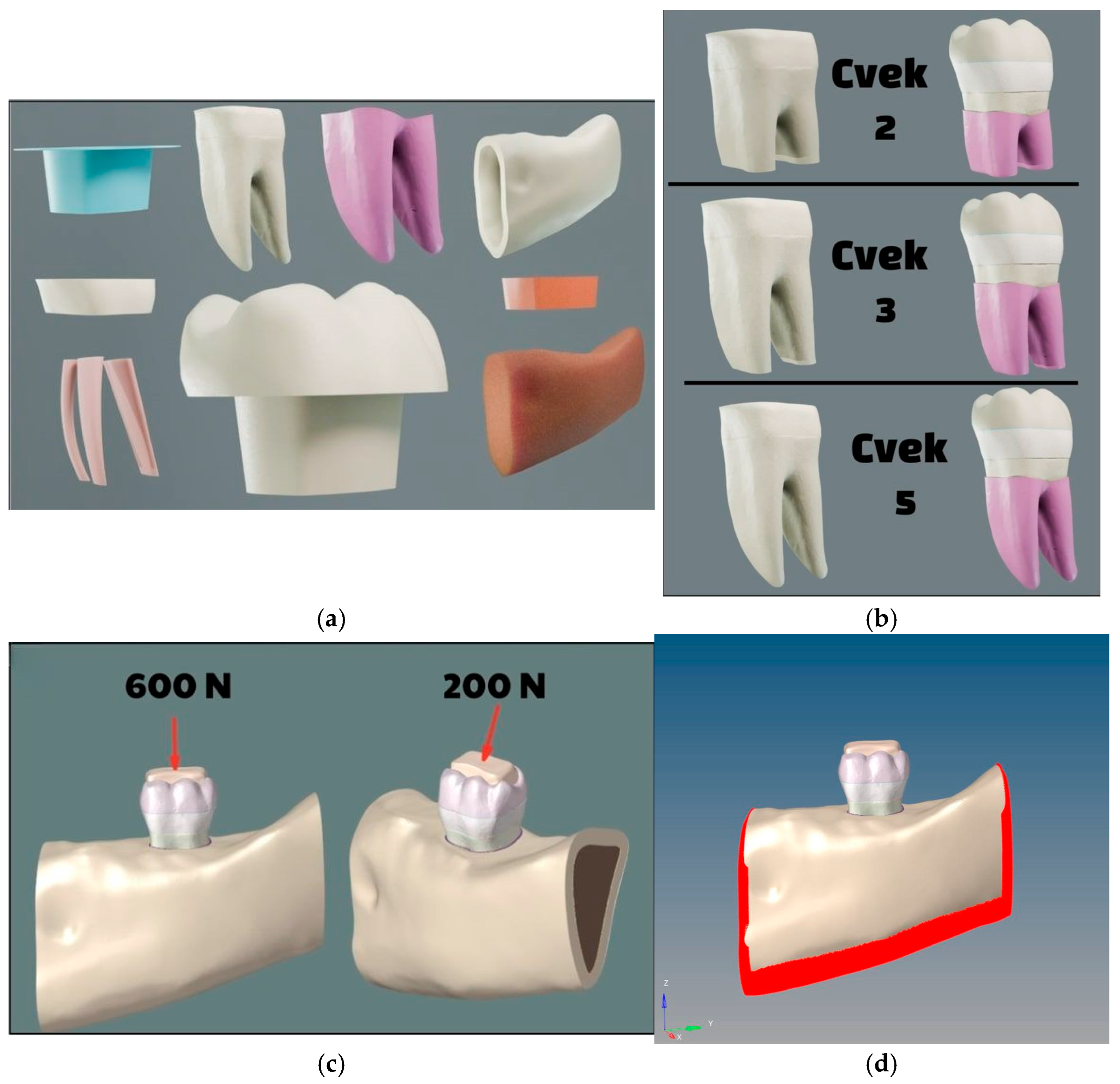
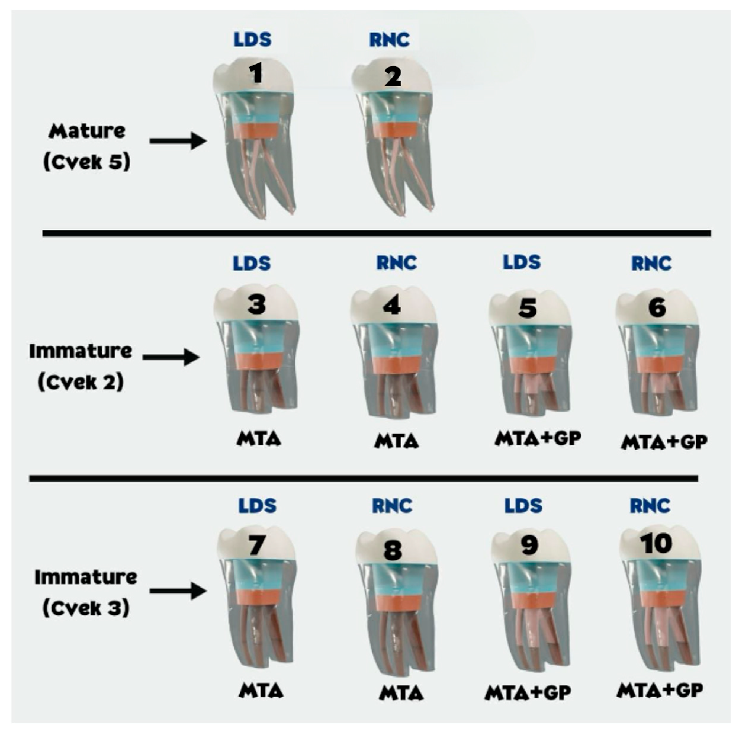
This virtual study used FEA to examine stress accumulation and distribution in immature mandibular molar remnants with significant tissue loss following endocrown application. The study simulated mature and two immature root development stages, using two different materials for endocrowns. The null hypotheses assert that there are no significant differences in the accumulated stress peaks within the residual hard tissues with respect to (i) stages of root development, (ii) apexification techniques, (iii) force directions, and (iv) materials used for endocrown fabrication.
4. Discussion
Cvek categorized teeth into five groups based on the degree of root development, with the first four groups classified as immature teeth and the fifth group as mature teeth [
24]. In this classification, the first group indicates the onset of root development, during which endodontic treatment is infrequently required. Class 4 describes teeth with roots that are almost fully developed in length and possess an open root apex. These teeth are classified as immature and continue to undergo maturation; however, they are not generally regarded as deficient in biomechanical strength. Therefore, this study focused on Classes 2 and 3, as they frequently occur during root development and present challenges in treatment. Studies of immature maxillary incisors and mandibular premolars favoured Class 3 specimens with a crown-to-root ratio near 1:1 and a length 3 mm shorter than mature teeth [
12,
17,
18,
19]. It has been evaluated that the choice of endocrown may be a possible restorative option in endodontically treated teeth with incomplete root formation and excessive coronal substance loss. An investigation was conducted into the variations in stress peaks and distribution patterns accumulated within the remnants of mature teeth following endocrown application. Molar teeth were selected because there is a lack of studies involving immature teeth in this group and due to the relatively high frequency of endocrown placement necessitated by material loss. Endocrown restorations have demonstrated reliable outcomes in mature teeth; therefore, mature tooth specimens were employed as the control group. The validity of the comparisons was assessed by evaluating differences between immature and mature teeth.
Von Mises stress serves as a failure criterion that reflects the distribution of energy within a material or structure and identifies areas of high energy concentration [
14]. It functions as a marker for potential damage [
11]. It measures forces along the X, Y, and Z axes by combining tensile, shear, and compressive stresses [
15]. Maximum principal stress identifies structural weak points and potential failure zones caused by tensile stress [
14]. Positive values represent tensile stress, while negative values represent compressive stress [
23]. This is the primary stress in brittle materials [
9]. For ductile materials, failure is typically defined by the von Mises stress criterion [
23]. Dental tissues possess substantial compressive strength; however, they tend to be brittle when subjected to tensile stress [
12]. Both von Mises stress and maximum principal stress are applicable to residual hard tissues, which may respond differently to bending and compressive forces [
21]. Some studies report only von Mises [
9,
10,
11,
15,
20] or only maximum principal stress in enamel and dentin [
25], while others evaluate both stress types together [
12,
14,
16]. Therefore, maximum and principal stress served as benchmarks for endocrown restoration. While results for both root dentin and the overall tooth structure were provided, von Mises stress was primarily used as the main stress criterion [
20].
The procurement or development of realistic, clinically accurate immature models is a key priority in finite element analysis (FEA) research. Immature tooth specimens can be further prepared using models from anatomical atlases [
11]. An immature tooth model can also be derived from a mature CBCT sample by reducing the apical length [
20]. Some researchers have compared two tooth samples—one immature and one mature—using CBCT scans [
12]. In Cvek classification, root length and apical opening are modeled distinctly across different classes during simulation. A 3 mm root length difference is assumed between Cvek 3 and mature teeth [
12]. For samples that simulated revascularized teeth, dentin thickening was estimated by projecting a specific amount of thickening from data on immature tooth samples [
12]. In in vitro studies, researchers simulated the model by using Peeso burs to enlarge the apex [
21]. In our study, we used a CBCT image of an 8-year-old patient with a Cvek 3 pattern to accurately model clinical conditions for this group. Additional groups were prepared in accordance with this example to standardise variables including crown length and dimensions, root dentin length, dentin thickness, canal dimensions, taper, and 3D geometry. Naturally, not every clinical case will display the same anatomies and sizes, as variations are inevitable. It is important to remember that the findings discussed are specific to this model.
Although conventional root canal therapy for immature teeth can be challenging, it remains both feasible and effective. However, due to the delicate root anatomy and physiology following treatment, there is an ongoing risk of fracture [
2,
17]. Apexification with an MTA apical barrier leads to incomplete maturation, leaving thin canal walls and weak root dentin [
14]. An in vitro investigation demonstrated that obturating the canal with MTA in immature central incisors resulted in enhanced fracture resistance relative to canals left unfilled; however, this resistance remained lower than that observed in the mature control group [
18]. In an FEA study of maxillary incisors, the apexification group showed lower peak stress than the empty canal immature tooth, but higher than the mature tooth [
12]. Immature teeth with empty canals showed a 64.8% decrease in strength, while apexification reduced weakness by 41.7% compared to mature teeth. Correlations have also been reported between the degree of root development and the occurrence of root fracture in teeth with immature apices [
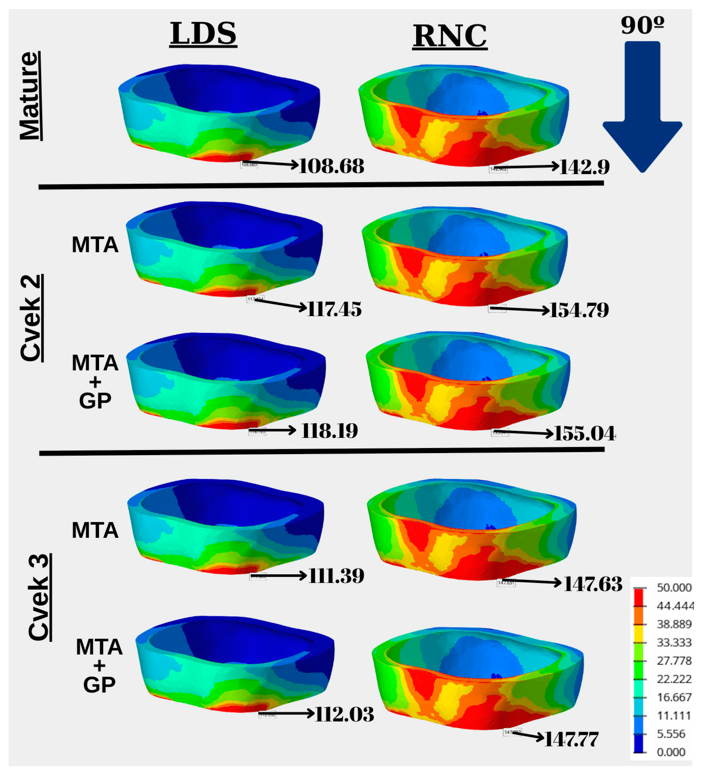
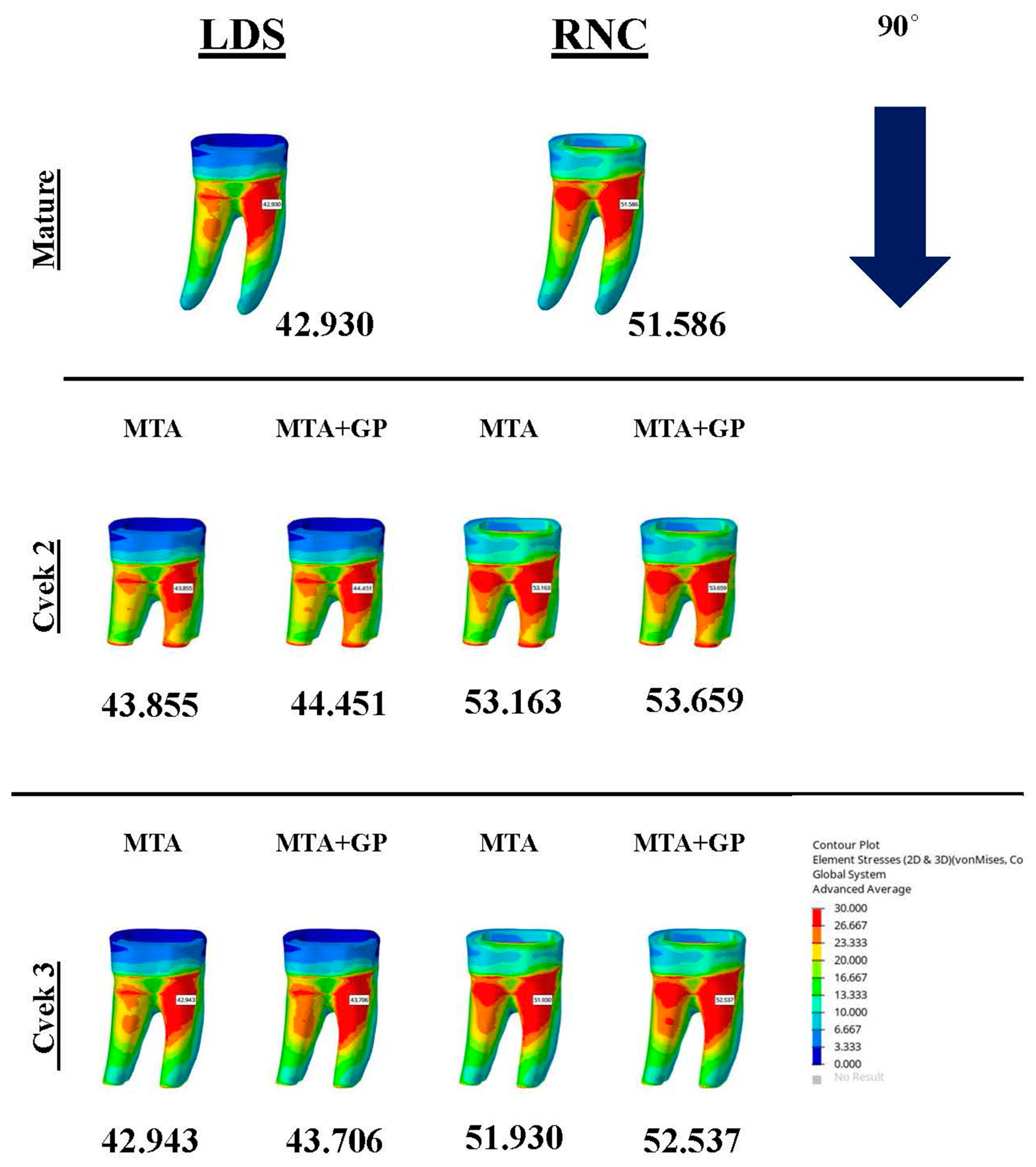
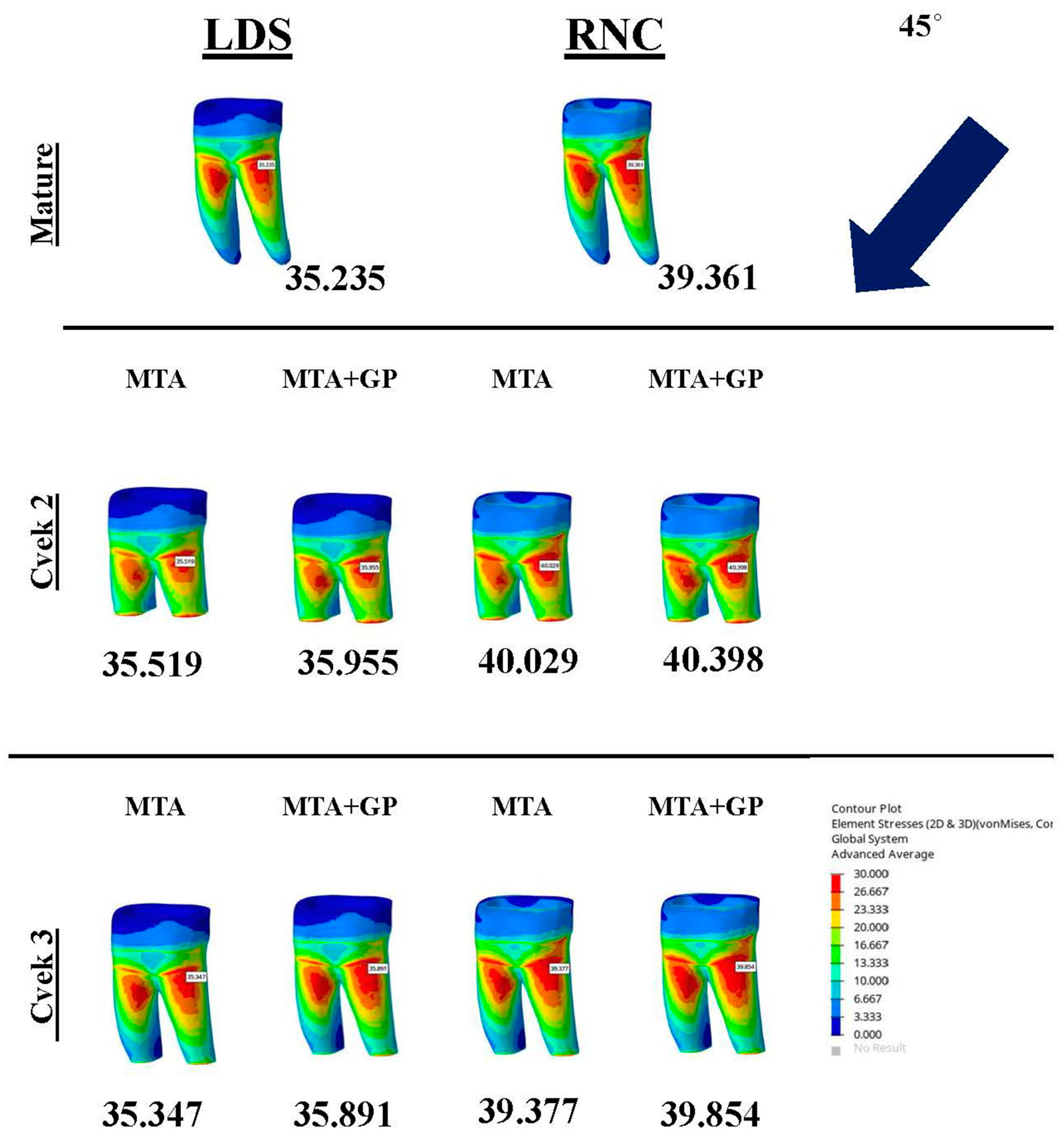
24]. The results of our study indicate that there was an increase in stress in the enamel of immature teeth, whereas the root dentin showed only a slight increase in stress. Selecting a restorative option that offers cuspal coverage, such as an endocrown, facilitates balanced distribution of stress and absorption, thereby safeguarding the structurally compromised remnants of immature teeth. Although stress levels in the remnants were higher than in the mature teeth (control group), and the first null hypothesis was rejected, the peak stresses remained below fracture and dentin risk thresholds. The Mohr–Coulomb ratios were well below the critical limit. Nevertheless, dentin tissue exposed to axial force—especially in the Cvek 2 groups—showed increased ratios, indicating a slightly elevated risk within these groups. Likewise, the maximum principal stress measured in Cvek 2 groups was higher than in other groups, highlighting the need for clinical caution.
Studies have examined the use of MTA to completely fill the root canal as a method to restore the structural integrity of non-vital immature teeth [
11,
19]. Variations in elastic modulus, the ratio of strain to stress, cause changes in stress under load. Studies indicate that using MTA to fill the entire root canal results in reduced stress in the cervical region of mandibular premolars, while increasing stress in the mid-coronal region [
11]. Some studies have reported no observable difference in maxillary incisors [
19]. The elastic modulus of gutta-percha is lower than that of dentin, which alters the resulting stress pattern [
11,
21]. A recent review found no clear evidence supporting full root canal obturation with MTA due to inconsistent results [
2]. In this study, the apexification technique was found to have very little impact on stress accumulations according to the generated models. Thus, the second null hypothesis was accepted. However, as the aforementioned studies focused on different tooth groups and restricted non-carious substance loss solely to the access cavities, direct comparison was not possible.
MTA is widely recommended for apexification of immature apices because of its superior properties [
17]. Biodentine has emerged as an effective material for apexification procedures [
3]. An in vitro study found that immature teeth filled with either MTA or Biodentine had similar fracture peak loads [
18]. In a FEA study of immature premolars, stress values in Biodentine groups were similar whether MTA was used with gutta-percha or alone in the canal [
11]. A FEA study on central incisors found comparable stress peak values in the MTA and Biodentine apexification groups, with the MTA group showing slightly lower values [
20]. Aslan et al. [
15] conducted a stress analysis on mandibular molar teeth with intact structure following apical resection of the mesial root and observed no significant difference between MTA and Biodentine when used as retrograde filling materials. Consistent with these findings, the present study elected to utilize a single apexification material for its methodology. Upon comprehensive examination of the study’s parameters, four distinct variables were evaluated: the degree of root development stage, endocrown material, apexification technique, and force load direction. Including an extra variable in the study would raise the number of models from 20 to 40, which would result in a larger set of parameters. Therefore, only MTA was selected for analysis.
During the occlusal force simulation on the maxillary incisors, a load of 240 N is applied from the lingual direction at an angle of 120–130 degrees [
12,
20]. In apexified maxillary incisor teeth, stress is concentrated in the palatal cervical third [
12]. Stress tends to accumulate in this area because it is located at the center of the fulcrum and has a cervical constriction [
20]. Root fractures during apexification treatments occur mainly in the cervical third, confirming this observation [
24]. Mandibular premolars undergo oblique stress analysis with a 300 N load only [
11]. Vertical as well as oblique forces are significant in their impact on mandibular molars. Thus, analyses used two forces. Stress caused by non-axial forces is more destructive due to their combined vertical and horizontal components [
9]. Under oblique forces, stress is primarily distributed within the pericervical dentin region. Due to the leverage effect, the lingual coronal third is subjected to increased stress exposure [
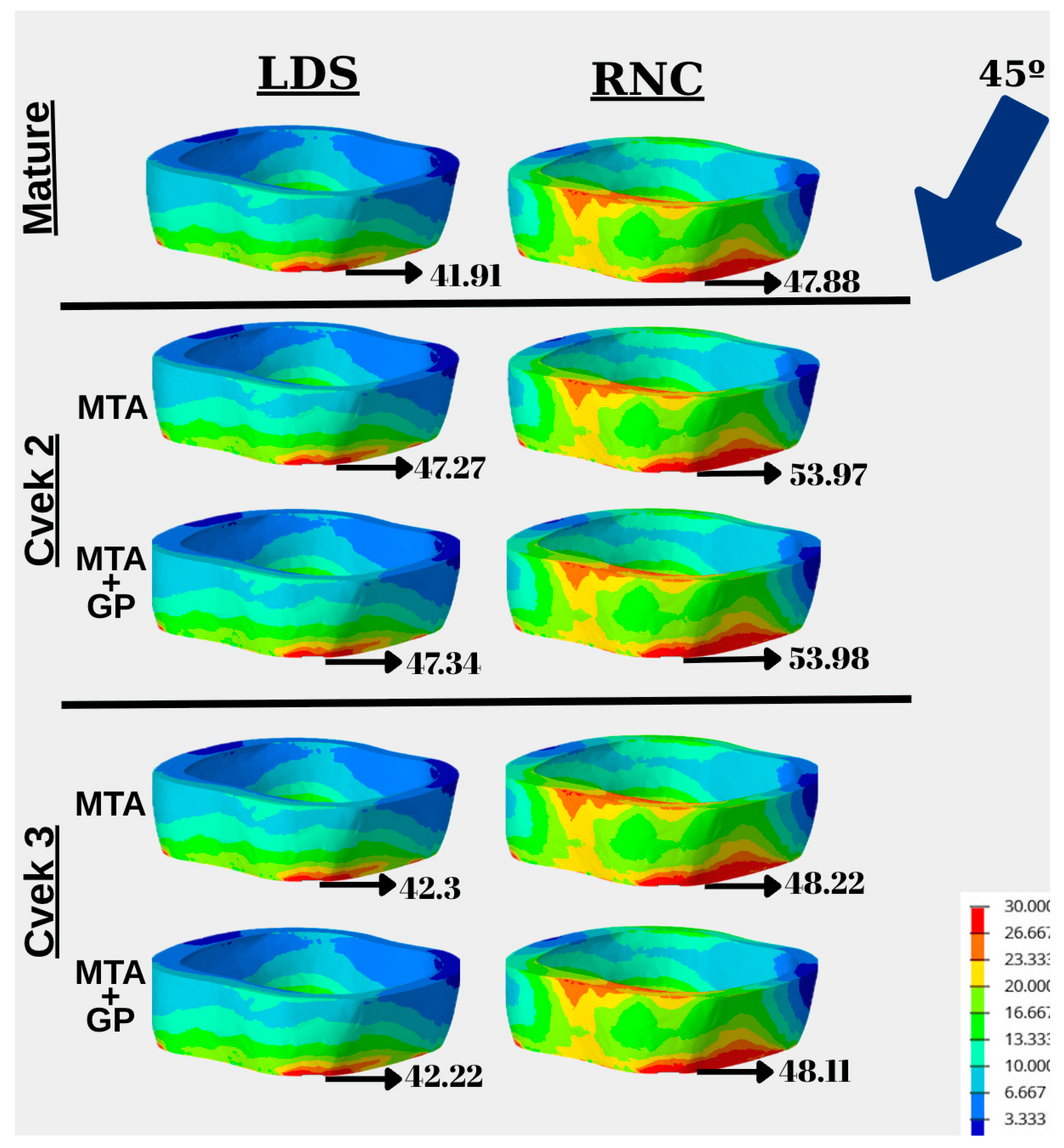
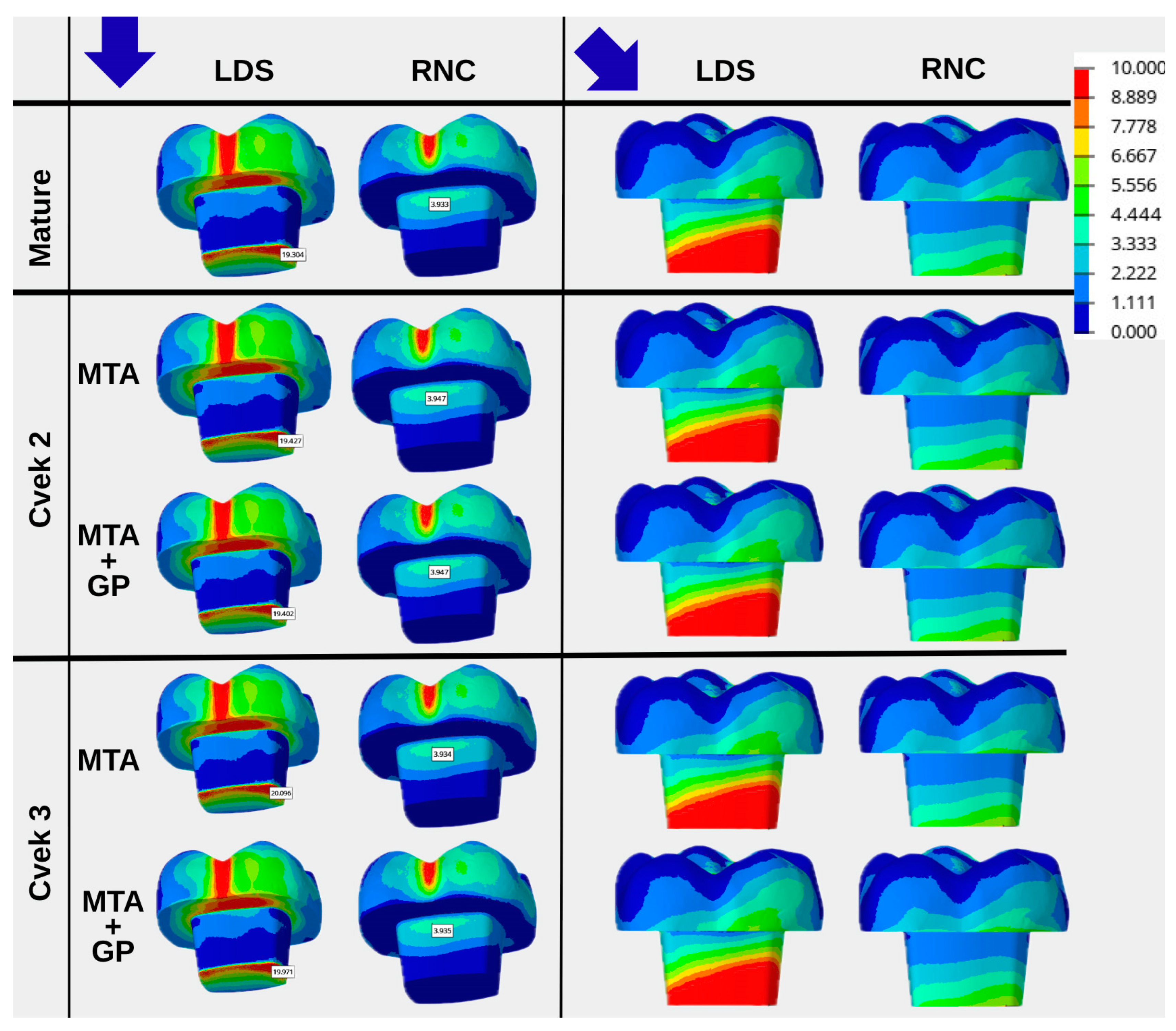
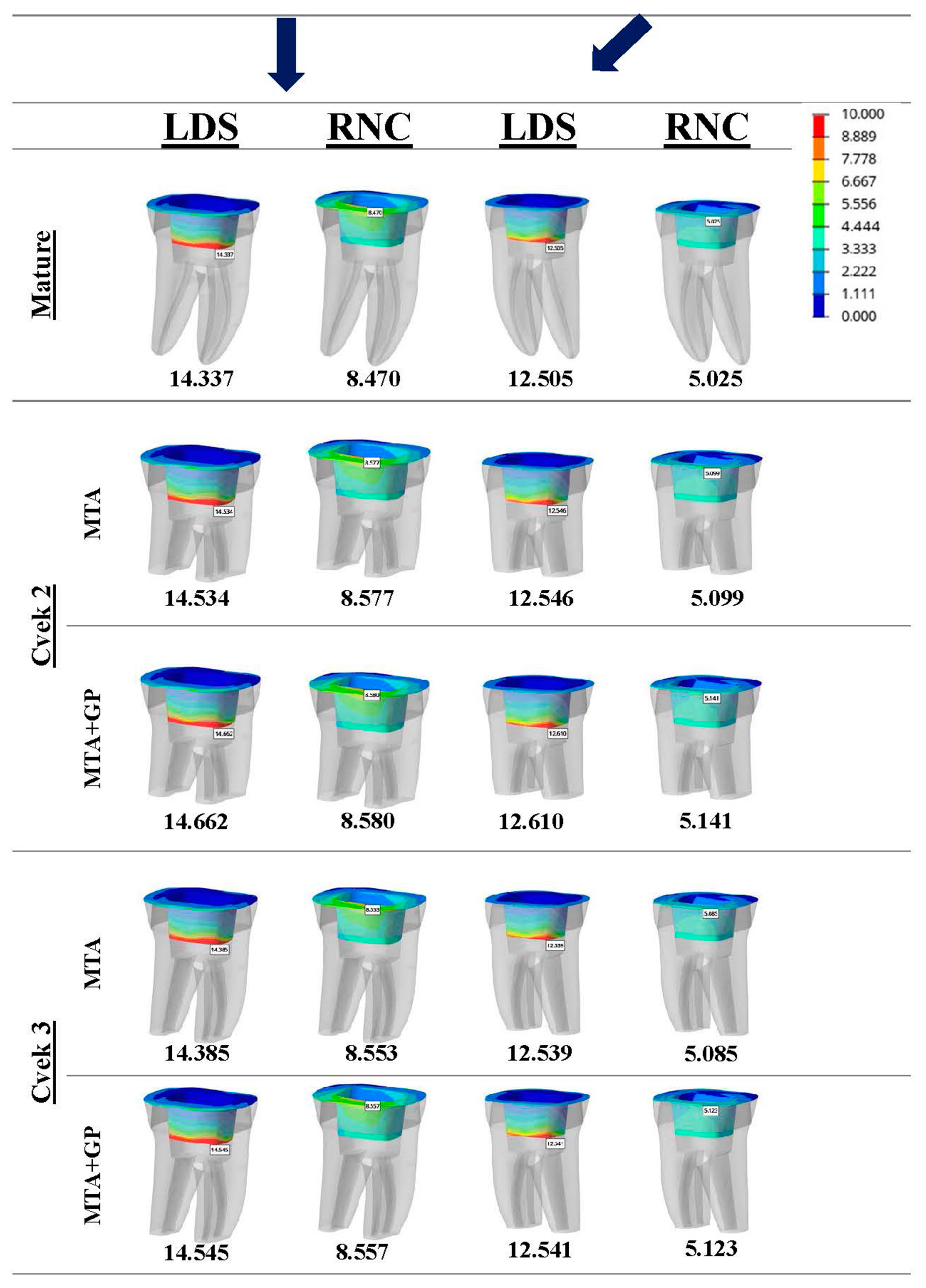
16]. This study found that oblique force generated higher stress peaks in the endocrown restoration. Whereas vertical force produced greater von Mises stress concentrations in the remnants. The fourth null hypothesis was not supported. Lower stress intensity under oblique force likely results from using a 200 N oblique load, compared to a higher 600 N vertical occlusal load. A study applying both vertical and oblique forces of 600 N demonstrated that oblique loading resulted in greater von Mises, tensile, and compressive stress concentrations across all structures [
9]. Higher maximum principal stress peaks and Mohr–Coulomb ratios were observed in root dentin, indicating the damaging impact of oblique forces. Stress accumulation is primarily observed in the lingual region, within the coronary third, and surrounding the root canal orifice. Consequently, potential fractures in these teeth are most likely to occur in these specific areas.
Masticatory forces exhibit inter-individual variability and typically range from 10 to 1000 newtons [
15]. On average, men have a maximum bite force of 630 N, while women typically reach up to 430 N [
5]. Selecting the right treatment and materials is particularly important when abnormal conditions like bruxism or trauma raise occlusal forces [
5,
26]. The selection of a high axial force of 600 N in this study is intended to evaluate the models under adverse conditions, such as those encountered during parafunctional activities in posterior teeth, where elevated force values can be observed. To better simulate real occlusal forces and clinical conditions, food pieces were placed on the models’ biting surfaces [
16]. The objective was to replicate the closing phase of the mastication process by employing a sliding contact between the dental surface and the food material [
16]. Adding a food layer before applying vertical or oblique force is based on the idea that, during chewing, stresses are distributed across regions rather than concentrated at single points [
25].
The effectiveness of residual dentin structure-oriented, minimally invasive endocrown placement in caries-affected immature teeth has not yet been evaluated. Restorative treatment planning is essential for root-filled teeth, especially since immature teeth have reduced structural integrity and other challenges, making effective restoration choices crucial [
23]. Indirect restorations are preferred for depulped teeth with significant substance loss [
16]. Greater pulp chamber height in immature teeth allows endocrowns to have a larger bonding surface within the chamber. A primary factor influencing the longevity of endocrowns is the uniform distribution of biomechanical stress [
5,
8]. Lithium disilicate ceramics are the most used material [
8,
22]. RNCs represent advanced hybrid endocrown materials that integrate the advantageous characteristics of both ceramic and composite resins, enabling more uniform stress distribution [
10]. Its modulus of elasticity, similar to dentin, helps absorb stress and reduces the incidence of catastrophic root fractures compared to LDS [
8]. However, FEA studies indicate that LDS accumulates stress within the restoration and transmits less stress to residual tooth tissues because of its high elastic modulus [
9]. Conversely, studies indicate that low modulus materials, such as RNC, may deform when subjected to stress and are unable to fully prevent power transfer [
16]. Similarly, in our study, the greatest difference in stress peak and distribution among the models was due to the materials, leading to rejection of the fourth null hypothesis. The LDS material showed greater and higher stress accumulation in endocrown. By comparison, remnants accumulated significantly less stress. The identification of this material, recognised as a leading option in clinical practice, indicates that it may be utilised safely in cases involving high-risk teeth, such as those with immature development.
FEA does not aim to replicate the full biological complexity of dental tissues, but rather to provide a controlled computational framework for comparative biomechanical assessment under standardized conditions. FEA helps reveal failure patterns by analysing stress and strain [
23]. However, these analyses typically rely on assumptions that may involve simplifications, idealisations, or disregarding various details [
11]. Replicating intraoral tissues in in vitro models is challenging, and the absence of standardization often results in varying outcomes across different studies [
20]. FEA addresses the lack of standardization by removing confounding factors, which leads to consistent results when initial conditions are the same. However, because dentofacial tissues have complex anatomy with varied layers and textures, it is not possible to achieve completely accurate analyses [
20]. Although dentin and enamel are typically inhomogeneous and anisotropic, they are treated as homogeneous and isotropic during analysis [
16]. This assumption does not align with the complex nature of biological tissues, including enamel, dentin, pulp, periodontal ligament, and bone, which are intrinsically heterogeneous, anisotropic, and demonstrate the capacity for biological adaptation. In parallel, restorative and endodontic materials such as gutta-percha, MTA, and resin-based compounds also cannot be accurately characterized by uniform distribution or fully homogeneous properties. Linear elastic FEA models, such as the one used in our study, capture overall stress distribution but cannot detect crack initiation or propagation [
27]. Consequently, the results of this finite element analysis should be considered primarily for relative comparison of stress distribution patterns and material-dependent trends, rather than as absolute indicators of clinical stress magnitudes or failure thresholds.
Although more advanced interface modeling approaches, such as frictional contact definitions or cohesive zone models, may better simulate adhesive failure mechanisms, their implementation requires reliable interface-specific parameters, including interfacial strength, fracture energy, and slip behavior. These parameters are highly dependent on material composition, surface treatment, cement type, and clinical application technique, and remain difficult to define consistently for all evaluated interfaces.
Moreover, incorporating such interface models would substantially increase computational complexity and introduce additional sources of uncertainty, potentially obscuring the primary objective of the present study, which was to perform a controlled comparative evaluation of stress distribution patterns across different root development stages, apexification techniques, and endocrown materials. Therefore, a fully bonded contact assumption was adopted to ensure numerical stability and maintain consistency across all models, while acknowledging that adhesive failure mechanisms were not explicitly simulated.
Another limitation of FEA models is that boundary condition definitions may influence absolute peak stress values. More flexible constraint strategies, such as base-only fixation or elastic foundation–based approaches, could potentially alter stress magnitudes. However, implementing such approaches would require additional assumptions regarding boundary stiffness, damping behavior, and physiological variability, which remain difficult to define consistently across models. Boundary condition variations may shift peak stress values, but the overall stress distribution patterns and relative comparisons between different geometrical configurations remain largely consistent. Accordingly, the comparative trends observed in the present study are considered robust. From a sensitivity perspective, reducing the extent of the fixed regions or relocating them further apically would be expected to increase global model compliance and lead to moderate reductions in absolute peak dentin stress values. Nevertheless, such variations are primarily anticipated to affect stress magnitudes rather than stress distribution patterns. Consequently, the relative comparisons between different root development stages, apexification techniques, and endocrown materials are therefore considered consistent across the evaluated models. Advanced approaches, such as the extended finite element method, have been proposed to overcome some of these challenges [
27]. Nevertheless, the present study is subject to certain methodological constraints, including the application of static loading conditions. Therefore, interpret the software-generated results of this study carefully. Further studies, including in vitro and clinical trials, are needed to support these findings. Furthermore, Cvek Class 4 was excluded from the study groups, and MTA was selected as the sole apexification material. Another issue is that new root development models are often based on just one existing model. In clinical practice, the diversity in root canal anatomies and dimensions restricts the model’s applicability across all cases.