Abstract
(1) Background: The restoration of posterior proximal carious lesions remains a clinical challenge, particularly in achieving a correct reconstruction of the interdental contact point. The choice of matrix system significantly influences the clinical outcome. (2) Methods: A cross-sectional study was conducted using an online questionnaire distributed to a sample of 129 dental practitioners between May and June 2024. The survey assessed clinicians’ preferences and experiences regarding the use of sectional versus circumferential matrix systems in posterior restorations. (3) Results: The majority of respondents (56.6%) preferred sectional matrix systems, which were associated with a higher rate of achieving an optimal contact point (p < 0.05) and an adequate emergence profile (94.6% vs. 82.9%). Sectional matrices were also associated with fewer open contacts than circumferential systems. (4) Conclusions: Sectional matrix systems are perceived as superior in terms of achieving effective proximal contact and an optimal emergence profile. The findings support the use of these systems in Class II restorations; however, more rigorous standards are needed for evaluating interdental contact quality.
1. Introduction
Proximal carious lesions in the lateral area represent a constant challenge in dental practice, given the complexity of treatment and the need for an effective conservative approach. In the modern era of dentistry, where technologies and materials have significantly advanced, research and clinical practice have evolved to offer innovative and personalized solutions for such lesions.
The carious lesion is a pathological process, a microbial infection and affection of the hard dental tissue, closely followed by destruction and loss of said tissue, and, finally, resulting in endodontic and periodontal affection [1]. Harndt defines dental caries as “a chronic destructive process that occurs without any inflammatory signs, generating dental tissue necrosis, and in the end, pulp and periodontal tissue inflammation” [2].
The determining factor for caries development is dental plaque [3,4]. It consists of a diverse microbial biofilm, with dynamic acidogenic processes, that in time demineralizes the enamel surface [5,6,7]. The acidogenic attack is promoted by poor oral hygiene and a diet rich in sugars [8,9].
Interdental cavities may be prevented and oral health may be improved by using preparations made from natural extracts, such as mouthwashes, jellies enhanced with plant extracts, or dental floss soaked in antibacterial treatments [10,11].
Incipient proximal caries can be treated with resin infiltration. It is a minimally invasive treatment that uses an etching agent to remove the outer hyper-mineralized enamel layer, thus facilitating the penetration of the resin. The resin infiltration re-establishes the healthy aspect of enamel, filling and strengthening the enamel structure [12]. This treatment successfully halts the caries progression, and studies show it is stable in time [13,14].
If incipient proximal lesions are not treated in time with minimally invasive methods such as resin infiltration, they may progress and lead to advanced carious processes with significant loss of dental tissue. In such cases, restorative treatment becomes necessary, and the use of matrix systems is essential for the proper re-establishment of proximal contact points and the emergence profile.
Proximal carious lesions represent a constant issue in dental practice, considering the complexity of the treatment and the necessity of a conservatory approach.
Restoring the interdental contact point in the posterior area is a significant challenge in dentistry. Correct contact points are essential for maintaining the integrity of the dental arches, preventing problems related to food impaction, periodontal disease, and distribution of masticatory forces.
Aim of the Study
The development of properly executed dental restorations from all perspectives brings numerous benefits, and the reasons for choosing this topic can be explored in several directions: a deep understanding of the impact of restoration on the patient’s quality of life, re-establishing the contact point, bringing the tooth into occlusion, and ensuring durability not only solves the problem of caries but also contributes to the patient’s comfort and the harmonious integration of the treatment into their daily life. Properly performed fillings in carious lesions help avoid later complications such as tooth sensitivity, recurrent caries, and the need for repeated interventions.
The aim of this paper is to raise awareness of the professional responsibility involved in providing quality restorative dental treatment, which not only addresses the current issue but also ensures long-term oral health.
This research was also conducted to evaluate and compare the effectiveness of different matrix systems used for the restoration of posterior interdental contact points. By collecting and analyzing data from practitioners, the study aimed to determine which of these matrix systems provides the best results in achieving optimal contact points.
2. Materials and Methods
2.1. Study Design and Population
The study was based on an online questionnaire developed through the Google Forms platform, sent to 150 people, namely, dentists of different specializations in Dentistry. The questionnaires were distributed via social media platforms for one month between 10 May and 10 June 2024. Participation was voluntary, and all information was kept confidential and anonymous. This study was approved by the Ethics Committee of the Faculty of Medicine and Pharmacy, University of Oradea, No. CEFMF/2 from 30 April 2024.
2.2. The Questionnaire
It included 22 questions related to profession, cavity preparation techniques, preferred filling materials, matrix systems used, methods of verifying the correctness of contact point restoration, evaluation of the contact points they reproduced, and evaluation of the emergence profiles of the restorations.
The questionnaire consisted of three main parts. Dentists were invited to participate voluntarily in this study, with the completion of the questionnaire serving as informed consent and agreement to participate. No personal identifying information, such as names or email addresses, was collected, and all responses remained anonymous to encourage honesty and transparency.
Section 1—Demographic Information (questions 1–4): The first section included four questions covering professional status, age of practice, gender, and location of the dental office.
Section 2—Common Proximal Carious Lesions and Applied Technique (questions 5–12): The second section comprised 8 questions focused on various aspects of the most common cases encountered in practice regarding proximal carious lesions and the technique used.
Section 3—Types of Matrices Used in Proximal Carious Lesion Restoration (questions 13–22): The third section comprised 11 questions targeted at dentists’ preferences for specific matrix types in re-establishing the contact point in proximal carious lesions and the assessment of their accuracy through various methods, such as post-restoration radiographic and dental floss check.
We used closed, single-choice format questions, and the average time required for completion was about 3–5 min.
2.3. Data Analysis
Data analysis was performed using Microsoft Excel (Microsoft Corporation, 2018) and R software (version 4.3.1). Data description was carried out through tables and graphs for both qualitative and quantitative variables. The non-parametric Chi-square test was used to assess the statistical significance of differences between types of matrices, and a significance level of p < 0.05 was considered statistically significant.
The total required sample size was calculated using a significance level of 0.05, a test power of 0.9, and a medium effect size of 0.3, resulting in a minimum of 117 observations.
2.4. Limitations of the Study
This study has some limitations, primarily due to the relatively small number of respondents, which is likely a result of the limited willingness of medical personnel to complete online questionnaires. Additionally, the cross-sectional design of this study reflects practitioners’ perceptions at a single point in time. Longitudinal studies would be useful to explore not only the long-term stability of proximal contacts but also the possible indirect effects of matrix type on clinical outcomes such as plaque accumulation, gingival health, and the interaction with matrix rigidity and composite type.
3. Results
A total of 129 individuals participated in this survey, most of whom were general dentists (55.0%), the majority with less than 10 years of professional experience (77.5%), and practicing in an urban setting (87.6%). In terms of gender, the majority of participants were female, representing 79.8% of the total (Table 1).

Table 1.
Distribution of dentists according to demographic characteristics.
In the case of proximal carious lesions, 62.8% of respondents believe that both walls are affected equally, 20.9% opt for the distal wall, and 16.3% for the mesial wall. In our study, 44.2% of individuals most frequently encountered the following situation: caries located at the contact point, undermining but not interrupting the marginal enamel ridge. The second most frequent situation, reported by 39.53% of respondents, involved a process in which the caries undermined and disrupted the marginal enamel ridge, with visible substance loss on the occlusal surface. In the least common cases, caries was located beneath the contact point without affecting the marginal enamel ridge, being distal to the marginal enamel ridge, as reported by 16.3% of individuals (Table 2).

Table 2.
Distribution of proximal carious lesion characteristics and locations.
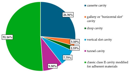
Regarding the cavity preparation technique, the majority of participants prefer the classic modified class II cavity for adhesive materials (66 participants, 51.16%), followed by the “box” cavity preferred by 34 individuals (26.36%), the “vertical slot” cavity chosen by 10 participants (7.75%), then the “tunnel” cavity preferred by 12 respondents (9.30%), and, lastly, the “gallery” or “horizontal slot” cavity and the “drop” cavity, each preferred by less than 5% of respondents (Figure 1).

Figure 1.
Cavity preparation technique.
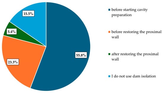
Among the respondents in our study, more than half consider the optimal moment to apply the rubber dam system to be before starting cavity preparation. Thirty participants prefer applying it before restoring the proximal wall, seven after restoring the proximal wall, and, unfortunately, twenty participants do not use rubber dam isolation at all (Figure 2).

Figure 2.
Timing of the dam system application.
For proximal restorations, the most commonly used filling material is composite resin, according to the responses of 120 participants. This is followed by glass ionomer cements, compomers, and giomers, which are used by less than 10% of participants. For restoring the proximal wall, high-viscosity composite is used by 51.9% of respondents, while low-viscosity composite is used by 48.1% (Table 3).

Table 3.
Materials used in proximal restorations and composite viscosity preferences.
When asked why they use flowable composite, most participants (n = 85) in the study cited better adaptation to cavity walls as their reason, followed by avoiding air voids, with 30 responses. Five respondents chose it to save time, two for stress relief, and seven either did not respond or specified that they do not exclusively use flowable composite.
On the other hand, in response to the question “Why don’t you use flowable composite?”, 50 participants indicated they avoid this material due to its low mechanical strength, 18 mentioned that it cannot be shaped with manual instruments, 15 do not use it because of the complicated application process (flowable composite must be applied in thin layers to compensate for high polymerization shrinkage), 12 cited greater occlusal wear, and 1 person noted that there are flowable composites with improved strength and shaping capabilities that can be used anywhere.
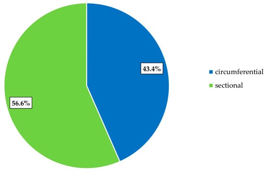
The results show that 73 participants (56.6%) preferred to use the sectional matrix system, while 56 participants (43.4%) chose the circumferential matrix system (Figure 3).

Figure 3.
Preferences in the use of matrices.
Among professionals using sectional matrices, 38 (29.5%) use the Palodent V3 ring system with sectional metal matrix (Dentsply Sirona, Charlotte, NC, USA), 21 (16.3%) use the classic ring system with pre-contoured sectional metal matrix, 16 (12.4%) use ivory matrix holders with perforated metal matrices, 13 (10.1%) use the Slot Springclip matrix holder system with saddle-type matrices (TOR VM, Moscow, Russia), and 11 (8.5%) respondents each use the Springclip matrix holder with pre-contoured saddle matrices (TOR VM, Moscow, Russia), and the Garrison ring with sectional metal matrix (Garrison Dental Solutions LLC, Spring Lake, MI, USA). Less than 15% use the Delta ring with pre-contoured sectional metal matrix (TOR VM, Moscow, Russia), the Slot ring with pre-contoured pony-type metal matrix (TOR VM, Moscow, Russia), the MD ring with pre-contoured sectional metal matrix (TOR VM, Moscow, Russia), the Polydentia system (Polydentia SA, Mezzovico-Vira, Switzerland), or pre-contoured matrices.
On the other hand, among professionals using circumferential matrices, 35 (27.1%) use the Supercap metal matrix (Kerr, Kloten, Switzerland), 28 (21.7%) use metal matrix with Tofflemire matrix holder (Kerr, Kloten, Switzerland), 24 (18.6%) use 360-degree metal matrices, and 16 (12.4%) and 20 (15.5%) of respondents use Onmimatrix metal matrices (Ultradent Products, Inc., South Jordan, UT, USA), Supercap celluloid matrices (Kerr, Kloten, Switzerland), or ProMatrix metal matrices (Astek Innovations Ltd., Altrincham, UK). Respondents in our study reported using wooden wedges (61.2%), followed by colored plastic wedges (30.2%), and, less commonly, transparent plastic wedges (6.2%) and elastic wedges (2.3%).
Regarding the tightness of the contact achieved, statistically significant differences were observed between the two types of matrices (p < 0.05). Most respondents reported optimal tightness for both types of matrices, but 15.5% considered that circumferential matrices produced an open contact, while only 6.2% felt the same about sectional matrices (Table 4).

Table 4.
Description of contact tightness achieved with sectional and circumferential matrix systems.
The emergence profile produced with a sectional matrix was reported as optimal by 94.6% of respondents and inadequate by 5.4%. On the other hand, the emergence profile produced with a circumferential matrix was reported as optimal by 82.9% of respondents and inadequate by 17.1%, with a statistically significant difference observed between the two types of matrices (p < 0.05) (Table 5).

Table 5.
Description of contact tightness and emergence profile achieved with sectional and circumferential matrix systems.
At the end of the procedure, 57 individuals (44.2%) take a radiograph to verify the accuracy of the contact point restoration, while 72 individuals (55.8%) do not. Additionally, to check the tightness of the contact after restoration, 123 individuals (95.3%) use dental floss, whereas 6 individuals (4.7%) do not use it.
4. Discussion
The success of posterior dental composite restorations depends on the skills of the operator, the characteristics of the material used, and the application techniques employed. Studies have shown that pre-contoured sectional matrix strips provide resin restorations with superior contour and emergence profile compared to conventional circumferential matrix strips. Research confirms that the use of pre-contoured sectional matrix strips in combination with a separating ring provides a more airtight tooth contact by the interdental separation achieved by the ring and the reproduction of the natural proximal contours and emergence profiles of the teeth [15].
A cross-sectional study including 171 patients and 227 first molars found that the mean decayed mesial surfaces (0.92 ± 0.85) were significantly higher than the mean decayed distal surfaces (0.73 ± 0.81) [16], the results being different from those we obtained in our research, with the majority of respondents considering that both were affected equally (62.8%), the distal wall being more affected in the perception of the study participants (20.9%) than the mesial one (16.2%).
In another cross-sectional study, Muñoz-Sandoval et al. (2022) [17] investigated the prevalence of cavitated caries in proximal lesions based on a direct clinical assessment of previously detected radiographic lesions in permanent molars and premolars. They concluded that the low proportion of cavitated lesions supports the cautious recommendation of invasive approaches in the management of proximal carious lesions, thus highlighting the need for radiographic investigations before initiating restorative dental treatment [17].
The tunnel restoration has been suggested as a conservative alternative to the conventional box preparation for treating proximal caries by Chu et al. (2013) [18]. The primary advantage of tunnel restoration compared to conventional box or slot preparations is its more conservative approach, which helps preserve the marginal ridge and thereby enhances tooth strength and structural integrity [18]. On the other hand, when used with a flowable liner, tunnel preparations resulted in fewer gaps at the proximal margins and provided tighter contact points than traditional box-only preparations. Clinically, tunnel restorations demonstrated significantly improved proximal contact quality compared to box-only restorations according to Preusse et al. (2021) [19].
In our study, respondents ranked the box cavity as the second most commonly used technique, with the classic modified Class II cavity for adhesive materials being the most frequently used. Modified proximal cavity designs for adherent materials offer several advantages over traditional cavity preparations because dentin bonding can be influenced by the type of cavity preparation, as shown in a recent article written by Abuzenada et al. (2025) [20]. Kumar et al. (2017) recommended an alternative approach—Clark’s Class II cavity preparation—which offers effectiveness comparable to the conventional Class II box preparation, while also preserving more tooth structure, allowing for more precise tooth preparation, ensuring good bond strength, and enhancing esthetics [21].
Effective control of moisture and microbial contamination is essential for the success of restorative procedures. The rubber dam is a widely used method for achieving isolation during dental restorative treatments [22]. Moreover, according to Erhamza et al. (2025), dental professionals are at higher risk of infection due to exposure to fluids and aerosols [23]. The rubber dam helps isolate the treatment area and reduce aerosol dispersion. In this context, we consider that the rubber dam isolation should be applied before cavity preparation, the results of our study supporting this approach.
Krishnakumar et al. (2024) showed that Class I restorations had significantly higher survival rates than Class II restorations, regardless of the restorative material used—whether high-viscosity glass ionomer cement or composite resin [24].
An important challenge in using composite resins for the restoration of proximal walls in Class II cavities is the accurate reconstruction of the proximal surface and the proper formation of a well-contoured interproximal contact point [25,26]. The use of high-viscosity materials (packable composites) and special instruments for occlusal and proximal shaping has proven to be more effective than medium-viscosity materials (flowable composites), while the multi-layer application technique yielded better results, according to the findings of Kampouropoulos et al. (2010) [27], which are consistent with the preferences expressed by the respondents in our study.
The results of the current survey showed that 56.6% of the individuals preferred to use the sectional matrix system and only 43.4% chose the circumferential matrix system, which means that sectional matrices are much easier to use. From a mechanistic perspective, sectional matrices are designed to closely adapt to the natural contours of proximal surfaces. When used in combination with separating rings, they allow for slight interproximal separation, which can facilitate more precise composite placement and achieve a tighter proximal contact. The pre-contoured shape of the matrix also promotes the reproduction of an optimal emergence profile, which may contribute to improved restorative morphology compared to conventional circumferential matrices. However, these findings were contradicted by another study in which 83.3% of dental students preferred to use circumferential matrix systems [28].
Within the circumferential matrix systems in the current study, the Supercap metal matrix was used most frequently (27.1%). For the sectional matrix systems, 29.5% of the participants used the Palodent V3 ring system, due to their ability to reproduce and maintain an airtight interdental contact. Colored plastic wedges and wood wedges were most commonly used for placement of posterior restorations. Wooden wedges have been favored by many dentists due to their low cost and their ability to expand in contact with oral fluids, thus facilitating interdental separation and adaptation of the matrix band to the gingival level [29].
In a previous survey investigating the techniques used by UK general dental practitioners for posterior composite placement, it was found that 155 dentists (61%) used the circumferential metal matrix system with a wooden wedge, 74 dentists (29%) used a clear matrix system, and only 25 dentists (10%) used the sectional metal matrix with a wooden wedge. These data, collected 15 years ago, reflect the period when sectional matrix systems were not yet universally included in evidence-based guidelines and amalgam restorations were preferred over composite posterior restorations [30]. Sectional matrix systems are delicate because of their rounded contours and reduced thickness, which can lead to deformation or bending of the matrix material during placement, rendering them unusable [27].
One of the most commonly used methods for evaluating interproximal contacts involves passing dental floss through the contact areas. In a study conducted by Teich et al. (2014), it was stated that the force required to pass the floss through the contact area serves as a parameter for assessing the quality of contact between adjacent teeth. This method was originally described by G.V. Black, who used waxed silk for the same purpose [31]. The same authors stated that an optimal contact is characterized by a ‘snap’ sensation when the floss passes through the contact point [31].
The results of our study showed statistically significant differences (p < 0.05) between the optimal contact points obtained using the two types of matrices studied—sectional and circumferential. Most respondents reported optimal tightness for both types of matrices; however, 15.5% considered that circumferential matrices resulted in an open contact, while only 6.2% reported the same for sectional matrices.
Nevertheless, further research and standardized methods are needed to accurately assess the contact point and contact area tightness, as the current literature does not clearly outline a standardized evaluation protocol.
In the current study, dentists reported that sectional matrix systems produced optimal contact points with adequate emergence profiles in a proportion of 94.6%, while optimal contacts obtained with circumferential matrices were 82.9%. Consequently, our survey indicates that respondents perceive the emergence profile obtained using sectional matrices as superior to that achieved with circumferential matrices, based on their clinical experience and opinion. These results are similar to those of Tolba et al. (2023), who demonstrated that contact tightness is significantly better compared to that obtained with circumferential matrices [32].
A combined clinical and radiographic evaluation following each proximal restoration is recommended to minimize the risk of failure, according to Karimi et al. (2015) [33]. Postoperative radiographs are advised to assess the contour of interproximal restorations, as they enable the detection of overcontoured areas (with excess restorative material) or undercontoured areas (with insufficient material) at the level of the proximal contact point. Fortes et al. (2017) stated that the quality of dental restorations is directly proportional to the maintenance of both superficial and deep marginal periodontal health [34]. By adhering to a well-defined protocol throughout the stages of dental treatment, improved case management can be achieved, thereby preventing potential dental complications [35]. However, only 44.2% of the participants in our study reported using postoperative radiographs. Considering the findings from the scientific literature and the results of our research, we recommend that dentists verify the accuracy of the contact point using both dental floss and postoperative radiographs.
5. Conclusions
The present study highlighted practitioners’ preferences and perceptions of matrix system effectiveness in posterior Class II cavity restorations. The majority of respondents favored sectional matrix systems, which were perceived to be associated with a higher rate of achieving optimal proximal contact points and adequate emergence profiles compared to circumferential matrix systems. These differences were statistically significant for both contact quality and emergence profile reproduction.
Furthermore, a considerable variation was observed in the use of essential auxiliary techniques, such as rubber dam isolation and postoperative verification methods (radiographs, dental floss), reflecting a lack of standardized clinical protocols. This variability may negatively impact the long-term success of restorations and associated periodontal health.
Therefore, we conclude that sectional matrix systems appear to be preferred by the surveyed practitioners in Class II restorations, reflecting practitioners’ perceptions of higher effectiveness. Additionally, these findings suggest that developing standardized clinical protocols and promoting ongoing professional education may help support effective assessment of proximal restorations and the adoption of modern restorative techniques.
Author Contributions
Conceptualization, G.I.P.C. and A.V.T.; methodology, G.I.P.C. and A.V.T.; software, R.H., R.A.C. and M.F.G.; validation, L.T., I.S. and G.C.; formal analysis, G.C.; investigation, A.V.T. and A.C.; resources, A.M.P.; data curation, I.S.; writing—original draft preparation, G.I.P.C. and R.A.C.; writing—review and editing, L.T. and G.C.; visualization, R.A.C. and I.S.; supervision, G.C. and R.H.; project administration, G.I.P.C. All authors have read and agreed to the published version of the manuscript.
Funding
The APC was funded by the University of Oradea, Oradea, Romania.
Institutional Review Board Statement
Not applicable.
Informed Consent Statement
Not applicable.
Data Availability Statement
Data sharing is not applicable to this article as no datasets were generated or analyzed during the current study.
Acknowledgments
The authors would like to thank the University of Oradea for supporting the payment of the invoice through an internal project.
Conflicts of Interest
The authors declare no conflicts of interest.
References
- Marcov, E.C.; Bodnar, D.C.; Marcov, N. Manual de Odontoterapie Restauratoare; Editura Universitară “Carol Davila”: București, Romania, 2020. [Google Scholar]
- Adrian, S. Tratamentul Minim Invaziv al Cariei Dentare; Princeps Edit: Iași, Romania, 2002. [Google Scholar]
- Ionescu, E. Manual Pentru Rezidențiat; Editura Universitară “Carol Davila”: București, Romania, 2021; Volume 1. [Google Scholar]
- Pitts, N.B.; Zero, D.T.; Marsh, P.D.; Ekstrand, K.; Weintraub, J.A.; Ramos-Gomez, F.; Tagami, J.; Twetman, S.; Tsakos, G.; Ismail, A. Dental Caries. Nat. Rev. Dis. Primers 2017, 3, 17030. [Google Scholar] [CrossRef]
- Seneviratne, C.J.; Zhang, C.F.; Samaranayake, L.P. Dental Plaque Biofilm in Oral Health and Disease. Chin. J. Dent. Res. 2011, 14, 87–94. [Google Scholar]
- Valm, A.M. The Structure of Dental Plaque Microbial Communities in the Transition from Health to Dental Caries and Periodontal Disease. J. Mol. Biol. 2019, 431, 2957–2969. [Google Scholar] [CrossRef]
- Marsh, P.D.; Moter, A.; Devine, D.A. Dental Plaque Biofilms: Communities, Conflict and Control. Periodontology 2000 2011, 55, 16–35. [Google Scholar] [CrossRef] [PubMed]
- Puleio, F.; Fiorillo, L.; Gorassini, F.; Iandolo, A.; Meto, A.; D’aMico, C.; Cervino, G.; Pinizzotto, M.; Bruno, G.; Portelli, M.; et al. Systematic Review on White Spot Lesions Treatments. Eur. J. Dent. 2022, 16, 41–48. [Google Scholar] [CrossRef] [PubMed]
- Shimada, Y.; Sato, T.; Inoue, G.; Nakagawa, H.; Tabata, T.; Zhou, Y.; Hiraishi, N.; Gondo, T.; Takano, S.; Ushijima, K.; et al. Evaluation of Incipient Enamel Caries at Smooth Tooth Surfaces Using SSOCT. Materials 2022, 15, 5947. [Google Scholar] [CrossRef]
- Potra Cicalău, G.I.; Vicaș, L.G.; Ciavoi, G.; Ghitea, T.C.; Csaba, N.; Cristea, R.A.; Miere, F.; Ganea, M. A Natural Approach to the Prevention and Treatment of Gingivitis and Periodontitis: A Review of Pomegranate’s Bioactive Properties. Life 2024, 14, 1298. [Google Scholar] [CrossRef] [PubMed]
- Ganea, M.; Ioana, P.C.G.; Ghitea, T.C.; Ștefan, L.; Groza, F.; Frent, O.D.; Nagy, C.; Iova, C.S.; Schwarz-Madar, A.F.; Ciavoi, G.; et al. Development and Evaluation of Gelatin-Based Gummy Jellies Enriched with Oregano Oil: Impact on Functional Properties and Controlled Release. Foods 2025, 14, 479. [Google Scholar] [CrossRef]
- Prada, A.M.; Potra Cicalău, G.I.; Ciavoi, G. A Review of White Spot Lesions: Development and Treatment with Resin Infiltration. Dent. J. 2024, 12, 375. [Google Scholar] [CrossRef]
- Flynn, L.N.; Julien, K.; Noureldin, A.; Buschang, P.H. The Efficacy of Fluoride Varnish vs a Filled Resin Sealant for Preventing White Spot Lesions during Orthodontic Treatment. Angle Orthod. 2022, 92, 204–212. [Google Scholar] [CrossRef]
- Knösel, M.; Eckstein, A.; Helms, H.J. Long-Term Follow-Up of Camouflage Effects Following Resin Infiltration of Post Orthodontic White-Spot Lesions In Vivo. Angle Orthod. 2019, 89, 33–39. [Google Scholar] [CrossRef] [PubMed]
- Saber, M.H.; Loomans, A.C.; El Zohairy, A.; Dörfer, C.E.; El-Badrawy, W. Evaluation of Proximal Contact Tightness of Class II Resin Composite Restorations. Oper. Dent. 2010, 35, 37–43. [Google Scholar] [CrossRef]
- Hasan, A.; Khan, J.A.; Taqi, M.; Naz, F.; Waqar, S.; Mehak, N.; Mehmood, H. Predictors for Proximal Caries in Permanent First Molars: A Multiple Regression Analysis. J. Contemp. Dent. Pract. 2019, 20, 818–821. [Google Scholar] [CrossRef]
- Muñoz-Sandoval, C.; Gambetta-Tessini, K.; Botelho, J.N.; Giacaman, R.A. Detection of Cavitated Proximal Carious Lesions in Permanent Teeth: A Visual and Radiographic Assessment. Caries Res. 2022, 56, 171–178. [Google Scholar] [CrossRef]
- Chu, C.H.; Mei, M.L.; Cheung, C.; Nalliah, R.P. Restoring Proximal Caries Lesions Conservatively with Tunnel Restorations. Clin. Cosmet. Investig. Dent. 2013, 5, 43–50. [Google Scholar] [CrossRef] [PubMed]
- Preusse, P.J.; Winter, J.; Amend, S.; Roggendorf, M.J.; Dudek, M.C.; Krämer, N.; Frankenberger, R. Class II Resin Composite Restorations—Tunnel vs. Box-Only In Vitro and In Vivo. Clin. Oral Investig. 2021, 25, 737–744. [Google Scholar] [CrossRef]
- Abuzenada, B. Impact of Different Cavity Preparation Techniques on Dentin Bond Strength. Bangladesh J. Med. Sci. 2025, 24, 104–108. [Google Scholar] [CrossRef]
- Kumar, T.; Sanap, A.; Bhargava, K.; Aggarwal, S.; Kaur, G.; Kunjir, K. Comparative Evaluation of the Bond Strength of Posterior Composite with Different Cavity Configurations and Different Liners Using a Two-Step Etch and Rinse Adhesive System: In Vitro Study. J. Conserv. Dent. 2017, 20, 166–169. [Google Scholar] [CrossRef]
- Miao, C.; Yang, X.; Wong, M.C.; Zou, J.; Zhou, X.; Li, C.; Wang, Y. Rubber Dam Isolation for Restorative Treatment in Dental Patients. Cochrane Database Syst. Rev. 2021, 5, CD009858. [Google Scholar] [CrossRef]
- Sezen Erhamza, T.; Küçük, K.C.; Malikov, İ. Comparative Effects of Rubber Dam and Traditional Isolation Techniques on Orthodontic Bracket Positioning: A 3D Digital Model Evaluation. Appl. Sci. 2025, 15, 2552. [Google Scholar] [CrossRef]
- Krishnakumar, K.; Kalaskar, R.; Kalaskar, A.; Bhadule, S.; Joshi, S. Clinical Effectiveness of High-Viscosity Glass Ionomer Cement and Composite Resin as a Restorative Material in Primary Teeth: A Systematic Review of Clinical Trials. Int. J. Clin. Pediatr. Dent. 2024, 17, 221–228. [Google Scholar] [CrossRef]
- Brackett, M.G.; Contreras, S.; Contreras, R.; Brackett, W.W. Restoration of Proximal Contact in Direct Class II Resin Composites. Oper. Dent. 2006, 31, 155–156. [Google Scholar] [CrossRef]
- Keogh, T.P.; Bertolotti, R.L. Creating Tight, Anatomically Correct Interproximal Contacts. Dent. Clin. N. Am. 2001, 45, 83–102. [Google Scholar] [CrossRef]
- Kampouropoulos, D.; Paximada, C.; Loukidis, M.; Kakaboura, A. The Influence of Matrix Type on the Proximal Contact in Class II Resin Composite Restorations. Oper. Dent. 2010, 35, 454–462. [Google Scholar] [CrossRef] [PubMed]
- Ahmad, M.Z.; Gaikwad, R.N.; Arjumand, B. Comparison of Two Different Matrix Band Systems in Restoring Two-Surface Cavities in Posterior Teeth Done by Senior Undergraduate Students at Qassim University, Saudi Arabia: A Randomized Controlled Clinical Trial. Indian J. Dent. Res. 2018, 29, 459–464. [Google Scholar] [CrossRef] [PubMed]
- Gupta, R.; Hegde, J.; Prakash, V.; Srirekha, A. Concise Conservative Dentistry and Endodontics; Elsevier Health Sciences: New York, NY, USA, 2019. [Google Scholar]
- Gilmour, A.S.M.; Latif, M.; Addy, L.D. Placement of Posterior Composite Restorations in United Kingdom Dental Practices: Techniques, Problems, and Attitudes. Int. Dent. J. 2009, 59, 148–154. [Google Scholar] [CrossRef]
- Teich, S.T.; Joseph, J.; Sartori, N.; Heima, M.; Duarte, S. Dental Floss Selection and Its Impact on Evaluation of Interproximal Contacts in Licensure Exams. J. Dent. Educ. 2014, 78, 921–926. [Google Scholar] [CrossRef]
- Tolba, Z.O.; Oraby, E.; Abd El Aziz, P.M. Impact of Matrix Systems on Proximal Contact Tightness and Surface Geometry in Class II Direct Composite Restoration In Vitro. BMC Oral Health 2023, 23, 535. [Google Scholar] [CrossRef] [PubMed]
- Karimi, Z.; Kessa, S.; Chala, S.; Abdallaoui, F. Évaluation de la Qualité des Restaurations Proximales aux Matériaux Plastiques: Étude Radiographique. Odonto-Stomatol. Trop. 2015, 38, 5–12. [Google Scholar]
- Fortes, J.; Dallavilla, F.; Tirapelli, C.; Watanabe, P.; Brito, A.; Oliveira-Santos, C. Radiographic Assessment of the Quality of Dental Restorations and Their Relationship with Periodontal Radiographic Changes. Rev. Odonto Ciênc. 2017, 32, 17. [Google Scholar] [CrossRef][Green Version]
- Potra Cicalău, G.I.; Todor, L.; Cristea, R.A.; Hodișan, R.; Marcu, O.A.; Țig, I.A.; Daina, L.G.; Ciavoi, G. Integrated Dental Practice Management in Romania: A Cross-Sectional Case–Control Study on the Perceived Impact of Managerial Training on Efficiency, Collaboration, and Care Quality. Healthcare 2025, 13, 1631. [Google Scholar] [CrossRef] [PubMed]
Disclaimer/Publisher’s Note: The statements, opinions and data contained in all publications are solely those of the individual author(s) and contributor(s) and not of MDPI and/or the editor(s). MDPI and/or the editor(s) disclaim responsibility for any injury to people or property resulting from any ideas, methods, instructions or products referred to in the content. |
© 2025 by the authors. Licensee MDPI, Basel, Switzerland. This article is an open access article distributed under the terms and conditions of the Creative Commons Attribution (CC BY) license (https://creativecommons.org/licenses/by/4.0/).