Abstract
Background: Rapid maxillary expansion is a common therapy when a palatal transversal discrepancy occurs. Different anchorage solutions have been proposed to obtain an effective skeletal expansion, even for adult patients. The aim of the present research was to evaluate the dentoskeletal effects of a hybrid expander and multi-bracket therapy, considering three groups of patients with different cervical vertebral maturation (CVM) stages. Materials and Methods: The study evaluated 68 consecutively treated patients. The age of the patients varied from 7 to 27 years old (mean age 13.45). The sample was divided into the following three groups based on CVM stage at the start of treatment: Group 1 (CS1–CS2) included 21 patients (mean age 10.21, SD 1.34), Group 2 (CS3–CS4) included 28 patients (mean age 13.37, SD 1.37) and Group 3 (CS5–CS6) was composed of 19 patients (mean age 17.14, SD 3.48). Each patient underwent orthodontic therapy where the first step was a palatal expansion by means of a hybrid expander; afterwards, the therapy was completed with a multi-bracket appliance. Expansion and torque values were observed at the end of treatment on digital models. Results: Significant intragroup differences in transversal expansion were found over time for all parameters in all groups. No significant differences were found among groups for longitudinal changes. No significant differences were found among groups for longitudinal changes of torque. Conclusions: The tooth–bone-borne maxillary expander and multi-bracket produced a significant clinical expansion with negligible dental compensation. The effect of the maxillary expansion and multi-bracket therapy showed no differences among the maturation groups in regard to transversal diameter changes and torque values.
1. Introduction
Transverse maxillary deficiency is a common orthodontic and orthopedic problem, affecting patients with different characteristics [1,2,3,4], such as the posterior crossbite, either mono-lateral or bilateral. The therapeutic effects include both dental and skeletal modifications [5,6,7], and these effects may vary considering the age of the patient [8,9,10,11] and the therapy options.
The recent introduction of miniscrew-supported expanders has made alternatives to the traditional tooth-borne device available. These alternatives include the hybrid expander, including both dental and miniscrew support [12,13,14], and the bone-borne expander without any dental support [15,16].
The use of miniscrew-supported expanders finds a rationale in those cases where the obliteration of the palatal suture tends to be complete or, alternatively, when dental tipping effects are undesired. In such cases, the use of dental-supported expanders would result in significant dental shifting [17]. Temporary anchorage devices inserted in the anterior palate are extremely reliable, with a success rate of approximately 98% [18], and safe [19,20,21].
Recently published articles reported the potential of the hybrid expander in different situations [22,23], but no studies have considered the effects of this approach at different ages so far. The age at which the midpalatal suture ossifies and transverse palatal growth ceases is extremely variable. Obliterations of the suture have been found in 16-year-old females and 18-year-old males [24], but ossification has only been found in half of the 15- to 20-year-old population [25]. The earliest ossification of the midpalatal suture in a 21-year-old male has also been reported, whereas the oldest unossified midpalatal suture was found in a 54-year-old patient. Ossification was observed in only 40 per cent of patients aged between 23 and 30. It may be assumed that the obliteration of the midpalatal suture in radiographs does not correlate with chronological age [26,27,28].
A palatine suture maturation staging method has been proposed to avoid the side effects of rapid maxillary expansion failure [29], and correspondence with CVM stages has been established as well [30].
The aim of the present research is to evaluate the dental-alveolar effects of a hybrid expander and multi-bracket therapy in three groups of consecutively treated patients, divided by CVM stages. The hypothesis is that different age and different skeletal maturation stages can possibly alter the results of miniscrew-supported rapid maxillary expansion.
2. Materials and Methods
The present retrospective study was conducted on patients who were treated consecutively by the same operator. The collected data were anonymously recorded, and the statistical analysis was conducted blindly (blindness was obtained by eliminating every reference to the group from the elaboration file).
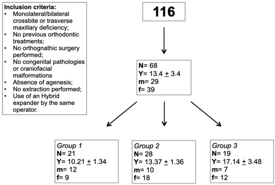
This study includes 116 initial patients, with ages ranging from 7 to 46 years (mean age 14.51 years). The criteria adopted for the inclusion in the study were as follows: the presence of maxillary transversal deficit; no previous orthodontic treatment; no orthognathic surgery performed; no extractions; absence of agenesis, congenital pathologies and cranio-maxillofacial malformations; use of a hybrid device for palatal expansion using two miniscrews placed in the anterior area of the palate; and a complete set of pre- and post-therapy models. Therapy was defined as treatment with maxillary expansion first and subsequent multi-bracket therapy.
Based on these criteria, 48 patients were excluded, and 68 were evaluated for the study. The ages of the patients included in the study varied from 7 to 27 years old (mean age 13.45). The sample was divided into three groups based on CVM stage at the start of treatment, using the CVM method as an assessment of midpalatal suture maturation [24]. Group 1 (CS1–CS2) included 21 patients (mean age 10.21, SD 1.34), Group 2 (CS3–CS4) included 28 patients (mean age 13.37, SD 1.37) and Group 3 (CS5–CS6) was composed of 19 patients (mean age 17.14, SD 3.48) (Figure 1).

Figure 1.
Study flow chart.
TeleRx and STL models of dental arches were obtained for each patient, both at the beginning and the end of the treatment.
Each patient underwent an orthodontic therapy where the first step was a palatal expansion by means of a hybrid expander; afterwards, the therapy was completed with a multi-bracket appliance (BioQuick, Forestadent, Germany).
The type of screw chosen for each device was Orthopal (8 mm length, 1.7 mm diameter). The number of activations of the hybrid device varied from 1 to 3 activations per day. Activation days varied from 7 to 23 days, until the operator was evaluated to have reached a proper upper transversal dimension.
All the patients were treated with an MBT prescription bracket for the upper incisors and standard Edgewise prescription for the posterior teeth.
Expansion values were observed at the end of treatment on digital models.
The following dental elements of the upper jaw were considered in the evaluation of the dental transversal variations: canine, premolars and first molar. Three points were considered for each element:
- Cusp: buccal cusp was considered. For the first molar, the point was placed on the mesio-vestibular cusp.
- Centroid: the center of the occlusal table.
- Lingual point: the most palatal point at the level of the tooth collar.
The distance in millimeters (Figure 2) was calculated between the respective points on the teeth of the contralateral arch on the 3D models, both pre- and post-treatment.

Figure 2.
Centroid interdental distance, cusp interdental distance and lingual point interdental distance.
The measurements were performed by two operators. A sample of 10 patients was examined to detect the accuracy of the measurements. Both operators evaluated the cusp–cusp, centroid–centroid and lingual point–lingual point distances at the level of the first upper molars. The obtained values were compared to each other, using Pearson’s correlation index. A correlation index value equal to +1 indicates a perfect positive correlation. A correlation index equal to 0.998 was obtained.
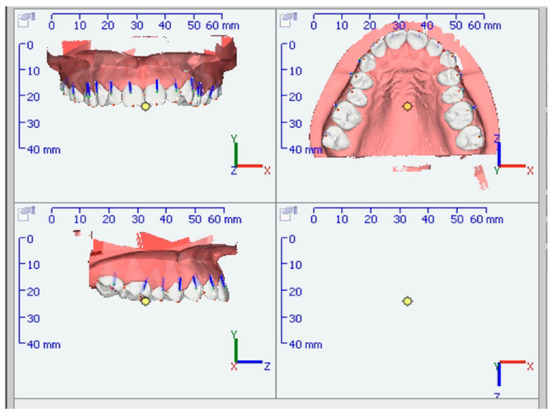
Dental torque values were observed at the end of treatment on digital models. The torque value was obtained through the OnyxCeph software (https://onyxceph.eu/en/ Image Instrument (accessed on 18 January 2024), Chemnitz, Germany) following these steps:
- The crowns of the dental elements were selected with the ‘Segmentation’ option.
- When the crowns were selected, the following commands were entered: ‘Segment crowns’ > ‘Separate crowns’ > ‘Complete separation’.
- Using the ‘Aligner’ option and selecting the ‘permanent teeth’ preference, the values were obtained by the ‘Tooth movement’ command (Figure 3).
 Figure 3. Dental torque analysis trough segmentation.
According to Andrews’ definition, torque is the inclination of the crown, represented by the angle between a vertical line perpendicular to the occlusal plane and a tangent passing through the center of the crown on the buccal face (i.e., the facial axis point, or FA). Graphic tests were carried out to ensure the reliability of the program used for this study. The following operations were performed on a sample of 10 random patients:
- 4.
- The segmented STL models were opened in OnyxCeph (Image Instrument, Chemnitz, Germany).
- 5.
- Using the ‘Segmentation’ tool, the soft tissues and all the dental elements were removed from the model except for element 11 and element 26.
- 6.
- The file was uploaded to Paint (Microsoft, Redmond, WA, USA). The occlusal plane and the vertical straight line perpendicular to the horizontal plane were drawn.
- 7.
- The plans were checked with the options ‘Show’ > ‘Scale’ of Paint program (Microsoft, Redmond, WA, USA).
- 8.
- The FA point, according to Andrews, was positioned in the center of the crown.
- 9.
- The tangent was drawn at the center of the crown on the vestibular face.
- 10.
- The image was then added to an online program to calculate angles (https://www.ginifab.com/feeds/angle_measurement/online_protractor.it-it.php, accessed on 1 June 2022), where it was possible to measure the angle between the vertical line and the tangent to the center of the crown on the vestibular face.
Element 26 has been kept as a reference for the occlusal plane, while element 11 has been measured.
An intraclass correlation test was performed between the values obtained using the OnyxCeph software (https://onyxceph.eu/en/, accessed on 18 January 2024) and the values obtained using the graphic method. The intraclass correlation coefficient (ICC) obtained was 0.9134.
Sample Size
Nineteen patients achieved 80% power to detect a difference of 2.02 mm in terms of intercuspal width at the first molar, with a significance level (alpha) of 0.05 using a two-sided paired t-test and assuming a standard deviation of 2.9 mm (data on post-treatment, intercuspal molar width were found in [1]; calculations were performed with the R v3.4.2 software).
3. Results
Statistical Analysis
Data were checked for normality. Continuous variables are given as means ± standard deviations (SD) and medians with interquartile range, and categorical variables are shown as the number or percentage of subjects.
The baseline differences among the groups were tested with one-way analysis of variance with group as a factor, or with the Kruskal–Wallis test.
Intragroup differences over time were tested with a paired t-test or Wilcoxon’s signed-rank test. One-way analysis of variance, or the Kruskal–Wallis test, was performed again to investigate the association of differences over time with the groups. Post hoc comparisons were carried out with the Bonferroni method.
Differences with a p-value < 0.05 were selected as significant. Data were acquired and analyzed in the R v3.4.4 software [31].
Data from 68 patients were analyzed. The mean age was 13.4 years. The minimum age was 7 years, and the maximum age was 27 years. The mean therapy duration was 16.5 months. The demographic and clinical characteristics of the groups are shown in Table 1. No miniscrew failures were observed.

Table 1.
Demographic and clinical characteristics of groups. M: male; F: female.
The mean amount of activation of the expansion screw was 7.63 ± 1.74 mm (range, 3–13.8 mm), and the duration of the expansion ranged from 7 to 23 days.
The baseline parameter differences between groups are shown in Table 2 and Table 3. No significant differences were found at baseline, except for III cent and VI L (p = 0.015 and 0.031, respectively), and for the torque of 1.6 (p = 0.047).

Table 2.
Baseline characteristics of transverse measurements in whole population (N = 68). C: cuspid; Cent: centroid; L: lingual. Results are expressed as mean ± standard deviation or median [interquartile range]; p-value = one-way analysis of variance with group as factor, or Kruskal–Wallis test p-value. Post hoc multiple comparisons report the Bonferroni method p-value.

Table 3.
Baseline characteristics of torque measurements in whole population (N = 68). Results are expressed as mean ± standard deviation or median [interquartile range]; p-value = one-way analysis of variance with group as factor, or Kruskal–Wallis test p-value. Post hoc multiple comparisons report the Bonferroni method p-value.
In terms of transversal expansion, significant intragroup differences over time were found for all parameters in all groups (Table 4).

Table 4.
Differences over time in expansion measurements (N = 68). Results are expressed as mean ± standard deviation or median [interquartile range]; intergroup p-value = one-way analysis of variance with group as factor or Kruskal–Wallis test p-value. Intragroup T1–T0 p-value = paired t-test p-value, or Wilcoxon’s signed rank test p-value. III: canine; IV: first premolar; V: second premolar; VI first molar.
No significant differences were found among groups in regard to longitudinal changes.
Significant intragroup differences over time were found for the torque of 1.5 in Group 2 (p = 0.009) and Group 3 (p < 0.001), for the torque of 1.4 in Group 3 (p = 0.001), for the torque of 2.4 in Group 1 (p = 0.006), Group 2 (p = 0.036) and Group 3 (p = 0.005) and for the torque of 2.5 in Group 2 (p = 0.028, Table 5). No significant differences were found among groups with regard to longitudinal changes of torque.

Table 5.
Differences over time in torque measurements (N = 68). Results are expressed as mean ± standard deviation or median [interquartile range]; intergroup p-value = one-way analysis of variance with group as factor or Kruskal–Wallis test p-value. Intragroup T1-T0 p-value = paired t-test p-value, or Wilcoxon’s signed rank test p-value.
4. Discussion
The objective of the present study was to evaluate the differences, if any, between the different CVM-stage groups of patients who underwent the same therapeutical protocol, including the use of a tooth–bone-borne maxillary expander and multi-bracket therapy.
4.1. Treatment Effects at Different CVM Stages
The recent appearance of miniscrew-supported palatal expanders, both tooth–bone-borne (tooth and miniscrews) and bone-borne devices, has widened the opportunity to correct transversal maxillary deficiency in adult patients. Previously published articles suggested that the midpalatal suture interdigitation becomes closer with age and skeletal maturity [32,33]. They report that routine clinical applications of tooth-borne expanders are limited to younger and growing patients, with a different device design. The use of skeletal anchorage with two or four miniscrews as points of resistance to the applied expansion force allows a skeletal effect in adult patients as well, with minimum or negligible dental compensation.
Studies conducted using CBCT to measure maxillary expansion in adult patients produced different results, compared to tooth-borne palatal expanders with miniscrew-supported devices. Molar-supporting expanders allow dentoalveolar expansion with significant molar buccal inclination (3.65° Lin et al.) and maxillary bending of 3.65°, resulting in a poor skeletal effect. Bone-borne devices showed limited dental and maxillary bending, allowing a significant bone expansion at the sutural level [12,18]. These results are similar to those found in the present study, where the expansion produced almost no effect on the first molar torque values (values ranged between −0.08 to 2.15). More changes were found at the premolar level, with values as high as 8.05°, but even at this level, no differences were found among the three groups. At the canine level, no significant differences were found intergroup or over time, except for the right canine in Group 1, which showed an approximately 9° change.
Effects of hybrid expanders on young and older patients were evaluated by Vassar et al. in 2016, comparing CBCTs before and after appliance removal; the therapy proved to be effective and gave positive skeletal effects with mild molar tipping, even though the older subjects appeared to have more dental tipping [34]. The tooth–bone-borne appliance could provide a more skeletally directed force vector and produce positive effects in >16-year-old patients as well. In contrast with those results, the analysis between groups in the present study showed no statistically significant differences at any level, considering both the torque values and the transversal parameters. This outcome suggests that, even in Group 3, an efficient maxillary expansion can be achieved. Note that no miniscrew failures were registered in the whole sample. Even considering the mean values of the transversal changes over time, a similar expansion was observed at the level of the first molars, and a one-millimeter-higher value was observed in Group 1 (10-year-old patients) compared to Group 3 patients (17 years) at the canine level. These differences to previously published articles may be related to the use of digital models instead of CBCT analysis. Interestingly, even the torque values of the first molars showed no differences over time among groups. It was noticed that the torque values determine a transversal component that can be trigonometrically derived.
Baccetti et al. compared CVM 1–3 and CVM 4–6 patients treated with a Haas expander and multi-bracket therapy, and found that, in the long run, the group treated later had no permanent increase in the skeletal width of the maxilla [8]. Thus, a significant part of the expansion was a dentoalveolar effect. This result was different for the early treated group, where, in the long run, the same therapy produced permanent increases in the transverse dimensions, both dental and skeletal. These results are in contrast with the data obtained in the present study, where no differences were reported among groups for any of the evaluated parameters. The type of anchorage, dental or skeletal, using temporary anchorage devices, could play a fundamental role in determining such differences.
A recently published clinical trial showed that examining the effect of the tooth–bone-borne appliance on younger patients found no evident clinically significant differences between the tooth-borne appliance and tooth–bone-borne devices 1 year after expansion [35].
The overall approach regarding which anchorage is better when addressing a maxillary expansion could be as follows. For mixed-dentition patients, a conventional tooth-borne appliance led to optimum maxillary skeletal expansion. In permanent-dentition patients less than 20 years old, tooth–bone-borne or bone-borne appliances guarantee an efficient skeletal expansion with negligible dental effects and a high success rate.
4.2. Limits of the Study
To evaluate the therapy effects on the maxillary expansion, measurements were conducted on digital models at different levels: canine, premolars and molars. A CBCT analysis could be a more detailed instrument for detecting the maxillary skeletal effect properly. Torque values were recorded before and after treatment to complete the data analysis on dental movement and evaluate dental tipping after this therapy. No data can be extracted for the expander effect only, since the whole therapy was considered.
Groups were established according to the CVM method [8]: CVM 1–2: Group 1, CVM 3–4: Group 2 and CVM 5–6: Group 3. Earlier CVM stages (CS1, CS2 and CS3) can be used as reliable indicators for the absence of fusion of the midpalatal suture. However, this approach could have some limits regarding the efficacy of identifying a partial or total suture obliteration in post-pubertal patients (CS4 and CS5) for whom an individual assessment of the midpalatal suture with CBCT would be a more reliable option. However, the widespread use of CBCT before and after expansion is questionable.
5. Conclusions
The results of the present study lead to the following conclusions:
- The tooth–bone-borne maxillary expander produced a significant clinical expansion with negligible dental compensation.
- Dental compensation and torque values after expansion and multi-bracket therapy were similar among the three maturation groups.
- The effects of maxillary expansion and multi-bracket therapy showed no differences in terms of transversal diameter changes and torque values among maturation groups.
Author Contributions
B.L. clinical interventions; B.G. and R.P., data collection and analysis; S.D., statistics; M.M. (Marco Migliorati), supervision, research protocol and submission; M.M (Maria Menini). and P.P. writing, editing and project management. All authors have read and agreed to the published version of the manuscript.
Funding
This research received no external funding.
Institutional Review Board Statement
The study was conducted in accordance with the Declaration of Helsinki, and approved by the Institutional Review Board of Genova University (2022/51, November 2022).
Informed Consent Statement
Informed consent was obtained from all subjects involved in the study.
Data Availability Statement
Data are available upon request from the corresponding author.
Conflicts of Interest
The authors declare no conflicts of interest.
References
- McNamara, J.A. Maxillary transverse deficiency. Am. J. Orthod. Dentofac. Orthop. 2000, 117, 567–570. [Google Scholar] [CrossRef]
- Proffit, W.R.; Fields, H.W.; Moray, L.J. Prevalence of malocclusion and orthodontic treatment need in the United States: Estimates from the NHANES III survey. Int. J. Adult Orthod. Orthognath. Surg. 1998, 13, 97–106. [Google Scholar]
- Brunelle, J.A.; Bhat, M.; Lipton, J.A. Prevalence and distribution of selected occlusal characteristics in the US population, 1988–1991. J. Dent. Res. 1996, 75, 706–713. [Google Scholar] [CrossRef] [PubMed]
- Bilgic, F.; Gelgor, I.E.; Celebi, A.A. Malocclusion prevalence and orthodontic treatment need in central Anatolian adolescents compared to European and other nations’ adolescents. Dent. Press J. Orthod. 2015, 20, 75–81. [Google Scholar] [CrossRef]
- Haas, A.J. Rapid Palatal Expansion: A Recommended Prerequisite to Class III Treatment; Transactions European Orthodontic Society: London, UK, 1973; pp. 311–318. [Google Scholar]
- Tollaro, I.; Baccetti, T.; Franchi, L.; Tanasescu, C.D. Role of posterior transverse interarch discrepancy in Class II, Division 1 malocclusion during the mixed dentition phase. Am. J. Orthod. Dentofac. Orthop. 1996, 110, 417–422. [Google Scholar] [CrossRef] [PubMed]
- Ghoneima, A.; Abdel-Fattah, E.; Eraso, F.; Fardo, D.; Kula, K.; Hartsfield, J. Skeletal and dental changes after rapid maxillary expansion: A computed tomography study. Australas. Orthod. J. 2010, 26, 141–148. [Google Scholar] [CrossRef]
- Baccetti, T.; Franchi, L.; Cameron, C.G.; McNamara, J.A. Treatment timing for rapid maxillary expansion. Angle Orthod. 2001, 71, 343–350. [Google Scholar] [PubMed]
- Seif-Eldin, N.F.; Elkordy, S.A.; Fayed, M.S.; Elbeialy, A.R.; Eid, F.H. Transverse Skeletal Effects of Rapid Maxillary Expansion in Pre and Post Pubertal Subjects: A Systematic Review. Open Access Maced. J. Med. Sci. 2019, 7, 467–477. [Google Scholar] [CrossRef] [PubMed]
- Lagravere, M.O.; Major, P.W.; Flores-Mir, C. Long-term skeletal changes with rapid maxillary expansion: A systematic review. Angle Orthod. 2005, 75, 1046–1052. [Google Scholar]
- Angelieri, F.; Cevidanes, L.H.; Franchi, L.; Gonçalves, J.R.; Benavides, E.; McNamara, J.A., Jr. Midpalatal suture maturation: Classification method for individual assessment before rapid maxillary expansion. Am. J. Orthod. Dentofac. Orthop. 2013, 144, 759–769. [Google Scholar] [CrossRef]
- Lin, L.; Ahn, H.W.; Kim, S.J.; Moon, S.C.; Kim, S.H.; Nelson, G. Tooth-borne vs. bone-borne rapid maxillary expanders in late adolescence. Angle Orthod. 2015, 85, 253–262. [Google Scholar] [CrossRef]
- Mosleh, M.I.; Kaddah, M.A.; Abd Elsayed, F.A.; Elsayed, H.S. Comparison of transverse changes during maxillary expansion with 4-point bone-borne and tooth-borne maxillary expanders. Am. J. Orthod. Dentofac. Orthop. 2015, 148, 599–607. [Google Scholar] [CrossRef]
- Choi, S.H.; Shi, K.K.; Cha, J.Y.; Park, Y.C.; Lee, K.J. Nonsurgical miniscrew-assisted rapid maxillary expansion results in acceptable stability in young adults. Angle Orthod. 2016, 86, 713–720. [Google Scholar] [CrossRef]
- Cantarella, D.; Dominguez-Mompell, R.; Mallya, S.M.; Moschik, C.; Pan, H.C.; Miller, J.; Moon, W. Changes in the midpalatal and pterygopalatine sutures induced by micro-implant-supported skeletal expander, analyzed with a novel 3D method based on CBCT imaging. Prog. Orthod. 2017, 18, 34. [Google Scholar] [CrossRef] [PubMed]
- Cantarella, D.; Dominguez-Mompell, R.; Moschik, C.; Mallya, S.M.; Pan, H.C.; Alkahtani, M.R.; Elkenawy, I.; Moon, W. Midfacial changes in the coronal plane induced by microimplant-supported skeletal expander, studied with cone-beam computed tomography images. Am. J. Orthod. Dentofac. Orthop. 2018, 154, 337–345. [Google Scholar] [CrossRef]
- Moon, H.W.; Kim, M.J.; Ahn, H.W.; Kim, S.J.; Kim, S.H.; Chung, K.R.; Nelson, G. Molar inclination and surrounding alveolar bone change relative to the design of bone-borne maxillary expanders: A CBCT study. Angle Orthod. 2020, 90, 13–22. [Google Scholar] [CrossRef] [PubMed]
- Wilmes, B.; Ludwig, B.; Vasudavan, S.; Nienkemper, M.; Drescher, D. The T-Zone: Median vs. Paramedian Insertion of Palatal Mini-Implants. J. Clin. Orthod. 2016, 50, 543–551. [Google Scholar]
- Migliorati, M.; Drago, S.; Schiavetti, I.; Olivero, F.; Barberis, F.; Lagazzo, A.; Capurro, M.; Silvestrini-Biavati, A.; Benedicenti, S. Orthodontic miniscrews: An experimental campaign on primary stability and bone properties. Eur. J. Orthod. 2015, 37, 531–538. [Google Scholar] [CrossRef][Green Version]
- Ludwig, B.; Glasl, B.; Bowman, S.J.; Wilmes, B.; Kinzinger, G.S.M.; Lisson, J.A. Anatomical guidelines for miniscrew insertion: Palatal sites. J. Clin. Orthod. 2011, 45, 433–467. [Google Scholar] [PubMed]
- Karagkiolidou, A.; Ludwig, B.; Pazera, P.; Gkantidis, N.; Pandis, N.; Katsaros, C. Survival of palatal miniscrews used for orthodontic appliance anchorage: A retrospective cohort study. Am. J. Orthod. Dentofac. Orthop. 2013, 143, 767–772. [Google Scholar] [CrossRef]
- Caprioglio, A.; Fastuca, R.; Zecca, P.A.; Beretta, M.; Mangano, C.; Piattelli, A.; Macchi, A.; Iezzi, G. Cellular midpalatal suture changes after rapid maxillary expansion in growing subjects: A case report. Int. J. Mol. Sci. 2017, 18, 615. [Google Scholar] [CrossRef]
- Akin, M.; Akgul, Y.E.; Ileri, Z.; Basciftci, F.A. Three-dimensional evaluation of hybrid expander appliances: A pilot study. Angle Orthod. 2016, 86, 81–86. [Google Scholar] [CrossRef]
- Melsen, B. Palatal growth studied on human autopsy material. A histologic microradiographic study. Am. J. Orthod. Dentofac. Orthop. 1975, 68, 42–54. [Google Scholar] [CrossRef]
- Stockmann, P.; Schlegel, K.A.; Srour, S.; Neukam, F.W.; Fenner, M.; Felszeghy, E. Which region of the median palate is a suitable location of temporary orthodontic anchorage devices? A histomorphometric study on human cadavers aged 15–20 years. Clin. Oral Implant. Res. 2009, 20, 306–312. [Google Scholar] [CrossRef]
- Knaup, B.; Yildizhan, F.; Wehrbein, H. Age-related changes in the midpalatal suture. A histomorphometric study. J. Orofac. Orthop. 2004, 65, 467–474. [Google Scholar] [CrossRef]
- Schlegel, K.A.; Kinner, F.; Schlegel, K.D. The anatomic basis for palatal implants in orthodontics. Int. J. Adult Orthod. Orthognath. Surg. 2002, 17, 133–139. [Google Scholar]
- Wehrbein, H.; Yildizhan, F. The mid-palatal suture in young adults: A radiological-histological investigation. Eur. J. Orthod. 2001, 23, 105–114. [Google Scholar] [CrossRef] [PubMed]
- Angelieri, F.; Franchi, L.; Cevidanes, L.H.S.; Gonçalves, J.R.; Nieri, M.; Wolford, L.M.; McNamara, J.A., Jr. Cone beam computed tomography evaluation of midpalatal suture maturation in adults. Int. J. Oral Maxillofac. Surg. 2017, 46, 1557–1561. [Google Scholar] [CrossRef] [PubMed]
- Angelieri, F.; Franchi, L.; Cevidanes, L.H.; McNamara, J.A., Jr. Diagnostic performance of skeletal maturity for the assessment of midpalatal suture maturation. Am. J. Orthod. Dentofac. Orthop. 2015, 148, 1010–1016. [Google Scholar] [CrossRef] [PubMed]
- R Core Team. R: A Language and Environment for Statistical Computing. Available online: http://www.R-project.org/ (accessed on 1 June 2022).
- Wertz, R. Skeletal and dental changes accompanying rapid midpalatal suture opening. Am. J. Orthod. 1970, 58, 41–66. [Google Scholar] [CrossRef] [PubMed]
- Rungcharassaeng, K.; Caruso, J.M.; Kan, J.Y.; Kim, J.; Taylor, G. Factors affecting buccal bone changes of maxillary posterior teeth after rapid maxillary expansion. Am. J. Orthod. Dentofac. Orthop. 2007, 132, 428.e1–428.e8. [Google Scholar] [CrossRef] [PubMed]
- Vassar, J.W.; Karydis, A.; Trojan, T.; Fisher, J. Dentoskeletal effects of a temporary skeletal anchorage device-supported rapid maxillary expansion appliance (TSADRME): A pilot study. Angle Orthod. 2016, 86, 241–249. [Google Scholar] [CrossRef] [PubMed]
- Bazargani, F.; Lund, H.; Magnuson, A.; Ludwig, B. Skeletal and dentoalveolar effects using tooth-borne and tooth-bone-borne RME appliances: A randomized controlled trial with 1-year follow-up. Eur. J. Orthod. 2020, 43, 245–253. [Google Scholar] [CrossRef] [PubMed]
Disclaimer/Publisher’s Note: The statements, opinions and data contained in all publications are solely those of the individual author(s) and contributor(s) and not of MDPI and/or the editor(s). MDPI and/or the editor(s) disclaim responsibility for any injury to people or property resulting from any ideas, methods, instructions or products referred to in the content. |
© 2024 by the authors. Licensee MDPI, Basel, Switzerland. This article is an open access article distributed under the terms and conditions of the Creative Commons Attribution (CC BY) license (https://creativecommons.org/licenses/by/4.0/).