Abstract
Aim: The objective of this systematic review was to offer quantitative evidence regarding the influence of surface properties on the mechanical stability of miniscrews. Materials and Methods: The comprehensive search strategy involved querying databases, namely PubMed, Web of Science, and Scopus. PRISMA guidelines were followed to determine relevant studies according to specific eligibility criteria. The final search was conducted on 30 August 2023. In this systematic review, in vivo studies published in the English language were included. Results: A total of 364 articles were viewed, and 17 of them were considered for evaluation. Two of the articles are about human studies, while the rest are about animal studies. The number of miniscrew samples ranged between 18 and 144 (totaling 1097 with a mean of 64.52). Among the surface modifications in the articles, the sandblasting with large grit and acid etching (SLA) method was most frequently applied, followed by acid etching in second place. The control groups’ (machined surface) maximum removal torque (MRT) values varied from 2.05 to 50.50 Ncm, while maximum insertion torque (MIT) values varied from 7.23 to 19.25 Ncm. Conclusions: The development of novel applications to improve the surface properties and survival rates of miniscrews is ongoing. In future studies, emerging surface modifications should be evaluated clinically, taking into account their cost and associated harm to the environment.
1. Introduction
1.1. Rationale
In recent years, miniscrews temporarily implanted in alveolar bone have often been used to enhance skeletal anchorage during orthodontic treatment. Situations where patient cooperation is lacking, or adequate anchorage cannot be achieved with traditional extra-oral appliances, result in treatment disruptions [1,2]. Because of such disadvantages, miniscrews were introduced in the 1990s for use in orthodontic treatments [3]. Miniscrews are advantageous because they require minimal patient cooperation, are easy to apply, and are less costly than dental implants [4]. Miniscrews are used both for light and continuous (orthodontic) force applications and for heavy dynamic and rotational (orthopedic) force applications [5]. Therefore, it is important that they remain stable throughout treatment. Over the years, it has been revealed that the main advantage of using miniscrews is a reduction in anchorage loss. The most common complication is root injury in the interradicular area during placement [6]. Miniscrews have excellent mechanical properties and an acceptable failure rate, even when orthopedic appliances are used [7,8]. Despite its limited biocompatibility and low resistance to corrosion, the Ti-6Al-4V alloy has been used extensively for orthodontic miniscrews. While this substitution successfully addresses concerns related to mechanical strength, it adversely affects the osseointegration process [9].
Primary stability is the absence of mobility between implant and bone [10,11]. Mechanical locking occurs between the bone and implant depending on the characteristics of the implant, bone quality and quantity, and implantation techniques [12,13,14]. Studies have emphasized that sufficient primary stability is necessary in order to apply orthodontic forces on miniscrews [15,16]. If primary stability cannot be achieved, the desired healing around the screw cannot take place, resulting in premature screw loss [15]. Secondary stability is the stability provided as the bone structure forms around the miniscrew during the healing process [17]. The degree of primary stability formed after miniscrew implantation affects the successful development of secondary stability [18]. In a study evaluating implant stability, Raghavendra et al. reported that primary stability decreases and secondary stability increases after the implants are placed, and osseointegration occurs around the miniscrew as secondary stability increases [17].
Their small size reduces both root injury and the risk of damage to surrounding tissues, and also enables them to be used in many regions of the maxilla and mandible [3,19]. However, in situations requiring prolonged anchorage, due to the limited surface area and reduced bone contact of miniscrews, the success rate may decrease despite their advantages. Some studies have reported that miniscrews also exhibit partial osseointegration [20,21]. For this reason, strategies to accelerate the osseointegration of miniscrews are being developed to increase their survival rate [5,22]. These strategies involve changing the surface topography by roughening the miniscrew surface. Roughening at the microscopic level increases the surface area and mechanical locking, thereby improving stability [23]. However, surface etching processes at the nanoscopic level increase surface energy, matrix protein absorption, and the migration and proliferation of bone cells in the area, resulting in a stronger bone–implant bond [24]. These strategies include efforts such as acid etching; ultraviolet (UV) treatment; anodization; sandblasting with large grit and acid etching (SLA); sandblasting, large-grit, and anodic oxidation (SLAO); resorbable blasting media (RBM); and grit blasting (Figure 1) [25,26,27,28,29,30].
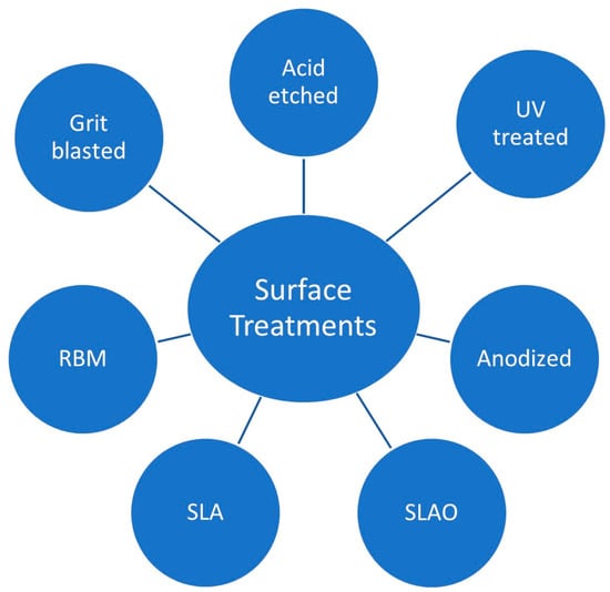
Figure 1.
Different surface treatments for orthodontic miniscrews.
1.2. Objectives
This systematic review focused on examining novel surface treatments developed to increase the mechanical stability of orthodontic miniscrews and quantitative effects of surface properties.
2. Materials and Methods
The execution of this study adhered to the quality reporting standards outlined in the PRISMA 2020 (Preferred Reporting Items for Systematic Reviews and Meta-Analyses) guidelines [31].
The research question of the present systematic review was defined according to the following PICO format:
P (Population/Patients): In vivo studies
I (Intervention): Miniscrews with differentiated surface properties
C (Comparison): Miniscrews with machined surfaces
O (Outcome): Alterations in the mechanical stability of miniscrews were assessed through parameters such as maximum insertion and removal torque, and Periotest values
2.1. Search Strategy
The comprehensive search strategy involved querying databases, namely PubMed, Web of Science, and Scopus, using search terms focused on miniscrews, mini-implants, stability, surface treatment, surface property, torque, insertion, removal, and Periotest. The final search was conducted on 30 August 2023.
Titles and abstracts were independently and redundantly reviewed for potential inclusion in the study. The interrater agreement, measured by an intraclass correlation coefficient, was found to be 0.95. Any conflicts that arose were resolved through consensus discussions between the two authors.
2.2. Eligibility Criteria
In vivo studies that evaluated the effects of surface treatments of orthodontic miniscrews on mechanical stability were eligible. These studies were published in the English language with no restriction regarding the date of publication. Both reviewers agreed upon the following exclusion criteria: in vitro studies, opinion articles, editorial letters, reviews, case reports, articles with no control group (machined surface), and studies without a full-text version available (Figure 2).
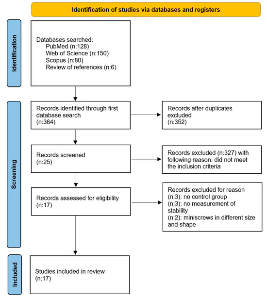
Figure 2.
The PRISMA 2020 flow diagram.
2.3. Data Collection Process and Data Items
The authors (H.Y., P.G.) selected articles meeting the inclusion criteria. Subsequently, data were gathered and recorded in a standardized Excel file.
2.4. Risk of Bias Assessment
In the initial phase of study inclusion, the authors (H.Y., P.G.) independently assessed the titles and abstracts of each study to minimize potential bias among reviewers. Risk of bias was assessed according to Cochrane’s reviewers’ handbook [32]. The main domains (random sequence generation, allocation concealment, blinding of participants and personnel, blinding of outcome assessment, incomplete outcome data, selective reporting, and other sources of bias) were identified as low, moderate, and high risk. The interrater agreement, measured by an intraclass correlation coefficient, was found to be 0.90. Any discrepancies regarding the inclusion or exclusion of a study were resolved through discussions between the authors.
2.5. Quality Assessment
The authors (H.Y., P.G.) conducted an assessment of the procedural quality for each study included in the article. The following assessment criteria were scored on a scale of 0 to 1 point, where a higher total score indicated better study quality.
- Group size at least 10 subjects: 1 point.
- Existence of control group: 1 point.
- Sample size calculation: 1 point.
- Detailed information of procedure MRT-MIT-PTV (at least 2 of them): 1 point.
- Force application: 1 point.
Any discrepancies regarding the scoring were resolved through discussion until a consensus was reached.
3. Results
3.1. Study Selection
As a result of the comprehensive evaluation, 364 studies were found in electronic databases including Pubmed (128), WOS (150), and Scopus (80), and via the review of references (6). In terms of duplication, 12 studies were excluded. A total of 352 studies were screened and title and abstract sections were read. After abstract screening, 327 studies were excluded because they did not meet the inclusion criteria. When the full text of the remaining 25 studies were analyzed, 8 studies were rejected. Three had no control group, three had no measurement of stability, and two had miniscrews of different size and shape. Finally, 17 articles were included in the systematic review. The publication dates of the included articles range from 2009 to 2022. Study selection is shown in Figure 2.
3.2. Study Characteristics
The selected data were included in this systematic review by evaluating author/year, the study type, the study groups, total number of miniscrews used, and the conclusion (Table 1). The studies were conducted between 2009 and 2022. Of the 17 in vivo studies, 2 were human studies and 15 were animal studies. Of the animal studies, eight were on rabbits, three were on rats, and four were on dogs. Different surface treatments were compared with the control groups. Anodized surfaces were compared in two of the studies. Anodization, cyclic precalcification, and heat treatment were evaluated in two different studies. Only one of the studies used a resorbable blasting media (RBM) surface. Four used acid etching, one used UV treatment, five used SLA, and two used a sandblasted and acid-etched group (SAE). Loading was applied in five studies. In addition, miniscrews were applied to diabetic patients in only one study. A total of 1097 miniscrews were used in the studies. Maximum insertion torque (MIT) measurements were used in nine studies to evaluate primary stability. Periotest values (PTV) were measured in three studies to evaluate the mobility of miniscrews. Maximum removal torque (MRT) values were measured in 15 studies to evaluate secondary stability.

Table 1.
General characteristics of included studies.
3.3. Main Study Outcome
The aim of this study was to determine the current surface treatments that improve the stability of orthodontic miniscrews and evaluate which surface treatments showed successful results compared to the control groups. The number of miniscrew samples ranged between 18 and 144 (totaling 1097 with a mean of 64.52) (Table 1). Among the surface modifications in the articles, the SLA method was most frequently applied, followed by acid etching in second place. The control groups’ (machined surface) MRT values varied from 2.05 to 50.50 Ncm, while MIT values varied from 7.23 to 19.25 Ncm (Table 2).
Table 2.
Details of included studies.
In the three papers evaluated, there was no clear difference in stability between the surface treatment group and the control group [25,37,40]. In two of these studies, acid etching was applied as surface treatment. In a human study by Park et al. [25], acid-etched and machined surface treatments were compared. In this study, the overall success rate of miniscrews was found to be 88.8%. The success rates of the two groups were found to be similar. When MIT, PTV (immediately after placement and 6 months after placement) values were analyzed, the acid-etched and machined surface treatments showed similar primary and secondary stability. Vilani et al. [40] also compared the stability of acid-etched and machined surface miniscrews in their animal study. They also evaluated the effect of force on these two different surfaces. High MIT and reduced PTV were observed in all groups, as well as a reduction in MRT in comparison with MIT. Whether force was applied or not, there were no significant differences in PTV, MIT or MRT in any of the groups. Choi et al. [37] compared anodized surfaces and machined surfaces in their study. Both groups were subjected to force loading and no significant differences were found in the MIT and MRT values measured at week 3 and week 12.
In the remaining 14 studies, differences were found between the stability of the machined surface and other surface treatments [27,28,30,33,34,35,36,38,39,41,42,43]. Unlike other acid-etched studies, Jang et al. [33] immersed miniscrews with acid-etched surfaces in calcium chloride solution. At 1 week after insertion, miniscrews with acid-etched surfaces showed higher MRT values than the control group, whether or not they were immersed in calcium chloride solution. At 4 and 7 weeks, the acid-etched group immersed in calcium chloride solution had the highest MRT value.
Espinar-Escalona et al. [30] found the MRT values of grit-blasted and grit-blasted + acid-etched surfaces significantly higher than acid-etched and machined surfaces. Moghaddam et al. [41] compared SAE-treated surfaces and machined surfaces in their human study. Although the MIT values of these two different surfaces were similar, the MRT value of the SAE surface group was found to be higher. Maino et al. [43] also compared SAE and machined surfaces. In addition, the effect of force on these surfaces was also evaluated. MRT was significantly higher for the machined surface in both the force and non-force groups than the SAE surface. Force did not significantly affect MRT for any surface or treatment. Sirisa-Ard et al. [38] compared SLA and machined surfaces in their study. MRT values measured at 0 and 8 weeks showed no significant difference between SLA and machined surfaces. Oh et al. [36] aimed to investigate the effects of surface-treated miniscrews in diabetic rabbits. In this study, SLA and machined surfaces were compared. MIT values showed no difference between the diabetic and control groups. In surface-treated miniscrews, MRT was higher in both the diabetic and control groups, but there was no significant difference between the diabetic and control groups in MIT, regardless of surface treatment. Kim et al. [39] aimed to evaluate the stability and resistance to rotational moments of early-loaded SLA and machined surface miniscrews. MIT values of SLA miniscrews were found to be lower than the machined surface screws. However, when MRT values were compared, there was no statistical difference between the two surfaces.
Karmarker et al. [27] compared anodized surface and machined surface miniscrews. While there was no difference between the two groups in terms of MIT values, MRT values were found to be significantly higher on the anodized surface. Oh et al. [34] evaluated the stability of miniscrews subjected to anodization, cyclic pre-calcification, and heat treatment. Compared to the control group, MRT values measured at 3 and 6 weeks were higher in the surface treatment group. Byeon et al. [35] evaluated the contribution of cyclic precalcification treatment to osseointegration and the bioactivity of miniscrews. The experimental groups of this study were as follows: anodized and heat-treated group; anodized, heat-treated and cyclic pre-calcification treatment group; and control group. When the MRT values were examined, the highest value was in the anodized, heat-treated and cyclic pre-calcification treatment group.
Cho et al. [28] compared the effects of SLAO and SLA surface treatments on the mechanical stability of miniscrews in vitro and in vivo. In the in vivo study, MRT values were significantly higher in the SLAO group than in the SLA and control group. In contrast, MIT values were higher in the control group compared to the SLA and SLAO groups.
Chang et al. [42] analyzed miniscrews with machined, SLA, and SL/NaOH (alkaline-etched) surfaces implanted in rabbit tibia and reported MRT in the SLA and SL/NaOH groups compared to the machined surface.
In a study by Gansukh et al. [29] evaluating the early stability of miniscrews with machined and RBM surfaces, MIT values were significantly higher with the machined surface compared to the RBM surface. In contrast, MRT values at week 2 were significantly lower in the machined surface group than in the RBM group, which was attributed to greater bone resorption in the early period. At week 4, there was no significant difference in MRT values between the groups.
Takahashi et al. [26] aimed to increase the osseointegration capabilities of miniscrews with UV-treated surfaces and evaluated their stability under loading. Whether force was applied or not, MRT values were significantly higher in UV-treated groups.
3.4. Quality Assessment and Risk of Bias
Two [37,43] of the articles included in the review were assessed as high quality with a score of 6/6 points. Two studies [30,35] were classified as low quality. Additionally, nine articles [26,27,28,29,33,34,38,40,42] were considered to have a moderate risk of bias, scoring between three and four points (Table 3). Red indicates high risk of bias, yellow indicates moderate risk of bias, and green indicates low risk of bias in Table 4.
Table 3.
Quality assessment.
Table 4.
Risk of bias assessment.
4. Discussion
Various surface modifications are applied to increase the osteoconductivity/osteoinductivity of miniscrews and consequently strengthen their bond with bone [44]. Surface modifications such as acid etching, sandblasting, grit blasting, and anodizing change the surface topography of miniscrews by creating porous and rough surfaces. These porous or rough surfaces increase stability and positively affect cellular responses [45,46]. Among the articles included in this systematic review, SLA was the most commonly used surface modification method, followed by acid etching.
In two meta-analyses on the effectiveness of miniscrews, the survival rate in the earlier study was 83.6%, which increased to 87.7% in the later study [47,48]. Miniscrew failure rates were reported as 11.5–15.9% by Alharbi et al. [49] and 11% by Cheng et al. [15]. Miniscrews are often preferred in clinical applications because they have a success rate over 80%. However, the effectiveness of miniscrews remains low compared to dental implants, which are more than 90% successful [50,51]. The most important factors affecting the success rate of miniscrews were primary and secondary stability [18].
4.1. Primary Stability
Motoyoshi et al. [16] recommended the insertion torque range for miniscrews as 5–10 Ncm in their study. Chaddad et al. [22] reported that insertion torque values above 15 Ncm would increase miniscrew stability. In this systematic review, MIT measurement was among the criteria included to evaluate primary stability. There are nine articles in which MIT measurement was used [25,27,28,29,36,37,39,40,41]. In only three of these articles, MIT values showed significant differences with the control group, and in all three, the MIT values of the control group were higher than the surface-treated group [28,29,39]. In the study of Cho et al. [28], MIT values were higher in the control group compared to the SLA and SLAO groups, which the authors suggested was because miniscrews with smooth surfaces cause more damage to the bone during insertion, whereas rough surfaces enable a better discharge of blood and bone particles. Only the study by Gansukh et al. [29] showed mean MIT values of RBM surface miniscrews in the range of 5–10 Ncm.
Periotest measurements are generally used to evaluate the mobility of teeth and osseointegration of dental implants [52]. The measurement results vary between −8 and +50. The lower the Periotest value the higher the stability, and the higher the Periotest value the lower the stability [53]. This value is also frequently used in in vitro and in vivo studies to analyze the primary and secondary stability of miniscrews [25,40,54]. In this systematic review, PTV measurement was used in three studies [25,26,40]. Takahashi et al. [26] reported that UV-treated groups showed lower stability than control groups in PTV measurements taken 2 weeks after the placement of miniscrews. Two other studies applied acid etching as a surface modification. There were no differences between the treated groups and the control groups in terms of initial PTV values [25,40].
4.2. Secondary Stability
MRT is the highest reaction force that occurs during the removal of the miniscrew from the bone [55]. MRT measurements in dental implants are generally considered a reliable method for the assessment of osseointegration. They have been frequently used in studies evaluating the secondary stability of orthodontic miniscrews [20].
When the acid-etched method was evaluated in this systematic review, in only one study the acid-etched group was found to be more stable than the control group. In the study by Jang et al. [33], the MRT values of the acid-etched group without immersion in calcium chloride were found to be higher than the control group. Miniscrews with acid-etched surfaces showed better stability when they were immersed in calcium chloride solution, or combined with sandblasting/grit-blasting methods.
There was a contradiction when comparing miniscrews with anodized surfaces in this study. In the study by Karmarker et al. [27], miniscrews with anodized surfaces were found to be more stable than the control group according to the MRT values measured 6 weeks after placement. However, in the study by Choi et al. [37], there was no difference between the control group and the anodized surfaces according to the MRT values measured at 3 weeks and 12 weeks. The use of anodized surfaces in combination with cyclic pre-calcification and heat treatment increased the secondary stability of miniscrews.
According to the study of Oh et al. [36], SLA surfaces are more stable than machined surfaces. In contrast to this article, Kim et al. [39] found no significant differences between SLA surfaces and machined surfaces. Similarly, Sirisa-Ard et al. [38] found no differences between the stability of SLA surfaces and machined surfaces. Cho et al. [28] found the combination of SLA surfaces with anodic oxidation to be more stable than SLA and the control group, according to MRT values.
Similar to the SLA method, the RBM method is also applied by acid etching and sandblasting to change the surface topography of titanium implants. The difference between these two methods is that the SLA method utilizes non-resorbable particles such as alumina for sandblasting, whereas the RBM method utilizes resorbable particles such as HA or calcium phosphate [56]. This provides a low risk of debris contamination and rapid osseointegration in the RBM technique [57]. In a study by Gansukh et al. [29], MRT values at week 2 were significantly lower in the machined surface group than in the RBM group, which was attributed to greater bone resorption in the early period. At week 4, there was no significant difference in MRT values between the groups. The authors suggested that although new bone formation occurred more on the machined surface than on the RBM surface after initial bone resorption, the torque values increased faster with the RBM surface.
4.3. Limitations
This systematic review evaluated the mechanical stability of surface-modified miniscrews. The limitation of this study was the variation between the studies evaluated. The loading or non-loading of miniscrews, and the amount, direction, and duration of force varied. The bone quality in the area where the miniscrew was applied and the variety of animals used in the animal experiments also added to the limitations. Although the measurement methods of mechanical stability in the present study were limited to MIT, MRT, and Periotest values, there were studies using different methods. In the evaluated studies, stability measurements were used in different combinations. When the risk of bias of these studies was evaluated, only two studies were high quality with a score of 6/6 points. Nine studies were considered to have a moderate risk of bias. According to the results of this study, future clinical and laboratory reports are needed, as well as randomized clinical trials.
4.4. Future Trends
Common surface modifications change the chemical and mechanical properties of miniscrews. However, roughening of the miniscrew surface poses a disadvantage both in terms of the risk of contamination and the difficulty of preventing bacterial colonization [58]. Poor oral hygiene leads to the accumulation of anaerobic bacteria around the miniscrew, resulting in the activation of immune cells such as neutrophils and macrophages that migrate to the implant site. This activation causes the release of proinflammatory cytokines such as IL-1β, IL-6, and TNF-α [59].
Studies on surface modifications of titanium implants have developed over the years. With advances in surface modification technologies, there has been a transition from traditional bioinert surfaces to biocompatible surfaces, and then to bioactive surfaces. As a satisfactory standard could not be reached with bioactive and bioinert coatings, recent studies have applied biomimetic coatings that simulate body fluids. These fourth-generation surface treatments are shedding light on future research [60].
Biomimetic calcium phosphate (BioCaP) coatings have been developed under physiological conditions (37 °C temperature and pH 7.4) under which bioactive agents such as bone morphogenetic protein-2 (BMP-2) can be deposited on the surface [61,62]. Li et al. used histomorphometric analyses to evaluate osteoconductivity by applying BioCaP (amorphous or crystalline) coatings with or without bovine serum albumin (BSA) on miniscrews [63]. Based on their results, the authors concluded that crystalline BioCaP coating is an effective method to accelerate osseointegration and increase the success rate of orthodontic miniscrews [63]. Li et al. aimed to improve the biocompatibility of stainless-steel miniscrews with the two-phase BioCaP coating they developed [64]. In their study, titanium discs were immersed in biomimetic modified Tyrode (BMT) solution for 24 h and stainless-steel discs were immersed in the same solution for 0, 12, 24, 36, or 48 h. The discs were then immersed in a supersaturated calcium phosphate solution for 48 h to form a crystal layer, and BSA was also added to this layer as a model protein during biomimetic mineralization. Their results demonstrated improved roughness and wettability of the smooth stainless-steel surface. Longer BMT coating time was also associated with increased cell seeding efficiency, cell proliferation, and cell spreading area on the surface. They concluded that the two-phase BioCaP application could improve the surface properties of stainless-steel miniscrews [64]. Rodriguez-Fernandez et al. aimed to create a polyethylene glycol (PEG) layer, using plasma coating to create a bacteriostatic effect on the surface of titanium miniscrews [65]. To achieve this layer, they activated the surfaces of titanium miniscrews with argon plasma and then with PEG plasma at different powers (100, 150, and 200 W) for 30 and 60 min. The PEG-coated samples showed more than 80% biocompatibility for fibroblast and osteoblastic cells. The miniscrews treated with only argon plasma activation showed an increase in bacterial colonization. The authors concluded that PEG significantly inhibits bacterial affinity due to its chemical configuration [65]. Bahrami et al. also examined the effects of the antimicrobial photosonodynamic treatment (aPSDT) of zinc oxide (ZnO) nanoparticle-coated miniscrews on anaerobic bacteria [66]. The study compared uncoated miniscrews treated with phosphate-buffered saline (PBS) or 0.2% chlorhexidine (CHX) with ZnO nanoparticle-coated miniscrews treated with PBS, light-emitting diode (LED) (aPDT), ultrasound (aSDT), or both LED and ultrasound (aPSDT). They reported that ZnO nanoparticle-coated miniscrews with surface aPSDT had the greatest antimicrobial effect against periopathogenic biofilm. Gene expression levels of proinflammatory cytokines such as IL-1β, IL-6, and TNF-α were also significantly lower in this group [66].
In recent years, these studies on the surface modifications of miniscrews have aimed to reduce bacterial retention on surfaces or accelerate osseointegration by increasing cell proliferation. ZnO nanoparticle-coated miniscrews with surface aPSDT and PEG-coated miniscrews were found to be successful in reducing bacterial colonization. BioCaP coating showed similar effects on both titanium and stainless-steel miniscrews; it has been found to accelerate osseointegration by increasing the cellular response. However, most of these studies were in vitro studies. Future clinical and laboratory reports are needed, as well as randomized clinical trials.
5. Conclusions
Surface modification is needed to strengthen the miniscrew–bone connection in areas with poor bone quality, to increase clinical success in patients with poor oral hygiene, and in orthodontic treatments where long-term and heavy forces are used. Conventional surface modifications increase the biomechanical stability of miniscrews. Although these methods are currently in use, there is still a search for new methods to improve the osseointegration capacity of miniscrews. These new methods include biomimetic coatings that simulate body fluids, antimicrobial agents, and drugs added to the miniscrew surface. In addition, current studies on the surface modification of miniscrews include not only miniscrews produced from titanium alloys, but also those produced from stainless steel.
Author Contributions
Conceptualization, H.Y.; methodology, P.G.; software, P.G.; validation, H.Y.; formal analysis, H.Y.; investigation, P.G.; resources, P.G.; data curation, P.G.; writing—original draft preparation, P.G.; writing—review and editing, P.G.; visualization, H.Y.; supervision, H.Y.; project administration, H.Y.; funding acquisition, H.Y. All authors have read and agreed to the published version of the manuscript.
Funding
This research received no external funding.
Institutional Review Board Statement
Not applicable.
Informed Consent Statement
Not applicable.
Data Availability Statement
The original contributions presented in the study are included in the article, further inquiries can be directed to the corresponding author.
Conflicts of Interest
The authors declare no conflicts of interest.
References
- Park, H.S.; Yoon, D.Y.; Park, C.S.; Jeoung, S.H. Treatment effects and anchorage potential of sliding mechanics with titanium screws compared with the Tweed-Merrifield technique. Am. J. Orthod. Dentofac. Orthop. 2008, 133, 593–600. [Google Scholar] [CrossRef]
- Favero, L.; Brollo, P.; Bressan, E. Orthodontic anchorage with specific fixtures: Related study analysis. Am. J. Orthod. Dentofac. Orthop. 2002, 122, 84–94. [Google Scholar] [CrossRef]
- Kanomi, R. Mini-implant for orthodontic anchorage. J. Clin. Orthod. 1997, 31, 763–767. [Google Scholar] [PubMed]
- Papadopoulos, M.A.; Tarawneh, F. The use of miniscrew implants for temporary skeletal anchorage in orthodontics: A comprehensive review. Oral Surg. Oral Med. Oral Pathol. Oral Radiol. Endod. 2007, 103, 6–15. [Google Scholar] [CrossRef]
- Ikeda, H.; Rossouw, P.E.; Campbell, P.M.; Kontogirogos, E.; Buschang, P.H. Three-dimensional analysis of peri-bone–implant contact of rough-surface miniscrew implants. Am. J. Orthod. Dentofac. Orthop. 2011, 139, 153–163. [Google Scholar] [CrossRef]
- Montasser, M.A.; Scribante, A. Root injury during interradicular insertion is the most common complication associated with orthodontic miniscrews. J. Evid. Based Dent. Pract. 2022, 22, 101688. [Google Scholar] [CrossRef] [PubMed]
- Xin, Y.; Wu, Y.; Chen, C.; Wang, C.; Zhao, L. Miniscrews for orthodontic anchorage: Analysis of risk factors correlated with the progressive susceptibility to failure. Am. J. Orthod. Dentofac. Orthop. 2022, 162, e192–e202. [Google Scholar] [CrossRef]
- Sfondrini, M.F.; Gandini, P.; Alcozer, R.; Vallittu, P.K.; Scribante, A. Failure load and stress analysis of orthodontic miniscrews with different transmucosal collar diameter. J. Mech. Behav. Biomed. Mater. 2018, 87, 132–137. [Google Scholar] [CrossRef]
- Serra, G.; Morais, L.; Elias, C.N.; Semenova, I.P.; Valiev, R.; Salimgareeva, G.; Pithon, M.; Lacerda, R. Nanostructured severe plastic deformation processed titanium for orthodontic mini-implants. Mater. Sci. Eng. C Mater. Biol. Appl. 2013, 33, 4197–4202. [Google Scholar] [CrossRef] [PubMed]
- Molly, L. Bone density and primary stability in implant therapy. Clin. Oral Implants Res. 2006, 17, 124–135. [Google Scholar] [CrossRef]
- Javed, F.; Romanos, G.E. The role of primary stability for successful immediate loading of dental implants. A literature review. J. Dent. 2010, 38, 612–620. [Google Scholar] [CrossRef] [PubMed]
- Wilmes, B.; Rademacher, C.; Olthoff, G.; Drescher, D. Parameters affecting primary stability of orthodontic mini-implants. J. Orofac. Orthop. 2006, 67, 162–174. [Google Scholar] [CrossRef] [PubMed]
- Song, Y.Y.; Cha, J.Y.; Hwang, C.J. Mechanical characteristics of various orthodontic mini-screws in relation to artificial cortical bone thickness. Angle Orthod. 2007, 77, 979–985. [Google Scholar] [CrossRef] [PubMed]
- Trisi, P.; De Benedittis, S.; Perfetti, G.; Berardi, D. Primary stability, insertion torque and bone density of cylindric implant ad modum Branemark: Is there a relationship? An in vitro study. Clin. Oral Implants Res. 2011, 22, 567–570. [Google Scholar] [CrossRef] [PubMed]
- Cheng, S.J.; Tseng, I.Y.; Lee, J.J.; Kok, S.H. A prospective study of the risk factors associated with failure of mini-implants used for orthodontic anchorage. Int. J. Oral Maxillofac. Implants 2004, 19, 100–106. [Google Scholar]
- Motoyoshi, M.; Yoshida, T.; Ono, A.; Shimizu, N. Effect of cortical bone thickness and implant placement torque on stability of orthodontic mini-implants. Int. J. Oral Maxillofac. Implants 2007, 22, 779–784. [Google Scholar]
- Raghavendra, S.; Wood, M.C.; Taylor, T.D. Early wound healing around endosseous implants: A review of the literature. Int. J. Oral Maxillofac. Implants 2005, 20, 425–431. [Google Scholar]
- Miyawaki, S.; Koyama, I.; Inoue, M.; Mishima, K.; Sugahara, T.; Takano-Yamamoto, T. Factors associated with the stability of titanium screws placed in the posterior region for orthodontic anchorage. Am. J. Orthod. Dentofac. Orthop. 2003, 124, 373–378. [Google Scholar] [CrossRef]
- Costa, A.; Raffainl, M.; Melsen, B. Miniscrews as orthodontic anchorage: A preliminary report. Int. J. Adult Orthodon. Orthognath. Surg. 1998, 13, 201–209. [Google Scholar] [PubMed]
- Favero, L.G.; Pisoni, A.; Paganelli, C. Removal torque of osseointegrated mini-implants: An in vivo evaluation. Eur. J. Orthod. 2007, 29, 443–448. [Google Scholar] [CrossRef] [PubMed]
- Vande Vannet, B.; Sabzevar, M.M.; Wehrbein, H.; Asscherickx, K. Osseointegration of miniscrews: A histomorphometric evaluation. Eur. J. Orthod. 2007, 29, 437–442. [Google Scholar] [CrossRef] [PubMed]
- Chaddad, K.; Ferreira, A.H.; Geurs, N.; Reddy, M.S. Influence of surface characteristics on survival rates of mini-implants. Angle Orthod. 2008, 78, 107–113. [Google Scholar] [CrossRef]
- Coelho, P.G.; Granjeiro, J.M.; Romanos, G.E.; Suzuki, M.; Silva, N.R.; Cardaropoli, G.; Thompson, V.P.; Lemons, J.E. Basic research methods and current trends of dental implant surfaces. J. Biomed. Mater. Res. B Appl. Biomater. 2009, 88, 579–596. [Google Scholar] [CrossRef]
- Wennerberg, A.; Albrektsson, T. A review of current knowledge, opinions and suggestions for possible common mechanisms behind the increased bone response reported to different types of modern oral implant surfaces. Int. J. Oral Maxillofac. Implants 2010, 25, 63–74. [Google Scholar]
- Park, H.J.; Choi, S.H.; Choi, Y.J.; Park, Y.B.; Kim, K.M.; Yu, H.S. A prospective, split-mouth, clinical study of orthodontic titanium miniscrews with machined and acid-etched surfaces. Angle Orthod. 2019, 89, 411–417. [Google Scholar] [CrossRef] [PubMed]
- Takahashi, M.; Motoyoshi, M.; Inaba, M.; Hagiwara, Y.; Shimizu, N. Enhancement of Orthodontic Anchor Screw Stability Under Immediate Loading by Ultraviolet Photofunctionalization Technology. Int. J. Oral Maxillofac. Implants 2016, 31, 1320–1326. [Google Scholar] [CrossRef] [PubMed][Green Version]
- Karmarker, S.; Yu, W.; Kyung, H.M. Effect of surface anodization on stability of orthodontic microimplant. Korean J. Orthod. 2012, 42, 4–10. [Google Scholar] [CrossRef] [PubMed]
- Cho, I.S.; Kim, S.K.; Chang, Y.I.; Baek, S.H. In vitro and in vivo mechanical stability of orthodontic mini-implants. Angle Orthod. 2012, 82, 611–617. [Google Scholar] [CrossRef]
- Gansukh, O.; Jeong, J.W.; Kim, J.W.; Lee, J.H.; Kim, T.W. Mechanical and Histological Effects of Resorbable Blasting Media Surface Treatment on the Initial Stability of Orthodontic Mini-Implants. Biomed. Res. Int. 2016, 2016, 7520959. [Google Scholar] [CrossRef]
- Espinar-Escalona, E.; Bravo-Gonzalez, L.A.; Pegueroles, M.; Gil, F.J. Roughness and wettability effect on histological and mechanical response of self-drilling orthodontic mini-implants. Clin. Oral Investig. 2016, 20, 1115–1120. [Google Scholar] [CrossRef]
- Page, M.J.; McKenzie, J.E.; Bossuyt, P.M.; Boutron, I.; Hoffmann, T.C.; Mulrow, C.D.; Shamseer, L.; Tetzlaff, J.M.; Akl, E.A.; Brennan, S.E. The PRISMA 2020 statement: An updated guideline for reporting systematic reviews. Int. J. Surg. 2021, 88, 105906. [Google Scholar] [CrossRef]
- Higgins, J.P.; Altman, D.G. Assessing Risk of Bias in Included Studies. In Cochrane Handbook for Systematic Reviews of Interventions; Cochrane Book Series; Wiley: Hoboken, NJ, USA, 2008; pp. 187–241. [Google Scholar]
- Jang, T.H.; Park, J.H.; Moon, W.; Chae, J.M.; Chang, N.Y.; Kang, K.H. Effects of acid etching and calcium chloride immersion on removal torque and bone-cutting ability of orthodontic mini-implants. Am. J. Orthod. Dentofac. Orthop. 2018, 154, 108–114. [Google Scholar] [CrossRef]
- Oh, E.J.; Nguyen, T.D.; Lee, S.Y.; Jeon, Y.M.; Bae, T.S.; Kim, J.G. Enhanced compatibility and initial stability of Ti6Al4V alloy orthodontic miniscrews subjected to anodization, cyclic precalcification, and heat treatment. Korean J. Orthod. 2014, 44, 246–253. [Google Scholar] [CrossRef]
- Byeon, S.M.; Kim, H.J.; Lee, M.H.; Bae, T.S. Enhancement of bioactivity and osseointegration in Ti-6Al-4V orthodontic mini-screws coated with calcium phosphate on the TiO2 nanotube layer. Korean J. Orthod. 2022, 52, 412–419. [Google Scholar] [CrossRef]
- Oh, N.H.; Kim, E.Y.; Paek, J.; Kook, Y.A.; Jeong, D.M.; Cho, I.S.; Nelson, G. Evaluation of stability of surface-treated mini-implants in diabetic rabbits. Int. J. Dent. 2014, 2014, 838356. [Google Scholar] [CrossRef]
- Choi, S.-H.; Jang, S.-H.; Cha, J.-Y.; Hwang, C.-J. Evaluation of the surface characteristics of anodic oxidized miniscrews and their impact on biomechanical stability: An experimental study in beagle dogs. Am. J. Orthod. Dentofac. Orthop. 2016, 149, 31–38. [Google Scholar] [CrossRef] [PubMed]
- Sirisa-Ard, A.; Woodroffe Michael, S.N.; Ahmed, K.; Dunstan, C.R.; Pearce, S.G.; Bilgin, A.A.; Dalci, O.; Darendeliler, M.A. Histomorphological and torque removal comparison of 6 mm orthodontic miniscrews with and without surface treatment in New Zealand rabbits. Eur. J. Orthod. 2015, 37, 578–583. [Google Scholar] [CrossRef]
- Kim, S.H.; Lee, S.J.; Cho, I.S.; Kim, S.K.; Kim, T.W. Rotational resistance of surface-treated mini-implants. Angle Orthod. 2009, 79, 899–907. [Google Scholar] [CrossRef] [PubMed]
- Vilani, G.N.; Ruellas, A.C.; Elias, C.N.; Mattos, C.T. Stability of smooth and rough mini-implants: Clinical and biomechanical evaluation—An in vivostudy. Dent. Press J. Orthod. 2015, 20, 35–42. [Google Scholar] [CrossRef] [PubMed][Green Version]
- Moghaddam, S.F.; Mohammadi, A.; Behroozian, A. The effect of sandblasting and acid etching on survival rate of orthodontic miniscrews: A split-mouth randomized controlled trial. Prog. Orthod. 2021, 22, 2. [Google Scholar] [CrossRef] [PubMed]
- Chang, C.S.; Lee, T.M.; Chang, C.H.; Liu, J.K. The effect of microrough surface treatment on miniscrews used as orthodontic anchors. Clin. Oral Implants Res. 2009, 20, 1178–1184. [Google Scholar] [CrossRef]
- Maino, B.G.; Di Blasio, A.; Spadoni, D.; Ravanetti, F.; Galli, C.; Cacchioli, A.; Katsaros, C.; Gandolfini, M. The integration of orthodontic miniscrews under mechanical loading: A pre-clinical study in rabbit. Eur. J. Orthod. 2017, 39, 519–527. [Google Scholar] [CrossRef]
- Qiu, Z.-Y.; Chen, C.; Wang, X.-M.; Lee, I.-S. Advances in the surface modification techniques of bone-related implants for last 10 years. Regen. Biomater. 2014, 1, 67–79. [Google Scholar] [CrossRef]
- Mistry, S.; Roy, S.; Maitra, N.J.; Roy, R.; Datta, S.; Chanda, A.; Sarkar, S. Safety and efficacy of additive and subtractive surface modification of Ti6Al4V endosseous implant in goat bone. J. Mech. Behav. Biomed. Mater. 2016, 57, 69–87. [Google Scholar] [CrossRef]
- Zahran, R.; Rosales Leal, J.; Rodríguez Valverde, M.; Cabrerizo Vílchez, M. Effect of hydrofluoric acid etching time on titanium topography, chemistry, wettability, and cell adhesion. PLoS ONE 2016, 11, e0165296. [Google Scholar] [CrossRef]
- Schätzle, M.; Männchen, R.; Zwahlen, M.; Lang, N.P. Survival and failure rates of orthodontic temporary anchorage devices: A systematic review. Clin. Oral Implants Res. 2009, 20, 1351–1359. [Google Scholar] [CrossRef] [PubMed]
- Papadopoulos, M.; Papageorgiou, S.; Zogakis, I. Clinical effectiveness of orthodontic miniscrew implants: A meta-analysis. J. Dent. Res. 2011, 90, 969–976. [Google Scholar] [CrossRef] [PubMed]
- Alharbi, F.; Almuzian, M.; Bearn, D. Miniscrews failure rate in orthodontics: Systematic review and meta-analysis. Eur. J. Orthod. 2018, 40, 519–530. [Google Scholar] [CrossRef] [PubMed]
- Chuang, S.; Tian, L.; Wei, L.; Dodson, T. Kaplan-Meier analysis of dental implant survival: A strategy for estimating survival with clustered observations. J. Dent. Res. 2001, 80, 2016–2020. [Google Scholar] [CrossRef] [PubMed]
- Gokcen-Rohlig, B.; Yaltirik, M.; Ozer, S.; Tuncer, E.D.; Evlioglu, G. Survival and success of ITI implants and prostheses: Retrospective study of cases with 5-year follow-up. Eur. J. Dent. 2009, 3, 42–49. [Google Scholar] [CrossRef] [PubMed][Green Version]
- Van Scotter, D.; Wilson, C. The Periotest method for determining implant success. J. Oral Implantol. 1991, 17, 410–413. [Google Scholar]
- Teerlinck, J.; Quirynen, M.; Darius, P.; van Steenberghe, D. Periotest: An Objective Clinical Diagnosis of Bone Apposition Toward Implants. Int. J. Oral Maxillofac. Implants 1991, 6, 55–61. [Google Scholar]
- Gezer, P.; Yilanci, H. Comparison of mechanical stability of mini-screws with resorbable blasting media and micro-arc oxidation surface treatments under orthodontic forces: An in vitro biomechanical study. Int. Orthod. 2023, 21, 100775. [Google Scholar] [CrossRef]
- Buser, D.; Nydegger, T.; Hirt, H.P.; Cochran, D.L.; Nolte, L.P. Removal torque values of titanium implants in the maxilla of miniature pigs. Int. J. Oral Maxillofac. Implants 1998, 13, 611–619. [Google Scholar]
- Hommerle, W.H.M. Bone-to implant contact of orthodontic implants in humans subjected to horizontal loadig. Clin. Oral Implants Res. 1998, 9, 348–353. [Google Scholar]
- Sanz, A.; Oyarzún, A.; Farias, D.; Diaz, I. Experimental study of bone response to a new surface treatment of endosseous titanium implants. Implant Dent. 2001, 10, 126–131. [Google Scholar] [CrossRef]
- Phutim-Mangkhalthon, A.; Teerakapong, A.; Tippayawat, P.; Morales, N.P.; Morkmued, S.; Puasiri, S.; Priprem, A.; Damrongrungruang, T. Anti-inflammatory effect of photodynamic therapy using guaiazulene and red lasers on peripheral blood mononuclear cells. Photodiagn. Photodyn. Ther. 2020, 31, 101747. [Google Scholar] [CrossRef] [PubMed]
- Misba, L.; Zaidi, S.; Khan, A.U. A comparison of antibacterial and antibiofilm efficacy of phenothiazinium dyes between Gram positive and Gram negative bacterial biofilm. Photodiagn. Photodyn. Ther. 2017, 18, 24–33. [Google Scholar] [CrossRef] [PubMed]
- Alves-Rezende, M.C.; Capalbo, L.C.; De Oliveira Limírio, J.P.; Capalbo, B.C.; Limírio, P.H.; Rosa, J.L. The role of TiO2 nanotube surface on osseointegration of titanium implants: Biomechanical and histological study in rats. Microsc. Res. Tech. 2020, 83, 817–823. [Google Scholar] [CrossRef] [PubMed]
- Liu, Y.; Groot, K.D.; Hunziker, E.B. Osteoinductive implants: The mise-en-scene for drug-bearing biomimetic coatings. Ann. Biomed. Eng. 2004, 32, 398–406. [Google Scholar] [CrossRef] [PubMed]
- Lin, X.; Chen, J.; Liao, Y.; Pathak, J.L.; Li, H.; Liu, Y. Biomimetic calcium phosphate coating as a drug delivery vehicle for bone tissue engineering: A mini-review. Coatings 2020, 10, 1118. [Google Scholar] [CrossRef]
- Li, M.; Wu, G.; Wang, M.; Hunziker, E.B.; Liu, Y. Crystalline biomimetic calcium phosphate coating on mini-pin implants to accelerate osseointegration and extend drug release duration for an orthodontic application. Nanomaterials 2022, 12, 2439. [Google Scholar] [CrossRef]
- Li, M.; Wang, M.; Wei, L.; Werner, A.; Liu, Y. Biomimetic calcium phosphate coating on medical grade stainless steel improves surface properties and serves as a drug carrier for orthodontic applications. Dent. Mater. 2023, 39, 152–161. [Google Scholar] [CrossRef] [PubMed]
- Rodriguez-Fernandez, J.C.; Pastor, F.; Barrera Mora, J.M.; Brizuela, A.; Puigdollers, A.; Espinar, E.; Gil, F.J. Bacteriostatic Poly Ethylene Glycol Plasma Coatings for Orthodontic Titanium Mini-Implants. Materials 2022, 15, 7487. [Google Scholar] [CrossRef] [PubMed]
- Bahrami, R.; Pourhajibagher, M.; Parker, S.; Esmaeili, D.; Bahador, A. Anti-biofilm and bystander effects of antimicrobial photo-sonodynamic therapy against polymicrobial periopathogenic biofilms formed on coated orthodontic mini-screws with zinc oxide nanoparticles. Photodiagn. Photodyn. Ther. 2023, 41, 103288. [Google Scholar] [CrossRef] [PubMed]


Disclaimer/Publisher’s Note: The statements, opinions and data contained in all publications are solely those of the individual author(s) and contributor(s) and not of MDPI and/or the editor(s). MDPI and/or the editor(s) disclaim responsibility for any injury to people or property resulting from any ideas, methods, instructions or products referred to in the content. |
© 2024 by the authors. Licensee MDPI, Basel, Switzerland. This article is an open access article distributed under the terms and conditions of the Creative Commons Attribution (CC BY) license (https://creativecommons.org/licenses/by/4.0/).