1. Introduction
Stiffness is the most common complication adversely affecting shoulder function postoperatively and is a crucial factor in shoulder pathology and associated conditions [
1]. An arthroscopic capsular release (ACR) is a key procedure for shoulder joint stiffness accompanied by rotator cuff tears or adhesive capsulitis that fails to respond to conservative treatment [
2]. Among other treatment options, an ACR is clinically favored because it poses a low risk of fracture and allows the simultaneous identification and treatment of intra-articular lesions [
2]. The improvement in range-of-motion (ROM) and pain score achieved with an ACR leads to patient satisfaction [
1,
2].
While an ACR provides excellent improvements in shoulder function, postoperative complications, such as iatrogenic axillary nerve injury or joint stiffness recurrence, have been reported [
2,
3]. The blood supply to the humeral head is sustained through intraosseous circulation facilitated by the anterior and posterior humeral circumflex arteries [
4]. The disruption of these pathways leads to osteonecrosis. Trauma and corticosteroid use commonly cause osteonecrosis of the humeral head of traumatic and atraumatic origins, respectively. Factors such as chronic alcohol consumption, systemic lupus erythematosus, coagulopathy, viral infection, radiotherapy, and chemotherapy induce osteonecrosis [
5]. Several case reports and series have presented the postoperative progression of osteonecrosis of the humeral head during other arthroscopic procedures in shoulder joints [
6]. For instance, recent case reports have described the development of osteonecrosis after arthroscopic rotator cuff surgery using anchors and surgical procedures for lesions in the biceps long-head tendon [
5,
7,
8,
9]. However, no case of osteonecrosis of the humeral head has been reported after an ACR.
Here, we report a clinical case of post-ACR osteonecrosis of the humeral head, which was followed up for 3 years.
2. Case Report
A 56-year-old male patient complained of severe ROM limitations and pain in his right shoulder joint that did not improve. He had a history of a dislocation of the right acromioclavicular joint due to a fall 9 months previously and had undergone open reduction and internal fixation of the joint using a hook plate at another hospital, followed by hardware removal 4 months after the initial surgery. Six months after the removal of the hook plate, he saw no improvement in the persistent stiffness or pain in his shoulder, despite appropriate rehabilitation protocols, including the use of Celebrex oral nonsteroidal anti-inflammatory drugs and physiotherapy. The patient had a history of consuming 40 g of alcohol ≥five times per week for 25 years but had no history of smoking. He was regularly monitored for chronic hepatitis B and was taking medications for primary hypertension.
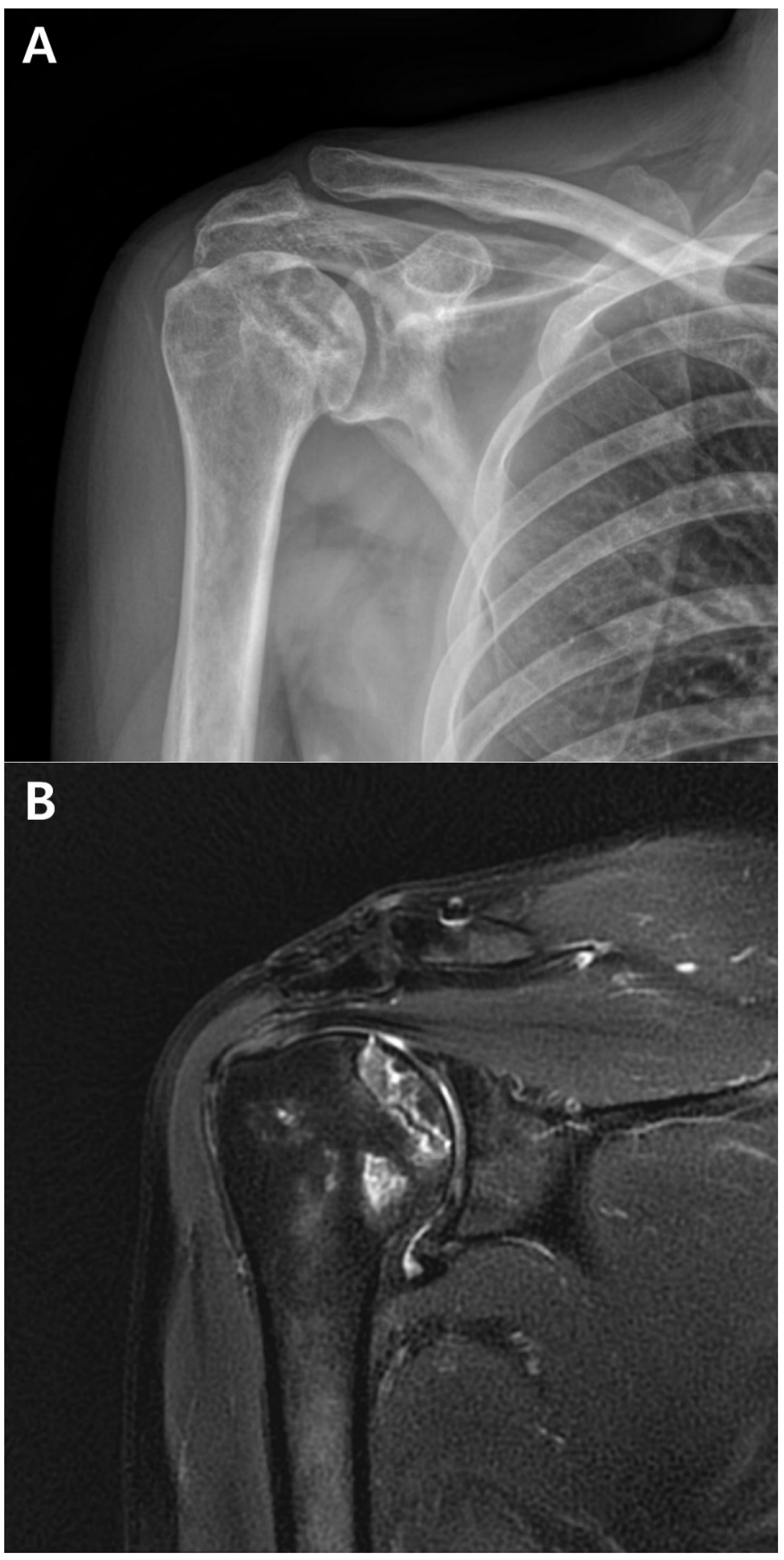
A postoperative scar was observed on the superior aspect along the distal clavicle and acromion, without definite tenderness, swelling, or redness. Preoperatively, the ROM of the affected shoulder was 90° for active forward flexion, 90° for abduction, 40° for external rotation, and at a sacral level for internal rotation. The passive and active ranges of motion were identical, attributed to the predominant cause of pain being stiffness rather than weakness. The visual analog scale (VAS) score was 4, the American Shoulder and Elbow Surgeons (ASES) score was 51, and the Constant–Murley Score (CMS) was 48. Preoperative shoulder radiographs showed acceptable alignment and joint congruency, with no bony spurs or subchondral lesions. The anteroposterior and axillary views of the shoulder radiograph showed an acceptable status of acromioclavicular joint reduction without arthritic changes. Magnetic resonance imaging (MRI) showed incomplete signal changes in the supraspinatus tendon, without a bony structural abnormality (
Figure 1). Given the patient’s persistent symptoms, which did not improve, the patient opted for an ACR with informed consent.
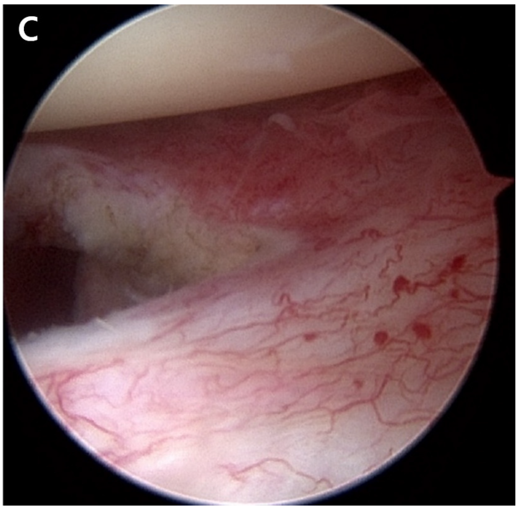
The ACR was performed in the lateral decubitus position under general anesthesia. The fluid used during the surgery was mixed with 1 mg of epinephrine per 3 L of normal saline, and the water pressure was maintained at 30–50 mmHg using an automatic infuser (10 K Fluid System, ConMed Linvatec, Largo, FL, USA). No abnormalities were observed in the rotator cuff, biceps long-head tendon, or glenohumeral joints upon arthroscopic examination. The ACR was performed according to the regular sequence, starting with the release of the rotator interval, middle glenohumeral ligament, and inferior and posterior capsules using a 3.0 mm 90° electrocautery device (Arthrocare, Smith & Nephew, Memphis, TN, USA) (
Figure 2). After the ACR, the ROM under general anesthesia was defined as 150° forward flexion, 150° abduction, at the level of the eighth thoracic vertebral body for internal rotation, and 80° external rotation.
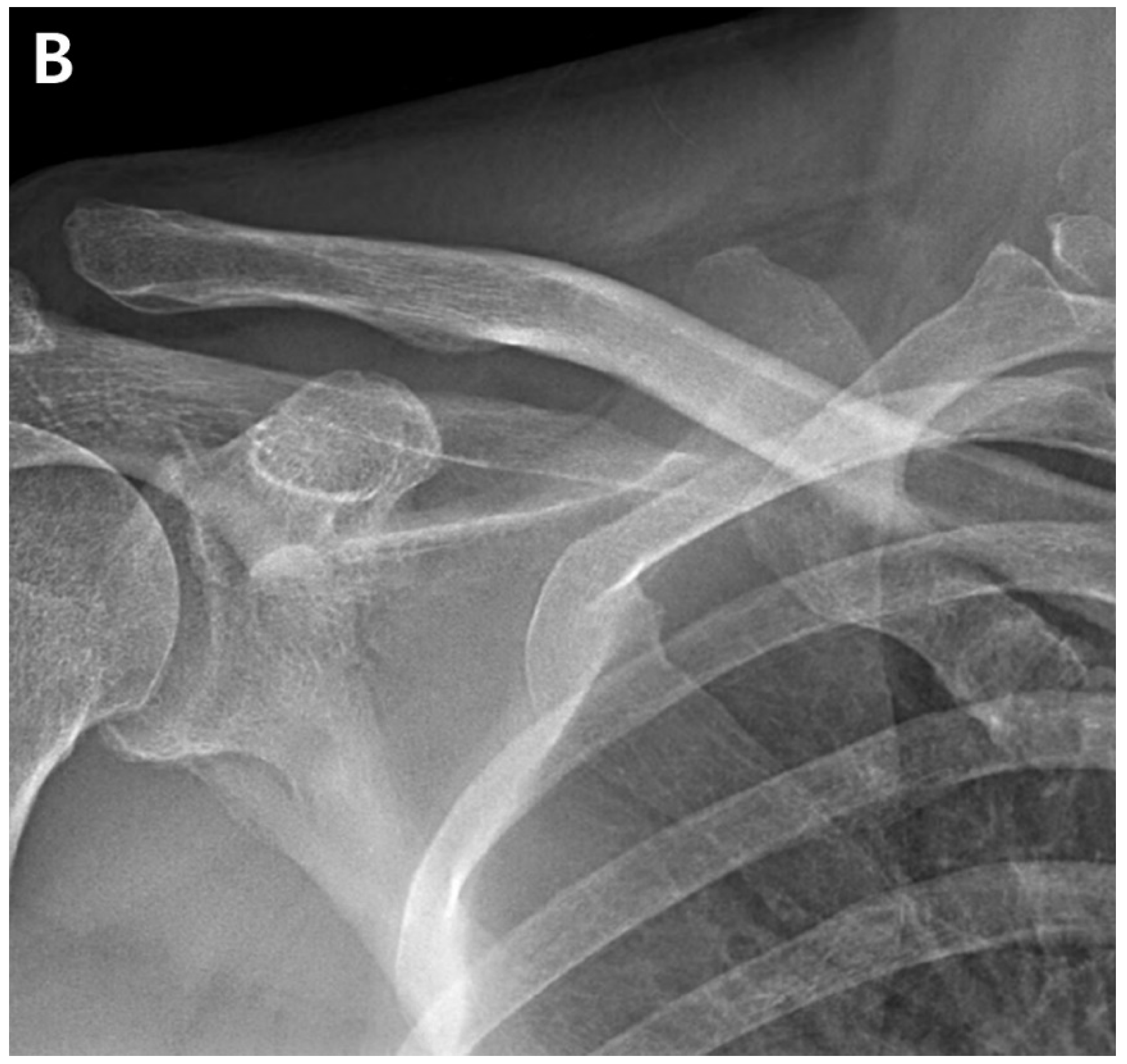
At 6-month post-operation, the functional outcomes were reported as a VAS score of 2, an ASES score of 79, and a CMS score of 75. The shoulder ROM was assessed as 135° forward flexion, 135° abduction, 70° external rotation, and internal rotation of the 10th thoracic vertebra. Radiographs showed an intact joint space and congruency (
Figure 3).
At 15 months post-operation, the patient developed recurring right shoulder pain. Subsequently, he sought treatment at another orthopedic hospital. He received two triamcinolone intra-articular injections and underwent four prolotherapy injections using a mixture of saline, dextrose, and lidocaine over a six-month period from 15 to 21 months post-operation. The patient returned to the hospital 21 months after the ACR with right shoulder pain. Plain shoulder anteroposterior radiography and MRI showed a subchondral cyst and a high bone marrow signal on the epiphysis of the superomedial area of the humeral head on T2-weighted images, suggesting avascular osteonecrosis at Cruess stage II (
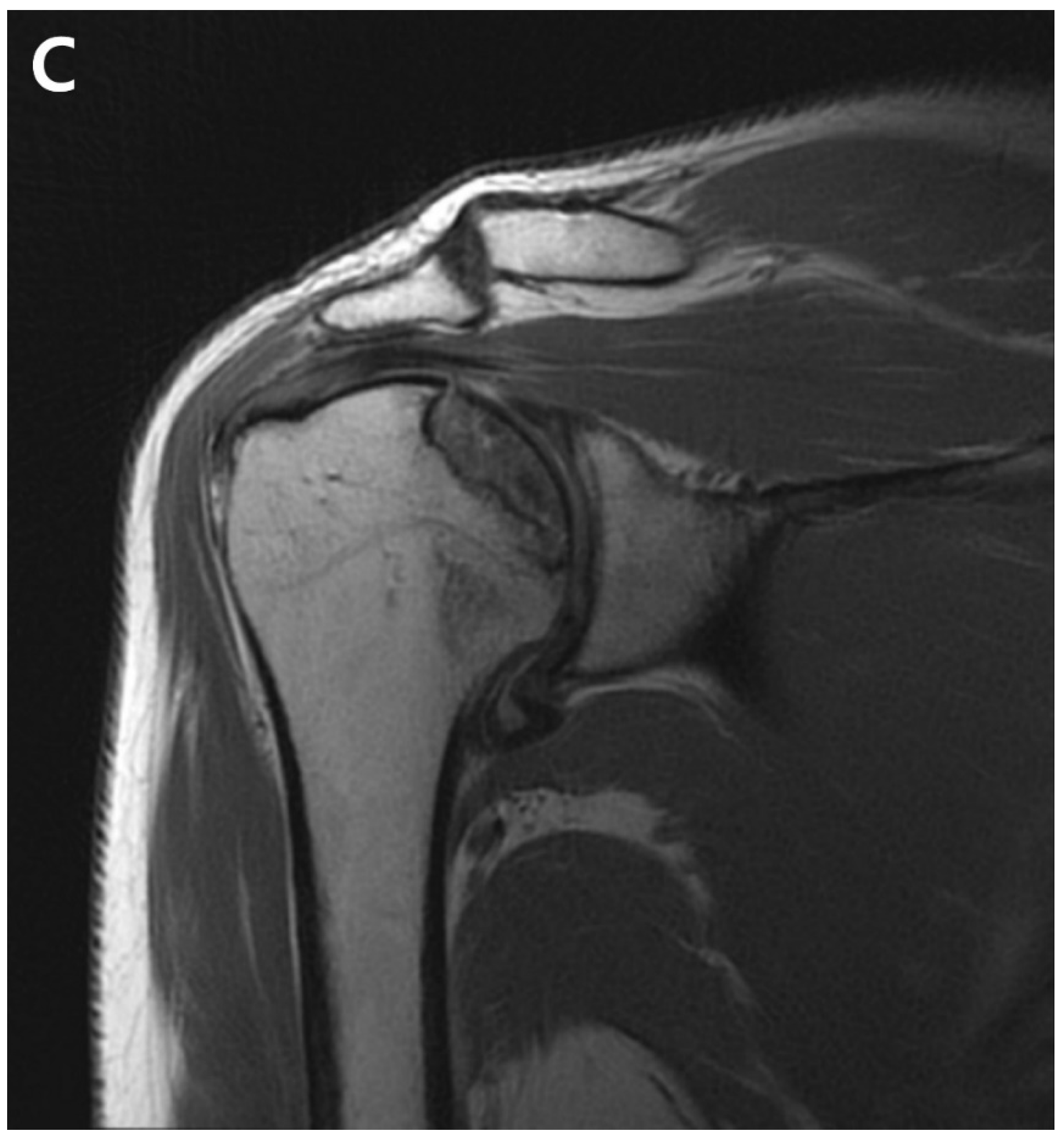
Figure 4). The patient denied any traumatic injury, radiotherapy, exacerbation of chronic hepatitis, alcohol consumption, or use of other medications. At 3 years post-operation, his VAS score was 3, his ASES score was 63, and his CMS score was 59, indicating increased discomfort. As the patient found the discomfort tolerable in terms of both work capacity and activities in daily life, we decided to maintain the current status and continue observation until osteonecrosis progressed (
Figure 5).
3. Discussion
An ACR is a reliable surgical procedure clinically performed in patients with persistent shoulder stiffness. However, the occurrence of osteonecrosis of the humeral head after arthroscopic surgery reflects critical damage to the shoulder joint, necessitating revision surgery [
4]. The case presented here involved a patient with predisposing risk factors for osteonecrosis, including a history of chronic alcohol consumption, repeated intra-articular glucocorticoid injections, and previous shoulder surgery.
In previous clinical studies, recurrent stiffness and axillary nerve injury have been reported as complications of an ACR [
10]. However, postoperative osteonecrosis has not been documented. Dilisio et al. hypothesized that associated surgical injuries could be caused by knots, the position of the suture anchor at the greater tuberosity or biceps, and the aggressive use of a radiofrequency ablation device [
7]. Goto et al. demonstrated that the use of metal or multiple anchors placed at the greater tuberosity injures the anterolateral branch of the anterior circumflex humeral artery (ACHA) [
8]. Damage to the arcuate artery can cause osteonecrosis of the humeral head in patients undergoing biceps tenodesis or tenotomy [
11]. In our case, the use of radiofrequency ablation may have been associated with the development of osteonecrosis.
Circulation to the humeral head is facilitated by anastomoses involving the ACHA, posterior circumflex humeral artery (PCHA), and thoracoacromial artery [
9]. The superomedial aspect of the humeral head is particularly prone to osteonecrosis because of compromised arterial perfusion from the interosseous artery, a branch of the ACHA [
8]. Given that our patient had undergone previous surgery for acromioclavicular joint dislocation, an iatrogenic injury to the thoracoacromial artery may have occurred [
4]. A pseudoaneurysm involving the acromial branch of the thoracoacromial artery near the anterior portal was reported during an arthroscopic rotator cuff repair [
4]. Potential injuries to the thoracoacromial artery and ACHA can occur during plate fixation or arthroscopic procedures. Recent research has demonstrated that the PCHA contributes significantly to the blood supply to the interosseous artery of the humeral head [
5,
12]. Although the PCHA may remain functional, the combined effects of intra-articular glucocorticoid injections and alcohol consumption may have critical effects, as detailed below.
Alcohol induces lipid deposition in osteocytes, increases adipogenesis, and decreases osteogenesis [
13]. Alcohol consumption has time- and dose-dependent effects on osteonecrosis [
13,
14]. Another clinical study showed that the occurrence of osteonecrosis was associated with a weekly ethanol consumption of >320 g per week for more than 6 months [
13,
14]. In this case report, the patient’s consistent alcohol consumption of 40 g ≥five times per week for 25 years, a time-dependent factor, may have contributed to the development of osteonecrosis. Although hepatitis B is not recognized as a risk factor, its impact on bone quality is notable in the context of liver cirrhosis [
13].
The complications of intra-articular steroid injections include septic arthritis, corticosteroid flares, elevated blood glucose levels, tendon rupture, chorioretinopathy, osteonecrosis, chondrotoxicity, and skin changes [
11]. Glucocorticoids can cause inadequate vascular supply, changes in blood flow, inflammatory processes, changes in lipid metabolism, and an imbalance between osteogenesis and adipogenesis in the bone marrow [
13,
15]. Intra-articular steroid injections are commonly used to treat adhesive capsulitis, resulting in reduced pain and an improved ROM [
15]. However, the administration of glucocorticoids can increase the risk of osteonecrosis by up to 20-fold, and the discontinuation of corticosteroid treatment does not completely reverse the pathogenic processes responsible for osteonecrosis [
13]. Thus, intra-articular steroid injections are considered a significant risk factor for humeral head necrosis.
This case report had several limitations. First, the index surgery for acromioclavicular dislocation was performed at another hospital; therefore, defined medical records or details of the surgery could not be identified. Second, the subjective masking of the patient in identifying his drinking history after the ACR was possible, and compliance with rehabilitation treatment, such as ROM exercises after surgery, could not be verified.


