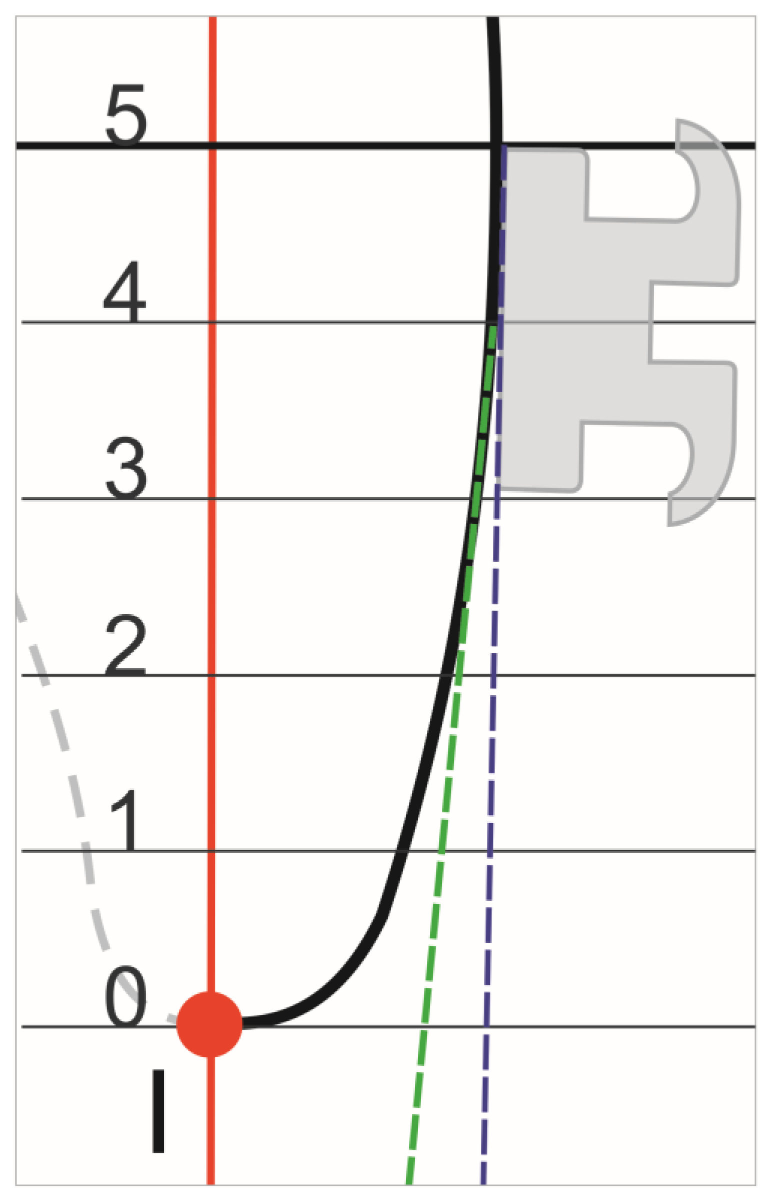
Featured Application
During bracket positioning, the individual variation in the tooth surface should be considered and evaluated, because the results of this work showed essential morphological differentiation of the CrVS of the maxillary incisors.
Abstract
Proper torque is an important element of orthodontic treatment. There are many factors affecting effective torque expression, e.g., the interplay between an archwire and a bracket, the precision during bracket positioning, and the morphology of the crown vestibular surface (CrVS) of the tooth. Our study focused on the impact of the maxillary incisor CrVS morphology on the torque exerted by the archwire–bracket interplay. Three-dimensional models of 50 patients acquired through the use of an intraoral scanner were used to examine the four maxillary incisors. A total of 200 teeth were examined. The influence of the tooth crown shape on the bracket position and the related torque change was analyzed with Ortho Analyzer software 2015 (3Shape, Copenhagen, Denmark). All calculations were made for full size archwires. Central incisors showed less variability in their vestibular surfaces than lateral incisors. For the central incisors, the mean values of the additional palatal root torque ranged from 0.6° to 1.6°. For the laterals, the mean values ranged from 1.4° of additional vestibular root torque to 3.5° of additional palatal root torque. The results showed essential morphological differentiation of the CrVS of the maxillary incisors. Therefore, when the bracket is positioned, the individual variation in the tooth surface should be considered and evaluated.
1. Introduction
Neglecting root torque control during orthodontic treatment usually leads to unstable occlusions and numerous complications, e.g., excessive tooth wear, the destruction of hard and soft periodontal tissues, root resorption [1], space reduction in dental arches, and dysfunction resulting from abnormal tooth guidance [2,3].
Difficulties in obtaining satisfactory treatment goals using the standard edgewise technique resulted in the development of brackets with built-in torque in the early 1960s. Ten years later, the straight wire appliance was introduced and the brackets with prescription in which 1st, 2nd, and 3rd order bends were systematically transferred from the wires to the brackets. It is commonly known that root torque may be provided either by twisted rectangular wire in a rectangular bracket slot [4,5] and/or by using brackets with prescriptions designed for the straight wire technique, which makes tooth position control possible in all three dimensions of space due to the angle between the bracket slot and its base [6]. However, even with sophisticated, contemporary brackets, torque expression may be affected by many factors, such as adopted treatment mechanics, variability of the mechanical properties of orthodontic wires and/or brackets, varying mechanical properties of the tooth surrounding structure, as well as variability of the tooth morphology [6,7,8,9,10,11,12,13,14]. Tooth morphology diversity has been proven in many studies [6,7,8,9,10,11]. Although the maxillary incisors show the smallest variability in shape [9], these teeth are also differentiated. Differences between the right and left central incisors have been demonstrated even in the same patient [6]. The morphology of the tooth crown vestibular surface (CrVS) has been examined using various approaches.
The study builds on the premise that individual tooth morphology can significantly influence orthodontic treatment outcomes, particularly in terms of the root torque control. Previous research has demonstrated variability of tooth morphology, but the application of three-dimensional scanning technology to analyze this variability is novel. The use of high-precision intraoral scanners, such as the TRIOS®3, represents a significant leap from traditional methods, offering sub-millimeter accuracy that is critical for assessing the complex geometries of the dental structures. Conventionally, CrVS was examined by means of a circle or a system of coordinates based on a cross-section of the teeth from plaster models [4,8,10,11,12,15,16]. To date, no modern 3D technologies have been used for this purpose. Therefore, the aim of this study was to apply intraoral scans to analyze the maxillary incisor CrVS morphology and to determine whether this anatomy affects the prescribed torque values.
2. Materials and Methods
The presented research is a prospective study, that obtained Wroclaw Medical University bioethics committee approval No KB–789/2020.
The control group included patients that were treated in private practice between 2016 and 2021.
The inclusion criteria were fully erupted maxillary incisors.
The exclusion criteria were malocclusions such as crossbite and severe crowding, past orthodontic treatment with fixed appliances, vestibular caries, filing, incisal edge wear, periodontal disease, gingival attachment loss, history of injury, abnormal formation of the enamel or external layer of the tooth crown.
Eventually, fifty patients participated: 37 women and 13 men, aged 18 to 35 years (median = 23; SD = 22.5). Each patient signed written consent to participate in the project.
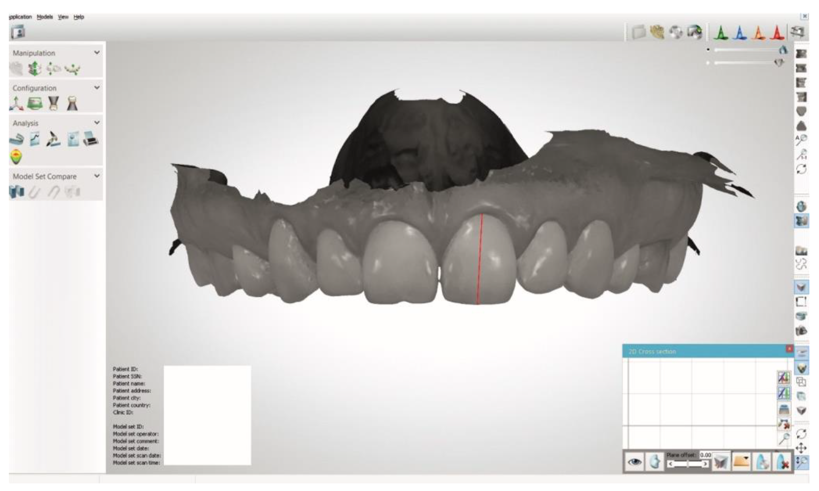
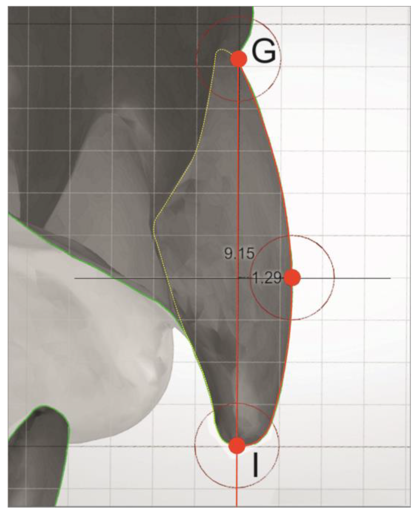
The patients’ dental arches were scanned with a TRIOS®3 intraoral scanner (3Shape, Copenhagen, Denmark). Subsequently, the long axis of the tooth crown determined using an Ortho Analyzer 2015 (3Shape, Copenhagen, Denmark) served as the place of the vertical cross-section (Figure 1). In the latter, the GI line was drawn using the “Length measurement” tool in “2D Cross-section” view. The line connected the “I” point, the lowest point on the incisal edge of the maxillary tooth, with the “G” point representing the gingival margin (Figure 2).
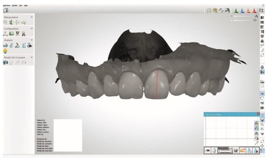
Figure 1.
Determination of the long axis of the tooth.
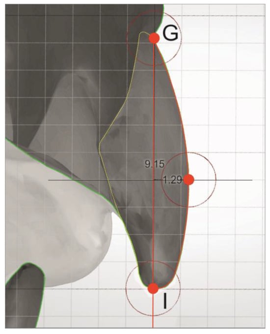
Figure 2.
Measurement between the reference GI line and tooth contour.
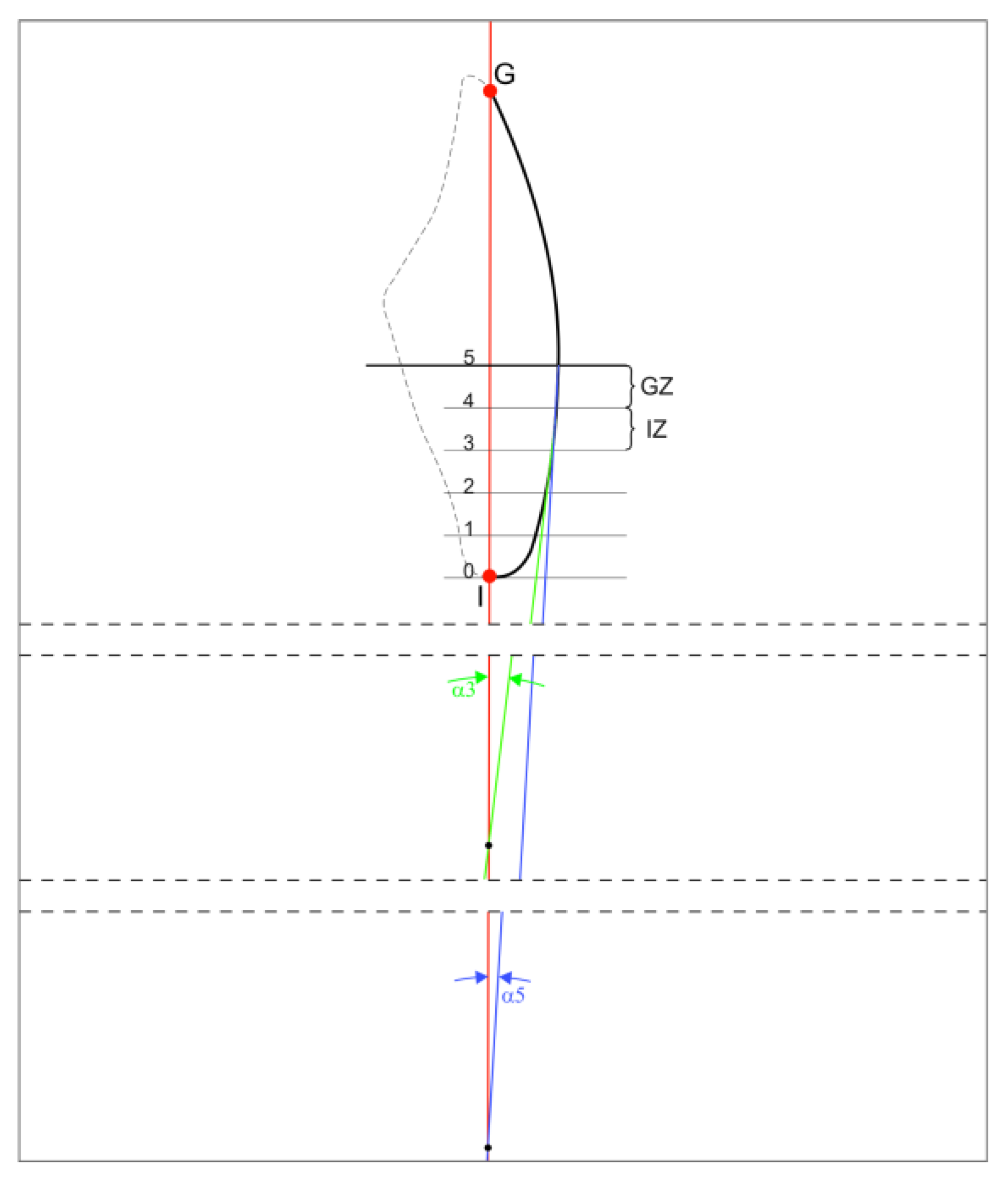
Subsequently, a grid of lines perpendicular to the GI axis and separated by a distance of 1 mm was constructed at 3, 4 and 5 mm distances from the “I” point. Using the “Distance measurement” tool, distance between CrVS and GI axis was measured at constructed points. Consequently, we determined two zones, incisal (IZ) and gingival (GZ), whose lateral limits were created by sections of the CrVS: “A” and “B”, respectively (Figure 3).
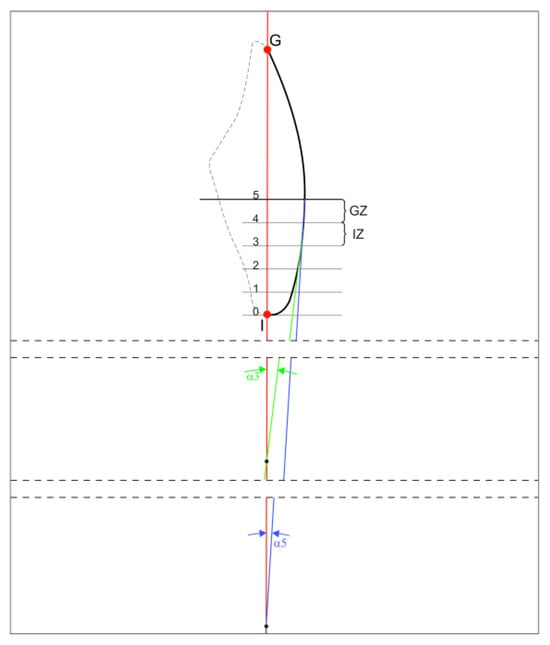
Figure 3.
Schematic of the α3 and α5 angles.
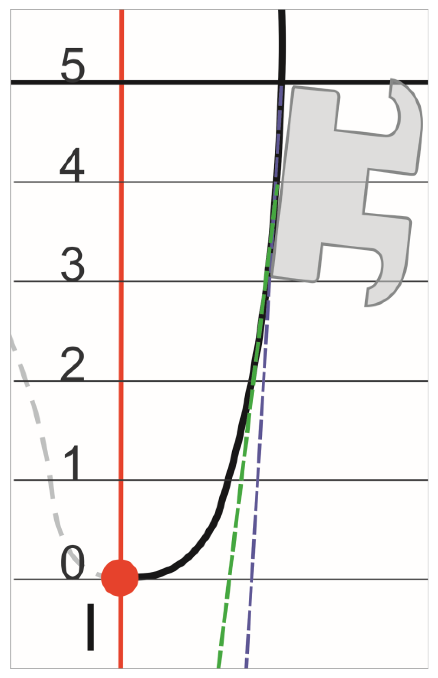
Two lines were drawn, one of which contained section “A” and the other section “B”, thus obtaining the α3 and α5 angles, respectively (Figure 3). Values of the α3 and α5 angles were calculated applying the mathematical functions, namely, tangents. It provided the change in the torque from the prescribed torque due to the anatomy of the CrVS. For the calculation of the torque alteration in the bracket whose default vertical position was 4 mm from the incisal edge, we referred to the α3 angle when the bracket base was parallel to Section A (Figure 4) and to the α5 angle when the bracket base was parallel to Section B (Figure 5).
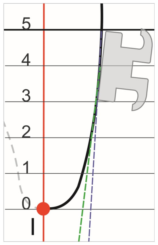
Figure 4.
Schematic of the bracket positioned at 4 mm with the base adjusted to Section A of the CrVS.
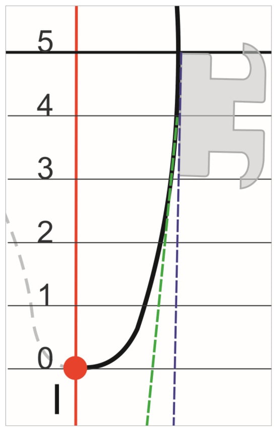
Figure 5.
Schematic of the bracket positioned at 4 mm with the base adjusted to Section B of the CrVS.
2.1. Error of the Method
To assess the method error and to avoid bias, two patients were scanned five times to define the possible impact of the scan precision on the results. Then, two different clinicians took all the measurements of the maxillary incisors on each scan at 1-week intervals.
2.2. Statistical Analysis
Statistical analysis was performed with STATISTICA software (version 13.3; StatSoft, Inc., Tulsa, OK, USA). ANOVA and post hoc HSD Tukey testing enabled assessment of the measurements together with the calculations; the repeatability and reproducibility module of STATISTICA was used to analyze the reproducibility of the measurements taken by the clinicians. The level of statistical significance was set at p < 0.05.
3. Results
Analysis of the measurement repeatability and reproducibility demonstrated that the error associated with the method did not affect the results. The repeatability and reproducibility of the measurements taken for α3 and α5 reached 1.3% (Table 1) and 0.7%, respectively (Table 2). This showed that the variability in the measurements strongly depended on the tooth shape in 98.7% in relation to α3 and in 99.3% in relation to α5.

Table 1.
Variance analysis of measurements at 3 mm from incisal edge.

Table 2.
Variance analysis of measurements at 5 mm from incisal edge.
In our findings, the mean values of the α3 and α5 angles in lateral incisors demonstrated statistically significant variation. Contrarily, the α angle measurements of the central incisors did not reveal any statistically significant differences (Table 3). Notably, the p-value for the left central incisor was marginally close to the conventional significance threshold, recorded at just above 0.05. In general, the lateral incisors demonstrated greater variability than the central incisors. In particular, the right lateral incisor was the most diverse, presenting mean values of α3 and α5 angles equal to −1.4° and +3.5°, respectively. The corresponding values of the α3 and α5 angles measured on the left lateral incisor reached −0.7° and +2.7°, respectively. The α3 and α5 values of the maxillary central incisors showed additional palatal root torque from + 0.6° to + 1.6° for the right central incisor and from +1.3° to + 1.4° for the left central incisor.

Table 3.
Average angular values of α angle (M) for the height of 3 mm and 5 mm from incisal edge.
4. Discussion
A diverse array of methods for evaluating tooth morphology is extensively documented in the orthodontic literature, reflecting the evolution of techniques and technologies in this field [4,8,10,11,15,16,17]. In an early methodological approach, Andrews pioneered the study of vestibular crown morphology in the incisal–gingival dimension [15,17]. This method involved the use of a template with circles of varying diameters, which were aligned against the tooth contour to gauge its morphology. While innovative for its time, this approach presents inherent limitations. Specifically, the labial/buccal tooth surfaces, with their complex curvatures and nuances, are not accurately represented by mere circular sections. This simplification could potentially lead to a loss of critical data, which is vital in orthodontic diagnostics and treatment planning.
Building upon these early methods, Miethke and Melsen [10] introduced a more refined technique. They utilized photocopied, magnified, and meticulously trimmed plaster models of teeth. This method marked a significant step forward in terms of precision, as it allowed for a more detailed examination of tooth morphology. However, it is important to consider that the process of trimming these models might have introduced subtle alterations to the original shape of the measured teeth, thus potentially affecting the accuracy of the results. This highlights a recurring challenge in morphological studies: balancing the need for detailed representation with the preservation of the tooth’s original form.
In a more recent advancement, Kong et al. [8] employed Cone Beam Computed Tomography (CBCT) to acquire detailed images of extracted teeth. This technology marked a significant leap forward in terms of imaging capabilities, offering a high level of accuracy with a 76 µm resolution in the final image. The use of CBCT represents a paradigm shift in dental imaging, providing three-dimensional data and unparalleled detail, which is crucial for comprehensive morphological analysis. However, it is essential to note that even this advanced technology does not match the precision achieved by the latest intraoral scanners. Specifically, the TRIOS®3 intraoral scanner has demonstrated an exceptional accuracy of 6.9 µm, setting a new standard in the field. Moreover, this error limit of 6.9 µm, compared with the range from 9.8 to 45.2 µm demonstrated by other intraoral scanners available on the market at the time of the examination, was the lowest value [18].
This study’s comprehensive analysis of the variability in maxillary incisor CrVS morphology finds a notable alignment with the findings reported by Kong et al. [8] and Miethke and Melsen [10]. These studies, including ours, consistently observed a greater degree of CrVS variability in the lateral upper incisors as compared to the central incisors. This observation is significant as it underscores a consistent pattern in maxillary incisor morphology across different research methodologies. However, it is imperative to exercise caution when drawing direct comparisons between these studies and our own. The differences in research methodologies, ranging from the tools used to the sample sizes could potentially introduce a degree of bias, affecting the validity of a direct comparison.
The implications of our findings become particularly salient when examining the prescribed torque in orthodontic treatments for lateral incisors. Here, the prescribed torque varies from +3° in the Andrews system, moving to +8° in the Roth system, and reaching up to +10° in the MBT system. Our study observed that the mean change in lateral incisor torque ranged from −1.4° to 3.5°, with a standard deviation from 3.0° to 4.1°. This finding is indicative of a potentially significant impact of the CrVS on the treatment outcomes for lateral incisors. The variability in torque prescriptions and the observed changes in our study highlight the need for a more tailored approach and the customization of orthodontic appliances considering the unique morphological characteristics of lateral incisors.
The results for the central incisors showed no statistical significance. Despite this, the scenario is different for central incisor brackets. The range of prescribed torque is broad, spanning from +7° in the Andrews prescription to +12° in the Roth prescription, and extending to +17° in the MBT system. Our study’s findings indicate a mean torque change from 0.6° to 1.6° with a standard deviation between 3.3° and 4.3°. This suggests that even if the results were statistically significant, the CrVS of the central incisors may not have a substantial impact on the overall treatment results.
Furthermore, it is essential to emphasize that our conclusions are drawn from the analysis of mean values, which provides a general overview but may not capture individual variations. Our study revealed that the vestibular surface morphology changed the torque value by 4° to 7° in 30% of the analyzed incisors and by more than 7° in 6.5% of cases. These variations are not just statistically significant but could also lead to clinically apparent changes in the torque prescribed in the bracket. This degree of change underscores the importance of individualized treatment planning in orthodontics, where the unique morphological characteristics of each tooth are taken into consideration to optimize treatment effectiveness and efficiency.
The inherent challenge in accurately determining the long axis of the tooth has been a significant limitation in our study, as it precluded us from providing a definitive quantitative value for the incisor inclination that results from bracket placement. This aspect is crucial in orthodontic treatment planning, as the long axis plays a key role in determining the optimal position of teeth. Despite this limitation, our research offers valuable insights into the influence of tooth surface morphology on the position of orthodontic brackets. This relationship is indirectly related to the root torque, a critical factor in achieving desired orthodontic outcomes. Our findings suggest that alterations in torque are likely due to the interplay between the bracket base and the crown surface.
We have demonstrated through our study that introducing specific angles, referred to as α3 or α5, during the bracket positioning process can lead to additional palatal or vestibular root torque in the lateral incisors. These findings are significant as they highlight the nuanced effects that bracket positioning can have on tooth movement. The torque discrepancy observed in our study may have a substantial impact on treatment outcomes, especially in terms of the final positioning of the lateral incisor. This aspect is critical as the lateral incisor position can influence the overall aesthetic and functional results of orthodontic treatment.
However, it is imperative to consider that addressing torque issues in orthodontic treatment is a multifaceted challenge. While our study emphasizes the importance of the morphology of the labial surface of the tooth as a significant factor [7,8], it is just one piece of a larger puzzle. The interaction between the archwire and the bracket slot, as well as the precision during bracket positioning, are equally important factors that must be considered [7,19,20,21,22]. These elements collectively contribute to the expression of torque and, by extension, the overall effectiveness of the orthodontic treatment. The precision of bracket placement, the compatibility of the archwire with the bracket slot, and the individual variations in tooth morphology all converge to determine the final orthodontic outcome. Therefore, a comprehensive understanding and consideration of these factors are essential for orthodontists in planning and executing effective treatment strategies.
The dynamic interaction between the archwire and the bracket slot, commonly referred to as torque play, has been meticulously examined by Dalstra and Melsen [22]. Their findings underscore its significant impact on treatment outcomes, revealing that the angular effect of this interaction can be as substantial as 36.1°. Consequently, it appears imperative for orthodontic practitioners to mitigate the influence of torque play to ensure consistent treatment results. To achieve this, the use of full-size archwires in conjunction with stainless steel ligatures is recommended during the final stages of treatment. This approach is suggested as it potentially offers a more precise control of tooth position, thereby aligning more closely with the intended treatment plan and reducing variability introduced by torque play.
In our study, we specifically focused on the area 3 to 5 mm from the incisal edge for bonding brackets on the incisors, following the standard practice referenced in the orthodontic literature [23]. Furthermore, we determined that a 2 mm scope was appropriate for our analysis. This decision was based on the observation that the majority of bracket base heights, measured from the gingival edge to the incisal edge of a bracket, typically slightly exceed 2 mm. Our study highlights the inherent challenges in achieving the ideal position of a directly bonded bracket. Often, the process of bracket placement inadvertently introduces α3 or α5 angles, which can have significant implications for the treatment’s effectiveness. This complexity is further emphasized by the findings of Balut et al. [21], who demonstrated that even when brackets are placed by experienced orthodontists, there is an average vertical position variation of 0.34 mm, with a considerable standard deviation of 1.80 mm. This variability, although seemingly small, can have profound effects on the final orthodontic outcome, particularly in terms of root torque.
The influence of diverse tooth morphology on bracket position is well-documented [7,8,10,11,20], and our findings add to this body of evidence, suggesting that inaccuracies in bonding can critically affect the finishing stage of orthodontic treatment. This underscores the importance of precision in bracket placement, as even minor deviations can lead to significant changes in the intended root torque. Moreover, it is important to acknowledge the existence of preformed brackets with a certain curvature given to the base surface, designed to accommodate various tooth morphologies. However, these may not entirely negate the errors due to individual variations in crown morphology. To address these issues, our study suggests that indirect bonding may offer a more reliable alternative to direct bonding methods [24,25,26]. Indirect bonding allows for greater precision in bracket placement, potentially reducing the variability in treatment outcomes. Furthermore, treatment with custom-made aligners represents an important strategy, as they are designed to fit the individual crown morphology of each patient, thereby minimizing errors related to variations in tooth shape and size. However, the capacity to control incisor torque using aligners remains limited [27].
The unequal distribution between the sexes, while reflective of the distribution found in orthodontic practices, may still be considered a limitation of the study and should be taken into account when interpreting the results.
Future research avenues are promising and could significantly advance the field of orthodontics. One such direction is the development of customized brackets that are specifically designed based on individual CrVS morphology. The potential use of 3D printing technology in this context is particularly exciting, as it offers the possibility of creating highly personalized orthodontic appliances. This approach could lead to a paradigm shift in orthodontic treatment, moving towards a more individualized and patient-specific methodology. Additionally, further exploration into the effectiveness of indirect bonding methods in achieving precise bracket placement is warranted. Such research could lead to a better understanding of how to minimize variability in treatment outcomes and improve overall treatment efficiency.
The implications of our current findings are far-reaching. They not only suggest the possibility of more personalized orthodontic treatments, with brackets and wires designed to fit the unique morphology of each patient’s teeth, but also hint at the potential for reducing the overall time required for orthodontic procedures. This personalized approach could significantly improve treatment effectiveness, patient satisfaction, and treatment efficiency. Furthermore, the methodologies and insights gained from this study could be pivotal in the development of orthodontic software. Such software could enhance the precision of orthodontic diagnostics and treatment planning, leading to more predictable and successful outcomes. Ultimately, our study contributes to the ongoing evolution of orthodontic practices, emphasizing the need for precision, personalization, and technological integration in the pursuit of optimal patient care.
5. Conclusions
The utilization of modern diagnostic tools in our study has once again highlighted the significant diversity in the CrVS morphology of incisors. This variability is a critical factor, as it can lead to the introduction of uncontrolled and unwanted root torque during orthodontic treatment. The occurrence of this phenomenon is closely tied to the individual characteristics of the patient’s CrVS. Therefore, it becomes imperative for orthodontic practitioners to consider and evaluate these individual variations in tooth surface morphology when positioning brackets.
This assessment underscores the need for a more customized approach to orthodontic treatment. Such customization could involve the preparation of individualized brackets or the use of indirect bonding trays tailored to the patient’s specific dental morphology. Additionally, the application of precise adjustments to the archwire, such as additional twisting, may be required to achieve the optimal position of the incisor roots. Our study’s findings advocate for a more personalized approach in orthodontics, where treatment strategies are adapted to the unique dental characteristics of each patient, thereby enhancing the efficacy and efficiency of orthodontic care.
Author Contributions
Conceptualization, L.M.; methodology, L.M. and M.W.; validation, L.M., J.L. and B.K.; investigation, M.W.; resources, M.W.; data curation, M.W.; writing—original draft preparation, M.W.; writing—review and editing, L.M. and J.L.; visualization, M.W.; supervision, J.L. and B.K.; project administration, M.W.; funding acquisition, M.W. and B.K. All authors have read and agreed to the published version of the manuscript.
Funding
This research received no external funding.
Institutional Review Board Statement
The study was conducted in accordance with the Declaration of Helsinki and approved by the Bioethics Committee of Wroclaw Medical University (No. KB-789/2020) on 7 December 2020.
Informed Consent Statement
Informed consent was obtained from all subjects involved in the study.
Data Availability Statement
The data presented in this study are available on request from the corresponding author due to privacy.
Conflicts of Interest
The authors declare no conflicts of interest.
References
- Casa, M.A.; Faltin, R.M.; Faltin, K.; Sander, F.G.; Arana-Chavez, V.E. Root resorptions in upper first premolars after application of continuous torque moment intra-individual study. J. Orofac. Orthop. 2001, 62, 285–295. [Google Scholar] [CrossRef]
- Sangcharearn, Y.; Ho, C. Maxillary incisor angulation and its effect on molar relationships. Angle Orthod. 2007, 77, 221–225. [Google Scholar] [CrossRef]
- Jeffrey, P. Okeson Management of Temporomandibular Disorders and Occlusion, 8th ed.; Mosby: St. Louis, MI, USA, 2020. [Google Scholar]
- Rauch, E.D. Torque and its application to orthodontics. Am. J. Orthod. 1959, 45, 817–830. [Google Scholar] [CrossRef]
- Johnson, E. Selecting custom torque prescriptions for the straight-wire appliance. Am. J. Orthod. Dentofac. Orthop. 2013, 143, S161–S167. [Google Scholar] [CrossRef] [PubMed]
- Gioka, C.; Eliades, T. Materials-induced variation in the torque expression of preadjusted appliances. Am. J. Orthod. Dentofac. Orthop. 2004, 125, 323–328. [Google Scholar] [CrossRef] [PubMed]
- Papageorgiou, S.N.; Sifakakis, I.; Keilig, L.; Patcas, R.; Affolter, S.; Eliades, T.; Bourauel, C. Torque differences according to tooth morphology and bracket placement: A finite element study. Eur. J. Orthod. 2016, 39, 411–418. [Google Scholar] [CrossRef] [PubMed]
- Kong, W.D.; Ke, J.Y.; Hu, X.Q.; Zhang, W.; Li, S.S.; Feng, Y. Applications of cone-beam computed tomography to assess the effects of labial crown morphologies and collum angles on torque for maxillary anterior teeth. Am. J. Orthod. Dentofac. Orthop. 2016, 150, 789–795. [Google Scholar] [CrossRef] [PubMed]
- Wheeler, R.C. Dental Anatomy, Physiology, and Occlusion; WB Saunders: Philadelphia, PA, USA, 1974. [Google Scholar]
- Miethke, R.R.; Melsen, B. Effect of variation in tooth morphology and bracket position on first and third order correction with preadjusted appliances. Am. J. Orthod. Dentofac. Orthop. 1999, 116, 329–335. [Google Scholar] [CrossRef]
- Germane, N.; Bentley, B.E.; Isaacson, R.J. Three biologic variables modifying faciolingual tooth angulation by straight-wire appliances. Am. J. Orthod. Dentofac. Orthop. 1989, 96, 312–319. [Google Scholar] [CrossRef]
- Kuc, A.E.; Kotuła, J.; Nahajowski, M.; Warnecki, M.; Lis, J.; Amm, E.; Kawala, B.; Sarul, M. Methods of Anterior Torque Control during Retraction: A Systematic Review. Diagnostics 2022, 12, 1611. [Google Scholar] [CrossRef]
- Sarul, M.; Rutkowska-Gorczyca, M.; Detyna, J.; Zięty, A.; Kawala, M.; Antoszewska-Smith, J. Do Mechanical and Physicochemical Properties of Orthodontic NiTi Wires Remain Stable In Vivo? Biomed Res Int. 2016, 2016, 5268629. [Google Scholar] [CrossRef]
- Connizzo, B.K.; Sun, L.; Lacin, N.; Gendelman, A.; Solomonov, I.; Sagi, I.; Grodzinsky, A.; Naveh, G. Nonuniformity in Periodontal Ligament: Mechanics and Matrix Composition. J. Dent. Res. 2021, 100, 179–186. [Google Scholar] [CrossRef] [PubMed]
- Andrews, L.F. Straight wire. In Age Concept and Appliance; L.A. Wells: San Diego, CA, USA, 1989. [Google Scholar]
- van Loenen, M.; Degrieck, J.; De Pauw, G.; Dermaut, L. Anterior tooth morphology and its effect on torque. Eur. J. Orthod. 2005, 27, 258–262. [Google Scholar] [CrossRef] [PubMed]
- Andrews, L.F. The straight wire appliance explained and compared. J. Clin. Orthod. 1967, 10, 174–195. [Google Scholar]
- Hack, G.D.; Patzelt, S.B.M. Evaluation of the accuracy of six intraoral scanning devices: An in-vitro investigation. ADA Prof. Prod. Rev. 2015, 10, 1–5. [Google Scholar]
- Magesh, V.; Harikrishnan, P.; Kingsly Jeba Singh, D. Finite element analysis of slot wall deformation in stainless steel and titanium orthodontic brackets during simulated palatal root torque. Am. J. Orthod. Dentofac. Orthop. 2018, 153, 481–488. [Google Scholar] [CrossRef] [PubMed]
- Sardarian, A.; Danaei, S.M.; Shahidi, S.; Boushehri, S.G.; Geramy, A. The effect of vertical bracket positioning on torque and the resultant stress in the periodontal ligament—A finite element study. Prog. Orthod. 2014, 15, 50. [Google Scholar] [CrossRef]
- Balut, N.; Klapper, L.; Sandrik, J.; Bowman, D. Variations in bracket placement in the preadjusted orthodontic appliance. Am. J. Orthod. Dentofac. Orthop. 1992, 102, 62–67. [Google Scholar] [CrossRef]
- Dalstra, M.; Eriksen, H.; Bergamini, C.; Melsen, B. Actual versus theoretical torsional play in conventional and self-ligating bracket systems. J. Orthod. 2015, 42, 103–113. [Google Scholar] [CrossRef]
- Armstrong, D.; Shen, G.; Petocz, P.; Darendeliler, M.A. A comparison of accuracy in bracket positioning between two techniques—Localizing the centre of the clinical crown and measuring the distance from the incisal edge. Eur. J. Orthod. 2007, 29, 430–436. [Google Scholar] [CrossRef]
- Schmid, J.; Brenner, D.; Recheis, W.; Hofer-Picout, P.; Brenner, M.; Crismani, A.G. Transfer accuracy of two indirect bonding techniques—An in vitro study with 3D scanned models. Eur. J. Orthod. 2018, 40, 549–555. [Google Scholar] [CrossRef] [PubMed]
- Castilla, A.E.; Crowe, J.J.; Moses, J.R.; Wang, M.; Ferracane, J.L.; Covel, D.A. Measurement and comparison of bracket transfer accuracy of five indirect bonding techniques. Angle Orthod. 2014, 84, 607–614. [Google Scholar] [CrossRef] [PubMed]
- Duarte, M.E.A.; Gribel, B.F.; Spitz, A.; Artese, F.; Miguel, J.A.M. Reproducibility of digital indirect bonding technique using three-dimensional (3D) models and 3D-printed transfer trays. Angle Orthod. 2020, 90, 92–99. [Google Scholar] [CrossRef] [PubMed]
- Simon, M.; Keilig, L.; Schwarze, J.; Jung, B.A.; Bourauel, C. Treatment outcome and efficacy of an aligner technique—Regarding incisor torque, premolar derotation and molar distalization. BMC Oral Health 2014, 14, 68. [Google Scholar] [CrossRef]
Disclaimer/Publisher’s Note: The statements, opinions and data contained in all publications are solely those of the individual author(s) and contributor(s) and not of MDPI and/or the editor(s). MDPI and/or the editor(s) disclaim responsibility for any injury to people or property resulting from any ideas, methods, instructions or products referred to in the content. |
© 2024 by the authors. Licensee MDPI, Basel, Switzerland. This article is an open access article distributed under the terms and conditions of the Creative Commons Attribution (CC BY) license (https://creativecommons.org/licenses/by/4.0/).

