Abstract
The invasive blue crab Portunus segnis, which was collected from two sites on the Gulf of Gabès, is the subject of this work. This study is based on demonstrating the accumulation capacity of P. segnis by measuring the concentrations of cadmium, zinc, lead, and copper in the gills and hepatopancreas. The enzymatic activities of catalase, glutathione-S-transferase, reduced glutathione, and lipid peroxidase were assessed in this region for the first time. The main results show that the metals have high bioaccumulation potentials in P. segnis tissues between different sites. The possible adaptation of P. segnis in the Gulf of Gabès and the variations in the studied biomarkers and metal concentrations at different sites confirm the usefulness of the invasive blue crab as a sentinel species.
1. Introduction
The aquatic environment is the ultimate container for pollutants. These include metal–elemental inorganic fertilizers that enter the aquatic environment, often through atmospheric deposition, the erosion of the geological matrix, or as a result of anthropogenic activities (e.g., the discharge of industrial, domestic, and mining wastes) and the use of pesticides and pesticides [1]. Metal source pollution is one of the greatest threats facing the world today. In fact, heavy metals pose serious ecological problems due to their toxicity and potential to accumulate in a variety of aquatic organisms, thereby causing damaging effects on the ecological balance of the aquatic environment. Therefore, this situation requires awareness of the vulnerability of these environments, regular monitoring, and the implementation of preventive measures.
However, the sound and scientific use of biological indicators, especially in environmental biological assessments (monitoring the state of the environment or the effectiveness of compensation or restoration measures) has not been recognized until recently. This discipline was originally based on the use of biological indicators, which can provide information about the contamination of the environment by chemicals. On the other hand, the identification of the bioavailability of contaminants and their accumulation in the tissues of these organisms requires the use of certain ecotoxicological tools such as biomarkers. The purpose of these tools is to provide information on the harmful effects of environmental exposures manifested at the individual level. However, due to the abundance of pollutants, the complexity of their effects, and the many interactions between them, environmental diagnosis must be based on the use of a multi-biomarker approach [2,3,4].
Like polluted aquatic environments, the Mediterranean marine environment is one of the most polluted semi-enclosed basins in the world, especially with respect to metallic pollutants. According to previous studies, about 49–82% of copper, 68–76% of zinc, 21–65% of lead, and 75–92% of cadmium are deposited in the Mediterranean Sea in soluble forms and can thus be easily accumulated by organisms [5].
One of the nations in the Mediterranean region in which the importance of metals to aquatic environments is growing is Tunisia, particularly in coastal regions where chemical manufacturing is most significant. Numerous studies revealed that across these coastal areas, the Gulf of Gabès constitutes an ecosystem under intense industrial stress and is therefore likely to be polluted with various contaminants such as metallic trace elements [6,7].
Numerous aquatic species have been applied in diverse ecotoxicological investigations to suggest the status of the pollution of ecosystems. Crustaceans, which are noted for their high accumulative potential, are among them [8]. The blue crab Portunus segnis (Forskl, 1775) was among the first lessepsian intruders to be identified in Egyptian waters 30 years after the Suez Canal was completed in 1868 [9]. It then established itself in coastal environments across the Levantine Basin, from Turkey to Egypt [10]. This crab species has been found in various Mediterranean locations such as the Aegean Sea, Sicily, and the northern Tyrrhenian Sea [11], as well as in Tunisian coastal waters and the surrounding region in recent years [10], such as Malta [12]. P. segnis was initially discovered in Tunisia in the Gulf of Gabès in 2014 [13]; it quickly spread throughout the Gulf, comprising Djerba Island and the northern beaches, namely, the Gulf of Hammamet, and it has also been found in Libya on occasion [14,15].
In this context, this work aims to use an invasive species of the Gulf of Gabès, P. segnis, as a bioindicator of the quality of the marine environment. This study aims to mirror the state of contamination of the aquatic surroundings in the Gulf of Gabès by pollutants through the determination of the heavy metal contents of the tissues of P. segnis, especially the gills and hepatopancreas, and the use of four biomarkers of pollution: catalase enzymatic activity, glutathione-S-transferase, reduced glutathione concentration, and an estimation of lipid peroxidation. To the best of our knowledge, this is the first study to biomonitor the two studied Tunisian aquatic ecosystems via combined chemical and biomarker analyses, using Portunus segnis as sentinel species.
2. Materials and Methods
2.1. Presentation of the Studied Sites
The Gulf of Gabès is located in the southeast of Tunisia and is characterized by a very wide continental shelf and a very gentle slope. It begins from Chebba (35.3° north latitude) in the north and ends at the Tunisia–Libya border in the south, with a total length of about 700 km. It is bordered by the Kerkennah Archipelago to the west, the shallow waters of the Kerkennah Islands to the south, and the mainland to the south. The Gulf basin is shallow, only 50 m deep at 110 km offshore.
The bottoms are generally sandy to muddy and are composed mainly of carbonate sediments [16]. The region is characterized by a pre-Saharan and arid to semi-arid climate, with generally high temperatures and a relatively low rainfall of around 200 mm per year [17]. In addition, in this gulf, the salinity of the seawater oscillates between 37.2 and 38 psu (practical salinity unit), with an average of 37.52 ± 0.29 psu. This is due to the low levels of precipitation, intense evaporation, and the low amount of runoff water [18]. Thus, as its geomorphological and climatic conditions are very favorable, the Gulf of Gabès is one of the most productive areas of the Mediterranean. Indeed, more than 42% of the production of the national fisheries occurs in this area [19].
2.2. Selection of Sampling Sites
The selection of sampling sites is a critical stage of any pollution impact assessment. For this purpose, two sites belonging to the Gulf of Gabès were selected, with different degrees of contamination (Figure 1):
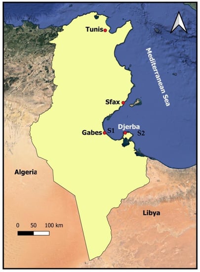
Figure 1.
Location of Portunus segnis sampling sites in the Gulf of Gabès. S1: Ghannouch site (industrial area); S2: Djerba Site (touristic area).
- -
- Ghannouch (respective geographical coordinates, 33°55′ N and 10°5′ E) is the area considered the most polluted due to the establishment of the chemical group, suggesting that the marine ecosystem and therefore the fauna and flora are threatened by liquid, solid, and gaseous industrial discharges [20].
- -
- Sidi Salem (Djerba), the island of Djerba, is characterized by seasonal mass tourism, and the main sources of pollution are still anthropogenic waste.
Twenty adult specimens (n = 20) were collected from each area in November 2021. They were captured with the aid of neighborhood fishers around ten kilometers south of Gabès and around 20 km north of Djerba at a depth of roughly 1 m, which is the typical depth of a crab burrow. The live crabs were transported to the laboratory in buckets filled with aerated seawater as quickly as possible. Once the cephalothorax was opened on ice, the digestive glands and gills were quickly removed. The organs were dried and powdered for metal analysis, frozen at −20 °C, ground in a mortar with liquid N2, and stored at −80 °C until biochemical testing. Three pools were prepared from each sampling station for each collected tissue.
2.3. Chemical Analysis
The tissue samples were dried as the first phase of the technique by incubating them in a G-Therm115 oven at 65 °C for 24 h to maintain a constant dry weight. The samples were placed on spit for mineralization after the dehydration phase was finished, and the dry weight of each sample was determined.
The samples were placed on spit for mineralization after the dehydration phase was finished, and the dry weight of every specimen was determined. The method outlined by Annabi et al. [21] was used to analyze the chosen chemicals, Cd, Zn, Pb, and Cu. By using an acid attack, organic matter was removed during the mineralization step. After each sample had dried, not more than 10 mg was taken from it, and then 3 mL of concentrated nitric acid (HNO3) was added. After being diluted to 30 mL with ultrapure water, the solution generated from the digestion process was maintained at 4 °C until it was analyzed via flame atomic absorption spectrometry, utilizing an atomic absorption spectrophotometer (Avanta GBC spectrometer, Australia) with an air–acetylene mixture.
Calibration curves were calculated using Agilent standard solutions for Cd, Pb, Cu, and Zn. They were prepared using serial dilutions of 3% nitric acid (HNO3) and ultrapure water with a stock solution of 1000 mg L−1 of each metal. Working standards (Cd 0, 3, 5, and 15 μg/mL; Pb 0, 10, 15, and 20 μg/mL; Zn 0, 3, 5, 15, and 50 μg/mL; Cu 0, 15, 20, and 25 μg/mL) were used to calibrate the devices. Using external standards with coefficients for calibration curves greater than 0.99, the heavy metals of interest in this study were quantified. For every 10 samples analyzed, a blank solution was also analyzed. The samples were analyzed in triplicate (the variation coefficient was less than 10%), and the findings were computed using the mean and standard deviation.
The crab samples were spiked with known quantities of heavy metals to carry out the repeatability test and the verification of the analytical samples. The samples were examined in triplicate for each run. The same procedure utilized for the initial samples was employed to digest and evaluate the heavy-metal-spiked crab samples. Recoveries for the tested chemicals using the provided method ranged from 77 to 95%. Recovery correction was carried out for each sample.
Utilizing ultrapure water, aqueous solutions of the reagents and standards were prepared. Every chemical utilized was of the analytical reagent standard. Glass materials were cleaned and submerged for 24 h in a tank containing 3% nitric acid to prevent contamination. Each time a glass material was used, it was then cleaned with ultrapure water and dried in an oven.
2.4. Biomarker Analyses
Samples for enzymatic assays were weighed using a precision balance and then ground in a 0.1 M potassium phosphate buffer, pH 7.2, at a rate of 4 mL of buffer per gram of tissue. The cell suspension was then centrifuged (4000 rpm/min, 4 °C, 10 min), and the resulting supernatant was collected in tubes and stored at −20 °C until the biochemical assays were performed.
2.4.1. Tissue Preparation
Gill and hepatopancreas tissue samples were weighed for the oxidative biomarker analyses using a precision balance and then ground in a cold 0.1 M potassium phosphate buffer, pH 7.2, at a rate of 4 mL of buffer per gram of tissue. The centrifugation step of the cell suspension was performed at 4000 rpm/min and 4 °C for 10 min. After this step, the obtained supernatants were collected in tubes and stored at −20 °C until analysis.
2.4.2. Total Protein Quantification
The protein contents were determined using bovine serum albumin as a standard protein [22]. To perform this method, 2.5 mL of Bradford solution was mixed with 10 µL of enzyme extract. The mixture was then blended thoroughly and incubated at room temperature for 2 min. Finally, the absorbance was recorded at 595 nm, and the protein concentration is expressed in µg/µL.
2.4.3. Glutathione S-Transferase (GST) Determination
GST is an enzyme that aids in the detoxification of xenobiotics and/or protection against the toxic metabolites produced by the breakdown of macromolecules after exposure to oxidative stress. GSTs are phase II metabolic enzymes whose function is to conjugate to a glutathione molecule a wide variety of pollutants to allow for their elimination. These enzymes are therefore triggered by the presence of certain pollutants in the environment, such as metals, and are therefore considered biomarkers of pollution. In addition, the major role of GST is the mitigation of oxidative stress generated by trace metal elements (TMEs) [23]. The technique adopted used to assay GST activity was that of Habig et al. [24]. This consisted of presenting the enzyme contained inside the homogenate with a substrate (2,4-chloro dinitrobenzene (CDNB)) which reacts without problems with many forms of GST and glutathione. The conjugation reaction of these two products catalyzed by GST results in the formation of a new molecule, 1-glutathione-2,4-dinitrobenzene (GST-DNB), which absorbs light at a 340 nm wavelength. The value of the optical density measured is directly proportional to the amount of conjugate formed and is related to the intensity of GST activity. Assays were performed via the homogenization of 100 µL of the biological extract with 200 µL of 0.1 M NaPi (pH 6.5), 150 µL of 10 mM CDNB, and 900 µL of 10 mM glutathione. The obtained mixture was incubated at 37 °C, and then the absorbance was measured every minute at 340 nm with the extinction coefficient of 9.6 mM−1 cm−1. The specific activity of GST is expressed in nanomoles of CDNB per minute per milligram of protein (nmol CDNB/min/mg protein).
2.4.4. Catalase (CAT) Determination
CAT is an enzyme specialized in the detoxification of hydrogen peroxide and its transformation into an oxygen and a water molecule. It is a biomarker of oxidative stress and is sensitive to certain contaminants that induce this type of stress, such as toxic metals [25]. The enzymatic activity of CAT was measured in the fractions of the gills and of the hepatopancreas, according to the method of Aebi [26], at 240 nm using a UV/visible spectrophotometer via the variation in the optical density, following the dismutation of hydrogen peroxide. The reaction mixture contained 950 µL of 0.05 M potassium phosphate buffer (pH 7), 500 µL of 30 mM H2O2, and 50 µL of enzyme extract. Every 20 s for 2 min, a spectrophotometer with an extinction value of 0.043 mM−1 cm−1 recorded the decline in absorbance at 240 nm. The specific activity of CAT is expressed in international units IU/mg of protein (µmoles of H2O2 destroyed/min/mg of protein).
2.4.5. Reduced Glutathione (GSH) Determination
The determination of reduced glutathione was carried via the colorimetric method using Ellman’s reagent (DTNB) [27]. The principle is based on the oxidation reaction of GSH with 5,5-dithiodis-2-nitrobenzoic acid (DTNB), thus releasing thionitrobenzoic acid (TNB), which has an absorbance at 412 nm. To perform this method, a mixture of 500 µL of the homogenate and 500 µL of sulfosalicylic acid (4%) was centrifuged at 3500 rpm for 10 min. Subsequently, 200 μL of the supernatant was added to 1 mL of the phosphate buffer (0.1 M, pH 7.4) and 400 μL of 10 mM DTNB. The mixture was left for 5 min at room temperature, and the optical densities were read via a spectrophotometer at 412 nm with the extinction coefficient of 13.600 mM−1 cm−1. The concentration of GSH is expressed in nanomoles per milligram of protein (nmol/mg of protein).
2.4.6. Malonedialdehyde Level (MDA) Determination
The MDA assay was carried out as described by Ohkawa [28], via the colorimetric method in the presence of thiobarbituric acid (TBA), which allows for the detection of small amounts of lipid peroxides and particularly free MDA. Thus, the detection of MDA present in biological samples was based on the reaction during which two molecules of TBA react with one molecule of MDA, which results in the formation of a red chromogen absorbing at 535 nm in an acidic medium. The intensity of the red coloration increases with the concentration of MDA. It should be noted that the MDA-TBA complex is only stable for 3 h in the dark and at room temperature. The operating mode was as follows: 100 mL of tissue extract was homogenized with 2 mL of butylated hydroxytoluene (BHT) in methanol, 50 mL of 0.37% TBA, 50 mL of 15% TCA, and 50 mL of 0.25 N HCl. Next, the obtained mixture was heated at 100 °C for 15 min. After the heating process, the mixture was chilled in ice for 10 min before being centrifuged at 3000 rpm for 10 min. At 535 nm, the optical density was read using the molar extinction coefficient (ε = 154 mM−1 cm−1). The MDA concentrations are measured in nmol of MDA/mg of protein (nmol/mg).
2.5. Statistics
All biomarker determination data (GST, CAT, GSH, and MDA) and metal concentrations levels are shown as means ± SDs (standard deviations). The normality of the data was checked prior to statistical analysis. Using the StatView software tool (Statview 5.0), the findings were analyzed using a one-way ANOVA and Fisher’s least significant difference technique (LSD). If p < 0.05, the value was judged statistically significant.
3. Results and Discussion
3.1. Metal Analysis
Figure 2a,b show the findings of the Cd, Cu, Zn, and Pb determinations in the gills and hepatopancreata of P. segnis. The studies confirmed the presence of these elements in the tissues of the target crab at heterogeneous levels. The Zn levels were higher at both study locations (Table 1). According to the findings, the heavy metal levels at the two research locations and in the two tissues (the gills and hepatopancreas) followed a concentration scale in the following decreasing order: Zn > Cd > Cu > Pb (Figure 2a,b). Statistical analyses tests for the levels of the four trace elements at the Ghannouch site revealed statistically significant differences in the P. segnis gills (F = 42.68; p < 0.01) and hepatopancreas (F = 42.68; p < 0.01) and in the hepatopancreas (F = 72.57; p < 0.01). In addition, the Zn, Cd, Cu and Pb contents at the Djerba site showed statistically significant differences for the gills (F = 20.31; p < 0.01) and hepatopancreas (F = 15.12; p < 0.01) (Table 1).
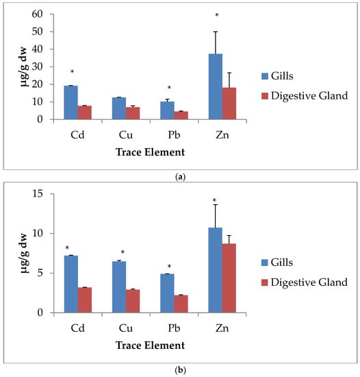
Figure 2.
Heavy metal content s(Cd, Cu, Pb, and Zn) in P. Segnis tissues (n = 20) from Ghannouch site (a) and from Djerba site (b). *: statistically significant difference between the trace element contents in the gills and hepatopancreas (p < 0.01).

Table 1.
Metal data (mg/kg dw) in P. segnis at the two studied sites for the gills and hepatopancreas.
Figure 3a reveals that the highest concentrations in the gills were found in the individuals collected from Ghannouch, near the industrial zone of Gabès, for the four studied metals, Cd (19.21 ± 0.1 µg/g), Cu (12.53 ± 0.078 µg/g), Pb (10.15 ± 1.38 µg/g), and Zn (37.36 ± 12.57 µg/g) compared to the values noted in the individuals coming from Djerba, which were Cd (7.2 ± 0.049 µg/g), Cu (6.458 ± 0.122 µg/g), Pb (2.212 ± 0.054 µg/g), and Zn (10.722 ± 2.9 µg/g). For the values obtained from the hepatopancreas, the individuals from the Ghannouch site also presented the highest levels (Figure 3b) of metallic trace elements, Cd (7.74 ± 0.14 µg/g), Cu (6.96 ± 0.79 µg/g), Pb (4.536 ± 0.24 µg/g), and Zn (18.14 ± 8.38 µg/g), in comparison with the contents in the tissues of the individuals from Djerba, Cd (3.18 ± 0.04 µg/g), Cu (2.91 ± 0.087 µg/g), Pb (2.212 ± 0.054 µg/g), and Zn (8.71 ± 1.03 µg/g). In fact, the statistical analyses between the two study stations showed statistically significant differences in gill levels for Cd (F = 23.32; p < 0.01), Cu (F = 92.35; p < 0.01), Zn (F = 62.24; p < 0.01), and Pb (F = 23.96; p < 0.01), as well as for the levels in the hepatopancreas for Cd (F = 74.39; p < 0.01), Cu (F = 17.11; p < 0.01), Zn (F = 54.31; p < 0.01), and Pb (F = 36.06; p < 0.01) (Figure 3a,b).
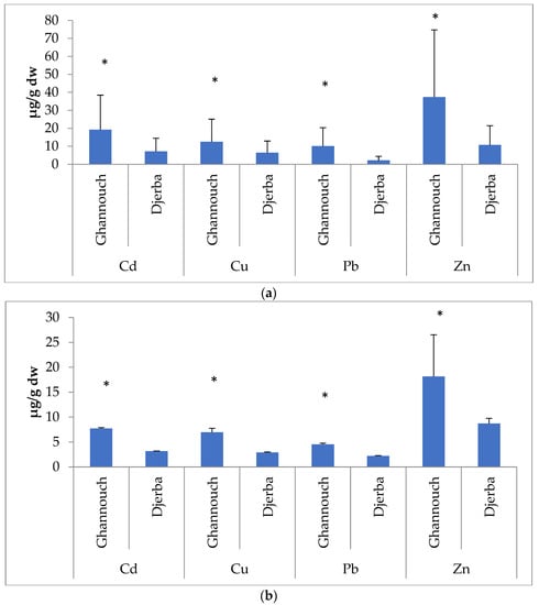
Figure 3.
Trace element contents (µg/g dry weight) in the gills (a) and the digestive glands (b) between the two studied sites (n = 20). *: statistically significant difference for the metal contents between the two sites (p < 0.01).
Only Italy [29] and Turkey [30,31] have provided data on metal levels in Mediterranean Portunus segnis populations. With a few exceptions, the majority of these research analyses were performed solely on soft tissues. The content of trace metal elements in crustaceans is estimated to be higher in the exoskeleton than in other tissues, as cadmium (with an ionic radius of 0.97 and 0.99 Å, respectively) and other elements such as manganese (Mn), lead, and zinc are gradually accumulated in the exoskeleton via calcium uptake routes [32]. Indeed, numerous investigations on brachyuran crustaceans have found that concentrations of metals in the exoskeleton are higher than in soft tissues [33,34].
Numerous environmental and biological factors, including molting, govern the trends of trace element bioaccumulation in crustaceans’ soft tissues [35,36]. This is an important phase because it provides a detoxifying mechanism for heavy metals to be eliminated on a regular basis [37]. Metals have been shown to have distinct bioaccumulation patterns and can accumulate in various tissues at different concentrations. As an example, cadmium accumulated mostly in the midgut and gills, copper in the gills, and zinc in the gills/muscle of Carcinus maenas treated with metals for 32 days [38].
According to the results obtained in our study, the Cd and Pb levels were higher than the MOH, EC, and FAO standards [39,40,41], while for the other two elements (Cu and Zn), the values do not exceed the prescribed standards (Table 2). It should be noted that copper and zinc are essential for the metabolism and physiology of crustaceans, and their accumulation is generally independent of environmental concentrations [42,43]. However, when concentrations reach abnormally high levels, the ions accumulate in cytosolic proteins such as metallothionein [44]. The zinc concentrations (Table 2) in all studied samples in this study were higher than the concentrations of the other metals in the same samples of P. segnis. These high levels in crabs can be attributed to the biological interest in Zn for the organism. Indeed, Zn is an essential element for the metabolic process and is associated with certain Zinc transporter metalloproteins (such as metallothioneins). The physiological functions of these metalloproteins are related to their affinities for essential cations, especially copper and zinc, and to their antioxidant characteristics. They also play a major role in cellular homeostasis by controlling the bioavailability of certain essential metals.

Table 2.
Data relating to some metal levels (mg/kg dw) in the tissues of aquatic organisms, according to standards [39,40,41].
Based on the data obtained in our study, we noted that the gill tissues at both studied sites had the highest concentrations of trace elements compared to the hepatopancreata (Table 3). These results are consistent with previous studies conducted on other crab species. A previous analysis [45] showed that Potamonauteswarreni crab gills contained more Cd and Cu than the hepatopancreas. Indeed, the work of Pan and Zhang [46] on the Charybdis japonica crab also revealed that the gills are more sensitive than the digestive glands to exposure to pollutants. Moreover, DNA strand breaks were positively correlated with Cd exposure only in the gill tissue. Our results are also consistent with the work performed by Corrêa et al. [47] on the Ucides cordatus crab. This work showed that metals accumulated in a concentration-dependent manner, with the highest levels found in the gills, followed by the hepatopancreas. Different species accumulate metals at different rates, depending on how they respond to metal exposure [48,49]. Within species that are closely related, as well as between genera, the processes of metal accumulation in aquatic crustaceans vary; some are net accumulators, whereas others are regulators. When an organism’s rate of absorption exceeds its rate of excretion, net accumulation develops. The size of the individual organism can also have an impact on metal bioaccumulation in crabs. For both freshwater and marine crabs, smaller animals do indeed accumulate more metals [50,51].
Some work on the contamination of crab tissues by heavy metals has been reported in the literature. The values recorded in different regions of the world have been mentioned in Table 3. The concentrations of copper reported in our study in the gills of P. segnis individuals from Ghannouch and Djerba are lower than those reported in the samples of crabs collected from the Bizerte Lagoon, the Monastir Coast, and Gargour in Sfax [52,53]. The levels of the same element in the hepatopancreata of P. segnis in the two studied sites (Ghannouch and Djerba) are also lower than those recorded in crab samples from Canada [54], Australia [35], and the United States [55]. However, the concentration in the hepatopancreas samples of individuals from Ghannouch is similar to the concentration found in crab samples from Brazil [56].
The zinc contents in the gills of crabs from Ghannouch and Djerba are lower than those reported in the samples of crabs collected from the Bizerte Lagoon, Monastir Coast and Gargour in Sfax [52]. The zinc concentrations in the hepatopancreata of P. segnis from Ghannouch and Djerba are lower than those recorded in crab samples from Canada [54], Australia [35], and the United States [55]. However, the concentration of this element in the hepatopancreas samples of crabs from the Ghannouch site was similar to the concentration found in crab samples from Brazil [56].
For cadmium, the values noted in the gills of P. segnis from Ghannouch and Djerba are higher than those reported in the samples of crabs (Carcinus maenas) collected from the Lagoon of Bizerte, the Coast of Monastir, and Gargour in Sfax [52]. The cadmium content in the hepatopancreata of individuals from Ghannouch is higher than the values recorded in crabs from Canada [54], Australia [35], the United States [55], and Brazil [56], while its concentration in the hepatopancreata of the Djerba crabs is similar to that reported in samples from Canada [54], Australia [35], and the United States [55]. However, it is higher than in the samples from Brazil [56].
The concentrations of lead in the gills of crabs from the two sites Ghannouch and Djerba are lower than those observed in crab samples collected from the Bizerte Lagoon and Monastir Coast [52]. The content of this element in the gills of crabs from Djerba is close to that of crabs collected in the site of Gargour in Sfax [52]. In the hepatopancreata of Ghannouch individuals, the Pb contents are close to the concentrations reported in C. maenas from the Bizerte lagoon. However, for the values observed in the hepatopancreata of P. segnis individuals from both from Ghannouch and Djerba, the levels are higher than those obtained in the samples from the coast of Monastir and from Gargour in Sfax (Table 3). Regarding the data from the study by Ben-Khedher et al. [53] on C. maenas captured from Bizerte lagoon, the Pb concentrations are higher than those in the gills and hepatopancreata except for the levels in the gills of individuals from the Sfax site (Table 3). Comparing our data with other studies in other localities, we notice that the Pb levels in our samples are higher.
It has been shown that accumulation of trace elements in marine crabs might arise via saltwater or food [8]. The contribution of each route of uptake is linked to the bioavailability of these minerals. Cu and Zn concentrations in water are fairly low in the Gulf of Gabès region [21,57] for a Mediterranean-wide comparison, whereas sediments can be moderately to highly enriched in these two elements, as well as other heavy metals, based on the area [58,59]. As a result, as various previous investigations have shown, a transfer mechanism through sediments can be considered the primary predictor of reported accumulation patterns [60,61]. As a result, the contamination can be attributed to anthropogenic origins.


Table 3.
Average contents of Cu, Zn, Cd, and Pb (mg/kg dw) in crab tissues from the literature.
Table 3.
Average contents of Cu, Zn, Cd, and Pb (mg/kg dw) in crab tissues from the literature.
| Species | Site | Tissues | Cu | Zn | Cd | Pb | Reference |
|---|---|---|---|---|---|---|---|
| Scylla serrata | Canada | Digestive Gland | 637 | 208 | 3.1 | - | [59] |
| Pseudocarcinus gigas | Australia | Digestive Gland | 52 | 108 | 2.2 | - | [62] |
| Menippemer scenario | South Carolina-United States | Digestive Gland | 308 | 46 | 4.6 | - | [63] |
| Ucidescordatus | Brazil | Digestive Gland | 6.6 | 8.89 | 0.16 | - | [64] |
| Xenograpsustestudinatus | Taiwan | Whole tissue | 53–290 | 119–610 | 0.49–1.29 | 1.83–2.64 | [65] |
| Eriocheir sinensis | Poland | Whole tissue | 3.17–21.9 | 1.15–26.6 | 0.016–0.025 | 0.29–0.61 | [60] |
| Carcinusmaenas | Bizerte Lagoon, Tunisia | Gills | 5.12–28.98 | 5.39–13.78 | 1.08–1.99 | 5.82–8.75 | [13] |
| Digestive Gland | 15.16–31.93 | 5.3–25.11 | 0.67–2 | 5.01–8.56 | |||
| Carcinusmaenas | Bizerte Lagoon, Tunisia | Gills | 857.3 | 627.1 | 0.61 | 21.1 | [39] |
| Monastir Coast, Tunisia | 376.5 | 83.4 | 0.38 | 15.57 | |||
| Sfax Coast, Tunisia | 238.9 | 88.8 | 0.52 | 4.94 | |||
| Bizerte Lagoon, Tunisia | Digestive Gland | 863.5 | 985.1 | 21.5 | 4.5 | ||
| Monastir Coast, Tunisia | 396.9 | 102.4 | 0.62 | 0.67 | |||
| Sfax Coast, Tunisia | 205.1 | 99.9 | 3.04 | 0.14 | |||
| Portunussegnis | Gulf of Gabès (Zarrat), Tunisia | Muscle | 206.45 | 590.04 | 0.21 | 0.4 | [6] |
| Gonad | 584.85 | 36.46 | 0.23 | 0.14 | |||
| Exoskeleton | 65.78 | 11.4 | 0.14 | 0.06 | |||
| Portunus pelagicus | Shuwaikh, Kuwait | Gills | 79.1 | 29.1 | 0.28 | 3.29 | [66] |
| Hepatopancreas | 68.6 | 18.9 | 0.43 | 4.69 | |||
| Chiromantes eulimene | Bhizolo Canal Richards Bay Harbour, South Africa | Gills | 106.74 | 219.8 | 9.72 | 23.24 | [67] |
| Digestive Gland | 153.6 | 140.57 | 7.12 | 8.94 | |||
| Scylla serrata | Punnakayal, Tuticorin, Southeast Coast of India | Gills | 3.63 | 14.26 | 0.14 | 0.19 | [68] |
| Hepatopancreas | 0.21 | 39.73 | 0.51 | 0.15 | |||
| Portunus pelagicus | Northern Bay of Bengal | Gills | 21.06 | - | 1.01 | 1.67 | [69] |
| Hepatopancreas | 12.46 | - | 2.82 | 2.07 | |||
| Cardisoma armatum | Kribi mangrove areas, Cameroon | Gills | 3.87 | 64.77 | 0.18 | 0.09 | [70] |
| Hepatopancreas | 7.88 | 51.34 | 0.35 | 2.07 | |||
| Portunus trituberculatus | Coast of Zhejiang Province, China | Gills | 35.1 | 16.4 | 0.2 | 0.08 | [71] |
| Hepatopancreas | 7.5 | 20.1 | 1.1 | 0.06 | |||
| Portunus segnis | Gulf of Gabès (Ghannouch), Tunisia | Gills | 12.53 | 37.36 | 19.21 | 10.15 | Present Study |
| Digestive Gland | 6.96 | 18.14 | 7.74 | 4.536 | |||
| Djerba Island, Tunisia | Gills | 6.45 | 10.72 | 7.2 | 4.9 | ||
| Digestive Gland | 2.91 | 8.71 | 3.18 | 2.21 |
-: not determined.
3.2. Biomarker Analysis
3.2.1. Catalase Activity
A comparison of CAT activities at the two studied sites shows that this activity is more important at the Ghannouch site (Figure 4). Indeed, the results of the ANOVA indicate that there is a significant difference between the two studied sites for the hepatopancreas (F = 71.25; p < 0.05) and for the gills (F = 17.9; p < 0.05). The data also showed statistically significant differences between the two tissues within the same site (Djerba: F = 114.68; p < 0.01 and Ghannouch: F = 24.8; p < 0.05), with higher activities found in the gills. Depending on the type, concentration, and exposure duration of the metal, antioxidant enzymes may be stimulated or inhibited. These results may be in favor of an induction by pollution, caused by industrial discharges in Ghannouch, on the antioxidant activity of CAT. This is in agreement with several previous works that showed an increase in CAT enzymatic activities in Carcinus maenas, which were exposed to pollutants in the Monastir region [72] and in the Bizerte region [52], and in crab species exposed to metals [73,74].
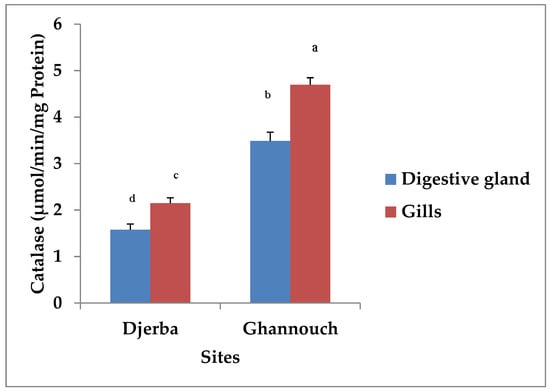
Figure 4.
Catalase activity (µmol/min/mg Protein) in digestive gland and gill tissues of P. segnis from different localities. Different letters indicate statistically significant differences.
These results can be explained by the fact that exposure to certain pollutants such as TMEs leads to an increase in the production of reactive oxygen species (ROS), such as the superoxide anion (O2•−), hydrogen peroxide (H2O2), and the hydroxyl radical (•OH), which can alter the defense system of aquatic organisms [75]. In such a situation, increases in certain antioxidant enzymes may occur. These enzymes participate in the neutralization of these ROS and thus in the reduction of the damage caused by the contaminants. In this respect, Dellali et al. [76] affirmed that the enzymatic activity of CAT increases after exposure to certain pollutants.
In addition, our results also show a significant difference in CAT activity between the two studied tissues. Indeed, it is higher in the gills than in the hepatopancreas, with activity levels of about 4.7 µmol H2O2/min/mg protein and 3.49 µmol H2O2/min/mg protein, respectively, in Ghannouch, and 2.15 µmol H2O2/min/mg protein and 1.58 µmol H2O2/min/mg protein, respectively, in Djerba. In fact, our results show a profile in the same direction between the enzymatic activity of CAT and the accumulation of heavy metals at the level of the gills. These results are in agreement with several other scientific studies, such as the study by Jerome et al. [77], which showed an increase in this activity in the gills of Callinectes amnicola crabs exposed to industrial effluents.
3.2.2. Glutathione S-Transferase Activity
Our results show the existence of a significant difference in GST activity, not only between the two studied sites but also between the two tissues examined in this study (Figure 5). GST showed important activity at the level of the gills compared to the hepatopancreas for the samples from Ghannouch (F = 20.87; p < 0.01) and Djerba (F = 37.21; p < 0.01). Between the two studied sites, GST activity was statistically more significant in the gills (F = 22.12; p < 0.01) and hepatopancreas (F = 37.21; p < 0.05).
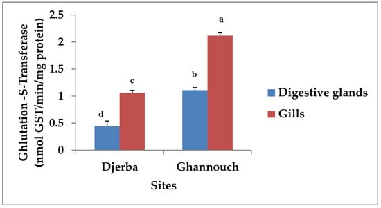
Figure 5.
Glutathione S-transferase (GST) activity (nmol GST/min/mg Protein) in P. segnis (n = 20) from Ghannouch and Djerba. Different letters indicate statistically significant differences.
The induction of GST enzymatic activity in the Ghannouch individuals is suggested to be a response to environmental stress caused by the contamination of the marine environment by heavy metals that are accumulated in crab tissues, particularly the gills. Indeed, our study shows an important activity of this enzyme in the individuals from the Ghannouch site, which present accumulations of Cd, Zn, Cu, and Pb at the level of the gills. Several studies have affirmed increases in the enzymatic activity of GST in crabs, fish, and mussels coming from sites contaminated by TMEs [65]. Furthermore, our data are in agreement with those of Barata et al. [64], who found an increase in GST activity in Daphnia magna following exposure to cadmium, and to those reported by Frías-Espericueta et al. [73] and by De Jesus et al. [74] for crab species exposed to metals.
The results of the present study can be partly explained by the bioavailability of heavy metals in the marine environment of Ghannouch, especially Cd, Zn, Cu, and Pb, which can generate disturbances that affect the homeostasis of individuals. To defend against and minimize this damage, an acceleration of the activities of certain enzymes, including GST, can protect the organisms and improve their overall health.
3.2.3. Malonedialdehyde Determination
The ANOVA results indicate an increase in MDA concentration in the crabs from Ghannouch. There is a significant difference between the two localities for both gill and hepatopancreas tissues, respectively (F = 54.22 and F = 42.51; p < 0.01) (Figure 6). Furthermore, according to our data for both sites, the concentration of MDA is higher in gills (Djerba: F = 51.46 and Ghannouch: F = 80.32; p < 0.01) than in the hepatopancreas. This increase in MDA levels was observed in individuals from the site with the highest levels of metals in the gills. In the same context, the studies of Bonneris et al. [78] confirm our results. They showed that following a 21-day exposure to Cd, MDA levels were elevated in the mussel M. edulis. This elevation was also demonstrated in Pyganodon grandis [79] and Bathymodiolus azoricus [80] captured from polluted environments and in crab species exposed to metals [73]. Therefore, we can suggest that an increase in MDA levels in individuals from Ghannouch is due to the bioavailability of heavy metals in the environment.
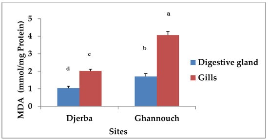
Figure 6.
Comparison of MDA levels (nmol/mg Protein) in digestive glands and gills of P. segnis captured from the studied sites. Means that do not share the same alphabetic sign are significantly different.
Indeed, MDA is a biomarker generated secondarily after lipid peroxidation caused by an alteration of the plasma membrane through the attack of polyunsaturated fatty acids. This membrane lipoperoxidation seems to depend on the organ that accumulates more metals [81]. Lipid peroxidation is followed by structural changes in biological membranes [80] or other elements containing lipids [82]. This leads to a loss of permeability and the inactivation of receptors and membrane enzymes [83]. These functional disturbances can lead to cell death, and lipid peroxidation is an endogenous source of DNA damage [63].
A previous study by Giarratano et al. [84] revealed an increase in MDA levels in Aulacomyaatra mussels after exposure to iron, aluminum, and cadmium. The same is true for the work of Fahmy et al. [85], who revealed the induction of increased MDA levels in the hemolymph and soft tissues of snails treated with zinc. Radwan et al. indicated in their study [86] that there is a positive correlation between heavy metals and lipid peroxidation due to the presence of metal ions in the digestive gland of snails that catalyze the Fenton reaction and increase the risk of cell damage.
3.2.4. Determination of Reduced Glutathione
The results obtained in Figure 7 show an increase in GSH concentration in teh crabs from Djerba. There is a significant difference between the two localities (p < 0.05) (Figure 7). In addition, according to our results, we notice that for both sites, the concentration of GSH is higher in the hepatopancreas than in the gills.
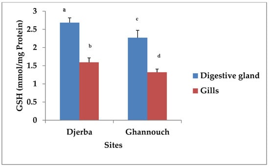
Figure 7.
Glutathione content (mmol/mg Protein) in digestive glands and gills tissues of P. segnis from the studied sites. Means that do not share the same alphabetic sign are significantly different.
Indeed, GSH is the main intracellular non-enzymatic antioxidant agent [87]. The enzyme that catalyzes the conjugation of GSH to a wide variety of endogenous and exogenous electrophilic compounds is GST, which possesses the capacity of detoxification, thus playing a role in cellular protection from oxidative stress [88]. The conjugation of metribuzin metabolites with GSH, followed by the conversion of mercapturic acid derivatives, seems to play a major role in detoxification and excretion [89]. GSH presents the majority state of glutathione in cells; an increase in the oxidized form (GSSG) of GSH indicates the presence of an oxidative stress state [90]. The transformation of oxidized glutathione into reduced glutathione is achieved via glutathione reductase (GR) to activate GPx [91]. GSH plays a multifactorial role in the antioxidant defense mechanism. It is a direct scavenger of free radicals and a necessary co-substrate for GPx and glutathione-s-transferase activity. Therefore, changes in GSH levels can be considered particularly sensitive indicators of oxidative stress [92].
The decrease in GSH levels at the Ghannouch site, which is the site with the highest trace element levels, is in agreement with several other previous studies [93,94,95] which showed that GSH, in addition to other biochemical markers, seems to be a potential antagonist against metals. According to El-Shenawy et al. [86], the decrease in the GSH level is attributed to the increased consumption of this peptide for the synthesis of heavy-metal-binding proteins such as metallothioneins. In addition, Singaram et al. [96] reported that the decrease in the GSH level is also due to the increased utilization of GSH by glutathione peroxidase (GPx) to catalyze the reduction of H2O2. Leomanni et al. [97] also showed that the early induction of GPx activity plays a main role in the anticipated decrease in GSH.
4. Conclusions
The aim of this paper was to estimate the consequences of pollution on an invasive crab species, Portunus segnis, which is widely distributed on the Tunisian coasts.
The results of the analyses revealed a significant difference between the two studied populations in terms of both the biochemical parameters studied and the concentrations of the various trace elements measured in two tissues: the gills and hepatopancreas.
Results of the metal analysis revealed that Portunus segnis accumulates metallic trace elements in a variable manner. The general order of heavy metal bioaccumulation analyzed in the gills and hepatopancreas tissues in our study is: Zn > Cd > Cu > Pb. The bioaccumulation by the gills (37.36 ± 12.57 vs. 10.72 ± 2.9 for Zn, 19.21 ± 0.1 vs. 7.2 ± 0.049 for Cd, 12.53 ± 0.078 vs. 6.45 ± 0.12 for Cu, and 10.15 ± 1.38 vs. 4.9 ± 0.01 for Pb) is greater than that of the hepatopancreas (18.14 ± 8.38 vs. 8.71 ± 1.03 for Zn, 7.74 ± 0.14 vs. 3.18 ± 0.04 for Cd, 6.96 ± 0.79 vs. 2.91 ± 0.08 for Cu, and 4.536 ± 0.24 vs. 2.21 ± 0.05 for Pb) for the studied sites, Ghannouch and Djerba, respectively. Finally, the crabs living near the industrial zone, Ghannouch, accumulated more heavy metals in their tissues (gills and hepatopancreas) than those obtained from Djerba, the area furthest from the source of pollution.
The study of biochemical biomarkers in the gills and hepatopancreata of crabs has shown that crabs from Ghannouch, the most polluted area, have higher levels of biomarkers compared to those from Djerba. Indeed, the enzymatic activities of catalase and glutathione-S-transferase as well as the contents of malonedialdehyde and reduced glutathione all present significant differences between the two studied stations.
The variations in the levels of the studied biomarkers reflect the alarming state of this ecosystem and suggest that Portunus segnis is setting up a relatively effective detoxification system following metal contamination. As a result, this species could be used as a bioindicator of pollution in biomonitoring programs to assess the quality of aquatic environments in the Golf of Gabès.
Author Contributions
Formal analysis, W.B.A. and A.A.; Methodology, A.D., W.B.A. and A.A.; Resources, A.A.; Supervision, A.A.; Validation, A.A. All authors have read and agreed to the published version of the manuscript.
Funding
The authors declare that no funds, grants, or other support were received during the preparation of this manuscript.
Institutional Review Board Statement
Not applicable.
Informed Consent Statement
Not applicable.
Data Availability Statement
Not applicable.
Acknowledgments
The Life Sciences Department of the Faculty of Sciences of Gabès, University of Gabès, provided the reagents and technical scientific support for which the authors are very thankful. The authorsalso acknowledge the support of the study from Tunisia’s Ministry of Higher Education and Scientific Research.
Conflicts of Interest
The authors declare no conflict of interest.
References
- Reddy, M.S.; Mehta, B.; Dave, S.; Joshi, M.; Karthikeyan, L.; Sarma, V.K.S.; Basha, S.; Ramachandraiah, G.; Bhatt, P. Bioaccumulation of heavy metals in some commercial fishes and crabs of the Gulf of Cambay. India Curr. Sci. 2007, 92, 1489–1491. [Google Scholar]
- Blaise, C.; Gagné, F.; Pellerin, J.; Hansen, P.-D.; Trottier, S. Molluscan Shellfish Biomarker Study of the Quebec, Canada, Saguenay Fjord with the Soft-Shell Clam, Mya arenaria: Molluscan Shellfish Biomarker StudyStudy. Environ. Toxicol. 2002, 17, 170–186. [Google Scholar] [CrossRef] [PubMed]
- Galloway, T.S.; Brown, R.J.; Browne, M.A.; Dissanayake, A.; Lowe, D.; Jones, M.B.; Depledge, M.H. A Multibiomarker Approach To Environmental Assessment. Environ. Sci. Technol. 2004, 38, 1723–1731. [Google Scholar] [CrossRef]
- Mayon, N.; Bertrand, A.; Leroy, D.; Malbrouck, C.; Mandiki, S.N.M.; Silvestre, F.; Goffart, A.; Thomé, J.-P.; Kestemont, P. Multiscale Approach of Fish Responses to Different Types of Environmental Contaminations: A Case Study. Sci. Total Environ. 2006, 367, 715–731. [Google Scholar] [CrossRef]
- UNEP/MAP. Draft Transboundary Diagnostic Analysis for the Mediterranean Sea, 1997 (TDA/MED) (UNEP(OCA)MED IG.ll/inf.7). Presented at MEDITERRANEAN ACTION PLAN Tenth Ordinary Meeting of the Contracting Parties to the Convention for the Protection of the Mediterranean Sea against Pollution and Its Protocols Tunis, Tunis, Tunisia, 18–21 November 1997. [Google Scholar]
- Hamza-Chaffai, A. Health Assessment of a Marine Bivalve Ruditapes decussatus from the Gulf of Gabès (Tunisia). Environ. Internat. 2003, 28, 609–617. [Google Scholar] [CrossRef]
- Smaoui-Damak, W.; Hamza-Chaffai, A.; Berthet, B.; Amiard, J.C. Preliminary Study of the Clam Ruditapes decussatus Exposed In Situ to Metal Contamination and Originating from the Gulf of Gabès, Tunisia. Bull. Environ. Contam Toxicol. 2003, 71, 961–970. [Google Scholar] [CrossRef] [PubMed]
- Rainbow, P.S. Trace Metal Concentrations in Aquatic Invertebrates: Why and so What? Environ. Pollut. 2002, 120, 497–507. [Google Scholar] [CrossRef]
- Fox, H.M. The Migration of a Red Sea Crab through the Suez Canal. Nature 1924, 113, 714–715. [Google Scholar] [CrossRef]
- Türeli, C.; Çelik, M.; Erdem, Ü. Comparison of meat composition and yield of blue crab (Callinectes sapidus RATHBUN, 1896) and sand crab (Portunus pelagicus Linne, 1758) caught in Iskenderun Bay, North-East Mediterranean. Turk. J. Vet. Anim. Sci. 2000, 24, 195–204. [Google Scholar]
- Rabaoui, L.; Arculeo, M.; Mansour, L.; Tlig-Zouari, S. Occurrence of the lessepsian species Portunus segnis (Crustacea: Decapda) in the Gulf of Gabés (Tunisia): First record and new information on its biology and ecology. Cah. Biol. Mar. 2015, 56, 169–175. [Google Scholar]
- Deidun, A.; Sciberras, A. A Further Record of the Blue Swimmer Crab Portunus segnis Forskal, 1775 (Decapoda: Brachyura: Portunidae) from the Maltese Islands (Central Mediterranean). BIR 2016, 5, 43–46. [Google Scholar] [CrossRef]
- Rifi, M.; Ounifi-BenAmor, K.; Ben Souissi, J.; Zaouali, J. Première mention du crabelessepsien Portunus segnis (Forskål, 1775) (Décapode, Brachyoure, Portunidae) dans les eaux marines Tunisiennes. In Proceedings of the du 4ème Congrès Franco-Maghrébin et 5èmes Journées Franco-Tunisiennes de Zoologie, Korba, Tunisie, 13–17 November 2014; p. 9. [Google Scholar]
- Bdioui, M. Premier signalement du crabe bleu Portunus segnis (Forskal, 1775) dans le Sud du Golfe de Hammamet (Centre-Est de la Tunisie). Bull. Inst. Natn. Scien. Tech. Mer de Salammbô. 2016, 43, 183–187. [Google Scholar]
- Ounifi-Ben Amor, K.; Rifi, Μ.; Ghanem, R.; Draeif, I.; Zaouali, J.; Ben Souissi, J. Update of Alien Fauna and New Records from Tunisian Marine Waters. Medit. Mar. Sci. 2015, 17, 124. [Google Scholar] [CrossRef]
- Burollet, P.F.; Clairefond, P.; Winnock, E. Géologie méditerranéenne, Tome VI, numéro 1. In La Mer Pélagienne: Etude Sédimentologique et écologique du Plateau Tunisien et du Golfe de Gabès; Département des sciences de la terre. Centre St Charles 13331 Marseille Cedex 3. Université de Provence: Marseille, France, 1979; p. 345. [Google Scholar]
- Hamza, A. Le Statut du Phytoplancton Dans le Golfe de Gabès. Ph.D. Thesis, Faculté des Sciences de Sfax, Sfax, Tunisie, 2003; p. 297. [Google Scholar]
- Drira, Z. Contribution à la Compréhension du Fonctionnement du Golfe de Gabès: Etude des Caractéristiques Dynamiques et Structurales des Communautés Phytozooplanctoniques. Ph.D. Thesis, Université de Sfax, Sfax, Tunisie, 2009; p. 229. [Google Scholar]
- Hattab, T.; Ben RaisLasram, F.; Albouy, C.; Romdhane, M.S.; Jarboui, O.; Halouani, G.; Cury, P.; Le Loch, F. An Ecosystem Model of an Exploited Southern Mediterranean Shelf Region (Gulf of Gabes, Tunisia) and a Comparison with Other Mediterranean Ecosystem Model Properties. J. Mar. Res. 2013, 128, 159–174. [Google Scholar] [CrossRef]
- Alaya-Ltifi, L.; Chokri, M.A.; Selmi, S. Breeding Performance of Passerines in a Polluted Oasis Habitat in Southern Tunisia. Ecotoxicol. Environ. Safe 2012, 79, 170–175. [Google Scholar] [CrossRef]
- Annabi, A.; Bardelli, R.; Vizzini, S.; Mancinelli, G. Baseline Assessment of Heavy Metals Content and Trophic Position of the Invasive Blue Swimming Crab Portunus segnis (Forskål, 1775) in the Gulf of Gabès (Tunisia). Mar. Pollut. Bull. 2018, 136, 454–463. [Google Scholar] [CrossRef]
- Bradford, M.M. A Rapid and Sensitive Method for the Quantitation of Microgram Quantities of Protein Utilizing the Principle of Protein-Dye Binding. Anal. Biochem. 1976, 72, 248–254. [Google Scholar] [CrossRef]
- Almeida, J.R.; Gravato, C.; Guilhermino, L. Challenges in Assessing the Toxic Effects of Polycyclic Aromatic Hydrocarbons to Marine Organisms: A Case Study on the Acute Toxicity of Pyrene to the European Seabass (Dicentrarchus labrax L.). Chemosphere 2012, 86, 926–937. [Google Scholar] [CrossRef]
- Habig, W.H.; Pabst, M.J.; Jakoby, W.B. Glutathione S-Transferases. J. Biol. Chem. 1974, 249, 7130–7139. [Google Scholar] [CrossRef]
- Turdi, S.; Li, Q.; Lopez, F.L.; Ren, J. Catalase Alleviates Cardiomyocyte Dysfunction in Diabetes: Role of Akt, Forkhead Transcriptional Factor and Silent Information Regulator 2. Life Sci. 2007, 81, 895–905. [Google Scholar] [CrossRef]
- Aebi, H. Catalase in vitro. Methods Enzymol. 1984, 105, 121–126. [Google Scholar] [CrossRef] [PubMed]
- Ellman, G.L. Tissue Sulfhydryl Groups. Arch. Biochem. Biophys. 1959, 82, 70–77. [Google Scholar] [CrossRef]
- Ohkawa, H.; Ohishi, N.; Yagi, K. Assay for Lipid Peroxides in Animal Tissues by Thiobarbituric Acid Reaction. Anal. Biochem. 1979, 95, 351–358. [Google Scholar] [CrossRef] [PubMed]
- Catalano, D.; Torchia, G.; Pititto, F.; Greco, R. Contaminazione da metalli del “granchio americano”, Portunus pelagicus (Linnaeus, 1758) nella Rada di Augusta (Sicilia orientale). Biol. Mar. Mediterr. 2006, 13, 696–699. [Google Scholar]
- Ayas, D. The Effects of Season and Sex on the Nutritional Quality of Muscle Types of Blue Crab Callinectes sapidus (Rathbun, 1896) and Swimming Crab Portunus segnis (Forskal, 1775). NESciences 2016, 1, 1–14. [Google Scholar] [CrossRef]
- Olgunoglu, M.; Olgunoglu, İ. Heavy Metal Contents in Blue Swimming Crab from the Northeastern Mediterranean Sea, Mersin Bay, Turkey. Pol. J. Environ. Stud. 2016, 25, 2233–2237. [Google Scholar] [CrossRef]
- Rainbow, P.S.; Furness, R.W. Heavy Metals in the Marine Environment. In Heavy Metals in the Marine Environment; Furness, R.W., Rainbow, P.S., Eds.; CRC Press: Boca Raton, FL, USA, 2018; pp. 1–4. ISBN 978135107315831. [Google Scholar]
- Gutiérrez, A.J.; Lozano, G.; Rubio, C.; Martín, V.; Hardisson, A.; Revert, C. Heavy Metals in Black Crabs in the Atlantic Coast (Tenerife, Spain)—Human Risk Assessment: Water. Clean Soil Air Water 2017, 45. [Google Scholar] [CrossRef]
- Nędzarek, A.; Czerniejewski, P.; Drost, A.; Harasimiuk, F.; Machula, S.; Tórz, A.; Masalski, P. The Distribution of Elements in the Body of Invasive Chinese Mitten Crabs (Eriocheir sinensis H. Milne-Edwards, 1853) from Lake Dąbie, Poland. J. Food Compos. Anal. 2017, 60, 1–9. [Google Scholar] [CrossRef]
- Turoczy, N.J.; Mitchell, B.D.; Levings, A.H.; Rajendram, V.S. Cadmium, Copper, Mercury, and Zinc Concentrations in Tissues of the King Crab (Pseudocarcinus gigas) from Southeast Australian Waters. Environ. Int. 2001, 27, 327–334. [Google Scholar] [CrossRef] [PubMed]
- Reichmuth, J.M.; Weis, P.; Weis, J.S. Bioaccumulation and Depuration of Metals in Blue Crabs (Callinectes sapidus Rathbun) from a Contaminated and Clean Estuary. Environ. Pollut. 2010, 158, 361–368. [Google Scholar] [CrossRef] [PubMed]
- Bondgaard, M.; Bjerregaard, P. Association between Cadmium and Calcium Uptake and Distribution during the Moult Cycle of Female Shore Crabs, Carcinus maenas: An In Vivo Study. Aquat. Toxicol. 2005, 72, 17–28. [Google Scholar] [CrossRef]
- Martín-Díaz, M.L.; Blasco, J.; Sales, D.; DelValls, T.A. Biomarkers Study for Sediment Quality Assessment in Spanish Ports Using the Crab Carcinus maenas and the Clam Ruditapes philippinarum. Arch. Environ. Contam Toxicol. 2007, 53, 66–76. [Google Scholar] [CrossRef]
- GB 5749-2006; Standards for Drinking Water Quality. Ministry of Health of the People’s Republic of China: Beijing, China, 2006.
- European Commission. Commission Regulation (EC) No 1881/2006 of the European parliament and the council of 19 December 2006 setting maximum levels for certain contaminants in foodstuffs. Off. J. Eur. Communities 2006, 18, L364. [Google Scholar]
- FAO. Compilation of legal limits for hazardous substances in fish and fishery products. FAO Fish Circ. 1983, 464, 5–100. Available online: http://trove.nla.gov.au/version/22206109 (accessed on 15 April 2023).
- Baden, S.P.; Eriksson, S.P. Role, routes and effects of manganese in crustaceans. Oceanogr. Mar. Biol. Annu. Rev. 2006, 44, 61–83. [Google Scholar]
- Mancinelli, G.; Papadia, P.; Ludovisi, A.; Migoni, D.; Bardelli, R.; Fanizzi, F.P.; Vizzini, S. Beyond the Mean: A Comparison of Trace- and Macroelement Correlation Profiles of Two Lacustrine Populations of the Crayfish Procambarus clarkii. Sci. Total Environ. 2018, 624, 1455–1466. [Google Scholar] [CrossRef]
- Amiard, J.; Amiardtriquet, C.; Barka, S.; Pellerin, J.; Rainbow, P. Metallothioneins in Aquatic Invertebrates: Their Role in Metal Detoxification and Their Use as Biomarkers. Aquat. Toxicol. 2006, 76, 160–202. [Google Scholar] [CrossRef]
- Schuwerack, P.-M.M.; Lewis, J.W.; Jones, P. The potential use of the South African river crab, Potamonauteswarreni, as a bioindicator species for heavy metal contamination. Ecotoxicology 2001, 10, 159–166. [Google Scholar] [CrossRef]
- Pan, L.; Zhang, H. Metallothionein, Antioxidant Enzymes and DNA Strand Breaks as Biomarkers of Cd Exposure in a Marine Crab, Charybdis Japonica. Comp. Biochem. Physiol. Part C Toxicol. Pharmacol. 2006, 144, 67–75. [Google Scholar] [CrossRef] [PubMed]
- Corrêa, J.D.; Da Silva, M.R.; Da Silva, A.C.B.; De Lima, S.M.A.; Malm, O.; Allodi, S. Tissue Distribution, Subcellular Localization and Endocrine Disruption Patterns Induced by Cr and Mn in the Crab Ucides Cordatus. Aquat. Toxicol. 2005, 73, 139–154. [Google Scholar] [CrossRef] [PubMed]
- Rainbow, P.S. Biomonitoring of trace metals in estuarine and marine environments. Aust. J. Ecotoxicol. 2006, 12, 107–122. [Google Scholar]
- Doherty, V.F.; Ogunkuade, O.O.; Kanife, U.C. Biomarkers of oxidative stress and heavy metal levels as indicators of environmental pollution in some selected fishes in Lagos, Nigeria. Am. Eurasian J. Agric. Envrion. Sci. 2010, 7, 359–365. [Google Scholar]
- Bjerregaard, P.; Depledge, M.H. Trace Metal Concentrations and Contents in the Tissues of the Shore Crab Carcinus maenas: Effects of Size and Tissue Hydration. Mar. Biol. 2002, 141, 741–752. [Google Scholar] [CrossRef]
- Peng, S.-H.; Hung, J.-J.; Hwang, J.-S. Bioaccumulation of Trace Metals in the Submarine Hydrothermal Vent Crab Xenograpsus testudinatus off Kueishan Island, Taiwan. Mar. Pollut. Bull. 2011, 63, 396–401. [Google Scholar] [CrossRef] [PubMed]
- Ghedira, J.; Jebali, J.; Banni, M.; Chouba, L.; Boussetta, H.; López-Barea, J.; Alhama, J. Use of Oxidative Stress Biomarkers in Carcinus Maenas to Assess Littoral Zone Contamination in Tunisia. Aquat. Biol. 2011, 14, 87–98. [Google Scholar] [CrossRef]
- Ben-Khedher, S.; Jebali, J.; Houas, Z.; Nawéli, H.; Jrad, A.; Banni, M.; Boussetta, H. Metals Bioaccumulation and Histopathological Biomarkers in Carcinus maenas Crab from Bizerta Lagoon, Tunisia. Envrion. Sci. Pollut. Res. 2014, 21, 4343–4357. [Google Scholar] [CrossRef] [PubMed]
- Mortimer, M.R. Pesticide and Trace Metal Concentrations in Queensland Estuarine Crabs. Mar. Pollut. Bull. 2000, 41, 359–366. [Google Scholar] [CrossRef]
- Reed, L.A.; Pennington, P.L.; Wirth, E. A Survey of Trace Element Distribution in Tissues of Stone Crabs (Menippe mercenaria) from South Carolina Coastal Waters. Mar. Pollut. Bull. 2010, 60, 2297–2302. [Google Scholar] [CrossRef]
- Pinheiro, M.A.A.; Silva, P.P.G.E.; Duarte, L.F.D.A.; Almeida, A.A.; Zanotto, F.P. Accumulation of Six Metals in the Mangrove Crab UcidesCordatus (Crustacea: Ucididae) and Its Food Source, the Red Mangrove Rhizophora Mangle (Angiosperma: Rhizophoraceae). Ecotoxicol. Environ. Safe 2012, 81, 114–121. [Google Scholar] [CrossRef]
- Bonanno, G.; Orlando-Bonaca, M. Trace Elements in Mediterranean Seagrasses: Accumulation, Tolerance and Biomonitoring. A Review. Mar. Pollut. Bull. 2017, 125, 8–18. [Google Scholar] [CrossRef]
- Ayadi, N.; Aloulou, F.; Bouzid, J. Assessment of Contaminated Sediment by Phosphate Fertilizer Industrial Waste Using Pollution Indices and Statistical Techniques in the Gulf of Gabes (Tunisia). Arab. J. Geosci. 2015, 8, 1755–1767. [Google Scholar] [CrossRef]
- El Zrelli, R.; Courjault-Radé, P.; Rabaoui, L.; Castet, S.; Michel, S.; Bejaoui, N. Heavy Metal Contamination and Ecological Risk Assessment in the Surface Sediments of the Coastal Area Surrounding the Industrial Complex of Gabes City, Gulf of Gabes, SE Tunisia. Mar. Pollut. Bull. 2015, 101, 922–929. [Google Scholar] [CrossRef] [PubMed]
- Jara-Marini, M.E.; Soto-Jiménez, M.F.; Páez-Osuna, F. Trophic Relationships and Transference of Cadmium, Copper, Lead and Zinc in a Subtropical Coastal Lagoon Food Web from SE Gulf of California. Chemosphere 2009, 77, 1366–1373. [Google Scholar] [CrossRef]
- Schneider, L.; Maher, W.A.; Potts, J.; Taylor, A.M.; Batley, G.E.; Krikowa, F.; Adamack, A.; Chariton, A.A.; Gruber, B. Trophic Transfer of Metals in a Seagrass Food Web: Bioaccumulation of Essential and Non-Essential Metals. Mar. Pollut. Bull. 2018, 131, 468–480. [Google Scholar] [CrossRef] [PubMed]
- El-Shenawy, N.S.; Mohammadden, A.; Al-Fahmie, Z.H. Using the Enzymatic and Non-Enzymatic Antioxidant Defense System of the Land Snail Eobania vermiculata as Biomarkers of Terrestrial Heavy Metal Pollution. Ecotoxicol. Environ. Safe 2012, 84, 347–354. [Google Scholar] [CrossRef]
- Marnett, L.J. Oxy Radicals, Lipid Peroxidation and DNA Damage. Toxicology 2002, 181–182, 219–222. [Google Scholar] [CrossRef]
- Barata, C.; Varo, I.; Navarro, J.C.; Arun, S.; Porte, C. Antioxidant Enzyme Activities and Lipid Peroxidation in the Freshwater Cladoceran daphnia magna Exposed to Redox Cycling Compounds. Comp. Biochem. Physiol. Part C Toxicol. Pharmacol. 2005, 140, 175–186. [Google Scholar] [CrossRef]
- Stegeman, J.J.; Marius, B.; Giulio, R.T.D.; Lars, F.; Fowler, B.A.; Sanders, B.Μ.; Veld, P.A.V. Molecular responses to environmental contamination: Enzyme and protein systems as indicators of chemical exposure and effect. In Biomarkers. Biochemical, Physiological and Histological Markers of Anthropogenic Stress; Huggett, R.J., Kimerle, R.A., Mehrle, P.M., Jr., Bergman, H.L., Eds.; Lewis Publishers: London, UK, 1992; pp. 235–335. [Google Scholar]
- Karam, Q.; Guermazi, W.; Subrahmanyam, M.N.V.; Al-Enezi, Y.; Ali, M.; Leignel, V.; Annabi-Trabelsi, N. Portunus pelagicus (Linnaeus, 1758) as a Sentinel Species to Assess Trace Metal Occurrence: A Case Study of Kuwait Waters (Northwestern Arabian Gulf). Toxics 2023, 11, 426. [Google Scholar] [CrossRef]
- Majola, N.; Mzimela, H.M.; Izegaegbe, J.I. Metal Bioaccumulation and Energy Biomarkers in Tissues of Two Populations of Chiromantes Eulimene from Richards Bay Harbour, South Africa. Sci. Afr. 2020, 10, e00558. [Google Scholar] [CrossRef]
- Yogeshwaran, A.; Gayathiri, K.; Muralisankar, T.; Gayathri, V.; Monica, J.I.; Rajaram, R.; Marimuthu, K.; Bhavan, P.S. Bioaccumulation of Heavy Metals, Antioxidants, and Metabolic Enzymes in the Crab Scylla serrata from Different Regions of Tuticorin, Southeast Coast of India. Mar. Pollut. Bull. 2020, 158, 111443. [Google Scholar] [CrossRef]
- Karar, S.; Hazra, S.; Das, S. Assessment of the Heavy Metal Accumulation in the Blue Swimmer Crab (Portunus pelagicus), Northern Bay of Bengal: Role of Salinity. Mar. Pollut. Bull. 2019, 143, 101–108. [Google Scholar] [CrossRef] [PubMed]
- Ngo-Massou, V.M.; Kottè-Mapoko, E.F.; Din, N. Heavy Metal Accumulation in the Edible Crab Cardisoma armatum (Brachyura: Gecarcinidae) and Implications for Human Health Risks. Sci. Afr. 2022, 16, e01248. [Google Scholar] [CrossRef]
- Liu, Q.; Liao, Y.; Xu, X.; Shi, X.; Zeng, J.; Chen, Q.; Shou, L. Heavy Metal Concentrations in Tissues of Marine Fish and Crab Collected from the Middle Coast of Zhejiang Province, China. Environ. Monit. Assess 2020, 192, 285. [Google Scholar] [CrossRef] [PubMed]
- Jebali, J.; Banni, M.; Almeida, E.A.D.; Boussetta, H. Oxidative DNA Damage Levels and Catalase Activity in the Clam Ruditapes Decussatus as Pollution Biomarkers of Tunisian Marine Environment. Environ. Monit. Assess 2007, 124, 195–200. [Google Scholar] [CrossRef]
- Frías-Espericueta, M.G.; Bautista-Covarrubias, J.C.; Osuna-Martínez, C.C.; Delgado-Alvarez, C.; Bojórquez, C.; Aguilar-Juárez, M.; Roos-Muñoz, S.; Osuna-López, I.; Páez-Osuna, F. Metals and Oxidative Stress in Aquatic Decapod Crustaceans: A Review with Special Reference to Shrimp and Crabs. Aquat. Toxicol. 2022, 242, 106024. [Google Scholar] [CrossRef]
- De Jesus, W.B.; Mota Andrade, T.D.S.D.O.; Soares, S.H.; Pinheiro-Sousa, D.B.; De Oliveira, S.R.S.; Torres, H.S.; Protazio, G.D.S.; Da Silva, D.S.; Santos, D.M.S.; De Carvalho Neta, A.V.; et al. Biomarkers and Occurrences of Heavy Metals in Sediment and the Bioaccumulation of Metals in Crabs (Ucides cordatus) in Impacted Mangroves on the Amazon Coast, Brazil. Chemosphere 2021, 271, 129444. [Google Scholar] [CrossRef]
- Khessiba, P.; Hoarau, M.; Gnassia-Bar, A. Biochemical Response of the Mussel Mytilus galloprovincialis from Bizerta (Tunisia) to Chemical Pollutant Exposure. Arch. Environ. Contam. Toxicol. 2001, 40, 222–229. [Google Scholar] [CrossRef]
- Dellali, M.; GnassiaBarelli, M.; Romeo, M.; Aissa, P. The Use of Acetylcholinesterase Activity in Ruditapes Decussatus and Mytilus Galloprovincialis in the Biomonitoring of Bizerta Lagoon. Comp. Biochem. Physiol. Part C Toxicol. Pharmacol. 2001, 130, 227–235. [Google Scholar] [CrossRef]
- Jerome, F.C.; Hassan, A.; Omoniyi-Esan, G.O.; Odujoko, O.O.; Chukwuka, A.V. Metal Uptake, Oxidative Stress and Histopathological Alterations in Gills and Hepatopancreas of Callinectes amnicola Exposed to Industrial Effluent. Ecotoxicol. Environ. Safe 2017, 139, 179–193. [Google Scholar] [CrossRef]
- Géret, F. Influence of Metal Exposure on Metallothionein Synthesis and Lipid Peroxidation in Two Bivalve Mollusks: The Oyster (Crassostrea gigas) and the Mussel (Mytilus edulis). Aquat. Living Resour. 2002, 15, 61–66. [Google Scholar] [CrossRef]
- Bonneris, E.; Perceval, O.; Masson, S.; Hare, L.; Campbell, P.G.C. Sub-Cellular Partitioning of Cd, Cu and Zn in Tissues of Indigenous Unionid Bivalves Living along a Metal Exposure Gradient and Links to Metal-Induced Effects. Environ. Pollut. 2005, 135, 195–208. [Google Scholar] [CrossRef] [PubMed]
- Bebianno, M.; Company, R.; Serafim, A.; Camus, L.; Cosson, R.; Fialamedoni, A. Antioxidant Systems and Lipid Peroxidation in from Mid-Atlantic Ridge Hydrothermal Vent Fields. Aquat. Toxicol. 2005, 75, 354–373. [Google Scholar] [CrossRef] [PubMed]
- Ferrat, L.; Pergent-Martini, C.; Roméo, M. Assessment of the Use of Biomarkers in Aquatic Plants for the Evaluation of Environmental Quality: Application to Seagrasses. Aquat. Toxicol. 2003, 65, 187–204. [Google Scholar] [CrossRef] [PubMed]
- Al-Mutairi, D.A.; Craik, J.D.; Batinic-Haberle, I.; Benov, L.T. Induction of Oxidative Cell Damage by Photo-Treatment with Zinc MetaN -Methylpyridylporphyrin. Free Radic. Res. 2007, 41, 89–96. [Google Scholar] [CrossRef] [PubMed]
- Pampanin, D.M.; Camus, L.; Gomiero, A.; Marangon, I.; Volpato, E.; Nasci, C. Susceptibility to Oxidative Stress of Mussels (Mytilus galloprovincialis) in the Venice Lagoon (Italy). Mar. Pollut. Bull. 2005, 50, 1548–1557. [Google Scholar] [CrossRef] [PubMed]
- Giarratano, E.; Gil, M.N.; Malanga, G. Biomarkers of Environmental Stress in Gills of Ribbed Mussel Aulacomya atra atra (Nuevo Gulf, Northern Patagonia). Ecotoxicol. Environ. Safe 2014, 107, 111–119. [Google Scholar] [CrossRef]
- Fahmy, S.R.; Abdel-Ghaffar, F.; Bakry, F.A.; Sayed, D.A. Ecotoxicological Effect of Sublethal Exposure to Zinc Oxide Nanoparticles on Freshwater Snail Biomphalaria alexandrina. Arch. Environ. Contam. Toxicol. 2014, 67, 192–202. [Google Scholar] [CrossRef]
- Radwan, M.A.; El-Gendy, K.S.; Gad, A.F. Biomarkers of Oxidative Stress in the Land Snail, Theba Pisana for Assessing Ecotoxicological Effects of Urban Metal Pollution. Chemosphere 2010, 79, 40–46. [Google Scholar] [CrossRef]
- Jacob, L. L’insuffisance Rénale Aiguë; Springer: Paris, France, 2007; pp. 1–342. [Google Scholar] [CrossRef]
- Schwab, M. Encyclopedia of Cancer; Springer: Berlin/Heidelberg, Germany, 2011. [Google Scholar] [CrossRef]
- Morgan, J. Evidence on the Developmental and Reproductive Toxicity of Metribuzin. DRAFT, California, 2001; p. 65. Available online: https://oehha.ca.gov/media/downloads/crnr/metribuzin.pdf (accessed on 15 April 2023).
- Pelletier, É.; Campbell, P.G.C.; Denizeau, F. Écotoxicologie Moléculaire: Principes Fondamentaux et Perspectives de Développement, 1st ed.; Presses de l’Université du Québec: Québec, QC, Canada, 2004; pp. 1–502. [Google Scholar] [CrossRef]
- Poortmans, J.R.; Boisseau, N. Biochimie des Activités Physiques, 2nd ed.; De BoeckSupérieur: Bruxelles, Belgium, 2003; p. 480. [Google Scholar]
- Taleb-Senouci, D.; Ghomari, H.; Krouf, D.; Bouderbala, S.; Prost, J.; Lacaille-Dubois, M.A.; Bouchenak, M. Antioxidant Effect of Ajuga Iva Aqueous Extract in Streptozotocin-Induced Diabetic Rats. Phytomedicine 2009, 16, 623–631. [Google Scholar] [CrossRef]
- Haq, Q.; Jan, A.; Ali, A. Glutathione as an Antioxidant in Inorganic Mercury Induced Nephrotoxicity. J. Postgrad. Med. 2011, 57, 72. [Google Scholar] [CrossRef]
- Jozefczak, M.; Remans, T.; Vangronsveld, J.; Cuypers, A. Glutathione Is a Key Player in Metal-Induced Oxidative Stress Defenses. Int. J. Mol. Sci. 2012, 13, 3145–3175. [Google Scholar] [CrossRef] [PubMed]
- Hore, M.; Saha, R.; Bhaskar, S.; Mandal, S.; Bhattacharyya, S.; Roy, S. Oxidative Stress Responses in Puntius sarana Collected from Some Environmentally Contaminated Areas of River Mahananda, Malda, West Bengal. Ecotoxicology 2023, 32, 211–222. [Google Scholar] [CrossRef] [PubMed]
- Singaram, G.; Harikrishnan, T.; Chen, F.-Y.; Bo, J.; Giesy, J.P. Modulation of Immune-Associated Parameters and Antioxidant Responses in the Crab (Scylla serrata) Exposed to Mercury. Chemosphere 2013, 90, 917–928. [Google Scholar] [CrossRef] [PubMed]
- Leomanni, A.; Schettino, T.; Calisi, A.; Gorbi, S.; Mezzelani, M.; Regoli, F.; Lionetto, M.G. Antioxidant and Oxidative Stress Related Responses in the Mediterranean Land Snail Cantareus Apertus Exposed to the Carbamate Pesticide Carbaryl. Comp. Biochem. Physiol. Part C Toxicol. Pharmacol. 2015, 168, 20–27. [Google Scholar] [CrossRef] [PubMed]
Disclaimer/Publisher’s Note: The statements, opinions and data contained in all publications are solely those of the individual author(s) and contributor(s) and not of MDPI and/or the editor(s). MDPI and/or the editor(s) disclaim responsibility for any injury to people or property resulting from any ideas, methods, instructions or products referred to in the content. |
© 2023 by the authors. Licensee MDPI, Basel, Switzerland. This article is an open access article distributed under the terms and conditions of the Creative Commons Attribution (CC BY) license (https://creativecommons.org/licenses/by/4.0/).