Article

Anti-Melanogenesis Effect of Rosa rugosa on α-MSH-Induced B16F10 Cells via PKA/CREB Pathway Activation

1
Department of Bioscience, Silla University, Busan 46958, Republic of Korea
2
Collaborative Innovation Center for Child Nutrition and Health Development, Chongqing University of Education, Chongqing 400067, China
3
Marine Biotechnology Center for Pharmaceuticals and Foods, Silla University, Busan 46958, Republic of Korea
4
Nutritional Education, Graduate School of Education, Silla University, Busan 46958, Republic of Korea
5
Department of Food and Nutrition, Silla University, Busan 46958, Republic of Korea
6
Division of Convergence on Marine Science, Korea Maritime and Ocean University, Busan 49112, Republic of Korea
*
Author to whom correspondence should be addressed.
Appl. Sci. 2023, 13(1), 184; https://doi.org/10.3390/app13010184
Submission received: 26 November 2022 / Revised: 20 December 2022 / Accepted: 20 December 2022 / Published: 23 December 2022

Abstract

:
Melanin protects the skin against UV damage, whereas excessive melanin accumulation causes abnormal pigmentation and even melanoma. It has been reported that Rosa rugosa exhibits antioxidant, anti-bacterial, anti-tumor, and anti-inflammation activities. The current study evaluated the melanogenesis-suppressing effect of R. rugosa extract and its solvent fractions (H2O, n-BuOH, 85% aq. MeOH, and n-hexane). The effect of R. rugosa on the extra/intra-cellular melanin and intracellular active tyrosinase levels, melanogenesis-related gene and protein expression, and PKA/CREB signaling pathway activation was investigated in α-MSH-induced B16F10 cells. The results showed that R. rugosa effectively suppressed melanin secretion and tyrosinase activity at non-cytotoxic concentrations. R. rugosa extract down-regulated the melanogenesis-related expression of genes and proteins of tyrosinase, microphthalmia-associated transcription factor (MITF), tyrosinase-related protein (TRP)-1, and TRP-2. Furthermore, R. rugosa effectively inhibited the phosphorylation of the PKA/CREB proteins. Finally, the total polyphenol content of R. rugosa crude extract and its H2O, n-BuOH, 85% aq. MeOH, and n-hexane solvent fractions were 1383.9 ± 44.5, 2004.7 ± 43.4, 7270.3 ± 54.5, 2064.1 ± 34.8, and 1091.1 ± 26.2 mg gallic acid equivalent/100 g extract, respectively. The anti-melanogenesis effect of R. rugosa was suggested to be exhibited by downregulating the PKA/CREB signaling pathway potentially due to a high content of polyphenols. Overall, R. rugosa crude extract and its solvent fractions could be considered sources of bioactive ingredients that can be used against hyperpigmentation.

1. Introduction

Melanin is the common name of pigments found in human skin. It is known to influence the skin color, eyes, and hair. It is known that melanin can protect the skin from UV radiation, while large amounts of melanin excretion cause abnormal skin pigmentation, freckles, melasma, and even skin melanoma [1]. Melanocytes are differentiated cells located mainly in the basal layer of the skin. They produce melanin in a specific organelle called the melanosome. Melanosomes provide a place for melanogenesis which is the name of the biochemical process to synthesize melanin pigments [2].
Tyrosinase is an oxidoreductase and the rate-limiting enzyme in melanin synthesis where it catalyzes the conversion of tyrosine to L-3,4-dihydroxyphenylalanine (DOPA). At the next step dopaquinone is formed via the oxidation of DOPA by tyrosinase [3]. Further down the process, the dopaquinone is further catalyzed to yield melanin in the presence of tyrosinase-related protein 1 (TRP-1) and TRP-2 [4]. The transcriptional regulation of the melanogenic genes and proteins such as tyrosinase and TRPs is mainly carried out by the microphthalmia-associated transcription factor (MITF) [5]. It has been reported that the cyclic adenosine monophosphate-dependent protein kinase A (PKA) and its downstream effector cyclic adenosine monophosphate response element-binding protein (CREB) are the main regulatory proteins in inducing melanogenesis [6]. Melanogenesis is influenced by several internal and external factors, namely, hormonal signaling, inflammatory cytokines, and UV irradiation. Although different mechanisms induce melanin synthesis, α-melanocyte-stimulating hormone (α-MSH) is one of the common downstream effectors and has been verified to stimulate melanin secretion in the presence of several stimuli [7]. Under the stimulation of α-MSH, the increased intracellular cAMP levels subsequently activate the cAMP-dependent protein kinase (PKA) via phosphorylation [8]. Next, the phosphorylated PKA activates CREB, which then causes an increased level of MITF transcription. Overexpressed MITF further stimulates the production of tyrosinase, TRP-1, and TRP-2 [9].
Excessive melanogenesis-induced pigmentation and pigmentary disorders have been focused on continually, and tyrosinase inhibitors have been widely studied as anti-melanogenic therapeutics all over the world [10]. Several chemical regents have been verified to possess anti-melanogenesis effects [11]. However, long-term use of these regents causes serious side effects including inflammation, cytotoxicity, skin irritation, and itchiness [12]. Therefore, finding and developing natural, safe, and effective whitening agents from natural sources is essential.
Rosa rugosa, also called Japanese or rugosa rose, is a plant originally from Asia [13]. Previous studies reported that R. rugosa exhibits antioxidant, anti-bacterial, anti-tumor, and anti-inflammation activities [14,15,16]. Different parts of R. rugosa possess various activities due to their rich amounts of phenolic compounds, terpenoids, tannins, or essential oils [17]. The R. rugosa seed oil has been reported to suppress the migration of acute monocytic leukemia THP-1 and lung adenocarcinoma A549 cells and reduce ROS formation [18]. In addition, it has been verified that polysaccharides from Rosa rugosa petals showed strong activities in radical scavenging and moisture retention in vitro [19]. Nevertheless, research on the whitening effect of R. rugosa in the literature is scarce. For the above reasons, the current research evaluated the anti-melanogenesis activity of R. rugosa crude extract and its fractions obtained through solvent fractionation (H2O, n-BuOH, 85% aq. MeOH, n-hexane). Anti-melanogenic properties were determined in B16F10 melanoma-like cells with α-MSH-induced melanogenesis Also, the potential anti-melanogenesis action mechanism of R. rugosa was explored by detecting the PKA/CREB signaling pathway.

2. Materials and Methods

2.1. Materials

The R. rugosa samples were kindly provided by the Division of Marine Bioscience, Korea Maritime and Ocean University (Busan, Korea). Briefly, the crude extract was initially fractionated by dissolving it in CH2Cl2 and adding H2O. The CH2Cl2 layer was then separated between n-hexane and 85% aq. MeOH while the aqueous layer was fractionated using n-BuOH and H2O to obtain different solvent fractions of R. rugosa crude extract. Dulbecco’s Modified Eagle Medium (DMEM), fetal bovine serum (FBS), L-glutamine solution, antibiotic solution (penicillin and streptomycin), phosphate-buffered saline (PBS), RIPA reagent, and QIAzol lysis reagent were obtained from Thermo Fisher Scientific (Waltham, MA, USA). L-3,4-dihydroxyphenylalanine(L-DOPA), 5-hydroxy-2-hydroxymethyl-4H-pyranone (kojic acid), α-melanocyte-stimulating hormone (α-MSH), and (3-4-5-Dimethylthiazol-2-yl)-2-5-diphenyl tetrazolium bromide (MTT) were purchased from Sigma-Aldrich (St. Louis, MO, USA). The primers used in the present experiment were synthesized by Bioneer (Daejeon, Korea). Antibodies recognizing tyrosinase (sc-20035), TRP-1(sc-166857), TRP-2 (sc-166716), and β-actin (sc-47778) were purchased from Santa Cruz Biotechnology (Dallas, TX, USA), and MITF (#978000), PKA (#4781), p-PKA (#9196), CREB (#9104), p-CREB (#9198), goat anti-rabbit IgG, HRP (#7076), and goat anti-mouse IgG, HRP (#7074) were obtained from Cell Signaling Technology (Danvers, MA, USA).

2.2. Cell Culture

A murine melanoma cell line (B16F10) was purchased from Korea Cell Line Bank (Seoul, Korea). During experiments cells were fed with DMEM containing 10% fetal bovine serum and 1% (m/v) antibiotic solution. The incubation condition was 5% CO2, 37 °C.

2.3. Cytotoxicity Assay

B16F10 cells were cultured in culture plates (transparent flat bottom 96-well) at an amount of 1.0 × 10⁴ cells/well and incubated for 24 h. Then, the cells were treated for 24 h with different doses of crude extract and solvent fractions (5, 10, and 20 μg/mL). After 24 h, the medium was removed and subsequently 100 μg/mL MTT solution was added to each well and the plates were incubated for 4 h. As the last step, 100 μL DMSO was added to each well after removing the MTT solution, and the optical density of each well was determined with a microplate reader at 560 nm (Multiskan GO, Tecan Austria GmbH, Grodig, Austria).

2.4. Intracellular Active Tyrosinase and Melanin Levels

Cells at a density of 3.4 × 10⁴ cells/well were cultured in 6-well plates. Depending on α-MSH stimulation and sample treatment, treatment groups were established as follows: blank group (B): untreated, unstimulated; control group (C): untreated stimulated with 100 nM α-MSH; kojic acid group (KA), stimulated with 100 nM α-MSH and treated with 10 μM kojic acid; crude extract and fractions: stimulated with 100 nM α-MSH and treated with corresponding sample at given concentration (5, 10, and 20 μg/mL). Fractions were obtained by separation of crude extract depending on its solubility in different solvents and named according to fractionating solvent: distilled water (H2O), butanol (n-BuOH), 85% aqueous methanol (85% aq. MeOH), and hexane (n-hexane). Plates were incubated for 24 h, and after the incubation period, melanogenesis in cells was stimulated by treating cells with 100 nM α-MSH for 24 h [20]. Subsequently cells were introduced to different concentrations of Rosa rugosa crude extract and its solvent fractions for a further 24 h. After treatment, wells were washed twice with 1×PBS, and 200 μL lysis buffer was added to each well. Plates then were kept at 4 °C for 30 min to induce lysis. Then the lysate was harvested and separated by centrifugation at 13,000 rpm for 10 min, and the supernatant was used to determine the intracellular tyrosinase activity, while the remaining cell pellet was used to detect melanin content. For active tyrosine levels, the protein content of the supernatant was determined using Bio-Rad Protein Assay Dye Reagent Concentrate (Bio-Rad Laboratories, Hercules, CA, USA). The reaction mixture was prepared by adding 0.1% (w/v) L-DOPA to the supernatant mixed in 0.1M sodium phosphate buffer (pH 7.0) in 96-well plates after the protein content of each sample normalized to 20 μg. The reaction mixture then was placed into a 37 °C incubator for 1 h. Using a microplate reader (Multiskan GO, Tecan Austria GmbH, Grodig, Austria), the absorbance at 490 nm was detected to measure tyrosinase activity. For intracellular melanin content, the cell pellet was resuspended in 200 μL 1N NaOH after it was washed with 75% ice-cold ethanol. After the resuspended cell pellet was heated for 10 min at 90 °C, the absorbance values of wells were detected at 405 nm (Multiskan GO, Tecan Austria GmbH, Grodig, Austria), and intracellular melanin content was measured according to the standard curve.

2.5. Determination of Secreted Melanin Levels

The cells were cultured in 96-well plates with a starting density of 1.0 × 10⁴ cells/well using phenol red-free DMEM. The treatment groups were previously described in Section 2.4. After sample treatment for 24 h, the absorbances of the culture medium were detected at 405 nm, and melanin content was calculated from absorbance values as a relative percentage of the untreated α-MSH stimulated cells (100%).

2.6. RT-qPCR Assay

The mRNA expression of target genes in melanogenesis was quantified using a previous report with slight modifications to the method [21]. Briefly, the total RNA of cells was extracted with QIAzol reagent, the cDNA was synthesized using a premixed cDNA kit (CellSafe, Yongin, Korea), and the amplification of cDNA was investigated with quantitative polymerase chain reaction analysis carried out with Luna Universal qPCR pre-mix (New England Biolabs, Ipswich, MA, USA) using a TP800 Thermal Cycler Dice™ Real-Time System (Takara Bio, Ohtsu, Japan) according to the manufacturer’s directions. β-actin was selected as an internal control gene. The amplification values were plotted as relative quantity calculated at cross point and given as relative change compared to the α-MSH-stimulated untreated control group. All amplification values were normalized against β-actin as the reference gene.

2.7. Immunoblotting

The cells were seeded and stimulated as previously noted in Section 2.4. Common Western blot protocols were employed to detect the melanogenesis-related protein levels [22]. Briefly, the total protein was extracted from cells using RIPA buffer and quantified with a commercial bicinchoninic acid protein assay kit (Thermo Fisher Scientific, Waltham, MA, USA). The same amount of protein (20 μg) in each group was loaded on a 10% SDS-PAGE gel, and the proteins were separated in the gels by electrophoresis at 100 V for 90 min. Separated proteins were then transferred onto polyvinylidene fluoride membranes using Trans-Blot Turbo transfer packs (Thermo Fisher Scientific) according to the manufacturer’s instructions. Blocking of the membrane background was carried out by keeping the membranes in 5% skim milk at room temperature for 1 h over a plate shaker, after which membranes were washed three times with 1×TBSt (Thermo Fisher Scientific, Waltham, MA, USA) and incubated with the primary antibodies at 4 °C overnight. Next, the membranes were incubated with secondary antibodies at room temperature for 1 h. Lastly, the target protein bands were visualized using WestGlowTM FENTO Chemiluminescent Substrate (Biomax, Seoul, Korea) and images were taken with CAS-400SM Davinch-Chemi ImageTM (Davinch-K).

2.8. Measurement of Total Polyphenol Content

To characterize the chemical composition of the samples to an extent, the polyphenolic content of in R. rugosa crude extract and its solvent fractions was determined according to the reported methods [23]. In brief, 1% of the above samples (100 μL) were added to 500 μL 1N Folin–Ciocalteau reagent, and kept at room temperature for 3 min, and then 7.5% Na2CO3 (400 μL) was slowly added to the solution. The above reaction system was incubated at room temperature for a further 90 min and centrifuged at 4 °C, 10,000 rpm for 10 min. Supernatants were used to detect the absorbance at 760 nm using a microplate reader (Multiskan GO, Tecan Austria GmbH, Grodig, Austria). Total polyphenol content was measured using a standard curve which was established using gallic acid as a standard, and the results were presented in milligram gallic acid equivalent per 100 g of extract (mg GAE/100 g extract).

2.9. Statistical Analysis

Results were given as means of three independent experiments ± SD and each experiment was finished with at least three repeats. The statistical difference between groups (p < 0.05) was analyzed with SPSS 21.0 software (IBM Corp., Armonk, NY, USA) with a one-way ANOVA test followed by the Duncan’s multiple range test.

3. Results

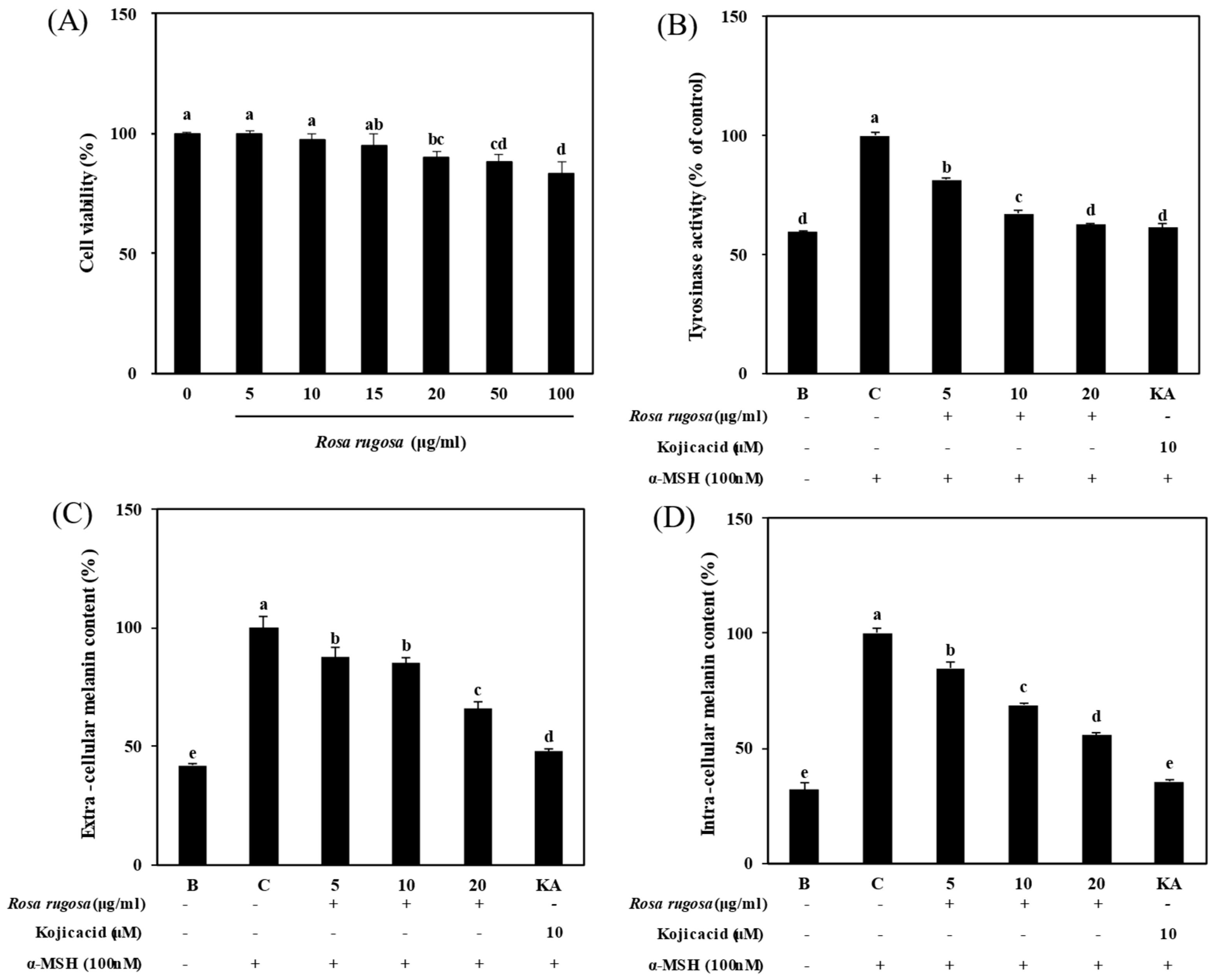
3.1. Effect of R. rugosa Crude Extract on α-MSH-Stimulated Melanogenesis

After being treated with different R. rugosa crude extract concentrations for 24 h, cytotoxicity of samples was tested using the MTT assay to measure viable cells. As shown in Figure 1A, the B16F10 cells showed high cell viability indicating an almost non-cytotoxicity at the 5, 10, 15, and 20 μg/mL concentrations of crude extract. Therefore, 5, 10, and 20 μg/mL concentrations were used for the further experiments. Furthermore, from Figure 1B–D, the crude extract treatment presented a dose-dependent inhibition on the active tyrosinase levels and extra-cellular and intra-cellular melanin contents of α-MSH-stimulated cells.

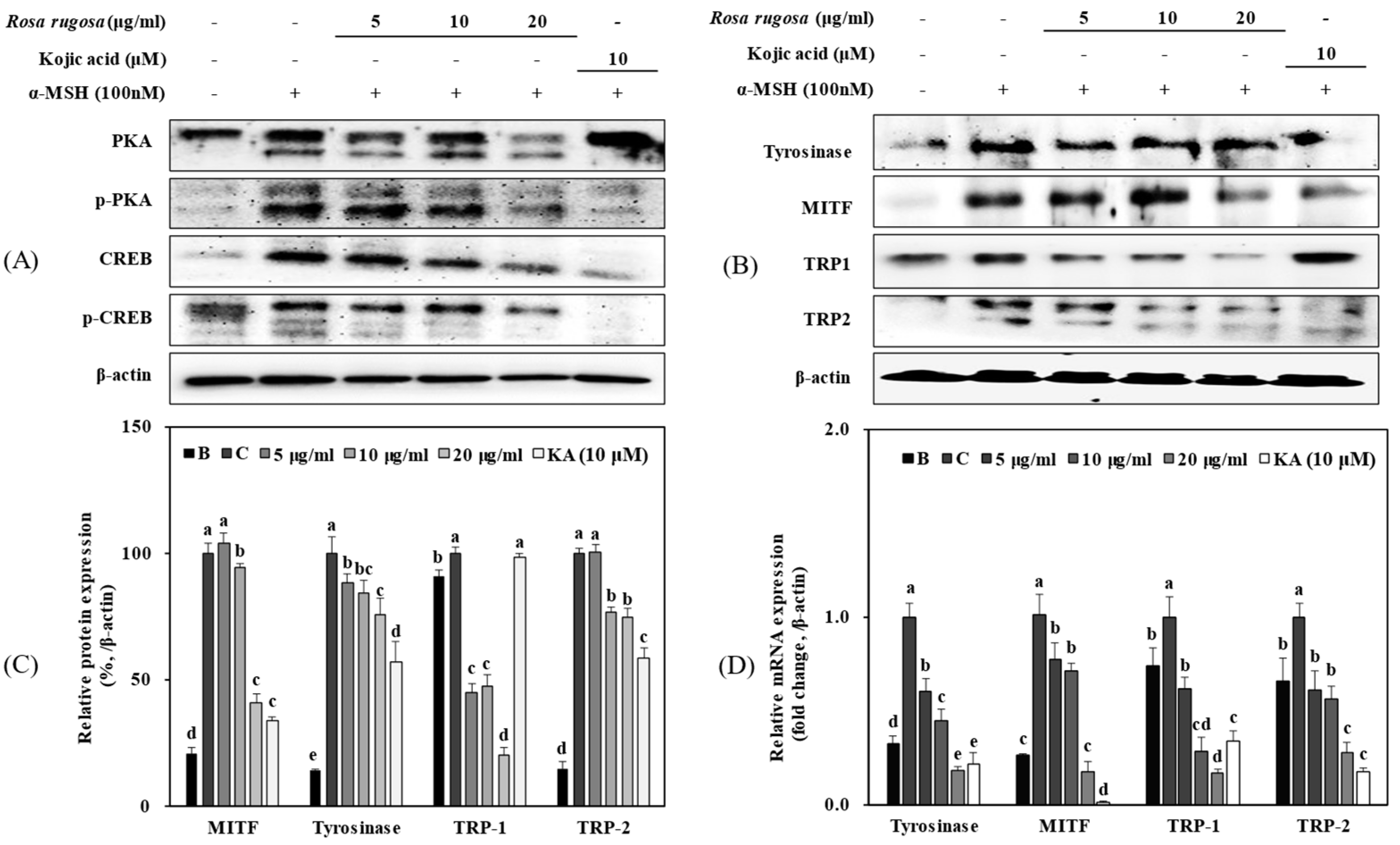
3.2. R. rugosa Crude Extracts Inhibit Melanogenesis by Regulating the PKA/CREB Signaling Pathway

To evaluate further, expression levels of the melanogenesis-related genes and proteins were determined following R. rugosa crude extract treatment in α-MSH stimulated B16F10 cells. In addition, the activation of the PKA/CREB signaling pathway was also investigated as a potential mechanism to regulate melanogenesis. As demonstrated in Figure 2, α-MSH stimulation activated the protein expression of PKA, P-PKA, CREB, and P-CREB (Figure 2A). In the case of PKA results, multiple bands were observed due to different subunits of PKA-C which the prime antibodies were designed to detect. Moreover, as reported earlier, primary antibodies against phosphorylated CREB often cross-react with phosphorylated cyclic-AMP-dependent transcription factor-1 which would result in the multiple bands observed in the results. Expectedly, the activation of the PKA/CREB signaling pathway was translated into an increase in the expression of the downstream proteins tyrosinase, MITF, TRP-1, and TRP-2 (Figure 2B). TRP-2 is expressed as glycoforms in the cells which led to multiple bands observed in the Western blot analysis. Compared to the untreated control group, the crude extract effectively decreased the expression levels of tyrosinase, MITF, TRP-1, and TRP-2. Decrease in these proteins was accompanied by suppressed activation of the PKA/CREB signaling pathway in a dose-dependent manner indicating that crude extract treatment decreased melanogenesis-related gene expression via suppressed PKA activation (Figure 2C). Consistent with the protein expression results, R. rugosa crude extract also down-regulated the mRNA expression levels of tyrosinase, MITF, TRP-1, and TRP-2 (Figure 2D), further confirming its effect on PKA activation.

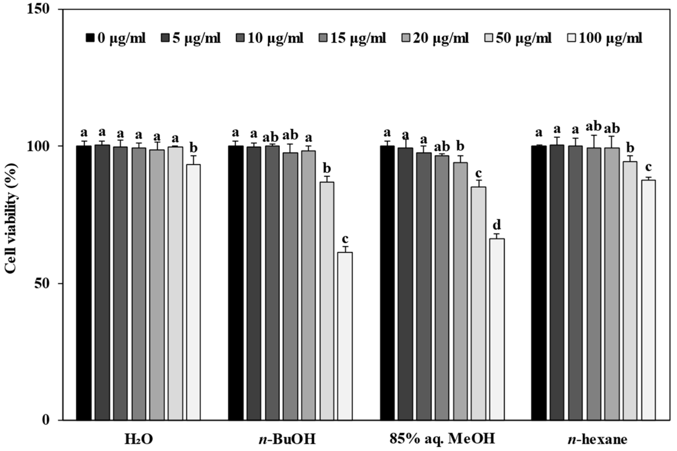
3.3. Effect of Solvent Fractions from R. rugosa Crude Extract on Cell Viability

Apart from the effect of R. rugosa crude extract, we also evaluated the anti-melanogenesis effect of H2O, n-BuOH, 85% aq. MeOH, and n-hexane solvent fractions, separated from the crude extract by solvent fractionation, in α-MSH-stimulated B16F10 cells. The results in Figure 3 show that all four fractions were not toxic to the cells at the concentrations ranging from 5 to 20 μg/mL; hence, 5, 10, and 20 μg/mL doses were used for the subsequent experiments.

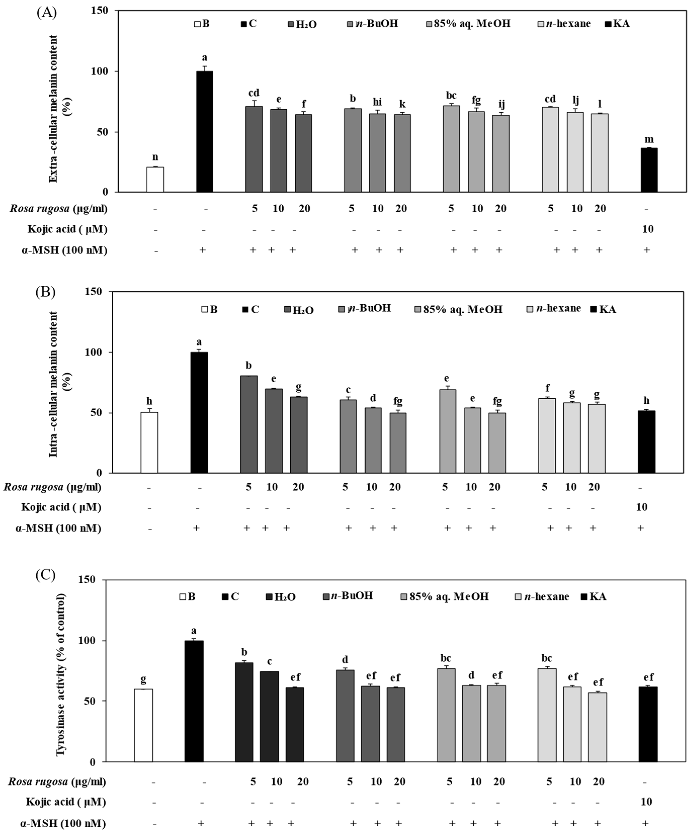
3.4. Effect of Solvent Fractions of R. rugosa Extract on Active Tyrosinase Levels and Extracellular and Intracellular Melanin Content

After treatment for 24 h at the concentrations of 5, 10, and 20 μg/mL with solvent fractions from R. rugosa extract, the extra/intracellular melanin contents were determined. As shown in Figure 4A,B, the four solvent fractions significantly inhibited both extracellular and intracellular melanin content of α-MSH-stimulated cells in a dose-dependent manner (p < 0.05). The tyrosinase activity in α-MSH-stimulated cells was also measured, following treatment with solvent fractions from R. rugosa extract. The result showed that the tyrosinase activity in the untreated α-MSH-stimulated control group was highest and that in the unstimulated untreated blank group was the lowest (Figure 4C). It is worth noting that the tyrosinase activity decreased significantly in the H2O, n-BuOH, 85% aq. MeOH, and n-hexane solvent fractions groups, in which the inhibition effect was most effective at 20 μg/mL. Therefore, the above results indicate that solvent fractions of R. rugosa extract contained potentially effective ingredients that could inhibit the synthesis of melanin.

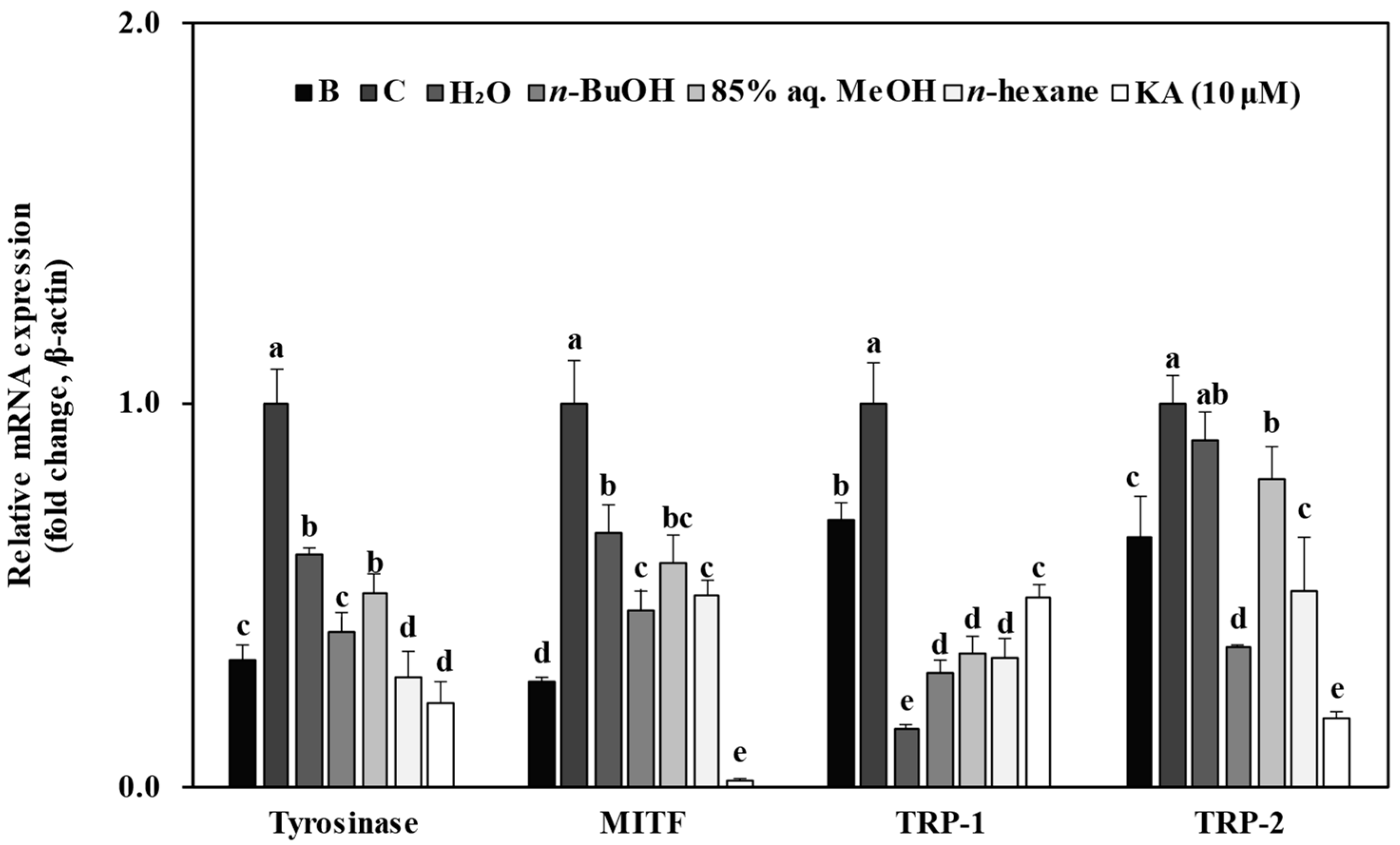
3.5. Effect of Solvent Fractions from R. rugosa Crude Extract on mRNA Expression Levels of Melanogenesis-Related Genes

To verify that the decrease in melanin and tyrosinase levels was related to the downregulation of melanogenesis-related genes, the expression levels of tyrosinase, MITF, TRP-1, and TRP-2 mRNA were determined using RT-qPCR. As demonstrated in Figure 5, the expression levels of the above genes in α-MSH-stimulated cells were notably higher than those of unstimulated cells (p < 0.05). However, the mRNA expression levels of tyrosinase, MITF, TRP-1, and TRP-2 were all down-regulated after the cells were treated with solvent fractions from R. rugosa crude extract, where the concentration of 20 μg/mL showed the most effective inhibition.

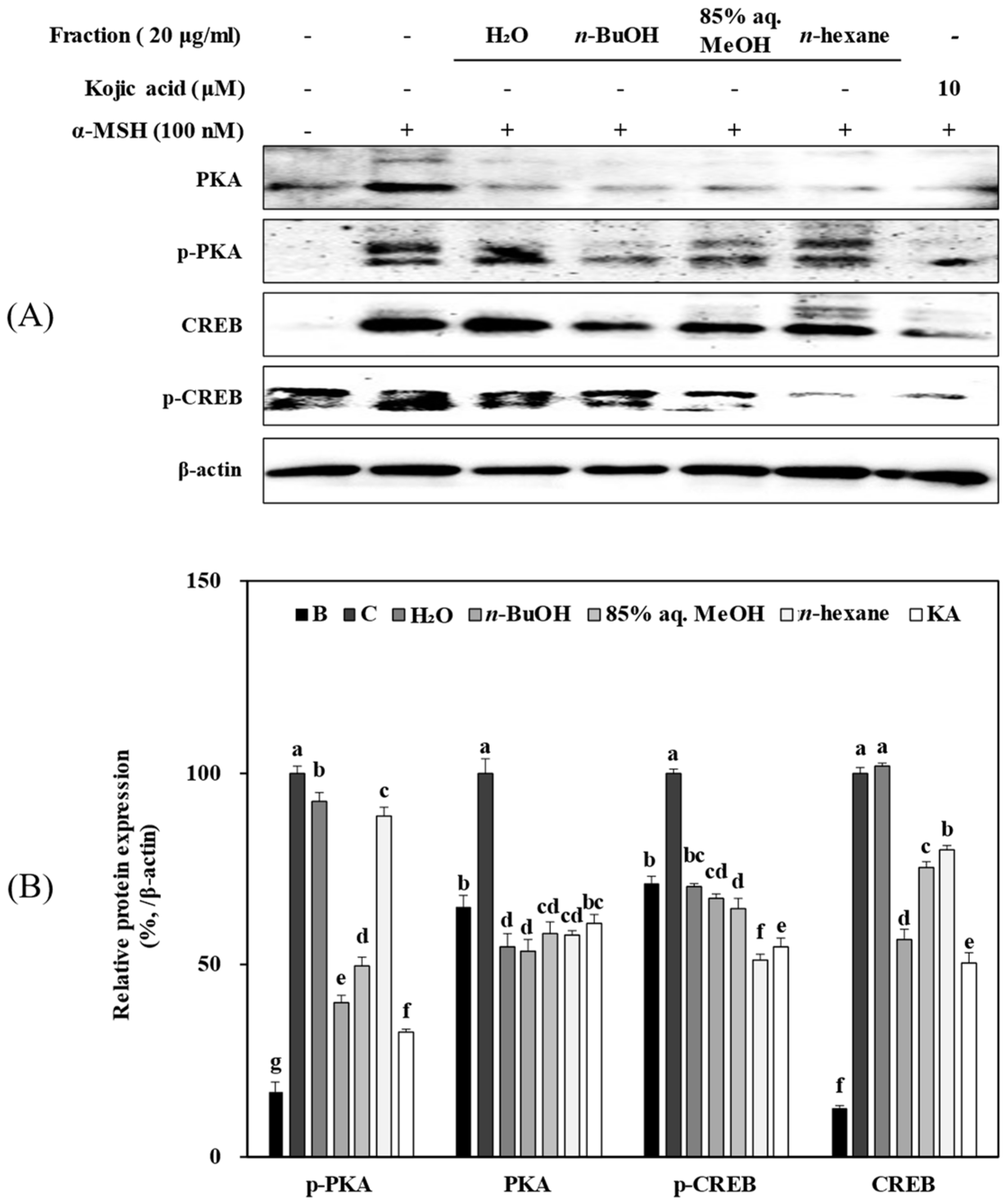
3.6. Solvent Fractions from R. rugosa Inhibit Melanogenesis via Supressed Activation of the PKA/CREB Pathway

To elucidate the anti-melanogenesis mechanism of H2O, n-BuOH, 85% aq. MeOH, and n-hexane solvent fractions from Rosa rugosa, their effect on the PKA/CREB signaling pathway and its downstream melanogenesis-related proteins (tyrosinase, MITF, TRP-1, and TRP-2) were investigated using Western blot analysis. As shown in Figure 6, the protein expression levels of PKA, p-PKA, CREB, and p-CREB were markedly up-regulated in the α-MSH-stimulated B16F10 cells compared to the unstimulated blank group. However, expression of these proteins was significantly suppressed following treatment with 20 μg/mL solvent fractions from R. rugosa, which indicates that fractions might possess the ability to inhibit the phosphorylation of PKA and subsequent activation of CREB to suppress melanin production. Furthermore, the protein expression levels of tyrosinase, MITF, TRP-1, and TRP-2 were also detected to determine whether the effect on PKA/CREB activation was translated into suppressed production of melanogenesis-related proteins. Consistent with the mRNA expression results, the H2O (Figure 7A), n-BuOH (Figure 7B), 85% aq. MeOH (Figure 7C), and n-hexane (Figure 7D) solvent fractions from R. rugosa dose-dependently decreased the protein levels of tyrosinase, MITF, TRP-1, and TRP-2. The above results reveal that solvent fractions from R. rugosa could attenuate overproduction of melanin by inhibiting its synthesis via suppression of the PKA/CREB pathway.

3.7. Total Polyphenol Content of Crude Extract of R. rugosa and Its Fractions

Total polyphenol contents of the samples were determined to investigate any relationship between the melanogenesis-inhibitory activity and phenolic compound content. As seen in Table 1, the total polyphenol content of R. rugosa crude extract and its fractions of H2O, n-BuOH, 85% aq. MeOH, and n-hexane were 1383.9 ± 44.5, 2004.7 ± 43.4, 7270.3 ± 54.5, 2064.1 ± 34.8, and 1091.1 ± 26.2 mg GAE/100 g extract, respectively. Among all fractions, n-BuOH contained the most phenolic compounds followed by the 85% aq. MeOH, H2O, and n-hexane fractions.

4. Discussion

R. rugosa is a widely utilized plant appearing in daily diets, cosmetics, and textile products due to its medical properties. Studies have shown that it owes these medical properties to various kinds of bioactive secondary metabolites, such as phenolic acids, flavonoids, and ascorbic acid [24]. The current results suggest that R. rugosa crude extract and its solvent fractions (H2O, n-BuOH, 85% aq. MeOH, and n-hexane) inhibited α-MSH-stimulated melanogenesis in B16F10 murine melanoma cells by suppressing the activation of the PKA/CREB signaling pathway.
Numerous studies have verified that tyrosinase is the rate-limiting enzyme for melanin synthesis, converting tyrosine to DOPA, which is an important intermediate during melanin synthesis [25]. Excess secretion of tyrosinase causes hyperpigmentation in the skin and leads to commercial problems in food industries because this enzyme attends to the unwanted color changes of vegetables, fruits, crustaceans, and fungi [26]. Therefore, searching for and developing tyrosinase inhibitors have drawn the attention of researchers in the fields of medicine, food, and cosmetics. It is widely accepted that inhibiting tyrosinase is the main action mechanism of chemicals that are used for their whitening properties, such as kojic acid, licorice flavonoid, α-arbutin, and other similar substances. However, reports also show that these agents also exhibit side effects such as cytotoxicity, skin irritation, and itchiness [27]. Many extracts or components from natural plants have shown a tyrosinase-inhibitory effect. A previous study reported that Euryale ferox seed extract showed inhibitory effects on both in vitro tyrosinase activity and cellular tyrosinase activity as well as melanin secretion [28]. Ishige okamurae extracts, Pueraria lobata stem extracts, and Camellia sinensis water extracts were also reported to possess anti-tyrosinase activities [29,30,31]. Consistent with these studies, our results indicate that both extract and solvent fractionate from R. rugosa significantly blocked melanin production by inhibiting α-MSH-induced cellular tyrosinase activity in B16F10 cells.
Based on the effect of R. rugosa extracts on extra/intra-cellular melanin content and active tyrosinase levels, the transcription and translation of melanogenesis-related genes, viz., tyrosinase, MITF, TRP-1, and TRP-2, were explored with RT-qPCR and Western blot. MITF is mainly responsible for regulating the expression of tyrosinase, TRP-1, and TRP-2, and its overexpression not only causes an increase in the expression of these proteins but also influences other melanocytic differentiation markers [32]. Therefore, MITF is usually looked at as a diagnostic marker for melanogenesis [33]. Expectedly, the present study found that R. rugosa crude extract and its H2O, n-BuOH, 85% aq. MeOH, and n-hexane fractions could effectively suppress the expression levels of MITF, tyrosinase, TRP-1, and TRP-2 in dose-dependent manners. A previous study revealed that the R. rugosa flower contains aqueous polyphenols in which quercetin glycoside was the main component [34]. Meanwhile, it has been reported that quercetin could inhibit UVA-induced melanogenesis through the Nrf2 signaling pathway in B16F10 cells [35]. R. rugosa is also rich in polysaccharides that showed hypoglycemic and immunomodulatory activities [36]. For these reasons, R. rugosa is a potential material to be researched and developed for anti-melanogenesis cosmetic products.
To elucidate the potential anti-melanogenesis mechanism of R. rugosa further in α-MSH-induced B16F10 cells, we analyzed the upstream melanogenesis-activating cascade: PKA/CREB signaling. As mentioned previously, the PKA/CREB signaling pathway plays an important role in melanogenesis. At the cellular level, increased cAMP activates PKA; subsequently, activated PKA facilitates the activation of CREB via phosphorylation. Phosphorylated CREB then stimulates the expression of its transcription targets MITF, tyrosinase, TRP-1, and TRP-2, which finally induce the melanocyte to secrete a large amount of melanin and initiate skin pigmentation [37,38]. Research revealed that pinostrobin could increase melanin content, tyrosinase activity, and melanogenesis-related markers by upregulating the PKA/CREB signaling pathways [39]. A natural citrus flavanone, 5-demethylnobiletin, was also confirmed to promote melanogenesis through the cAMP/CREB pathway [40]. Conversely, a long noncoding RNA, UCA1, was found to decrease the levels of melanogenesis-related gene expression by inhibiting the cAMP/PKA pathway [41]. The present results found that the protein expression levels of PKA, P-PKA, CREB, and P-CREB were notably upregulated after the cells were stimulated with α-MSH. However, the expression levels of the above proteins were downregulated after treating the α-MSH-stimulated B16F10 cells with R. rugosa crude extract and its solvent fractions at a concentration of 20 μg/mL. Among all tested fractions, the n-BuOH fraction showed the most consistent results to inhibit melanogenesis. In some tests other fractions showed higher inhibitory effect than n-BuOH but failed to exhibit similar effects in all areas, resulting in n-BuOH being the most effective to inhibit both melanin and tyrosinase content, PKA/CREB signaling, MITF, and TRPs. Nevertheless, these results indicate that R. rugosa crude extract and its solvent fractions may contain potential therapeutic ingredients against abnormal pigmentation diseases. Although kojic acid treatment as a positive control indicated that the effect of the samples tested in the current study was not fully comparable to that of kojic acid, the present samples were mixture of compounds that may or may not possess the anti-melanogenic properties. Therefore, future studies to elucidate the active ingredients of R. rugosa are expected to yield more effective compounds compared to fractions. Considering the side effects of continuous use of kojic acid and other tyrosinase inhibitors [27], finding novel natural bioactive agents will pave the way for safer treatment of hyperpigmentation. In this regard, the current results showed that R. rugosa is a potential source for novel anti-melanogenic compounds to produce more effective results than current tyrosinase inhibitors such as kojic acid.
To find the potential components that play an effective role in anti-melanogenesis activity in R. rugosa crude extract and its solvent fractions, the total polyphenol content was determined. The results showed that the total polyphenol content of R. rugosa crude extract and its H2O, n-BuOH, 85% aq. MeOH, and n-Hexane solvent fractions were 1383.9 ± 44.5, 2004.7 ± 43.4, 7270.3 ± 54.5, 2064.1 ± 34.8, and 1091.1 ± 26.2 mg GAE/100 g extract, respectively, in which the n-BuOH fraction possessed the highest total polyphenol content. Many studies have verified that plant polyphenols exert various kinds of positive effects on human health, such as antioxidant, anti-inflammation, anti-aging, anti-cancer, and antiallergenic activities, etc. [42,43]. A previous study detected a total of 531 polyphenol metabolites in rose hips with UPLC-MS/MS. Those results showed that rose hip extract contained phenolic acids (220), flavonoids (219), phlorotannins (50), lignans, and coumarins [44]. It has also been reported that polyphenols can ameliorate skin aging [45] and suppress tyrosinase activity [46]. Considering that currently the most effective tyrosinase inhibitor, hydroquinone, is a polyphenol, natural polyphenols are expected to be strong candidates to discover novel melanogenesis inhibitors [47]. In this context, results were parallel with the polyphenol content of the samples, where n-BuOH contained the highest polyphenol amount and exhibited the most consistent anti-melanogenesis effect. Therefore, the anti-melanogenesis effect of R. rugosa is suggested to arise from its high polyphenol content, which may exert a regulatory effect on the PKA/CREB-mediated melanin synthesis.

5. Conclusions

In conclusion, the present study reveals the melanogenesis-inhibitory effect of R. rugosa in α-MSH-stimulated B16F10 cells. The current results demonstrate that R. rugosa crude extract and its solvent fractions decreased melanin production and active tyrosinase levels by downregulating expressions of tyrosinase, MITF, TRP-1, and TRP-2. In addition, suppressing the PKA/CREB signaling pathway, possibly due to the activities of polyphenols, is suggested to be the mechanism by which these extracts exerted their anti-melanogenesis effect. These results suggest that R. rugosa crude extract and its solvent fractions may be potential therapeutic ingredients against abnormal pigmentation diseases.

Author Contributions

Conceptualization, Y.S. and C.-S.K.; Data curation, J.H.O., F.K. and H.L.; Formal analysis, X.Z. and F.K.; Funding acquisition, C.-S.K.; Investigation, X.Z. and J.H.O.; Methodology, H.L., Y.S. and C.-S.K.; Project administration, C.-S.K.; Resources, Y.S.; Software, J.H.O. and J.Y.; Supervision, C.-S.K.; Validation, F.K.; Visualization, X.Z., J.H.O., J.Y. and H.L.; Writing—original draft, X.Z. and F.K. All authors have read and agreed to the published version of the manuscript.

Funding

This research was supported by the Korea Institute of Marine Science and Technology Promotion (KIMST) and funded by the Ministry of Oceans and Fisheries, Korea (20220259).

Institutional Review Board Statement

Not applicable.

Informed Consent Statement

Not applicable.

Data Availability Statement

All data used to support the findings of this study are available from the corresponding author upon reasonable request.

Conflicts of Interest

The authors declare no conflict of interest.

References

  1. Zhou, S.; Yotsumoto, H.; Tian, Y.; Sakamoto, K. α-Mangostin suppressed melanogenesis in B16F10 murine melanoma cells through GSK3β and ERK signaling pathway. Biochem. Biophys. Rep. 2021, 26, 100949. [Google Scholar] [CrossRef] [PubMed]
  2. Merecz-Sadowska, A.; Sitarek, P.; Kowalczyk, T.; Zajdel, K.; Kucharska, E.; Zajdel, R. The Modulation of Melanogenesis in B16 Cells Upon Treatment with Plant Extracts and Isolated Plant Compounds. Molecules 2022, 27, 4360. [Google Scholar] [CrossRef] [PubMed]
  3. Pillaiyar, T.; Manickam, M.; Namasivayam, V. Skin whitening agents: Medicinal chemistry perspective of tyrosinase inhibitors. J. Enzym. Inhib. Med. Chem. 2017, 32, 403–425. [Google Scholar] [CrossRef] [PubMed] [Green Version]
  4. Avogadri, F.; Gnjatic, S.; Tassello, J.; Frosina, D.; Hanson, N.; Laudenbach, M.; Ritter, E.; Merghoub, T.; Busam, K.J.; Jungbluth, A.A. Protein Expression Analysis of Melanocyte Differentiation Antigen TRP-2. Am. J. Dermatopathol. 2016, 38, 201–207. [Google Scholar] [CrossRef] [PubMed]
  5. Zhou, S.; Riadh, D.; Sakamoto, K. Grape Extract Promoted α-MSH-Induced Melanogenesis in B16F10 Melanoma Cells, Which Was Inverse to Resveratrol. Molecules 2021, 26, 5959. [Google Scholar] [CrossRef]
  6. Wu, K.C.; Hseu, Y.C.; Shih, Y.C.; Sivakumar, G.; Syu, J.T.; Chen, G.L.; Lu, M.T.; Chu, P.C. Calycosin, a Common Dietary Isoflavonoid, Suppresses Melanogenesis through the Downregulation of PKA/CREB and p38 MAPK Signaling Pathways. Int. J. Mol. Sci. 2022, 3, 1358. [Google Scholar] [CrossRef]
  7. Ryu, S.; Johnson, A.; Park, Y.; Kim, B.; Norris, D.; Armstrong, C.A.; Song, P.I. The Alpha-Melanocyte-Stimulating Hormone Suppresses TLR2-Mediated Functional Responses through IRAK-M in Normal Human Keratinocytes. PLoS ONE 2015, 10, e0136887. [Google Scholar] [CrossRef]
  8. Jung, H.J.; Choi, D.C.; Noh, S.G.; Choi, H.; Choi, I.; Ryu, I.Y.; Chung, H.Y.; Moon, H.R. New Benzimidazothiazolone Derivatives as Tyrosinase Inhibitors with Potential Anti-Melanogenesis and Reactive Oxygen Species Scavenging Activities. Antioxidants 2021, 10, 1078. [Google Scholar] [CrossRef]
  9. Huang, H.C.; Wang, S.S.; Tsai, T.C.; Ko, W.P.; Chang, T.M. Phoenix dactylifera L. Seed Extract Exhibits Antioxidant Effects and Attenuates Melanogenesis in B16F10 Murine Melanoma Cells by Downregulating PKA Signaling. Antioxidants 2020, 9, 1270. [Google Scholar] [CrossRef]
  10. Zolghadri, S.; Bahrami, A.; Hassan Khan, M.T.; Munoz-Munoz, J.; Garcia-Molina, F.; Garcia-Canovas, F.; Saboury, A.A. A comprehensive review on tyrosinase inhibitors. J. Enzym. Inhib. Med. Chem. 2019, 34, 279–309. [Google Scholar] [CrossRef]
  11. Khongkarat, P.; Ramadhan, R.; Phuwapraisirisan, P.; Chanchao, C. Safflospermidines from the bee pollen of Helianthus annuus L. exhibit a higher in vitro antityrosinase activity than kojic acid. Heliyon 2020, 6, e03638. [Google Scholar] [CrossRef] [PubMed]
  12. Yun, H.Y.; Kim, D.H.; Son, S.; Ullah, S.; Kim, S.J.; Kim, Y.J.; Yoo, J.W.; Jung, Y.; Chun, P.; Moon, H.R. Design, synthesis, and anti-melanogenic effects of (E)-2-benzoyl-3-(substituted phenyl) acrylonitriles. Drug Des. Dev. Ther. 2015, 9, 4259–4268. [Google Scholar]
  13. Dashbaldan, S.; Rogowska, A.; Pączkowski, C.; Szakiel, A. Distribution of Triterpenoids and Steroids in Developing Rugosa Rose (Rosarugosa Thunb.) Accessory Fruit. Molecules 2021, 26, 5158. [Google Scholar] [CrossRef] [PubMed]
  14. Liaudanskas, M.; Noreikienė, I.; Zymonė, K.; Juodytė, R.; Žvikas, V.; Janulis, V. Composition and Antioxidant Activity of Phenolic Compounds in Fruit of the Genus Rosa L. Antioxidants 2021, 10, 545. [Google Scholar] [CrossRef]
  15. Liu, Y.; Zhi, D.; Wang, X.; Fei, D.; Zhang, Z.; Wu, Z.; Li, Y.; Chen, P.; Li, H. Kushui Rose (R. Setate x R. Rugosa) decoction exerts antitumor effects in C. elegans by downregulating Ras/MAPK pathway and resisting oxidative stress. Int. J. Mol. Med. 2018, 42, 1411–1417. [Google Scholar] [CrossRef] [Green Version]
  16. Kim, K.H.; Park, Y.J.; Jang, H.J.; Lee, S.J.; Lee, S.; Yun, B.S.; Lee, S.W.; Rho, M.C. Rugosic acid A, derived from Rosa rugosa Thunb.; is novel inhibitory agent for NF-κB and IL-6/STAT3 axis in acute lung injury model. Phytother. Res. PTR 2020, 34, 3200–3210. [Google Scholar] [CrossRef] [PubMed]
  17. Olech, M.; Nowacka-Jechalke, N.; Masłyk, M.; Martyna, A.; Pietrzak, W.; Kubiński, K.; Załuski, D.; Nowak, R. Polysaccharide-Rich Fractions from Rosa rugosa Thunb.-Composition and Chemopreventive Potential. Molecules 2019, 24, 1354. [Google Scholar] [CrossRef] [Green Version]
  18. Grajzer, M.; Wiatrak, B.; Gębarowski, T.; Matkowski, A.; Grajeta, H.; Rój, E.; Kulma, A.; Prescha, A. Chemistry, oxidative stability and bioactivity of oil extracted from Rosa rugosa (Thunb.) seeds by supercritical carbon dioxide. Food Chem. 2021, 335, 127649. [Google Scholar] [CrossRef]
  19. Zhang, C.; Zhao, F.; Li, R.; Wu, Y.; Liu, S.; Liang, Q. Purification, characterization, antioxidant and moisture-preserving activities of polysaccharides from Rosa rugosa petals. Int. J. Biol. Macromol. 2019, 124, 938–945. [Google Scholar] [CrossRef]
  20. Song, M.; Lee, J.; Kim, Y.J.; Hoang, D.H.; Choe, W.; Kang, I.; Kim, S.S.; Ha, J. Jeju Magma-Seawater Inhibits α-MSH-Induced Melanogenesis via CaMKKβ-AMPK Signaling Pathways in B16F10 Melanoma Cells. Mar. Drugs 2020, 18, 473. [Google Scholar] [CrossRef]
  21. Han, S.M.; Kim, J.M.; Hong, I.P.; Woo, S.O.; Kim, S.G.; Jang, H.R.; Park, K.K.; Pak, S.C. Whitening Effect of Watersoluble Royal Jelly from South Korea. Korean J. Food Sci. Anim. Resour. 2015, 35, 707–713. [Google Scholar] [CrossRef] [PubMed]
  22. Xin, X.J.; Zou, J.; Zou, T.; Shang, H.; Sun, L.Y. A Newly Authenticated Compound from Traditional Chinese Medicine Decoction Induces Melanogenesis in B16-F10 Cells by Increasing Tyrosinase Activity. Evid.-Based Complement. Altern. Med. Ecam 2018, 2018, 8485670. [Google Scholar] [CrossRef] [Green Version]
  23. Ma, S.; Kim, C.; Neilson, A.P.; Griffin, L.E.; Peck, G.M.; O’Keefe, S.F.; Stewart, A.C. Comparison of Common Analytical Methods for the Quantification of Total Polyphenols and Flavanols in Fruit Juices and Ciders. J. Food Sci. 2019, 84, 2147–2158. [Google Scholar] [CrossRef] [PubMed] [Green Version]
  24. Um, M.; Han, T.H.; Lee, J.W. Ultrasound-assisted extraction and antioxidant activity of phenolic and flavonoid compounds and ascorbic acid from rugosa rose (Rosa rugosa Thunb.) fruit. Food Sci. Biotechnol. 2017, 27, 375–382. [Google Scholar] [CrossRef]
  25. Garcia-Molina, P.; Garcia-Molina, F.; Teruel-Puche, J.A.; Rodriguez-Lopez, J.N.; Garcia-Canovas, F.; Muñoz-Muñoz, J.L. The Relationship between the IC50 Values and the Apparent Inhibition Constant in the Study of Inhibitors of Tyrosinase Diphenolase Activity Helps Confirm the Mechanism of Inhibition. Molecules 2022, 27, 3141. [Google Scholar] [CrossRef] [PubMed]
  26. da Silva, A.P.; Silva, N.F.; Andrade, E.; Gratieri, T.; Setzer, W.N.; Maia, J.; da Silva, J. Tyrosinase inhibitory activity, molecular docking studies and antioxidant potential of chemotypes of Lippia origanoides (Verbenaceae) essential oils. PLoS ONE 2017, 12, e0175598. [Google Scholar] [CrossRef] [PubMed] [Green Version]
  27. Azumi, J.; Takeda, T.; Shimada, Y.; Aso, H.; Nakamura, T. The Organogermanium Compound THGP Suppresses Melanin Synthesis via Complex Formation with L-DOPA on Mushroom Tyrosinase and in B16 4A5 Melanoma Cells. Int. J. Mol. Sci. 2019, 20, 4785. [Google Scholar] [CrossRef] [Green Version]
  28. Baek, S.H.; Nam, I.J.; Kwak, H.S.; Kim, K.C.; Lee, S.H. Cellular Anti-Melanogenic Effects of a Euryale ferox Seed Extract Ethyl Acetate Fraction via the Lysosomal Degradation Machinery. Int. J. Mol. Sci. 2015, 16, 9217–9235. [Google Scholar] [CrossRef] [Green Version]
  29. Li, X.; Yang, H.W.; Jiang, Y.; Oh, J.Y.; Jeon, Y.J.; Ryu, B. Ishophloroglucin A Isolated from Ishige okamurae Suppresses Melanogenesis Induced by α-MSH: In Vitro and In Vivo. Mar. Drugs 2020, 18, 470. [Google Scholar] [CrossRef]
  30. Gao, D.; Kim, J.H.; Kim, C.T.; Jeong, W.S.; Kim, H.M.; Sim, J.; Kang, J.S. Evaluation of Anti-Melanogenesis Activity of Enriched Pueraria lobata Stem Extracts and Characterization of Its Phytochemical Components Using HPLC-PDA-ESI-MS/MS. Int. J. Mol. Sci. 2021, 22, 8105. [Google Scholar] [CrossRef]
  31. Shin, S.; Kim, M.; Song, N.; Sun, S.; Choi, J.; Park, K. Antioxidant and Anti-Melanogenesis Effects of Colloidal Gold Camellia sinensis L. Extracts. Molecules 2022, 27, 5593. [Google Scholar] [CrossRef] [PubMed]
  32. Lin, J.Y.; Fisher, D.E. Melanocyte biology and skin pigmentation. Nature 2007, 445, 843–850. [Google Scholar] [CrossRef] [PubMed]
  33. Flesher, J.L.; Paterson-Coleman, E.K.; Vasudeva, P.; Ruiz-Vega, R.; Marshall, M.; Pearlman, E.; MacGregor, G.R.; Neumann, J.; Ganesan, A.K. Delineating the role of MITF isoforms in pigmentation and tissue homeostasis. Pigment. Cell Melanoma Res. 2020, 33, 279–292. [Google Scholar] [CrossRef] [PubMed]
  34. Zhang, J.; Xiao, Y.; Guan, Y.; Rui, X.; Zhang, Y.; Dong, M.; Ma, W. An aqueous polyphenol extract from Rosa rugosa tea has antiaging effects on Caenorhabditis elegans. J. Food Biochem. 2019, 43, e12796. [Google Scholar] [CrossRef] [PubMed]
  35. Chaiprasongsuk, A.; Onkoksoong, T.; Pluemsamran, T.; Limsaengurai, S.; Panich, U. Photoprotection by dietary phenolics against melanogenesis induced by UVA through Nrf2-dependent antioxidant responses. Redox Biol. 2016, 8, 79–90. [Google Scholar] [CrossRef] [Green Version]
  36. Yang, S.; Qu, Y.; Chen, J.; Chen, S.; Sun, L.; Zhou, Y.; Fan, Y. Bee Pollen Polysaccharide from Rosa rugosa Thunb. (Rosaceae) Promotes Pancreatic β-Cell Proliferation and Insulin Secretion. Front. Pharmacol. 2021, 12, 688073. [Google Scholar] [CrossRef]
  37. Lee, S.E.; Park, S.H.; Oh, S.W.; Yoo, J.A.; Kwon, K.; Park, S.J.; Kim, J.; Lee, H.S.; Cho, J.Y.; Lee, J. Beauvericin inhibits melanogenesis by regulating cAMP/PKA/CREB and LXR-α/p38 MAPK-mediated pathways. Sci. Rep. 2018, 8, 14958. [Google Scholar] [CrossRef] [Green Version]
  38. Tsang, T.F.; Chan, B.; Tai, W.C.; Huang, G.; Wang, J.; Li, X.; Jiang, Z.H.; Hsiao, W.L.W. Gynostemma pentaphyllum saponins induce melanogenesis and activate cAMP/PKA and Wnt/β-catenin signaling pathways. Phytomedicine 2019, 60, 153008. [Google Scholar] [CrossRef]
  39. Yoon, J.H.; Youn, K.; Jun, M. Discovery of Pinostrobin as a Melanogenic Agent in cAMP/PKA and p38 MAPK Signaling Pathway. Nutrients 2022, 14, 3713. [Google Scholar] [CrossRef]
  40. Wang, H.M.; Qu, L.Q.; Ng, J.P.L.; Zeng, W.; Yu, L.; Song, L.L.; Wong, V.K.W.; Xia, C.L.; Law, B.Y.K. Natural Citrus flavanone 5-demethylnobiletin stimulates melanogenesis through the activation of cAMP/CREB pathway in B16F10 cells. Phytomedicine 2022, 8, 153941. [Google Scholar] [CrossRef]
  41. Pei, S.; Chen, J.; Lu, J.; Hu, S.; Jiang, L.; Lei, L.; Ouyang, Y.; Fu, C.; Ding, Y.; Li, S.; et al. The Long Noncoding RNA UCA1 Negatively Regulates Melanogenesis in Melanocytes. J. Investig. Dermatol. 2020, 140, 152–163.e5. [Google Scholar] [CrossRef] [PubMed]
  42. Zhao, Y.; Jiang, Q. Roles of the Polyphenol-Gut Microbiota Interaction in Alleviating Colitis and Preventing Colitis-Associated Colorectal Cancer. Adv. Nutr. 2021, 12, 546–565. [Google Scholar] [CrossRef] [PubMed]
  43. Chiva-Blanch, G.; Badimon, L. Effects of Polyphenol Intake on Metabolic Syndrome: Current Evidences from Human Trials. Oxidative Med. Cell. Longev. 2017, 2017, 5812401. [Google Scholar] [CrossRef] [PubMed] [Green Version]
  44. Sun, Y.; Yang, Y.; Zhou, M.; Luo, L.; Pan, H.; Zhang, Q.; Yu, C. Widely Targeted Metabolic Profiling Reveals Differences in Polyphenolic Metabolites during Rosa xanthina f. spontanea Fruit Development and Ripening. Metabolites 2022, 12, 438. [Google Scholar] [CrossRef]
  45. Csekes, E.; Račková, L. Skin Aging, Cellular Senescence and Natural Polyphenols. Int. J. Mol. Sci. 2021, 22, 12641. [Google Scholar] [CrossRef]
  46. Kampatsikas, I.; Bijelic, A.; Pretzler, M.; Rompel, A. Three recombinantly expressed apple tyrosinases suggest the amino acids responsible for mono- versus diphenolase activity in plant polyphenol oxidases. Sci. Rep. 2017, 7, 8860. [Google Scholar] [CrossRef] [Green Version]
  47. Panzella, L.; Napolitano, A. Natural and bioinspired phenolic compounds as tyrosinase inhibitors for the treatment of skin hyperpigmentation: Recent advances. Cosmetics 2019, 6, 57. [Google Scholar] [CrossRef]
Figure 1. Effect of R. rugosa crude extract on cell viability, tyrosinase activity, and melanin content. (A) Cell viability; (B) Intracellular tyrosinase activity; (C) Extracellular melanin content; (D) Intracellular melanin content. a–e Bars with different letters in the same treatment group are significantly different (p < 0.05), while the same letters indicate no significance according to a one-way ANOVA test followed by the Duncan’s multiple range test. B: unstimulated untreated blank group; C: α-MSH-stimulated untreated control group; KA: kojic acid (positive control).
Applsci 13 00184 g001
Figure 2. Effect of R. rugosa crude extract on protein and mRNA expression levels of the melanogenesis- and PKA/CREB-signaling-pathway-related genes. (A) Protein expression bands of PKA, P-PKA, CREB, and P-CREB; (B) Protein expression bands of tyrosinase, MITF, TRP-1, and TRP-2; (C) Quantification of protein expression levels with densitometric analysis of Western blot bands; (D) The mRNA levels of tyrosinase, MITF, TRP-1, and TRP-2 given as relative percentages of mRNA levels in the stimulated untreated group. All mRNA levels were normalized against β-actin levels. The mRNA expression was measured using RT-qPCR. a–e Bars with different letters in the same treatment group are significantly different while the same letters mean no significance (p < 0.05) according to a one-way ANOVA test followed by the Duncan’s multiple range test. B: unstimulated untreated blank group; C: α-MSH-stimulated untreated control group; KA: kojic acid (positive control).
Applsci 13 00184 g002
Figure 3. Effect of H2O, n-BuOH, 85% aq. MeOH, and n-hexane solvent fractions on cell viability. a–d Bars with different letters in the same treatment group are significantly different (p < 0.05) while the same letters indicate no significance according to a one-way ANOVA test followed by the Duncan’s multiple range test.
Applsci 13 00184 g003
Figure 4. Effect of solvent fractions from R. rugosa crude extract on extra/intracellular melanin content and tyrosinase activity. (A) Extracellular melanin content; (B) Intracellular melanin content; (C) Intracellular tyrosinase activity. a–n Bars with different letters in the same treatment group are significantly different (p < 0.05) while the same letters indicate no significance according to a one-way ANOVA test followed by the Duncan’s multiple range test. B: unstimulated untreated blank group; C: α-MSH-stimulated untreated control group; KA: kojic acid (positive control).
Applsci 13 00184 g004
Figure 5. Effect of solvent fractions from R. rugosa crude extract on mRNA expression levels of tyrosinase, MITF, TRP-1, and TRP-2. The mRNA levels are given as a relative percentage of the stimulated untreated group. The mRNA expression was quantified using RT-qPCR and normalized against β-actin. a–e Bars with different letters in the same treatment group are significantly different (p < 0.05) while the same letters indicate no significance according to a one-way ANOVA test followed by the Duncan’s multiple range test. B: unstimulated untreated blank group; C: α-MSH-stimulated untreated control group; KA: kojic acid (positive control).
Applsci 13 00184 g005
Figure 6. Effect of solvent fractions from R. rugosa crude extract on the protein expression of PKA/CREB-signaling-pathway-related proteins. (A) Effect of H2O, n-BuOH, 85% aq. MeOH, and n-hexane solvent fractions on protein expression of PKA, p-PKA, CREB, and p-CREB at a concentration of 20 μg/mL in B16F10 cells; (B) Quantification of protein expression levels with densitometric analysis of Western blot bands a–g Bars with different letters in the same treatment group are significantly different (p < 0.05) while the same letters indicate no significance according to a one-way ANOVA test followed by the Duncan’s multiple range test. B: unstimulated untreated blank group; C: α-MSH-stimulated untreated control group; KA: kojic acid (positive control).
Applsci 13 00184 g006
Figure 7. Effect of H2O (A), n-BuOH (B), 85% aq. MeOH (C), and n-hexane (D) solvent fractions from R. rugosa crude extract on the protein expression of melanogenesis-related proteins (tyrosinase, MITF, TRP-1, and TRP-2) in α-MSH-stimulated B16F10 cells analyzed using Western blot. Quantification of protein expression levels was carried out with densitometric analysis of Western blot bands. a–f Bars with different letters in the same treatment group are significantly different (p < 0.05) while the same letters indicate no significance according to a one-way ANOVA test followed by the Duncan’s multiple range test. B: unstimulated untreated blank group; C: α-MSH-stimulated untreated control group; KA: kojic acid (positive control).
Applsci 13 00184 g007
Table 1. Total polyphenol content of crude extract of R. rugosa and its solvent fractions.
SampleCrude ExtractH2O fr.n-BuOH fr.85% aq. MeOH fr.n-Hexane fr.
Total polyphenol content 11383.9 ± 44.52004.7 ± 43.47270.3 ± 54.52064.1 ± 34.81091.1 ± 26.2
1 Unit: mg GAE/100 g extract.
Disclaimer/Publisher’s Note: The statements, opinions and data contained in all publications are solely those of the individual author(s) and contributor(s) and not of MDPI and/or the editor(s). MDPI and/or the editor(s) disclaim responsibility for any injury to people or property resulting from any ideas, methods, instructions or products referred to in the content.

