Article

Comparative Evaluation of Adequacy of Root Canal Treatment Performed by Interns and Undergraduate Dental Students—A Cross-Sectional, Retrospective Study

by
Mohammed Assayed Mousa
1,*,
Akram Mojidea Alshammari
2,
Abdulelah Zaid Alshammari
2,
Meshari M. Alsharari
2,
Muhammad Nadeem Baig
3,
Kumar Chandan Srivastava
4,
Deepti Shrivastava
3,
Mohammad Khursheed Alam
3,
Meshal Aber Alonazi
5 and
Hmoud Ali Algarni
5,*
1
Department of Prosthetic Dental Sciences, College of Dentistry, Jouf University, Sakakah 42421, Saudi Arabia
2
Dental Graduate, College of Dentistry, Jouf University, Sakakah 72345, Saudi Arabia
3
Department of Preventive Dentistry, College of Dentistry, Jouf University, Sakakah 72345, Saudi Arabia
4
Department of Oral & Maxillofacial Surgery & Diagnostic Sciences, College of Dentistry, Jouf University, Sakakah 72345, Saudi Arabia
5
Department of Restorative Dentistry, College of Dentistry, Jouf University, Sakakah 42421, Saudi Arabia
*
Authors to whom correspondence should be addressed.
Appl. Sci. 2022, 12(18), 9079; https://doi.org/10.3390/app12189079
Submission received: 9 July 2022 / Revised: 3 September 2022 / Accepted: 5 September 2022 / Published: 9 September 2022

Abstract

:
This study aimed to assess the errors that happened during root canal treatment (RCT) and compare the quality of root canal fillings (RCFs) performed by different academic grades of dental students at Jouf University. Materials and Methods: a retrospective audit study was conducted to check the errors that occurred during RCT performed by seniors and undergraduates. The quality of the RCFs were evaluated, using digital periapical radiographs depending on previously validated criteria. Data analyses were performed using Statistical Package for the Social Sciences. Descriptive statistics were used for expressing the frequencies of variables and the Chi-square test was used for comparing different variables with a 0.05 level of significance. Results: overall, 332 endodontically treated teeth (566 root canals) were finally chosen for the study. Out of these treated cases, 156 teeth (47%) had a total of 287 recorded errors, mainly in posterior teeth. From the total number of errors, short obturation was the highest error occurred (29%), followed by lack of apical seal (25.1%). Root perforation was the least reported error (2.8%), proceeded by broken instruments (5.6%). A total of 13.2% of the errors were teeth left with unfilled canals, while 7.7% were for an overextended filling. There were no significant differences between the quality of RCF performed by different academic grades. Conclusion: depending on the current study, the quality of RCTs performed by seniors and undergraduate students was 53% acceptable. A clear plan should be applied to improve the quality of treatment outcomes of undergraduate students by implementing more effective preclinical training courses, increasing the ration of supervisor-to-students, and using more advanced modalities during treatment.

1. Introduction

Root canal treatment (RCT) is an integral part of comprehensive dental management. Therefore, advances in endodontic instruments, materials, and sciences have been made to modern root canal therapy with a reported high degree of success rate [1]. A successful outcome of RCT depends on a variety of factors, including pulpal and periapical status, anatomy and configuration of root canal/s, quality of root canal/s shaping, and the quality of root canal filling procedure [2]. Before, during, and after RCT, using periapical radiographs is crucial for the detection of periapical lesions, canal/s anatomy and dimensions, the quality of obturation, and records [2,3]. An appropriate RCT is characterized by a uniformly tapered canal from the coronal part to the apex of the root, obturation with no voids, and the presence of homogenous filling materials 0.5–2 mm before the radiographic apex [4,5]. Many iatrogenic factors can compromise the prognosis of RCT, such as inadequacy of the preparation, ledge formation, perforation, overfilling, underfilling, breaking of the instrument within the canals, inadequate coronal seal, and inadequate apical seal [6,7,8]. It was found that each 1 mm loss of length of the root canal after 2 mm in teeth with apical periodontitis increases the RCT failure rate by 14% [9,10].
Dental students at Jouf University take a five-year Bachelor Dental Surgery course following the problem-based learning (PBL) system [11,12,13]. This Bachelor’s course is preceded by a preliminary year and followed by a one-year internship. The students undertake a full-year course in preclinical endodontics in the third academic year. This course involves 28 didactic hours and 56 training hours in a laboratory during which the students perform all the basic technical procedures of RCT on natural teeth. The clinical endodontic course starts in the fourth academic year and involves 5 h of theoretical lectures and 84 h of clinical sessions, during which students endodontically treat single- and multi-rooted teeth using hand root canal instruments and cold Gutta Percha condensation techniques with aid of digital periapical radiographs. The followed protocol during RCT includes identification of the working length using the conventional parallel and/or bisecting angle periapical radiographs, use of step-back preparation techniques, and, finally, the obturation is performed using the cold lateral compaction technique. To finish the clinical course in the fourth year, the student should accomplish a minimum of 12 cases that are equally distributed among anterior, premolars, and first molar teeth. During the fifth academic year, the student performs RCTs as part of the comprehensive management of four patients who need, at least, three disciplines among which, RCT must be. Fifth-year students perform the RCTs following the same protocol as the fourth academic year. In both fourth and fifth academic grades, endodontic treatment is always performed under the supervision of an endodontic consultants (or specialist) staff, with an average staff-to-student ratio being 1:5 and 1:8, respectively. During the internship clinical year, the senior students are allowed to treat the vital and non-vital teeth and re-treat the failed root canal treated teeth using hand conventional and pro-taper root canal preparatory instruments after using an electronic apex locator in the identification of working length (which they are not allowed to use before), under the supervision of the mentors with an average staff-to-student ratio 1:10, excluding the cases that are subjected to complication.
Because of the importance of RCT performed by dental students as a part of community service, several distributed studies regarding the evaluation of the technical quality of RCT performed by dental students have been conducted. Table 1 shows the results of a meta-analysis carried out by the authors (M.A.M., K.S., and M.B.) on studies performed in the last 10 years on the adequacy of RCT by undergraduates. The analysis was made to examine the literature describing the adequacy of RCT by undergraduates and to review the prevalence of iatrogenic errors that happened during RCT by different dental grades. Four electronic databases (Google Scholar, PubMed, Scopus, and Web of Science) were searched using the keywords “root canal treatment, endodontics, dental students, undergraduates”. Means and standard deviations were calculated depending on the means of each study. The analysis included all clinical studies by undergraduates in different continents in the period from January 2011 to December 2021. The preclinical, systematic reviews, and questionnaire studies were excluded. The studies were performed in Africa [14,15,16], Europe [17,18,19,20], America [21,22,23], and Asia [24,25,26,27,28,29,30,31,32,33,34,35,36,37,38,39]. Systematically, the studies were on 8288 teeth with 14,205 root canals. Most of these surveyed studies were mostly consistent in their aims and methodologies. They used periapical radiographs to assess the length, density, and taper of the RCT [14,15,16,17,19,21,23,31,32,33,34,35,36,37,38]. In addition, some studies added more criteria for evaluation to explore the technical errors that happened during treatment such as ledge, broken instruments, canal transportation, and perforation [18,23,25,26,27,28,29,30]. Some of the surveyed studies were more general and explored the overall quality of the RCT [15,17,21,25,31,32], while others were more specific, such as those comparing the quality of RCT with respect to tooth type and location [1,14,16,22,26,33,34,36,37,40], extracting the frequency of technical errors [17,18,23,27,28,29,30,41], identifying the academic grade of students who perform the treatment [24,35,38,39], and exploring the complications happening as a result of errors [20]. Although the studies showed significant variation in their outcomes, the overall quality of RCT performed by undergraduates was significantly high when comparing inadequate RCT (61.4% SD ± 20.2 and 38.6% SD ± 20.3, respectively). While the study for Fritz et al. showed the highest adequate RCT among dental students, 93.8% [21], the study performed by Ehsani et al. showed the lowest percentage of adequate RCT, 17.5% [38]. The maxillary anterior teeth, followed by premolar, showed higher RC quality compared to posterior teeth in most of the studies [15,16,20,22,25,26,35,37], while fewer of them showed no significant differences between anterior and premolars or anterior and posteriors [17,21]. The deficiency in the length of RCT was the highest error that happens during RCT as reported by some of the studies [16,18,21,30,34,39]. On the other hand, lack of density was reported as the highest error in other studies [26,27,29,31,32,33,36]. Leaving canals without filling was reported as the least common error [18], proceeded by broken instrument/s [23]. Other than one study [39], there was no assumed significant difference between different academic grades of undergraduate students during RCT [24,28,38].
However, most of the surveyed studies, including the excluded studies, were cross-sectional as it conducted in a limited period and were performed either on one academic grade or in general, without identifying the actual improvement of hand skills of students between different academic grades. This was the aim of the current study: to measure the prevalence and technical quality of RCF by senior and undergraduate students, the type and location of teeth which showed higher RCF errors, and the most occurred error/s, done by the different academic grades.

2. Materials and Methods

The current retrospective study was conducted in the College of Dentistry, Jouf University, involving the periapical radiographs of patients who were endodontically treated by undergraduates (4th- and 5th-year dental students) and intern students. Ethical approval was delivered to the study by the Local Committee of Bioethics (LCB) under reference number [11–04–41] in full accordance with the World Medical Association Declaration of Helsinki. The inclusion criteria of the study were as follows:
  • All mature permanent single and multirooted teeth that were treated or retreated with a root canal (RC)
  • The cases treated by senior and undergraduate students
  • The cases with at least three available X-ray records
The exclusion criteria were as follows:
  • The deciduous teeth
  • Permanent immature teeth
  • The teeth were treated with no available or unreadable radiographs
  • The teeth were treated in the specialty clinics of staff members
  • The teeth were already treated outside the outpatient clinics of the Jouf University
All electronic and paper files of the subjects who did seek dental treatment at Jouf University were accessed to express all subjects who underwent RCT in the last 7 years (the time of starting students’ clinics) till the beginning of the study in October 2021. Three senior undergraduate dental students (A.M.A., A.Z.A., M.M.A.) were responsible for the collection and identification of the files for patients who were treated with RCT. A further revision was conducted to identify the grade of the student who did the endodontic treatment. The files of cases which treated by 4th-year students, 5th-year students, and interns were coded by senior students to be distributed randomly to the consultants for final examinations. Examination of treated teeth was carried out to explore the canals recognition, seal, density, extension of RCT, periapical seal, broken instruments, and marginal adaptation of coronal restoration.
Examination of the density of the root filling was judged according to how homogeneous the radiopacity of the intracanal materials and adaptation of intracanal materials were. The length of each root filling was categorized as adequate when ending 2 mm short of the radiographic apex; ending more than 2 mm short, less than 2 mm short from the apex, or having the filling passing beyond the radiographic apex was considered unacceptable RCT [40]. Examination of the peri-apical area included the width of periodontal ligament spaces, continuity or loss of lamina dura, and healing or development of periapical radiolucency [41,42]. These criteria were based on the parameters and recommendations of the quality guidelines for endodontic treatment that have been declared by the European Society of Endodontology. Table 2 shows the criteria and standardization for calibration of RCT [4,43]. The consultants (M.A.A. and H.A.A.) were mutually responsible for evaluating pre- and post-operative intra-oral periapical radiographs of the completed RCT using the previously mentioned assessment criteria. The author (M.A.A.) was responsible for the evaluation of pre-obturation X-rays to identify errors such as perforation, ledges, and broken files while the author (H.A.A.) was responsible for recognizing the errors that happened during the seal of the canal/s, such as voids and apical seal, the lack of coronal seal and canal recognition, and vice versa. When disagreement in interpretation was noted between observers, radiographs were re-examined until an agreement was reached. Upon evaluation, each parameter was scored as 0 or 1, where 1 indicated the existence of error (inadequate quality) and 0 for the absence of error (adequate parameter) for each case. Data were collected for 3 months, and the analysis were performed using Statistical Package for the Social Sciences (SPSS; IBM, and Chicago, IL, USA); version 21. Descriptive statistics were used for expressing the frequencies of criteria and the Chi-square (χ2) test was used for comparing tooth types, tooth locations, and academic level of students while the difference was considered significant when the p-value was 5% or less.

3. Results

By the end of the last surveyed file, 3108 files (papers and electronic files) were completely evaluated, of these, 456 teeth were treated with RC. However, 332 endodontically treated teeth (566 root canals) were finally chosen for the study (72.8% of all reported cases by students after excluding the cases performed outside the outpatient clinics of the Jouf University and the cases by staff members in specialty clinics). The excluded cases were due to lack of data or inadequacy of the records.
Figure 1, Figure 2, Figure 3 and Figure 4 show the distribution of the treated teeth and encountered errors among different studied academic grades in respect of the number of teeth and the number of root canals. The 4th year showed the highest grade of RCT performance (n = 152; 45.8%), while the interns performed the least (n = 56; 16.9%). The maxillary incisors were the highest endodontically treated teeth (79; 23.8%), followed by premolars, while the mandibular incisors were the least treated teeth (10; 3%). The errors encountered were 47% (n = 156) of the treated teeth with overall 287 errors.
Table 3 shows the frequency and percentage of errors in RCF performed by 4th-year students according to various tooth types. While maxillary molars were the highest tooth-type showing inadequate extension of root canal/s (less than/more than 2 mm or passed one canal/s) and lack of apical seal (37.5%; p = 0.000 and 24%; p = 0.019, respectively), the mandibular molars were the highest tooth-type showing inadequate coronal seals (37.5%; p = 0.004). The maxillary premolars were the highest tooth-type associated with broken instruments and canals left without filling (57.1%; p = 0.005, and 28.6%; p = 0.002, respectively). Regarding the frequency of errors, short obturation followed by lack of apical seal was the highest recorded error in the 4th-year grade (n = 32; 23.4% and n = 25; 18.2%, respectively) while root perforation was the least recorded error (n = 4; 2.9%), proceeded by broken files (n = 16; 5.6%).
Table 4 shows the frequency and percentage of errors in RCF performed by 5th-year students according to various tooth types. While mandibular molars were the highest tooth-type that showed errors of voids (n = 3; 100%; p = 0.036), they formed the highest root perforation error with maxillary premolars, (n = 2; 33.3% for both; p = 0.001) and lack of apical seal with maxillary canines (n = 5; 50% for both; p = 0.01). Maxillary premolars showed the highest recorded errors of a broken instrument (66.7%; p = 0.039). Short obturation and lack of apical seal were the most recorded errors in the 5th grade (n = 43; 34.4% and n = 39; 31.2% respectively).
Regarding the interns, the mandibular molars showed the highest root perforation (50%; p = 0.043) and lack of coronal seal errors with no significant differences with maxillary molars (n = 2; 50%; for both; p = 0.01). Overextended obturation errors were reported significantly in maxillary incisors and molars (50%; p = 0.01) as shown in Table 5. Short obturation and lack of apical seal were the highest errors, followed by the unfilled canal errors.
The frequency and percentage of errors RCF performed by different academic grades according to various tooth types, as shown in Table 6, showed that the short obturation and inadequate apical seal were mostly encountered in the 5th year (70%; p = 0.001 and 22.9%; p = 0.045, respectively), while overextension and voids mostly happened with 4th-year students (42.9%; p = 0.025, 13.3%; p = 0.03). Intern students showed the highest unfilled canals and lack of coronal seal errors (50%; p = 0.001, 50%; p = 0.039). Among all grades, the short obturation followed by lack of apical seal was the highest recorded error (n = 85; 29.6% and n = 72; 25.1%, respectively), while perforation was the least recorded error (n = 8; 2.8%).
To compare different academic grades with respect to the number of the examined eight errors and because each tooth can encounter more than one iatrogenic error, the comparison can be obtained by extracting the percentage, by dividing the number of errors by the number of treated teeth which is multiplied by eight. This showed the percentage of encountering errors, which were 11.3%, 12.6%, and 12.5% in 4th year, 5th year, and the intern group, respectively, with apparently no significant differences between different grades.

4. Discussion

At Jouf University, RCT is started by an initial identification of root length, using the conventional parallel and/or bisecting angle technique (for the 4th and 5th academic grades) with help of an electronic apex locator (that can be used by interns). The preparation of the canals is performed following the protocol of step-back instrumentation technique using conventional hand stainless-steel K-files (for the 4th and 5th academic years) and hand pro-tapers (for interns) and irrigation with normal saline and sodium hypochlorite. Finally, the obturation is performed using the conventional Gutta Percha lateral compaction technique followed by direct restoration (such as composite or amalgam) or indirect restoration (such as inlay, onlay, post and core, and crown). For treatment of each tooth, at least 4 periapical radiographs must be taken before, during, and after treatment using a digital radiographic machine (Minray; Soredex; Tuusula, Finland) controlled by the radiograph software program (Digora for windows 2.8.109.465 network client: Strasbourg; France). Generally, the average academic staff-to-student ratio was 1:5, 1:8, and 1:10 for 4th and 5th academic years and interns, respectively, at the time of the study.
Iatrogenic errors during restorative treatments have a major role in poor prognosis and lack of durability of those treatments. Continuous assessment of the performance of students is critical for continuous improvement of the quality of treatment outcomes. The current study was conducted from the data of 72.8% of known subjects who received RCT by senior and undergraduate dental students at the College of Dentistry, Jouf University, from 2014 (the first clinical year of the College) to October 2021. Most of the excluded cases were due to a lack of or inadequate periapical radiographs, especially those cases being finished before the digital periapical radiographic machines become available in the College. The technical quality of RCT depended on the criteria that were approved by the European Society of endodontology in scoring the technical quality of RCT [43].
To reduce bias during evaluation to the minimum, specific categories, as mentioned in the methodology, were mutually distributed to the examiners by file number without identifying the academic grade of the students. Errors such as perforation, ledges, and breaking of files were evaluated by one consultant. Errors that happened during sealing of the canal/s and lack of coronal seal and canal recognition were evaluated by the second consultant [25].
Most of the known iatrogenic errors that may happen during RCT were covered in the present study. This study revealed that the quality of 53% of examined RCTs that were performed by seniors and undergraduates was adequate, while 47% of these cases showed one or more iatrogenic errors happened during treatment. These results were very close to studies that showed the adequacy of RCT performed by undergraduate students as ranging between 50% and 55% [14,15,16,29,37]. However, the results were contrary to other works that showed that RCT by undergraduates was adequate in more than 70% of cases [18,21,22,23], and those who reported that less than 30% were adequate [27,38]. This conflict can be explained by the variation in the criteria of evaluation, educational system, methodology, students’ hand skills, and sampling size calculation.
Adequate length (0–2 mm from the apex of the root) and homogeneity of the RC filling were proven to have a tremendous effect on the success and durability of RCT [5,44]. The current study showed that deficiency in the length of RCT (short length) was the highest among studied groups (29.6%). This result was highly comparable to other studies that showed the deficiency in the length of RCT was the highest among other errors, ranging from 25% to 30% [17,27]. However, these percentages were very high compared to different studies that showed 5–20% deficiency [14,26,32,45]. The reasons for this controversy between the current study and the studies that showed a very low incidence of inadequate length were mentioned in one of these studies [19]. This may be attributed to the strict protocol followed during RCT, such as judging the cases before the start of the treatment (if appropriate for undergraduate level or not), teaching principles, intimate supervision during every step of RCT, and considering redoing the suspected cases by the consultants, which is difficult to follow in every case due to a low consultant-to-student ratio at Jouf University [19].
Root perforation was the least reported error among all seniors and undergraduates (2.8%), preceded by broken instruments (5.6%). This agreed with other studies that reported root perforation as the least-common mishap during RCT [27,28,30]. However, this disagreed with the studies performed by other authors who reported the broken instrument as the least reported error preceded by the root perforation [23,34].
Generally, the errors were mostly associated with posterior teeth compared to anterior teeth (71.8%), with a slightly higher number in maxillary than mandibular teeth. This was consistent with several studies that expressed that the posterior teeth were more associated with accidents during RCT [15,20,29]. However, these findings contradicted some studies that showed the mandibular molars were more frequent in recording errors comparing maxillary molars [16,30], and the studies that showed no significant differences between anterior and posterior teeth [17,32]. The maxillary molars were the teeth most associated with errors in respect of overextension of RC filling, unfilled canals, and lack of coronal seal. The reason for the increase in the incidence of errors during treatment of posterior teeth may be attributed to the form and shape of the roots, the accessibility to the treated tooth, and students’ hand skills.
Grade-wise, there were no significant differences in the quality of endodontically treated teeth by different examined academic grades (12.5, 12.6, and 11.3% for intern, 5th, and 4th academic years). Although there is no study, as per our knowledge, to compare the quality of performed RCT between different grades collectively like the current study, some studies compared two variables. One of these studies found no significant differences in the quality of RCF performed by final year dental students and interns for all types of examined teeth, which was in agreement with the current study [24]. Other findings showed less encountered errors with advanced training (after 2 years of training) which is contrary to our study [25]. From the authors’ point of view, the reason for the lack of differences between different grades may be attributed to the application of the same techniques and protocols followed during RCT with no introduction of advanced modalities for higher grades, a decrease in the number of requirements in 5th year students and interns, types and complexity of the referred cases to interns, the cases needing re-treatment in interns which had lower success rate compared to initial cases, and a decrease in the number of the cases by interns compared to those in the 4th and 5th academic year. The decrease in the number of reported cases treated by seniors compared to undergraduates was mainly due to the limited number of seniors who work within the College during the internship year, as up to 50% of seniors can be assigned to work in Dental Specialty centers of the Ministry of Health of Saudi Arabia for no more than 6 months.
The principal aim of the present study was to evaluate the performance of seniors and undergraduate students when performing adequate RCT depending on available digital periapical radiographs. It is clear that the quality of RCT performed after graduation is dependent on how students have been taught during the undergraduate stage. The relatively lower percentage of acceptable RCT assessed in this study could be attributable to many reasons. These reasons may include the conventional techniques used for RCT in the clinics, the clinical requirements, lack of intimate supervision due to deficiency of the number of supervisors for the students, and lack of use of advanced modalities used in RCT, such as electronic apex locators by undergraduates, rotary instrumentations, magnification tools by all examined samples during RCT procedure, and type of used instruments and/or endodontic materials. The authors recommend implementing a clear plan that should be followed to improve the quality of RCT outcomes, such as adding more training in the preclinical restorative courses, especially on the posterior teeth, more intimate supervision of the seniors and undergraduate students by increasing the number of supervisors (hiring more staff), increasing the ratio of supervisor-student work by such a way to decrease the need for redoing the RCT, and following advanced protocol in the outpatient clinic. The plan could include earlier training of the 4th year grade students to use electronic apex locators and rotary instruments, training on the use of magnification tools, and considering redoing the cases that were subjected to mistakes during treatment by the endodontist.
Although many variables were considered in the current study in order to assess the prevalence of errors during the root canal treatment and factors influencing these errors, the sample size remains a limiting factor. Therefore, the results of the current study should be interpreted with caution and extrapolating the conclusion to a larger population should be carried out carefully. One of the limitations of the present study was using the periapical radiographs to the identification of the errors that happened during RCT. Some errors, such as ideal identification of root length, cannot be identified or recognized due to the inherited limitation of two-dimensional periapical radiographs. The study also did not include all errors that happened during treatment or the complications that may happen as consequences of these mishaps. Additionally, the lack of adequate documentation of many cases can be added to the limitation of the study.

5. Conclusions

Within the limitations of the current study, the following can be concluded.
  • The radiographic quality of RCT performed by senior and graduate students was acceptable in 53% of the treated cases.
  • The short length of root canal filling was the most recorded error (29%).
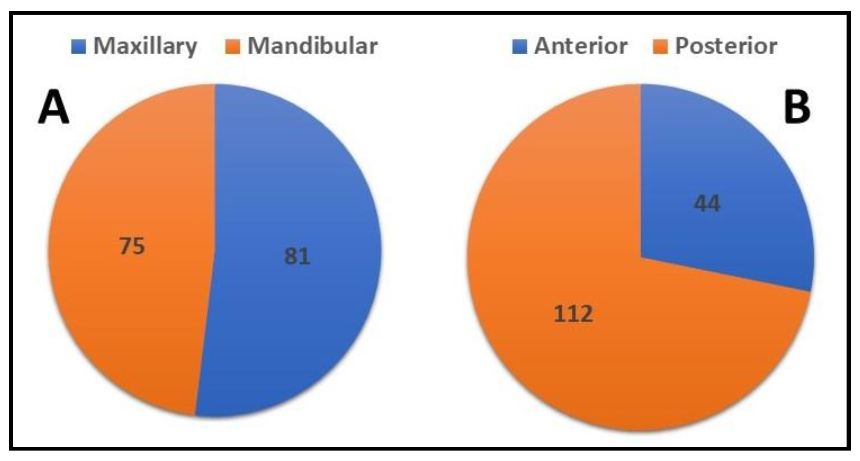
  • The posterior teeth were the tooth type most associated with the incidence of the errors.
  • There were no significant differences between the quality of RCT performed by different academic grades.
  • A clear plan should be applied to improve the quality of treatment outcomes for undergraduate students by implementing more effective preclinical training courses, increasing the supervision ratio, and using more advanced modalities during treatment.

Author Contributions

Conceptualization, M.A.M., K.C.S., D.S., M.K.A., M.A.A. and H.A.A.; methodology, M.A.M., A.M.A., A.Z.A., M.M.A., K.C.S., D.S., M.A.A. and H.A.A.; software, K.C.S., M.K.A., M.A.A. and H.A.A.; validation, D.S., M.A.A. and H.A.A.; formal analysis, K.C.S.; investigation, M.A.M., A.M.A., A.Z.A., M.M.A. and M.N.B.; writing—original draft preparation, M.A.M., K.C.S. and D.S.; writing—review and editing, M.A.M., A.M.A., A.Z.A., M.M.A., M.N.B., K.C.S., D.S., M.K.A., M.A.A. and H.A.A.; project administration, M.A.M. and H.A.A. All authors have read and agreed to the published version of the manuscript.

Funding

This research received no external funding.

Institutional Review Board Statement

The study was conducted in accordance with the Declaration of Helsinki and approved by the local committee for bioethics of Jouf University ([11-04-41]).

Informed Consent Statement

Not applicable.

Data Availability Statement

The data will be available on reasonable request from the corresponding authors.

Conflicts of Interest

The authors declare no conflict of interest.

References

  1. Kishen, A.; Peters, O.A.; Zehnder, M.; Diogenes, A.R.; Nair, M.K. Advances in endodontics: Potential applications in clinical practice. J. Conserv. Dent. 2016, 19, 199. [Google Scholar] [CrossRef] [PubMed]
  2. Soikkonen, K. Endodontically treated teeth and periapical findings in the elderly. Int. Endod. J. 1995, 28, 200–203. [Google Scholar] [CrossRef] [PubMed]
  3. Hansrani, V. Assessing root canal fillings on a radiograph—An overview. Br. Dent. J. 2015, 219, 481–483. [Google Scholar] [CrossRef] [PubMed]
  4. Endodontology ESo. Quality guidelines for endodontic treatment: Consensus report of the European Society of Endodontology. Int. Endod. J. 2006, 39, 921–930. [Google Scholar] [CrossRef] [PubMed]
  5. Stoll, R.; Betke, K.; Stachniss, V. The influence of different factors on the survival of root canal fillings: A 10-year retrospective study. J. Endod. 2005, 31, 783–790. [Google Scholar] [CrossRef] [PubMed]
  6. Chandra, A. Discuss the factors that affect the outcome of endodontic treatment. Aust. Endod. J. 2009, 35, 98–107. [Google Scholar] [CrossRef]
  7. Peters, O.A. Current challenges and concepts in the preparation of root canal systems: A review. J. Endod. 2004, 30, 559–567. [Google Scholar] [CrossRef] [PubMed]
  8. Kabak, Y.; Abbott, P. Prevalence of apical periodontitis and the quality of endodontic treatment in an adult Belarusian population. Int. Endod. J. 2005, 38, 238–245. [Google Scholar] [CrossRef]
  9. Chugal, N.M.; Clive, J.M.; Spångberg, L.S. Endodontic infection: Some biologic and treatment factors associated with outcome. Oral Surg. Oral Med. Oral Pathol. Oral Radiol. Endodontol. 2003, 96, 81–90. [Google Scholar] [CrossRef]
  10. Naito, T. Better success rate for root canal therapy when treatment includes obturation short of the apex. Evid. Based Dent. 2005, 6, 45. [Google Scholar] [CrossRef] [PubMed] [Green Version]
  11. Ganji, K.K.; Alam, M.K.; Gudipaneni, R.K.; Algarni, H.; Munisekhar, M.S.; Hamza, M.O.; Mousa, M.A.; Sghaireen, M.G. Do learning style preferences influence the cumulative gross point average and self directed learning hours in dental students: A preliminary study. BMC Med. Educ. 2022, 22, 493. [Google Scholar] [CrossRef] [PubMed]
  12. Srivastava, K.C.; Shrivastava, D.; Khan, Z.A.; Nagarajappa, A.K.; Mousa, M.A.; Hamza, M.O.; Al-Johani, K.; Alam, M.K. Evaluation of temporomandibular disorders among dental students of Saudi Arabia using Diagnostic Criteria for Temporomandibular Disorders (DC/TMD): A cross-sectional study. BMC Oral Health 2021, 21, 211. [Google Scholar] [CrossRef] [PubMed]
  13. Srivastava, K.C.; Shrivastava, D.; Nagarajappa, A.K.; Khan, Z.A.; Alzoubi, I.A.; Mousa, M.A.; Hamza, M.; David, A.P.; Al-Johani, K.; Sghaireen, M.G.; et al. Assessing the Prevalence and Association of Pulp Stones with Cardiovascular Diseases and Diabetes Mellitus in the Saudi Arabian Population—A CBCT Based Study. Int. J. Environ. Res. Public Health 2020, 17, 9293. [Google Scholar] [CrossRef]
  14. Awooda, E.M.; Siddig, R.I.; Alturki, R.S.; Sanhouri, N.M. Radiographic technical quality of root canal treatment performed by undergraduate dental students at the Academy Dental Teaching Hospital, UMST, Sudan. J. Int. Soc. Prev. Community Dent. 2016, 6, 554. [Google Scholar] [CrossRef]
  15. Elemam, R.F.; Abdul Majid, Z.S.; Groesbeck, M.; Azevedo, Á.F. Quality of root canals performed by the inaugural class of dental students at Libyan International Medical University. Int. J. Dent. 2015, 2015, 135120. [Google Scholar] [CrossRef] [PubMed]
  16. Elsayed, R.O.; Abu-bakr, N.H.; Ibrahim, Y.E. Quality of root canal treatment performed by undergraduate dental students at the University of Khartoum, Sudan. Aust. Endod. J. 2011, 37, 56–60. [Google Scholar] [CrossRef]
  17. Fong, W.; Heidarifar, O.; Killough, S.; Lappin, M.; El Karim, I. An audit on technical quality of root fillings performed by undergraduate students. Int. Endod. J. 2018, 51, e197–e203. [Google Scholar] [CrossRef]
  18. Vukadinov, T.; Blažić, L.; Kantardžić, I.; Lainović, T. Technical quality of root fillings performed by undergraduate students: A radiographic study. Sci. World J. 2014, 2014, 751274. [Google Scholar] [CrossRef]
  19. Rapo, H.; Oikarinen-Juusola, K.; Laitala, M.; Pesonen, P.; Anttonen, V. Outcomes of endodontic treatments performed by dental students-a follow-up study. J. Dent. Oral Biol. 2017, 2, 1046. [Google Scholar]
  20. Polyzos, N.K.; Sarris, K.G.; Pita, A.I.; Mikrogeorgis, G.V.; Lyroudia, K.M. Quality of root canal fillings performed by undergraduate students and the related factors on the treatment outcome: A 2-to 5-year follow-up. Eur. Endod. J. 2018, 3, 179. [Google Scholar] [CrossRef]
  21. Fritz, A.L.C.; Ribeiro, F.C.; Xavier, J.M.B.; Reis, C.M.; Demuner, C. Evaluation of Quality of Root Canal Fillings Performed by Undergraduate Students of a Brazilian University Avaliação da qualidade da obturação do canal radicular realizada por estudantes de graduação de uma Universidade brasileira. Braz. J. Health Rev. 2021, 4, 18964–18975. [Google Scholar] [CrossRef]
  22. Ribeiro, D.M.; Henckel, M.D.; Mello, F.W.; Felippe, M.C.S.; Felippe, W.T. Radiographic analysis the obturation’s quality in root canal treatment performed by a South Brazilian sample of undergraduate students. RGO-Rev. Gaúcha De Odontol. 2019, 67, e20190040. [Google Scholar] [CrossRef]
  23. da Silva, P.Z.; Ribeiro, F.C.; Xavier, J.M.B.; Pratte-Santos, R.; Demuner, C. Radiographic evaluation of root canal treatment performed by undergraduate students, part I; iatrogenic errors. Iran. Endod. J. 2018, 13, 30. [Google Scholar]
  24. Galhotra, V.; Rai, T.; Kaur, I. Assessment of quality of root canal fillings performed by undergraduate dental students. J. Adv. Med. Dent. Sci. Res. 2017, 5, 153. [Google Scholar]
  25. Alsulaimani, R.S.; Al-Manei, K.K.; Alsubait, S.A.; AlAqeely, R.S.; Al-Shehri, S.A.; Al-Madi, E.M. Effects of clinical training and case difficulty on the radiographic quality of root canal fillings performed by dental students in Saudi Arabia. Iran. Endod. J. 2015, 10, 268. [Google Scholar] [PubMed]
  26. Smadi, L.; Hammad, M.; El-Ma’aita, A. Evaluation of the Quality of Root Canal Treatments Performed by Dental Undergraduates: Is There a Need to Review Preclinical Endodontic Courses? Am. J. Educ. Res. 2015, 3, 1554–1558. [Google Scholar]
  27. AbuMostafa, A.; Ahmad, I.A.; Alenezy, G.; AlZoman, A. Quality of root canal filling performed by undergraduate students in a Saudi Dental College. J. Dent. Oral Hyg. 2015, 7, 64–70. [Google Scholar]
  28. AlRahabi, M.K. Evaluation of complications of root canal treatment performed by undergraduate dental students. Libyan J. Med. 2017, 12, 1345582. [Google Scholar] [CrossRef]
  29. Saatchi, M.; Mohammadi, G.; Sichani, A.V.; Moshkforoush, S. Technical quality of root canal treatment performed by undergraduate clinical students of Isfahan dental school. Iran. Endod. J. 2018, 13, 88. [Google Scholar]
  30. Yavari, H.; Samiei, M.; Shahi, S.; Borna, Z.; Abdollahi, A.A.; Ghiasvand, N.; Shariati, G. Radiographic evaluation of root canal fillings accomplished by undergraduate dental students. Iran. Endod. J. 2015, 10, 127. [Google Scholar]
  31. Eskandarloo, A.; Karkehabadi, H.; Hashemi, S.Z.H.; Ahmadi, M.; Hendi, S.S. Radiographic quality of root canal obturation performed by fifth year students of hamadan dental school. Iran. Endod. J. 2017, 12, 236. [Google Scholar] [PubMed]
  32. Chakravarthy, P.V.K.; Moorthy, J.R.K. Radiographic assessment of quality of root fillings performed by undergraduate students in a Malaysian Dental School. Saudi Endod. J. 2013, 3, 77. [Google Scholar] [CrossRef]
  33. Moradi, S.; Gharechahi, M. Quality of root canal obturation performed by senior undergraduate dental students. Iran. Endod. J. 2014, 9, 66. [Google Scholar]
  34. Mukhaimer, R. Radiographic technical quality of root canal fillings performed by dental students in Palestine. Indian J. Oral Sci. 2013, 4, 55. [Google Scholar] [CrossRef]
  35. Unal, G.C.; Kececi, A.D.; Kaya, B.U.; Tac, A.G. Quality of root canal fillings performed by undergraduate dental students. Eur. J. Dent. 2011, 5, 324–330. [Google Scholar] [CrossRef]
  36. Habib, A.A.; Doumani, M.D.; Nassani, M.Z.; Shamsy, E.; Jto, B.S.; Arwadi, H.A.; Mohamed, S.A. Radiographic assessment of the quality of root canal fillings performed by senior dental students. Eur. Endod. J. 2018, 3, 101. [Google Scholar] [CrossRef]
  37. Agwan, M.A.S.; Sheikh, A.; Shaikh, S. Qualitative Analysis of Root Canal Treatments performed by Undergraduate Students: A Retrospective Study. Pesqui. Bras. Em Odontopediatria E Clínica Integr. 2021, 21, e0260. [Google Scholar] [CrossRef]
  38. Ehsani, M.; Abesi, F.; Ghasemi, T. Technical Quality of Root Fillings Performed by Dental Students in Babol Dental School. J. Dent. Mater. Tech. 2014, 3, 66–70. [Google Scholar]
  39. Alghamdi, N.S.; Algarni, Y.A.; Ain, T.S.; Alfaifi, H.M.; AlQarni, A.A.; Mashyakhi, J.Q.; Alasmari, S.E.; Alshahrani, M.M. Endodontic mishaps during root canal treatment performed by undergraduate dental students: An observational study. Medicine 2021, 100, e27757. [Google Scholar] [CrossRef]
  40. Eckerbom, M. Prevalence and technical standard of endodontic treatment in a Swedish population. A longitudinal study. Swed. Dent. J. Suppl. 1993, 93, 1–45. [Google Scholar]
  41. Ödesjö, B.; Helldén, L.; Salonen, L.; Langeland, K. Prevalence of previous endodontic treatment, technical standard and occurrence of periapical lesions in a randomly selected adult, general population. Dent. Traumatol. 1990, 6, 265–272. [Google Scholar] [CrossRef] [PubMed]
  42. Frisk, F. Incidence, frequency, and prevalence. In Apical Periodontitis in Root-Filled Teeth: Endodontic Retreatment and Alternative Approaches; Springer: Berlin/Heidelberg, Germany, 2018; pp. 7–19. [Google Scholar]
  43. Endodontology ESo. Consensus report of the European Society of Endodontology on quality guidelines for endodontic treatment. Int. Endod. J. 1994, 27, 115–124. [Google Scholar] [CrossRef] [PubMed]
  44. Ng, Y.L.; Mann, V.; Gulabivala, K. A prospective study of the factors affecting outcomes of non-surgical root canal treatment: Part 2, tooth survival. Int. Endod. J. 2011, 44, 610–625. [Google Scholar] [CrossRef]
  45. Grayli, E.; Peyvandi, A.; Mallahi, M.; Naeemi, S.; Afshari, E. Radiographic Evaluation of Iatrogenic Errors of Root Canal Treatments Performed in an Undergraduate Dental Clinic. J. Dent. Mater. Tech. 2021, 10, 16–21. [Google Scholar]
Figure 1. Number of endodontic treatments performed (tooth type-wise) by students of different academic levels.
Applsci 12 09079 g001
Figure 2. Number of endodontic treatments performed (root canal-wise) by students of different academic levels.
Applsci 12 09079 g002
Figure 3. Graph showing overall cases performed and errors encountered.
Applsci 12 09079 g003
Figure 4. (A,B): distribution of errors encountered; (A): arch-wise; (B): location-wise.
Applsci 12 09079 g004
Table 1. Adequacy of root canal treatment conducted by dental students in the last 10 years.
The ContinentThe StudyLocationType of Study (Type of Course)Number of Treated Teeth (or Canals)Adequate (%)Not Adequate (%)
AfricaAwooda et al., 2016 [14]SudanIn-vivo (5th year)173 T (324 RC)55.5%44.5%
Elemam et al., 2015 [15]LibyaIn-vivo (4th and 5th year)128 T (284 RC)53.9%46.1%
Elsayed et al., 2011 [16]SudanIn-vivo (4th and 5th year)166 T (265 RC)52.7%47.3%
EuropeFong et al., 2018 [17]UKIn-vivo (4th and 5th year)222 T (381RC)66.4%33.6%
Vukadinov et al., 2014 [18]SerbiaIn-vivo (5th year)322 T (565RC)74.2%25.8%
Rapo et al., 2017 [19]FinlandIn-vivo (Undergrad)105 T (167 RC)65.7%34.3%
Polyzos et al., 2018 [20]GreeceIn-vivo (Undergrad)244 T (349 RC)40.4%59.6%
AmericaFritz et al., 2021 [21]BrazilIn-vivo (4th and 5th year)368 T (442 RC)93.8%6.2%
Ribeiro et al., 2019 [22]BrazilIn-vivo (4th and 5th year)274 T (381 RC)71.9%28.1%
da Silva et al., 2018 [23]BrazilIn-vivo (4th and 5th year)397 T (480 RC)92.7%7.3%
AsiaGalhotra et al., 2017 [24]IndiaIn-vivo (5th year and intern)135 T (246 RC)80.9%19.1%
Alsulaimani et al., 2017 [25]KSAIn-vivo (5th and 4th year)692 T (1081 RC)87.9%12.1%
Smadi et al., 2015 [26]JordanIn-vivo (4th and 5th year)213 T (327 RC)29.2%70.8%
AbuMostafa et al., 2015 [27]KSAIn-vivo (5th year)241 T (450 RC)26.1%73.9%
Alrahabi et al., 2017 [28]KSAIn-vivo (4th and 5th year)280 T (533 RC)68.9%31.1%
Saatchi et al., 2018 [29]IranIn-vivo (4th and 5th year)784 T (1674 RC)54.1%55.9%
Yavari et al., 2015 [30]IranIn-vivo (4th, 5th, and intern)620 T (1183 RC)72.1%27.9%
Eskandarloo et al., 2017 [31]IranIn-vivo (5th year)432 T (605 RC)70.8%29.2%
Chakravarthy et al., 2013 [32]MalaysiaIn-vivo (4th and 5th year)153 T (194 RC)61.3%38.7%
Moradi et al., 2014 [33]IranIn-vivo (4th and 5th year)200 T (411 RC)45%55%
Mukhaimer et al., 2013 [34]PalestineIn-vivo (4th and 5th year)612 T (1013 RC)71.7%28.3%
Unal et al., 2011 [35]TurkeyIn-vivo (4th and 5th year)460 T (833 RC)73.7%26.3%
Habib et al., 2018 [36]KSAIn-vivo (senior students)246 T (390 RC)32.6%66.4%
Agwan et al., 2021 [37]PakistanIn-vivo (5th year)92 T (170 RC)52.3%47.6%
Ehsani et al., 2014 [38]IranIn-vivo (4th and 5th year)325 T (727 RC)17.5%82.5%
Alghamdi et al., 2021 [39]KSAIn-vivo (4th and 5th year)404 T (730 RC)86%14%
Overall cases8288 T (14,205 RC)61.4%38.6%
Table 2. Criteria of calibration of performed root canal treatment.
VariableDefinitionCriteria
1Canal recognitionThe canal/s is recognized and filledAcceptable
One or more canals were not recognized Unacceptable
2Length of RCFRCF end 0–2 mm from the radiographic apexAcceptable
RCF end more than 2 mm from the apexUnderextended
RCF passed the radiographic apexOverextended
3Taper of RCFThe taper is consistent from the coronal to the apex of the treated toothAcceptable
The taper is lacking from the coronal to the apexUnacceptable
4Density of RCFNo voids are recognizable within the canalAcceptable
Voids are recognizable within the canalUnacceptable
5Coronal sealThe coronal part is adequately sealed by filling, adequate inserted post, or crowningAcceptable
Lack of coronal seal due to deficiency in the restoration Unacceptable
6Apical sealThe apex of tooth is apparently sealed with RCFAcceptable
Lack of the seal or voids is recognizable at the apex of the RCFAcceptable
7Broken instrumentNo broken instrument can be seen within the canalsAcceptable
One or more broken instrument is apparent within one or more canalsUnacceptable
RCF root canal filling.
Table 3. The frequency and percentage of errors in RCF performed by 4th year students according to various tooth types.
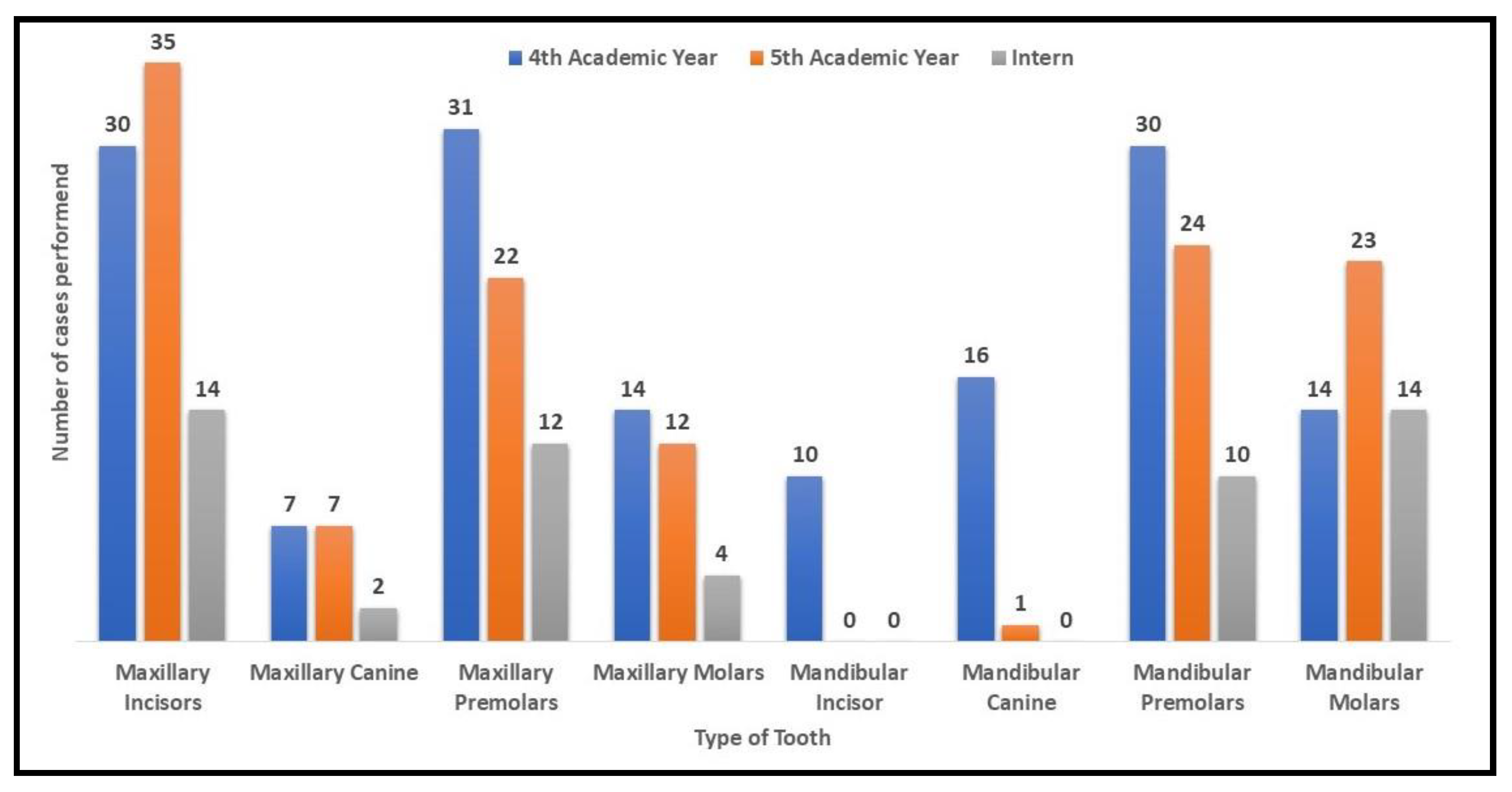
4th Year Students [n = 152 (45.8)]
Nature of Endodontic ErrorTooth Types p Value
Max. Incisors (n = 30)Max. Canine (n = 7)Max. PM (n = 31)Max. Molars (n = 14)Mandi. Incisor (n = 10)Mandi. Canine (n = 16)Mandi. PM (n = 30)Mandi. Molars (n = 14)n. Errors (%)
Root perforation002 (50)0002 (50)04 (2.9)0.525
Short obturation6 (18.8)2 (6.3)10 (31.3)2 (6.3)02 (6.3)6 (18.8)4 (12.5)32 (23.4)0.444
Over-extended obturation4 (25)006 (37.5)02 (12.5)04 (25)16 (11.7)0.000
Voids4 (25)02 (12.5)4 (25)02 (12.5)4 (25)016 (11.7)0.212
Canal Left Unfilled2 (14.3)2 (14.3)4 (28.6)0004 (28.6)2 (14.3)14 (10.2)0.002
No Coronal Seal2 (12.5)02 (12.5)2 (12.5)004 (25)6 (37.5)16 (11.7)0.004
No Apical Seal2 (8)2 (8)5 (20)6 (24)006 (24)4 (16)25 (18.2)0.019 *
Broken Instrument008 (57.1)02 (14.3)2 (14.3)02 (14.3)14 (10.2)0.005
Errors% 137 (90.13)
Note: Results are expressed endodontic errors encountered in number (%); * p < 0.05; € p < 0.01; ⁋ p < 0.001.
Table 4. The frequency and percentage of errors in RCF performed by 5th-year students according to various tooth types.
5th Year Students [n = 124 (37.3)]
Nature of Endodontic ErrorTooth Types p Value
Max. Incisors (n = 35)Max. Canine (n = 7)Max. PM
(n = 22)
Max. Molars (n = 12)Mandi. Canine (n = 1)Mandi. PM (n = 24)Mandi. Molars (n = 23)n. Errors (n = 125)
Root Perforation01 (16.7)2 (33.3)01 (16.7)02 (33.3)6 (4.8)0.000
Short Obturation8 (18.6)2 (4.7)6 (14)0017 (39.5)10 (23.3)43 (34.4)0.000
Over-extended obturation2 (50)000002 (50)4 (3.2)0.531
Voids0000003 (100)3 (2.4)0.036 *
Canal Left Unfilled3 (21.4)03 (21.4)004 (28.6)4 (28.6)14 (11.2)0.241
No Coronal Seal05 (50)00005 (50)10 (8)0.01 *
No Apical Seal8 (20.5)2 (5.1)7 (17.9)1 (2.6)012 (30.8)9 (23.1)39 (31.2)0.167
Broken Instrument004 (66.7)0002 (33.3)6 (4.8)0.039 *
Errors 125
Note: Results are expressed endodontic errors encountered in number (%); * p < 0.05; ⁋ p < 0.001.
Table 5. The frequency and percentage of errors in RCF performed by interns according to various tooth types.
Intern [n = 56 (16.9)]
Nature of Endodontic ErrorTooth Types p Value
Max. Incisors (n = 14)Max. Canine (n = 2)Max. PM (n = 12)Max. Molars (n = 4)Mandi. PM (n = 10)Mandi. Molars (n = 14)Errors (n = 42)
Root Perforation0000000 (0)NA
Short Obturation002 (16.7)04 (33.3)6 (50)12 (28.6)0.043 *
Over-extended Obturation2 (50)002 (50)004 (9.5)0.010 *
Voids0000000 (0)NA
Canal Left Unfilled2 (20)002 (20)2 (20.0)4 (40)10 (23.8)0.213
No Coronal Seal0002 (50)02 (50)4 (9.5)0.010 *
No Apical Seal002 (16.7)2 (16.7)4 (33.3)4 (33.3)12 (28.6)0.112
Broken Instrument0000000 (0)NA
Note: Results are expressed endodontic errors encountered in number (%); * p < 0.05; NA—Not applicable.
Table 6. The frequency and percentage of errors in RCF performed by different academic grades according to various tooth types.
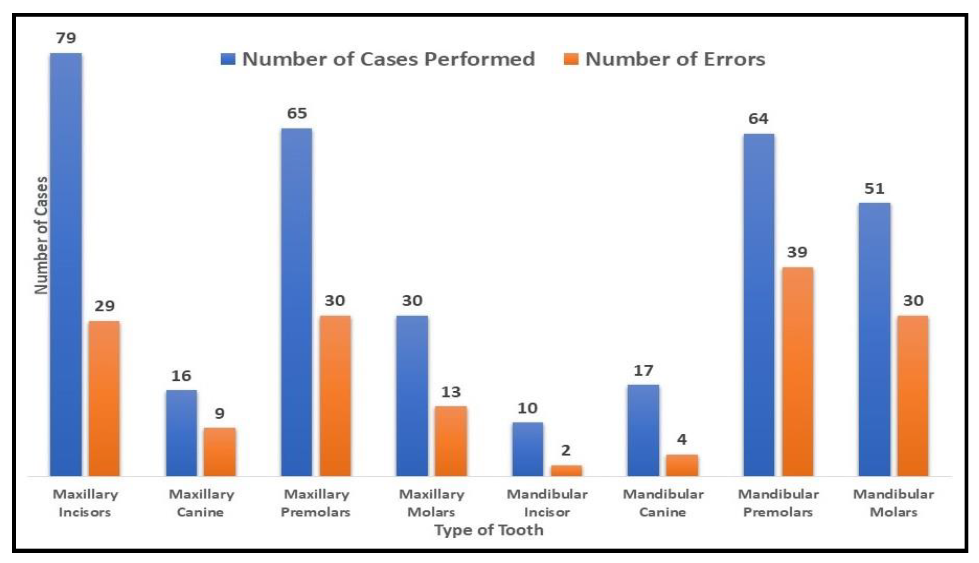
4th Yr. + 5th Yr. + Intern = 332 (n)
Nature of Endodontic ErrorTooth Types
Maxillary Incisors (30 + 35 + 14 = 79)Maxillary Canine (7 + 7 + 2 = 16)Maxillary Premolars (31 + 22 + 12 = 65)Maxillary Molars (14 + 12 + 4 = 30)Mandibular Premolars (30 + 24 + 10 = 64)Mandibular Molars (14 + 23 + 14 = 51)Sum of Errors (%)
Root Perforation4th Yr.002 (6.5)02 (6.7)04
5th Yr.012 (9.1)002 (8.7)4
Intern0000000
p valueNA0.5040.571NA0.310.2828 (2.8)
Short Obturation4th Yr.6 (20)2 (28.6)10 (32.3)2 (14.3)6 (20)4 (28.6)30
5th Yr.8 (22.9)2 (28.6)6 (27.3)017 (70.8)10 (43.5)43
Intern002 (16.7)04 (40)6 (42.9)12
p value0.1570.6830.5910.3160.00 €0.63285 (29.6)
Over-extended Obturation4th Yr.4 (13.3)006 (42.9)04 (28.6)14
5th Yr.2 (5.7)00002 (8.7)4
Intern2 (14.3)002 (50)004
p value0.513NANA0.025 *NA0.05322 (7.7)
Voids4th Yr.4 (13.3)02 (6.5)44 (13.3)016
5th Yr.000003 (13)3
Intern0000000
p value0.03 *NA0.3230.0720.0890.14419 (6.6)
Canal left unfilled4th Yr.2 (6.7)2 (28.6)4 (13)04 (13.3)2 (14.3)14
5th Yr.3 (8.6)03 (13.6)04 (16.7)4 (17.4)14
Intern2 (14.2)002 (50)2 (20)4 (28.6)10
p value0.780.010.130.00 €0.8670.59538 (13.2)
No Coronal Seal4th Yr.2 (6.7)02 (6.5)2 (14.3)4 (13.3)6 (42.9)16
5th Yr.02 (28.6)0005 (21.7)7
Intern0002 (50)02 (14.3)4
p value0.1910.230.3230.039 *0.0890.1927 (9.4)
No Apical Seal4th Yr.2 (6.7)2 (28.6)5 (16.7)6 (42.9)6 (20)4 (28.6)25
5th Yr.8 (22.9)2 (28.6)7 (31.8)1 (8.3)12 (50) 9 (39.1)35
Intern002 (16.7)2 (50)4 (40)4 (28.6)12
p value0.045 *0.6830.3790.1030.0640.72872 (25.1)
Broken Instrument4th Yr.008 (25.8)002 (14.3)10
5th Yr.004 (18.2)002 (8.7)6
Intern0000000
p valueNANA0.147NANA0.36416 (5.6)
Sum of errors287
Note: Results are expressed endodontic errors encountered in number (%); * p < 0.05; € p < 0.01; NA—Not applicable.
Publisher’s Note: MDPI stays neutral with regard to jurisdictional claims in published maps and institutional affiliations.

