Article

Benincasa hispida Extract Promotes Proliferation, Differentiation, and Mineralization of MC3T3-E1 Preosteoblasts and Inhibits the Differentiation of RAW 246.7 Osteoclast Precursors

Haram Central Research Institute, Cheongju 28160, Korea
*
Author to whom correspondence should be addressed.
Appl. Sci. 2022, 12(17), 8849; https://doi.org/10.3390/app12178849
Submission received: 4 July 2022 / Revised: 23 August 2022 / Accepted: 1 September 2022 / Published: 2 September 2022

Abstract

:
Owing to global population aging, instances of bone metabolic diseases have increased. Consequently, interest in natural and functional plant food products for the prevention and treatment of osteoporosis is also increasing. In this study, we determine the potential therapeutic effects of Benincasa hispida extract (HR1901-W) on osteoblast and osteoclast differentiation. The potential preventive effects of Benincasa hispida in osteoporosis have not previously been reported. We identified and analyzed 2-furoic acid, a chemical component of HR1901-W. We evaluated whether HR1901-W promoted osteogenesis in the MC3T3-E1 cell line and whether it inhibited the differentiation of RAW 264.7 macrophage cells (osteoblast precursors). We observed that HR1901-W promoted significantly high dose-dependent proliferation and extracellular matrix mineralization in MC3T3-E1 cells. In fact, increased cell proliferation was found to be associated with increased protein expression of factors related to osteoblast differentiation, including alkaline phosphatase, osteocalcin, and runt-related transcription factor 2. On the other hand, macrophage colony-stimulating factor (10 ng/mL) and nuclear factor-κB ligand (100 ng/mL) treated differentiated RAW264.7 macrophages exhibited a significant reduction in tartrate-resistant acid phosphatase activity. Taken together, our results indicate that HR1901-W is a promising candidate of functional materials that regulate the balance between bone-forming osteoblasts and bone-resorbing osteoclasts to prevent osteoporosis.

1. Introduction

A bone is a mineralized tissue of calcium and phosphorus; it is a storage warehouse of minerals, and it functions to support and protect the body [1,2]. Skeletal homeostasis is maintained when osteoclast-regulated bone resorption is balanced by osteoblast-regulated bone matrix formation. However, the bone quantity per unit volume decreases when bone resorption exceeds bone formation. This pathological scenario leads to the development of osteoporosis, a systemic skeletal disease [3,4]. Osteoporosis is accompanied by various complications, including bone fractures, cardiopulmonary dysfunction, and economic burden [5,6]. Clinically, patients with osteoporosis are administered estrogen hormones to promote osteoblastogenesis and increase the proliferation and differentiation of osteoblasts [7]. Alternatively, oral bisphosphonates are also provided as a treatment to inhibit osteoclastic activity, as they are potent antiresorptive agents [8,9]. Although these treatments are effective, their long-term use is not beneficial because of side effects such as headache, weight gain, breast cancer, and jaw osteonecrosis [10]. Therefore, studies investigating natural products that inhibit bone loss, promote bone formation, and have minimum side effects are being conducted.
Osteoblasts originate from the mesenchymal stem cells of the bone marrow. They are involved in the synthesis and mineralization of the bone matrix, wherein calcium and other minerals are deposited onto the bone or its extracellular matrix [11,12]. Notably, the differentiation of osteoblasts is mediated by the differential expression of specific factors. For instance, runt-related transcription factor 2 (Runx2) regulates the early stages of osteoblast differentiation by promoting the differentiation of pluripotent mesenchymal cells into immature osteoblasts [13,14]. Another key factor of osteoblast differentiation is alkaline phosphatase (ALP) that is synthesized by osteoblast precursors immediately following growth arrest [15,16]. Notably, ALP is an ectoenzyme that is attached to the outer surface of the osteoblast cell membrane. It facilitates calcium deposition on the bone by transporting inorganic phosphoric acid during mineralization [17,18]. While it initially forms the bone mass, it subsequently plays a role in bone remodeling and mineral metabolism [19,20].
Osteoclasts are multinucleated cells derived from the monocyte/macrophage lineage that resorb the mineralized matrices formed by osteoblasts. The macrophage colony-stimulating factor (M-CSF) and the receptor activator of nuclear factor-κB ligand (RANKL) stimulate the differentiation of immature osteoclasts into mature osteoclasts. These factors are recruited by hematopoietic stem cells to specific regions of the bone surface. Furthermore, they initiate bone resorption by stimulating osteoclastogenesis factors in the early stage of osteoclasts [21,22]. Tartrate-resistant acid phosphatase (TRAP) is specifically expressed when immature osteoclasts differentiate into multinucleated osteoclasts. Notably, its expression is used to confirm osteoclast proliferation [23].
Benincasa hispida (Thunb.) Cogn. (syn. Benincasa cerifera Savi) is an annual plant of the family Cucurbitaceae. It is a vegetable widely distributed in Asian countries and regions such as Southeast Asia, India, China, and Korea [24,25]. B. hispida fruit contains 93–96% moisture and is rich in nutrients such as vitamin C, vitamin B2, Na, and Ca [17,26,27]. Phenolic compounds such as astilbin, catechin, and naringenin have been isolated from B. hispida fruit [28]. Other bioactive compound constituents include triterpenes (alnusenol, multiflorenol, isomultiflorenol), sterols (lupeol, lupeol acetate, β-sitosterol), glycosides, saccharides, carotenes, β-sitosterin, tannins, and uronic acid [29,30]. However more studies should be done on the isolation and identification of chemical components contained in B. hispida. Several studies on the functionality of B. hispida fruit extracts have reported their antioxidant, anti-inflammatory, anti-ulcerogenic, antidiabetic, and anti-Alzheimer’s efficacy [31,32,33,34,35,36]. However, there have been no studies on the effect of B. hispida on the prevention of bone diseases, including osteoporosis.
Therefore, in this study, we identified and quantitatively analyzed the chemical components of B. hispida extract (HR1901-W). Furthermore, we evaluated the inhibitory effects of extract on the proliferation and differentiation of osteoblasts and osteoclasts in MC3T3-E1 cells (osteoblasts) and RAW 264.7 macrophage cells (osteoclast precursors). We thereby investigated their potential as a natural plant-based functional food to treat osteoporosis.

2. Materials and Methods

2.1. Chemicals

We purchased 2-furoic acid from Sigma (Sigma Aldrich, St. Louis, MO, USA). Methanol was used as a solvent and acetonitrile was used as a mobile phase as described by J.T. Baker (Phillipsburg, NJ, USA). Trifluoroacetic acid was purchased from Sigma Aldrich. α-Minimal Essential Medium (αMEM) and fetal bovine serum (FBS) were obtained from Gibco (Gibco, Waltham, MA, USA). Furthermore, we purchased Dulbecco’s Modified Eagle’s Medium (DMEM) and penicillin–streptomycin from Hyclone (HyClone Laboratodies Inc., Logan, UT, USA). The ALP, MTT, and TRAP assay kits were obtained from Abcam (Abcam, Cambridge, UK), DoGenBio (DoGenBio, Seoul, Korea), and BioVision (BioVision Inc., Milpitas, CA, USA), respectively. The antibodies used for immunoblotting were obtained from Cell Signaling Technology (Cell Signaling Technology, Danvers, MA, USA) and Thermo Fisher (Thermo Fisher Scientific, Milan, Italy), whereas the BSA and ECL kits were purchased from Bio-Rad (Bio-Rad, Hercules, CA, USA).

2.2. Sample Preparation

We purchased B. hispida in August 2020 from Korea and prepared samples at Haram Co. Ltd. (Cheongju, Chungbok, Korea). To the harvested fruits, we added distilled water that was 10 times the weight of the fruits and extracted the sample at 100 °C for 4 h. The extracts were filtered and concentrated to 10–13 Brix. They were then freeze-dried using a freeze-dryer (Labcono, Kansas City, MO, USA) to obtain B. hispida extract samples (HR1901-W).

2.3. Chemical Component Characterization

A quantity of 5 g of HR1901-W was dissolved in 10 mL of 50% methanol and filtered using a 0.45 μM polyvinylidene fluoride (PVDF) syringe filter (Shatman, Maidstone, UK). The filtrate was concentrated under reduced pressure. To the concentrated sample, 10 mL of methanol was added; then, the sample was sonicated for 10 min and centrifuged (3000 rpm/5 min). The supernatant was filtered using a 0.45 μM PVDF syringe filter and used as a microfractionation and NMR test solution. For the characterization of compounds, preparative HPLC and NMR spectra were obtained. Identification of the main chemical components was performed by modifying Tan et al. [37]. We isolated HR1901-W using a preparative HPLC instrument combined with a Waters e2695 separation module HPLC system, a Waters 2998 photodiode array detector (Waters Co., Milford, MA, USA), and a phenomenex luna C18 column (10 mm × 250 mm × 5.0 μm; Phenomenex, Torrance, CA). The mobile phase comprised solvent A, 0.1% trifluoroacetic acid in distilled water (v/v), and solvent B, acetonitrile. The parameters for the HPLC analysis were as follows: 0–2 min: 90% A, 10% B; 2–15 min: 70% A, 30% B; 15–16 min: 30% A, 70% B; 16–18 min: 30% A, 70% B; 18–19 min: 30% A, 70% B; 19–22 min: 10% A, 90% B; 22–23 min: 10% A, 90% B; 23–30 min: 90% A, 10% B gradient system. The analysis time was 30 min, the detection wavelength was 200–400 nm, the sample injection volume was 90 µL, the flow rate was 2.5 mL/min, and the column temperature was maintained at 30 °C. 1H NMR and 13C NMR spectra were recorded in CDDl3 using the JEOL JNM-ECX400 (1H-400 MHz, 13C-100 MHz) FT NMR systems (Tokyo, Japan) with Me4Si as the internal standard.
2-Furoic acid -1H-NMR (400 MHz, CDCl3, Me4Si) δ: 6.58 (7-H), 7.20 (8-H), 7.72 (6-H) 13C-NMR (100 MHz, CDCl3, Me4Si) δ: 113.05 (2-C), 119.18 (3-C), 146.56 (1-C), 148.21 (4-C), 159.45 (5-C).

2.4. HPLC Analysis of 2-Furoic Acid

The analysis of 2-furoic acid was performed by modifying Tan et al. [37]. We detected the presence of 2-furoic acid in the extracts using an HPLC instrument combined with a Waters 2998 separation module HPLC system, a Waters 996 photodiode array detector (Waters Co., Milford, MA, USA), and a Kromasil C18 column (4.6 mm × 250 mm × 5.0 μm; Tedia, Rio de Janeiro, Brazil). The mobile phase comprised solvent A, 0.1% trifluoroacetic in distilled water (v/v), and solvent B, acetonitrile. The parameters for the HPLC analysis were as follows: 0–5 min: 90% A, 10% B; 5–15 min: 70% A, 30% B; 15–20 min: 30% A, 70% B; 20–25 min: 30% A, 70% B; 25–30 min: 30% A, 70% B; 30–32 min: 10% A, 90% B; 32–33 min: 10% A, 90% B; 33–40 min: 90% A, 10% B gradient system. The analysis time was 40 min, the detection wavelength was 254 nm, the sample injection volume was 20 µL, the flow rate was 1.0 mL/min, and the column temperature was maintained at 30 °C. Additionally, we prepared stock solutions (100 μg/mL) of 2-furoic acid by dissolving 1 mg of each standard with 50% methanol in a 10 mL diaphragm flask. The stock solutions were prepared in 6.25–100 μg/mL concentrations. We weighed 200 mg of HR1901-W and dissolved it in 50% methanol in a 10 mL vial to obtain a concentration of 20,000 μg/mL. Subsequently, the test samples were injected in the HPLC column using a PVDF syringe filter.

2.5. Osteoblast Cell Culture

We purchased the MC3T3-E1 preosteoblast cell line from the Korean Cell Line Bank (KCLB, Seoul, Korea). We cultured the cells in αMEM medium with 10% FBS supplemented with 100 units/mL of penicillin–streptomycin at 37 °C in a 5% CO2 incubator (Thermo Fisher Scientific, Waltham, MA, USA).

2.6. Osteoblast Cell Proliferation

We incubated 5 × 10³ MC3T3-E1 cells/100 μL in a 96 well-plate at 37 °C in a 5% CO2 incubator for 24 h. These cells were then treated with HR1901-W and 2-furoic acid for 48 h. Cell proliferation was estimated via MTT assay using an MTT assay kit (DoGenBio, Seoul, Korea). The absorbances of the wells were measured at 450 nm using a microplate reader (Epoch, Biotek Instruments, Inc., Winooski, VT, USA).

2.7. Analysis of ALP Activity

We incubated 5 × 10³ MC3T3-E1 cells/250 μL in a 48 well-plate at 37 °C in a 5% CO2 incubator for 24 h. Thereafter, the cells were treated with HR1901-W and 2-furoic acid for 48 h. The cells were washed with phosphate-buffered saline (PBS) and lysed using lysis buffer. Subsequently, they were centrifuged at 15,800× g for 15 min to precipitate the cell membrane components, and we collected the supernatant. We used the ALP assay kit to quantify the ALP activity in the supernatant as per the manufacturer’s instructions.

2.8. Western Blotting

We estimated the expression levels of the proteins involved in osteoblast differentiation by Western blotting. Initially, 5 × 10⁴ MC3T3-E1 cells/1 mL were cultured in a 100 mm dish for 24 h. They were cultured in a differentiation induction medium that was supplemented with ascorbic acid (50 μg/mL) and β-glucan (10 mM). Once the cells differentiated, they were treated with the sample extracts, and the cells were then cultured again in a CO2 incubator for 24 h. Subsequently, we harvested the cells and washed them with PBS. Next, the cells were lysed with RIPA buffer, and the supernatant was collected post centrifugation (15,800× g, 15 min). The cellular protein concentration was quantified using a BSA kit. Thereafter, the quantified proteins were separated by electrophoresis on 10% sodium dodecyl sulfate polyacrylamide gel at 80–90 V. The separated proteins were transferred onto a PVDF membrane for 1 h at 100 V. The membrane was blocked with 5% skim milk at 37 °C for 1 h and was subsequently washed thrice with tris-buffered saline with 5% Tween-20 (TBS-T) for 15 min. The membrane was incubated with alkaline phosphatase (Cell Signaling, cat# 8681S), osteocalcin (Thermo Fisher, Cat# VS-4917R), and Runx2 (Cell Signaling, cat# 12556S) primary antibodies (1:200–1:1000) and subsequently washed with TBS-T. The membrane was then incubated with horseradish-peroxidase-conjugated anti-rabbit IgG (Cell Signaling, cat# 7074S) and anti-mouse IgG (Cell Signaling, cat# 7076S) antibodies for 1 h (1:3000). The membrane was once again washed with TBS-T. Protein expression levels were detected upon addition of the chemiluminescent ECL reagent to the membrane and quantified using an imaging densitometer (ImageJ bundled with 64-bit Java 1.8.0_112).

2.9. Osteoclast Cell Culture

The murine macrophage cell line RAW264.7 was purchased from the Korea Cell Line Bank (KCLB, Seoul, Korea). The cells were cultured in DMEM supplemented with 10% FBS and 100 units/mL penicillin–streptomycin at 37 °C in a 5% CO2 incubator.

2.10. Cell Viability

We seeded and incubated 5 × 10³ RAW264.7 cells/100 μL in a 96-well plate at 37 °C in a 5% CO2 incubator for 24 h. Thereafter, the cells were treated with HR1901-W for 24 h. We evaluated the cell viability using an MTT assay kit. The absorbance in each well was measured at 450 nm using a microplate reader (Epoch, Biotek Instruments, Inc., Winooski, VT, USA).

2.11. TRAP Assay

We seeded 5 × 10³ RAW264.7 cells/250 μL in a 48-well plate and treated them with HR1901-W. The treated cells were cultured in a medium supplemented with M-CSF (10 ng/mL) and RANKL (100 ng/mL) to induce osteoclast differentiation for 48 h. Following incubation, the cells were washed with PBS and lysed using a lysis buffer. They were subsequently centrifuged at 12,000 rpm for 15 min to precipitate the cell membrane components. We evaluated the TRAP activity in the resultant supernatant using a TRAP assay kit, in accordance with the manufacturer’s instructions.

2.12. Statistical Analyses

All the experimental data were statistically analyzed using GraphPad Prism 8.0 (Graph Pad Software, San Diego, CA, USA) software. Statistical significance was assessed via Student’s t-test and one-way analysis of variance (ANOVA). The results are expressed as the mean ± standard deviation (SD). p values of <0.05, <0.01, and <0.001 were considered statistically significant.

3. Results

3.1. Identification of Chemical Components in HR1901-W by Preparative HPLC and NMR

In order to identify the chemical components contained in HR1901-W, it was separated and fractionated by preparative HPLC. Nuclear magnetic resonance (NMR) was used for structural identification. The HPLC measurement wavelength was set to 254 nm, and the chromatography and spectra of the main components identified in the HR1901-W chromatogram were compared. The main component peak (corresponding to compound 1) in the 15 min was confirmed, and the maximum wavelength was shown at 252.5 nm (Figure S1b). The spectrum of HR1901-W showed a maximum wavelength at 252.5 nm. The 1H NMR spectrum of HR1901-W shows a peak observed at δ3.58–7.7, indicating the H-7, H-8, and H-6 protons of 2-furoic acid. Meanwhile, the 13C-NMR spectrum of HR1901-W shows a peak at 113.05–159.45 ppm, indicating the C-2, C-3, C-1, C-4, and C-5 carbon atoms of 2-furoic acid (Figure S1c,d). As a result of comparing the 1H-NMR and 13C-NMR spectra with the literature, this compound was estimated to be 2-furoic acid (Figure S1a) [38]. In this study, the chemical component of HR1901-W was set to 2-furoic acid by referring to the results of identification via UV and NMR.

3.2. Analysis of the 2-Furoic Acid Concentration in HR1901-W

We measured the concentrations of 2-furoic acid present in HR1901-W by HPLC. Figure 1 shows the UV spectra and chromatograms of 2-furoic acid and HR1901-W. The 2-furoic acid and HR1901-W showed the same maximum absorption wavelength of 249.8 nm in the UV 200 nm–400 nm wavelength range. 2-Furoic acid was separated from HR1901-W without interference from other substances and exhibited the same peak retention time. The 2-furoic acid concentration in HR1901-W was 0.37 ± 0.01 mg/dry weight in g (Table S1).

3.3. Effect of HR1901-W on the Proliferation of MC3T3-E1 Preosteoblasts and ALP Activity

We evaluated the effects of the extracts on the cell proliferation and differentiation of MC3T3-E1 preosteoblasts. As shown in Figure 2, we observed that varying concentrations of HR1901-W increased the proliferation of the MC3T3-E1 cells. In addition, the ALP activity was increased in the HR1901-W treatment. Genistein (20 μg/mL) was used as a positive control. Thus, we deduced that the extracts promote the mineralization of the bone extracellular matrix.

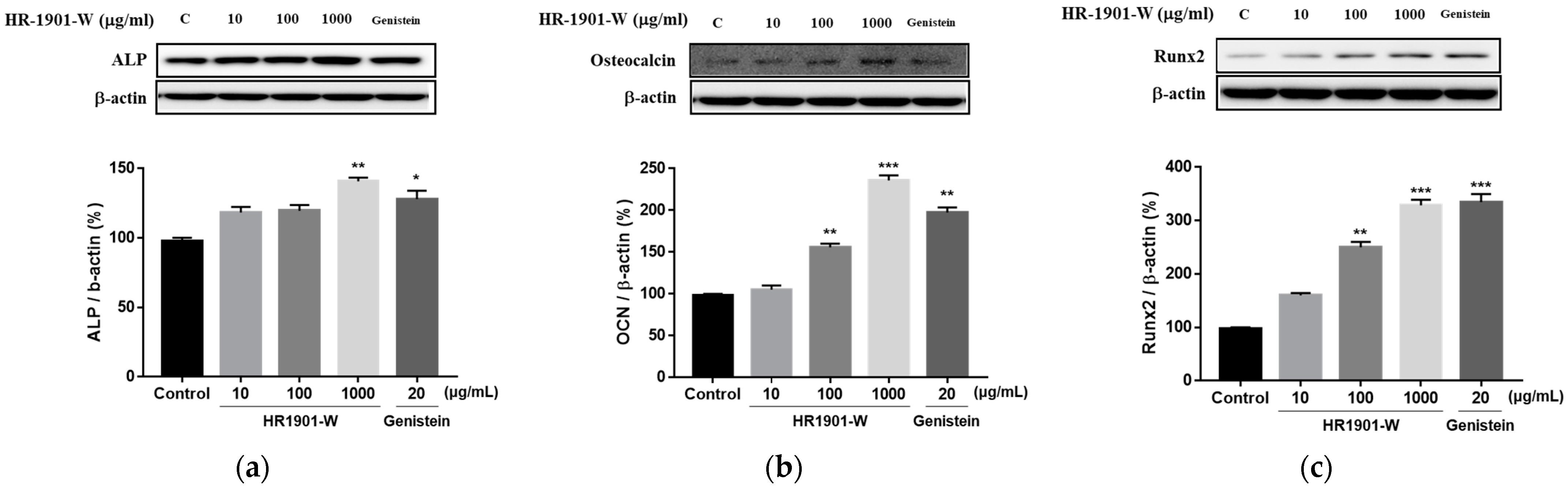
3.4. Effects of HR1901-W on Expression Levels of Factors Related to Osteoblast Differentiation

We determined the potential of HR1901-W to induce the differentiation of MC3T3-E1 preosteoblasts into osteoblasts. Furthermore, the total protein content of the cells was estimated by immunoblotting. As shown in Figure 3, we observed that the protein expression levels of ALP, OCN, and Runx2 were increased by the HR1901-W treatments when compared to the positive control, genistein. These results suggest that HR1901-W promotes osteoblast differentiation and osteocalcification by increasing the expression levels of ALP, OCN, and Runx2.

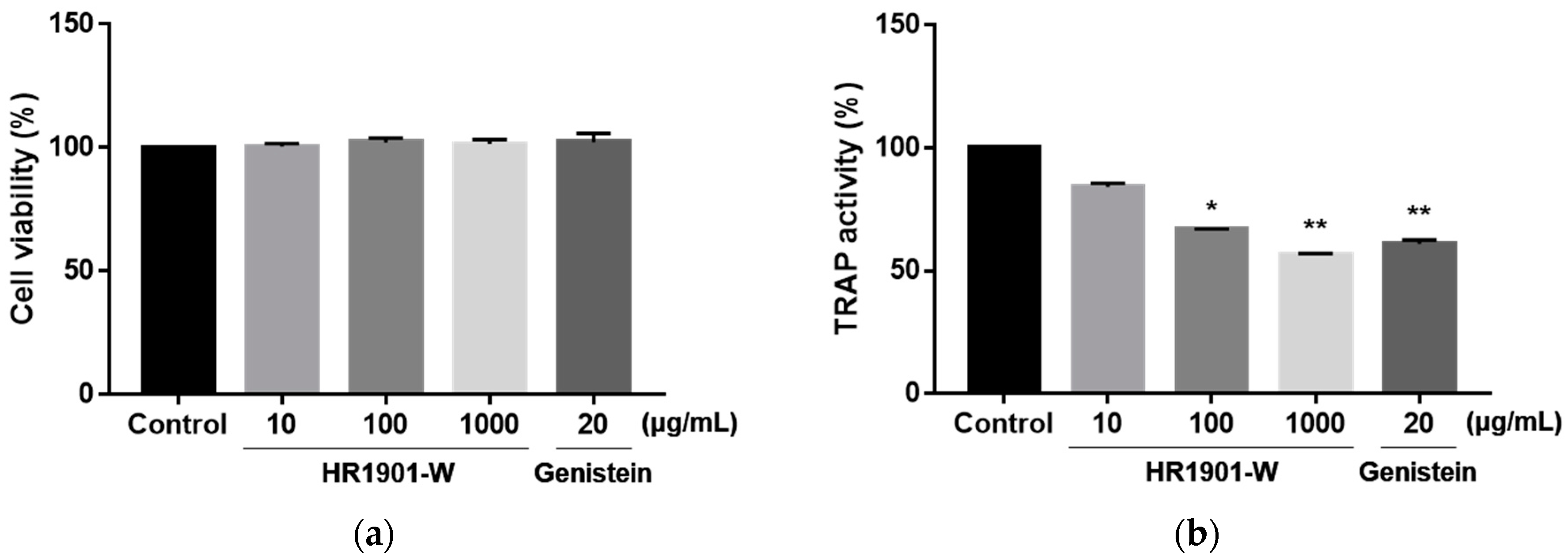
3.5. Effects of HR1901-W on Osteoclast Cell Viability and TRAP Activity

We evaluated the cytotoxicity of HR1901-W on RAW 264.7 cells. As shown in Figure 4, the different concentrations of HR1901-W did not affect the viability of the RAW264.7 cells. Osteoclasts are bone-resorbing cells that have TRAP and calcitonin receptors. Therefore, we determined the effect of the extracts on the differentiation of osteoclast progenitor cells into osteoclasts via TRAP assay. HR1901-W and genistein reduced the TRAP activity of the RAW264.7 cells. Therefore, HR1901-W inhibits TRAP activity to inhibit bone resorption.

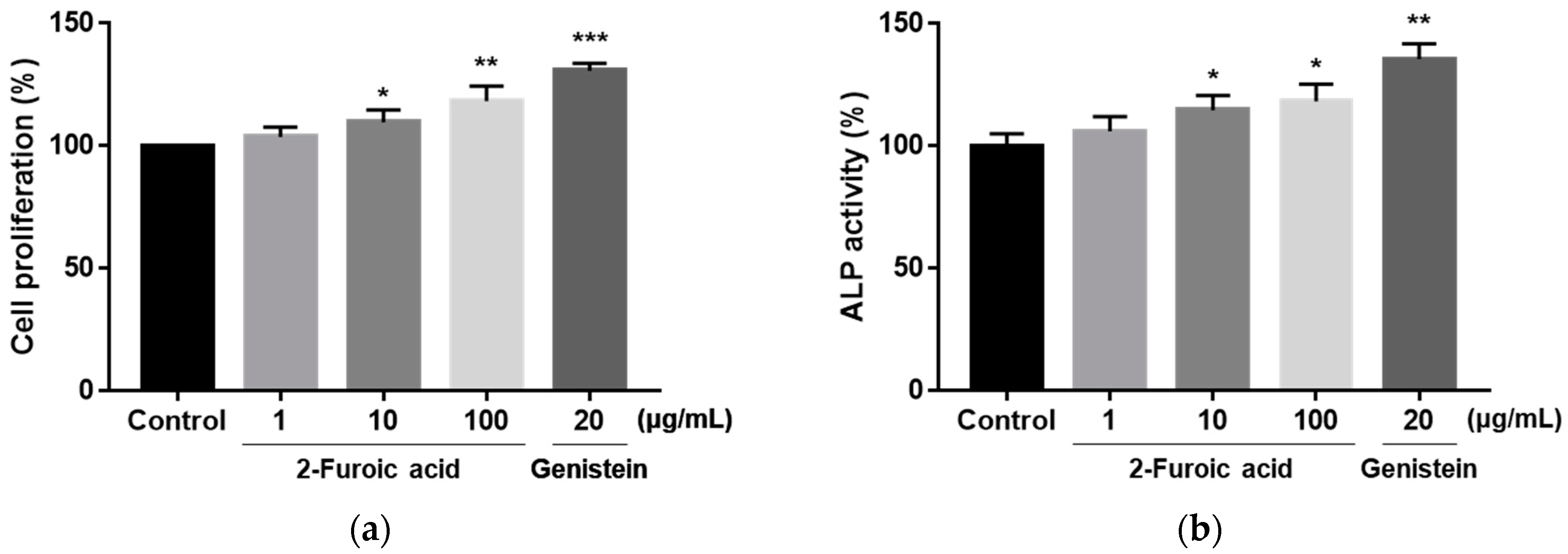
3.6. Effect of 2-Furoic Acid on the Proliferation of MC3T3-E1 Preosteoblasts and ALP Activity

We evaluated the effect of treatment with 2-furoic acid, contained in HR1901-W, on the cell proliferation of MC3T3-E1 preosteoblasts (Figure 5). 2-Furoic acid was found to increase MC3T3-E1 cell proliferation in a dose-dependent manner. Also, ALP activity was increased by treatment with 2-furoic acid. These results suggest that 2-furoic acid promotes the proliferation of osteoblasts.

4. Discussion

Bone remodeling is a process in which old or damaged bone is removed by osteoclasts and replaced with new bone formed by osteoblasts. Osteoblast-mediated bone formation and osteoclast-mediated bone resorption balance dysregulation lead to abnormal bone remodeling, leading to osteoporosis [39]. Throughout life, bone tissue undergoes continuous remodeling that requires the concerted action of bone-forming osteoblasts and bone-resorbing osteoclasts [40]. The current research trend for anti-osteoporotic treatment and prevention is to induce bone remodeling through the regulation of osteoblasts or osteoclasts using natural dietary compounds.
B. hispida (Thunb.) Cogn. is a cucurbitaceae, a food material with potential for functional food production. B. hispida fruit has pharmacological activities such as antioxidant, anti-inflammatory, gastroprotective, and neuroprotective properties [32,34,35,36,41]. The various pharmacological effects of B. hispida can be attributed to the presence of phenolic compounds. Previous studies reported the identification of astilbin, catechin, and naringenin in B. hispida fruit, and the quantification of gallic acid [28,42]. Despite the several health benefits associated with B. hispida, studies investigating its therapeutic efficacy on bone health have not yet been conducted. In this study, the chemical components of B. hispida extract (HR1901-W) were isolated and identified, and their ability to regulate factors related to the differentiation and proliferation of osteoblasts and osteoclasts was evaluated.
We analyzed the chemical components of HR1901-W. The main chemical component of HR1901-W was identified as 2-furoic acid (0.37 ± 0.01 mg/dry weight in g). 2-Furoic acid has been reported to be effective in reducing cholesterol and in controlling blood sugar level and osteoporosis [43,44,45]. The phytochemical constituents of Celastrus paniculatus seed 70% ethanol extract included 1.61% 2-furoic acid [46], and the main sugar of Castanea sativa Mill shells acetyl soluble extracts was 2-furoic acid (7.12%) [47]. Also, the content of 2-furoic acid in the aqueous extract of Agave tequilana Weber leaves was reported to be 1.8~3.5 mg/L [48,49].
Bone homeostasis is maintained by a balance between the synthesis of bone matrix by osteoblasts and resorption of the bone tissue by osteoclasts. We investigated the in vitro effects of HR1901-W on mouse osteoblast and osteoclast metabolism. The natural isoflavone phytoestrogen genistein was used as a positive control. The natural decrease in endogenous estrogen synthesis during menopause in women increases the risk of osteoporosis due to a decrease in bone mineral density and negative changes in bone microarchitecture [50,51]. Genistein has been shown to stimulate osteoblastic bone formation, inhibit osteoclastic bone resorption, and prevent bone loss in ovariectomized rats. In addition, the effect of decreasing bone resorption and increasing bone formation in postmenopausal women has been reported [52,53,54,55,56].
Osteoblasts secrete bone matrix proteins such as type I collagen, OCN, and ALP to produce proteins that constitute the bone [57,58,59]. Moreover, these factors are specific to early osteoblast differentiation, making them important factors that can determine the degree of differentiation of osteoblasts. They are expressed in high concentrations in the outer cell membrane of osteoblasts and mineralized tissues, and they are involved in the transport of inorganic phosphates during the mineralization process [60,61,62]. The osteoblast lineage is determined by the expression of Runx2 and β-catenin in multipotent mesenchymal stem cells. On the other hand, maturation of the committed osteoblast precursors is characterized by the activation and expression of ALP [63]. Mature osteoblasts are embedded in osteoids to form osteocytes, which are involved in the β-catenin-mediated synthesis of OCN and synthesis of the bone matrix to form bone mass. This is followed by bone remodeling and mineral metabolism [64,65]. HR1901-W significantly increased the proliferation and ALP activity of MC3T3-E1 osteoblasts. The expression of the osteoblast differentiation factors Runx2, ALP, and OCN was significantly inhibited by HR1901-W treatment in the MC3T3-E1 osteoblasts. We also confirmed that 2-furoic acid, the chemical component identified in HR1901-W, increased osteoblast proliferation and ALP activity. When given orally to ovariectomized rats, 2-furoic acid prevented bone loss by selectively inhibiting bone resorption activity by osteoclasts [45]. Plant extracts may contain many individual components that elicit a biological effect [66]. The physiological activity of plant extracts may be synergistic, additive, or antagonistic due to the mixture of various compounds [67,68,69]. Therefore, further studies are needed on the potential of 2-furoic acid to affect the bone health efficacy of HR1901-W.
Osteoclasts are large cells that resorb mineralized matrix by secreting acids and collagenases [70]. TARP-positive mononuclear cells differentiate into committed preosteoclasts that subsequently fuse to form multinuclear cells that eventually differentiate into mature osteoclasts [71]. Excessive osteoclastic activity is associated with decreased bone mass and increased fractures, both of which are characteristics of osteoporosis [72]. During bone resorption, osteoclasts attach to the mineralized bone matrix to dissolve minerals and secrete protons and proteases that digest the organic compounds of the bone [73,74,75]. Therefore, TRAP is widely used as a marker for osteoclast differentiation. We confirmed that HR1901-W inhibited osteoclasts from secreting TRAP in a dose-dependent manner.
In summary, we demonstrated that HR1901-W promotes bone formation by up-regulating the differentiation of MC3T3-E1 osteoblasts. It also inhibits the differentiation of RAW 264.7 cells, thereby inhibiting osteoclast formation. Although the present study studied factors related to the differentiation of osteoblasts and osteoclasts at the in vitro level, it is necessary to reproduce the mechanism and perform in vivo level studies in follow-up research. Taken together, HR1901-W is effective in promoting bone formation and inhibiting bone resorption, suggesting that it is a potential natural plant-based functional food for preventing osteoporosis.

Supplementary Materials

The following supporting information can be downloaded at: https://www.mdpi.com/article/10.3390/app12178849/s1, Figure S1: Structure of 2-furoic acid from HR1901-W (a). Chromatogram and PDA spectrum of Benincasa hispida extract (HR1901-W) separated and fractionated by preparative HPLC (b). HPLC 1H-NMR and 13C-NMR spectra (c,d) of HR1901-W; Figure S2: Original blots of Western blot. Original blots of Figure 4; Table S1: The content of 2-furoic acid in HR1901-W.

Author Contributions

Conceptualization, Y.-E.C.; methodology, J.-M.Y.; software, Y.-E.C.; validation, J.-M.Y.; formal analysis, J.-M.Y.; investigation, Y.-E.C.; resources, J.-H.C.; data curation, Y.-E.C.; writing—original draft preparation, Y.-E.C.; writing—review and editing, J.-H.C.; visualization, Y.-E.C.; supervision, J.-H.C.; project administration, J.-H.C.; funding acquisition, J.-H.C. All authors have read and agreed to the published version of the manuscript.

Funding

The research was funded by Ministry of SMEs and Startups, grant number S3004427.

Institutional Review Board Statement

Not applicable.

Informed Consent Statement

Not applicable.

Data Availability Statement

The data presented in this study are available within this article.

Conflicts of Interest

The authors declare no conflict of interest.

References

  1. Bonewald, L.F. The amazing osteocyte. J. Bone Miner. Res. 2011, 26, 229–238. [Google Scholar] [CrossRef] [PubMed]
  2. Datta, H.K.; Ng, W.F.; Walker, J.A.; Tuck, S.P.; Varanasi, S.S. The cell biology of bone metabolism. J. Clin. Pathol. 2008, 61, 577–587. [Google Scholar] [CrossRef] [PubMed]
  3. Chen, X.; Wang, Z.; Duan, N.; Zhu, G.; Schwarz, E.M.; Xie, C. Osteoblast–osteoclast interactions. Connect. Tissue Res. 2018, 59, 99–107. [Google Scholar] [CrossRef]
  4. Teitelbaum, S.L. Osteoclasts: What do they do and how do they do it? Am. J. Pathol. 2007, 170, 427–435. [Google Scholar] [CrossRef] [PubMed]
  5. Hernlund, E.; Svedbom, A.; Ivergård, M.; Compston, J.; Cooper, C.; Stenmark, J.; McCloskey, E.V.; Jönsson, B.; Kanis, J.A. Osteoporosis in the european union: Medical management, epidemiology and economic burden. Arch. Osteoporos. 2013, 8, 136. [Google Scholar] [CrossRef]
  6. Tolar, J.; Teitelbaum, S.L.; Orchard, P.J. Osteopetrosis. N. Engl. J. Med. 2004, 351, 2839–2849. [Google Scholar] [CrossRef]
  7. Freedman, K.B.; Kaplan, F.S.; Bilker, W.B.; Strom, B.L.; Lowe, R.A. Treatment of osteoporosis: Are physicians missing an opportunity? JBJS 2000, 82, 1063. [Google Scholar] [CrossRef]
  8. Marcus, R.; Wong, M.; Heath, H., III; Stock, J.L. Antiresorptive treatment of postmenopausal osteoporosis: Comparison of study designs and outcomes in large clinical trials with fracture as an endpoint. Endocr. Rev. 2002, 23, 16–37. [Google Scholar] [CrossRef]
  9. McClung, M.R.; Geusens, P.; Miller, P.D.; Zippel, H.; Bensen, W.G.; Roux, C.; Adami, S.; Fogelman, I.; Diamond, T.; Eastell, R.; et al. Effect of risedronate on the risk of hip fracture in elderly women. N. Engl. J. Med. 2001, 344, 333–340. [Google Scholar] [CrossRef]
  10. Woo, S.; Hellstein, J.W.; Kalmar, J.R. Systematic review: Bisphosphonates and osteonecrosis of the jaws. Ann. Intern. Med. 2006, 144, 753–761. [Google Scholar] [CrossRef] [Green Version]
  11. Nishimura, R.; Hata, K.; Matsubara, T.; Wakabayashi, M.; Yoneda, T. Regulation of bone and cartilage development by network between BMP signalling and transcription factors. J. Biochem. 2012, 151, 247–254. [Google Scholar] [CrossRef] [PubMed]
  12. Yang, L.; Tsang, K.Y.; Tang, H.C.; Chan, D.; Cheah, K.S. Hypertrophic chondrocytes can become osteoblasts and osteocytes in endochondral bone formation. Proc. Natl. Acad. Sci. USA 2014, 111, 12097–12102. [Google Scholar] [CrossRef] [PubMed]
  13. Komori, T. Regulation of osteoblast differentiation by Runx2. In Osteoimmunology; Springer: Berlin/Heidelberg, Germany, 2009; pp. 43–49. [Google Scholar]
  14. Komori, T. Regulation of bone development and extracellular matrix protein genes by RUNX2. Cell Tissue Res. 2010, 339, 189–195. [Google Scholar] [CrossRef] [PubMed]
  15. Franzen, A.; Heinegård, D. Isolation and characterization of two sialoproteins present only in bone calcified matrix. Biochem. J. 1985, 232, 715–724. [Google Scholar] [CrossRef]
  16. Owen, T.A.; Aronow, M.; Shalhoub, V.; Barone, L.M.; Wilming, L.; Tassinari, M.S.; Kennedy, M.B.; Pockwinse, S.; Lian, J.B.; Stein, G.S. Progressive development of the rat osteoblast phenotype in vitro: Reciprocal relationships in expression of genes associated with osteoblast proliferation and differentiation during formation of the bone extracellular matrix. J. Cell. Physiol. 1990, 143, 420–430. [Google Scholar] [CrossRef]
  17. Beertsen, W.; Van den Bos, T. Alkaline phosphatase induces the mineralization of sheets of collagen implanted subcutaneously in the rat. J. Clin. Investig. 1992, 89, 1974–1980. [Google Scholar] [CrossRef]
  18. Delmas, P.D.; Eastell, R.; Garnero, P.; Seibel, M.J.; Stepan, J.; Committee of Scientific Advisors of the International Osteoporosis Foundation. The use of biochemical markers of bone turnover in osteoporosis. Osteoporos. Int. 2000, 11, S2. [Google Scholar] [CrossRef]
  19. Mizokami, A.; Kawakubo-Yasukochi, T.; Hirata, M. Osteocalcin and its endocrine functions. Biochem. Pharmacol. 2017, 132, 1–8. [Google Scholar] [CrossRef]
  20. Watanabe, K.; Ikeda, K. Osteoblast differentiation and bone formation. Nihon Rinsho. Jpn. J. Clin. Med. 2009, 67, 879–886. [Google Scholar]
  21. Nakashima, T.; Hayashi, M.; Fukunaga, T.; Kurata, K.; Oh-Hora, M.; Feng, J.Q.; Bonewald, L.F.; Kodama, T.; Wutz, A.; Wagner, E.F.; et al. Evidence for osteocyte regulation of bone homeostasis through RANKL expression. Nat. Med. 2011, 17, 1231–1234. [Google Scholar] [CrossRef]
  22. Xiong, J.; Onal, M.; Jilka, R.L.; Weinstein, R.S.; Manolagas, S.C.; O’brien, C.A. Matrix-embedded cells control osteoclast formation. Nat. Med. 2011, 17, 1235–1241. [Google Scholar] [CrossRef] [PubMed] [Green Version]
  23. Karsdal, M.A.; Hjorth, P.; Henriksen, K.; Kirkegaard, T.; Nielsen, K.L.; Lou, H.; Delaissé, J.-M.; Foged, N.T. Transforming growth factor-β controls human osteoclastogenesis through the p38 MAPK and regulation of RANK expression. J. Biol. Chem. 2003, 278, 44975–44987. [Google Scholar] [PubMed]
  24. Chopra, R.N. Glossary of Indian Medicinal Plants; CSIR: New Delhi, India, 1956. [Google Scholar]
  25. Nanjing University of Traditional Chinese Medicine. Dictionary of Traditional Chinese Medicine; Nanjing University of Traditional Chinese Medicine: Nanjing, China, 2006. [Google Scholar]
  26. Sahu, P.K.; Sharma, D.; Nair, S.K. Performance of ash gourd genotypes for earliness and yield under chhattisgarh plains, india. Plant Arch. 2015, 15, 1157–1160. [Google Scholar]
  27. Siong, T.E.; Noor, M.I.; Azudin, M.N.; Idris, K. Nutrient Composition of Malaysian Foods; ASEAN Sub-Committee on Protein: Kuala Lumpur, Malaysia, 1988. [Google Scholar]
  28. Du, Q.; Zhang, Q.; Ito, Y. Isolation and identification of phenolic compounds in the fruit of benincasa hispida by HSCCC. J. Liq. Chromatogr. Relat. Technol. 2005, 28, 137–144. [Google Scholar] [CrossRef]
  29. Gill, N.S.; Dhiman, K.; Bajwa, J.; Sharma, P.; Sood, S. Evaluation of free radical scavenging, anti-inflammatory and analgesic potential of benincasa hispida seed extract. IJP-Int. J. Pharmacol. 2010, 6, 652–657. [Google Scholar] [CrossRef]
  30. Yoshizumi, S.; Murakami, T.; Kadoya, M.; Matsuda, H.; Yamahara, J.; Yoshikawa, M. Medicinal foodstuffs. XI. histamine release inhibitors from wax gourd, the fruits of benincasa hispida cogn. Yakugaku Zasshi J. Pharm. Soc. Jpn. 1998, 118, 188–192. [Google Scholar] [CrossRef]
  31. Grover, J.K.; Adiga, G.; Vats, V.; Rathi, S.S. Extracts of benincasa hispida prevent development of experimental ulcers. J. Ethnopharmacol. 2001, 78, 159–164. [Google Scholar] [CrossRef]
  32. Grover, J.K.; Rathi, S.S. Benincasa hispida: An anti-inflammatory agent with cytoprotective activity (abs). Can. J. Physiol. Pharmaol. 1994, 72, 269. [Google Scholar]
  33. Patil, R.N.; Patil, R.Y.; Ahirwar, B.; Ahirwar, D. Evaluation of antidiabetic and related actions of some indian medicinal plants in diabetic rats. Asian Pac. J. Trop. Med. 2011, 4, 20–23. [Google Scholar]
  34. Rachchh, M.A.; Jain, S.M. Gastroprotective effect of benincasa hispida fruit extract. Indian J. Pharmacol. 2008, 40, 271. [Google Scholar] [CrossRef]
  35. Roy, C.; Guha, D. Role of benincasa hispida linn. on brain electrical activity in colchicine induced experimental rat model of alzheimer’s disease: Possible involvements of antioxidants. World J. Pharm. Res. 2017, 6, 1439–1443. [Google Scholar]
  36. Mandal, U.; De, D.; Ali, K.M.; Biswas, A.; Ghosh, D. Effect of different solvent extracts of benincasa hispida T. on experimental hypochlorhydria in rat. J. Adv. Pharm. Technol. Res. 2012, 3, 41. [Google Scholar] [PubMed]
  37. Tan, Z.B.; Tonks, C.E.; O’Donnell, G.E.; Geyer, R. An improved HPLC analysis of the metabolite furoic acid in the urine of workers occupationally exposed to furfural. J. Anal. Toxicol. 2003, 27, 43–46. [Google Scholar] [PubMed]
  38. Vimalraj, V.; Vijayalakshmi, S.; Umayaparvathi, S.; Krishnan, A.R. Vibrational, NMR spectral studies of 2-furoic hydrazide by DFT and ab initio HF methods. Spectrochim. Acta Part A Mol. Biomol. Spectrosc. 2011, 78, 670–675. [Google Scholar]
  39. Feng, X.; McDonald, J.M. Disorders of bone remodeling. Annu. Rev. Pathol. 2011, 6, 121. [Google Scholar]
  40. Boyle, W.J.; Simonet, W.S.; Lacey, D.L. Osteoclast differentiation and activation. Nature 2003, 423, 337–342. [Google Scholar]
  41. Jiang, X.; Kuang, F.; Kong, F.; Yan, C. Prediction of the antiglycation activity of polysaccharides from Benincasa hispida using a response surface methodology. Carbohydr. Polym. 2016, 151, 358–363. [Google Scholar]
  42. Fatariah, Z.; Zulkhairuazha, T.Y.T.G.; Rosli, W.W. Quantitative HPLC analysis of gallic acid in Benincasa hispida prepared with different extraction techniques. Sains Malays. 2014, 43, 1181–1187. [Google Scholar]
  43. Hall, I.H.; Williams, W.L., Jr.; Rhyne, K.A.; Knowles, M. The hypolipidemic activity of furoic acid and furylacrylic acid derivatives in rodents. Pharm. Res. 1985, 2, 233–238. [Google Scholar]
  44. Hall, I.H.; Wong, O.T.; Reynolds, D.J.; Chang, J.J. Hypolipidemic Effects of 2-Furoic Acid in Sprague-Dawley Rats. Arch. Pharm. 1993, 326, 15–23. [Google Scholar]
  45. Kim, M.K.; Kim, H.D.; Park, J.H.; Lim, J.I.; Yang, J.S.; Kwak, W.Y.; Sung, S.Y.; Kim, S.H.; Lee, C.H.; Shim, J.Y.; et al. An orally active cathepsin K inhibitor, furan-2-carboxylic acid, 1-{1-[4-fluoro-2-(2-oxo-pyrrolidin-1-yl)-phenyl]-3-oxo-piperidin-4-ylcarbamoyl}-cyclohexyl)-amide (OST-4077), inhibits osteoclast activity in vitro and bone loss in ovariectomized rats. J. Pharmacol. Exp. Ther. 2006, 318, 555–562. [Google Scholar] [PubMed]
  46. Kandikattu, H.K.; Venuprasad, M.P.; Pal, A.; Khanum, F. Phytochemical analysis and exercise enhancing effects of hydroalcoholic extract of Celastrus paniculatus Willd. Ind. Crops Prod. 2014, 55, 217–224. [Google Scholar]
  47. Gullón, B.; Eibes, G.; Dávila, I.; Moreira, M.T.; Labidi, J.; Gullón, P. Hydrothermal treatment of chestnut shells (Castanea sativa) to produce oligosaccharides and antioxidant compounds. Carbohydr. Polym. 2018, 192, 75–83. [Google Scholar]
  48. Avila-Gaxiola, E.; Avila-Gaxiola, J.; Velarde-Escobar, O.; Ramos-Brito, F.; Atondo-Rubio, G.; Yee-Rendon, C. Effect of drying temperature on Agave tequilana leaves: A pretreatment for releasing reducing sugars for biofuel production. J. Food Process Eng. 2017, 40, e12455. [Google Scholar]
  49. Avila-Gaxiola, J.C.; Avila-Gaxiola, E. Ethanol production from Agave tequilana leaves powder by Saccharomyces cerevisiae yeast applying enzymatic saccharification without detoxification. Ind. Crops Prod. 2022, 177, 114515. [Google Scholar]
  50. Bonnick, S.L. Osteoporosis in men and women. Clin. Cornerstone 2006, 8, 28–39. [Google Scholar]
  51. Papaioannou, A.; Morin, S.; Cheung, A.M.; Atkinson, S.; Brown, J.P.; Feldman, S.; Hanley, D.A.; Hodsman, A.; Jamal, S.A.; Kaiser, S.M.; et al. 2010 clinical practice guidelines for the diagnosis and management of osteoporosis in Canada: Summary. CMAJ 2010, 182, 1864–1873. [Google Scholar] [CrossRef] [Green Version]
  52. Hooshmand, S.; Juma, S.; Arjmandi, B.H. Combination of genistin and fructooligosaccharides prevents bone loss in ovarian hormone deficiency. J. Med. Food 2010, 13, 320–325. [Google Scholar]
  53. Zittermann, A.; Geppert, J.; Zehn, N.; Gouni-Berthold, I.; Berthold, H.K.; Reinsberg, J.; Stehle, P.; Baier, S. Short-term effects of high soy supplementation on sex hormones, bone markers, and lipid parameters in young female adults. Eur. J. Nutr. 2004, 43, 100–108. [Google Scholar]
  54. Morabito, N.; Crisafulli, A.; Vergara, C.; Gaudio, A.; Lasco, A.; Frisina, N.; D’Anna, R.; Corrado, F.; Pizzoleo, M.A.; Cincotta, M.; et al. Effects of genistein and hormone-replacement therapy on bone loss in early postmenopausal women: A randomized double-blind placebo-controlled study. J. Bone Miner. Res. 2002, 17, 1904–1912. [Google Scholar]
  55. Filipović, B.; Šošić-Jurjević, B.; Ajdžanović, V.; Živanović, J.; Manojlović-Stojanoski, M.; Nestorović, N.; Ristić, N.; Trifunović, S.; Milošević, V. The phytoestrogen genistein prevents trabecular bone loss and affects thyroid follicular cells in a male rat model of osteoporosis. J. Anat. 2018, 233, 204–212. [Google Scholar] [CrossRef] [PubMed]
  56. Marini, H.; Minutoli, L.; Polito, F.; Bitto, A.; Altavilla, D.; Atteritano, M.; Gaudio, A.; Mazzaferro, S.; Frisina, A.; Frisina, N.; et al. Effects of the phytoestrogen genistein on bone metabolism in osteopenic postmenopausal women: A randomized trial. Ann. Intern. Med. 2007, 146, 839–847. [Google Scholar] [CrossRef] [PubMed]
  57. Mizuno, M.; Kuboki, Y. Osteoblast-related gene expression of bone marrow cells during the osteoblastic differentiation induced by type I collagen. J. Biochem. 2001, 129, 133–138. [Google Scholar] [CrossRef]
  58. Jensen, E.D.; Gopalakrishnan, R.; Westendorf, J.J. Regulation of gene expression in osteoblasts. Biofactors 2010, 36, 25–32. [Google Scholar] [CrossRef]
  59. Jafary, F.; Hanachi, P.; Gorjipour, K. Osteoblast differentiation on collagen scaffold with immobilized alkaline phosphatase. Int. J. Organ Transplant. Med. 2017, 8, 195. [Google Scholar] [PubMed]
  60. Brandao-Burch, A.; Utting, J.C.; Orriss, I.R.; Arnett, T.R. Acidosis inhibits bone formation by osteoblasts in vitro by preventing mineralization. Calcif. Tissue Int. 2005, 77, 167–174. [Google Scholar] [CrossRef]
  61. Ling, L.; Dombrowski, C.; Foong, K.M.; Haupt, L.M.; Stein, G.S.; Nurcombe, V.; van Wijnen, A.J.; Cool, S.M. Synergism between Wnt3a and heparin enhances osteogenesis via a phosphoinositide 3-kinase/akt/RUNX2 pathway. J. Biol. Chem. 2010, 285, 26233–26244. [Google Scholar] [CrossRef]
  62. Stein, G.S.; Lian, J.B.; Owen, T.A. Relationship of cell growth to the regulation of tissue-specific gene expression during osteoblast differentiation. FASEB J. 1990, 4, 3111–3123. [Google Scholar] [CrossRef]
  63. Neve, A.; Corrado, A.; Cantatore, F.P. Osteoblast physiology in normal and pathological conditions. Cell Tissue Res. 2011, 343, 289–302. [Google Scholar] [CrossRef]
  64. Huang, W.; Yang, S.; Shao, J.; Li, Y. Signaling and transcriptional regulation in osteoblast commitment and differentiation. Front. Biosci. A J. Virtual Libr. 2007, 12, 3068. [Google Scholar] [CrossRef] [Green Version]
  65. Chen, D.; Gong, Y.; Xu, L.; Zhou, M.; Li, J.; Song, J. Bidirectional regulation of osteogenic differentiation by the FOXO subfamily of forkhead transcription factors in mammalian MSCs. Cell Prolif. 2019, 52, e12540. [Google Scholar] [CrossRef] [PubMed]
  66. Enke, C.G.; Nagels, L.J. Undetected components in natural mixtures: How many? What concentrations? Do they account for chemical noise? What is needed to detect them? Anal. Chem. 2011, 83, 2539–2546. [Google Scholar] [CrossRef] [PubMed]
  67. Junio, H.A.; Sy-Cordero, A.A.; Ettefagh, K.A.; Burns, J.T.; Micko, K.T.; Graf, T.N.; Richter, S.J.; Cannon, R.E.; Oberlies, N.H.; Cech, N.B. Synergy-directed fractionation of botanical medicines: A case study with goldenseal (Hydrastis canadensis). J. Nat. Prod. 2011, 74, 1621–1629. [Google Scholar] [CrossRef] [PubMed]
  68. Stermitz, F.R.; Scriven, L.N.; Tegos, G.; Lewis, K. Two flavonols from Artemisa annua which potentiate the activity of berberine and norfloxacin against a resistant strain of Staphylococcus aureus. Planta Med. 2002, 68, 1140–1141. [Google Scholar] [CrossRef]
  69. Wagner, H.; Ulrich-Merzenich, G. Synergy research: Approaching a new generation of phytopharmaceuticals. Phytomedicine 2009, 16, 97–110. [Google Scholar] [CrossRef]
  70. Indo, Y.; Takeshita, S.; Ishii, K.; Hoshii, T.; Aburatani, H.; Hirao, A.; Ikeda, K. Metabolic regulation of osteoclast differentiation and function. J. Bone Miner. Res. 2013, 28, 2392–2399. [Google Scholar] [CrossRef]
  71. Suda, T.; Takahashi, N.; Udagawa, N.; Jimi, E.; Gillespie, M.T.; Martin, T.J. Modulation of osteoclast differentiation and function by the new members of the tumor necrosis factor receptor and ligand families. Endocr. Rev. 1999, 20, 345–357. [Google Scholar] [CrossRef]
  72. Karsenty, G.; Wagner, E.F. Reaching a genetic and molecular understanding of skeletal development. Dev. Cell 2002, 2, 389–406. [Google Scholar] [CrossRef]
  73. Arai, F.; Miyamoto, T.; Ohneda, O.; Inada, T.; Sudo, T.; Brasel, K.; Miyata, T.; Anderson, D.M.; Suda, T. Commitment and differentiation of osteoclast precursor cells by the sequential expression of c-fms and receptor activator of nuclear factor κB (RANK) receptors. J. Exp. Med. 1999, 190, 1741–1754. [Google Scholar] [CrossRef]
  74. Teitelbaum, S.L.; Ross, F.P. Genetic regulation of osteoclast development and function. Nat. Rev. Genet. 2003, 4, 638–649. [Google Scholar] [CrossRef]
  75. Li, D.; Zhang, Q.; Dong, X.; Li, H.; Ma, X. Treatment with hydrogen molecules prevents RANKL-induced osteoclast differentiation associated with inhibition of ROS formation and inactivation of MAPK, AKT and NF-kappa B pathways in murine RAW264. 7 cells. J. Bone Miner. Metab. 2014, 32, 494–504. [Google Scholar] [CrossRef] [PubMed]
Figure 1. HPLC chromatograms of 2-furoic acid (a) and HR1901-W (b). PDA spectra of 2-furoic acid (c) and HR1901-W (d) from the chromatogram.
Applsci 12 08849 g001
Figure 2. Effects of HR1901-W on the cell proliferation (a) and alkaline phosphatase (ALP) activity (b) of MC3T3-E1 cells. All values are expressed as the mean ± standard deviation. * p < 0.05, ** p < 0.01, and *** p < 0.001 when compared with the control.
Applsci 12 08849 g002
Figure 3. Effects of HR1901-W on the protein expression levels of alkaline phosphatase (ALP), osteocalcin (OCN), and runt-related transcription factor 2 (Runx2) in MC3T3-E1 cells. Respective immunoblot images of the protein expression levels of ALP, OCN, and Runx2 affected by HR1901-W. Relative protein levels of ALP, OCN, and Runx2 densitometrically quantified with β-actin in HR1901-W-treated MC3T3-E1 cells (ac). As a positive control, 20 μg/mL of genistein was used. All values are expressed as the mean ± standard deviation. * p < 0.05, ** p < 0.01, and *** p < 0.001 when compared with the control.
Applsci 12 08849 g003
Figure 4. Effect of HR1901-W on the cell viability (a) and tartrate-resistant acid phosphatase (TRAP) activity (b) of RAW 264.7 cells. All values are expressed as the mean ± standard deviation. * p < 0.05 and ** p < 0.01 when compared with the control.
Applsci 12 08849 g004
Figure 5. Effects of 2-furoic acid on the cell proliferation (a) and alkaline phosphatase (ALP) activity (b) of MC3T3-E1 cells. All values are expressed as the mean ± standard deviation. * p < 0.05, ** p < 0.01, and *** p < 0.001 when compared with the control.
Applsci 12 08849 g005
Publisher’s Note: MDPI stays neutral with regard to jurisdictional claims in published maps and institutional affiliations.

