Error in Figure
In the original publication, there was a mistake in Figure 2a as published.
We, the authors, wish to make the following correction to our paper [1]. In the Results and Discussion sections of the research paper Appl. Sci. 2020, 10, 6239, the authors correct the duplicate micro-computed tomographic images (week 2 and week 8) of Figure 2a. This correction will avoid misleading potential readers to connect the bone healing features of the implanted materials. The authors declare that this correction does not change the micro-computed tomographic analysis results and statements in the Results and Discussion sections of the research paper Appl. Sci. 2020, 10, 6239. The authors apologize for the incorrect image presentation.
The corrected Figure 2a appears below. The authors apologize for any inconvenience caused and state that the scientific conclusions are unaffected. The original publication has also been updated.
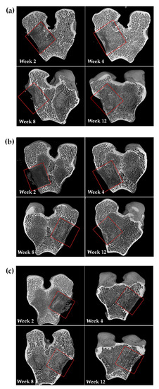
Figure 2.
Micro-computed tomographic (μ-CT images) showing the surgical area and the healing area of (a) blank, (b) control, and (c) α-CSH samples at each time point.
Reference
- Ou, K.-L.; Hou, P.-J.; Huang, B.-H.; Chou, H.-H.; Yang, T.-S.; Huang, C.-F.; Ueno, T. Bone Healing and Regeneration Potential in Rabbit Cortical Defects Using an Innovative Bioceramic Bone Graft Substitute. Appl. Sci. 2020, 10, 6239. [Google Scholar] [CrossRef]
Publisher’s Note: MDPI stays neutral with regard to jurisdictional claims in published maps and institutional affiliations. |
© 2022 by the authors. Licensee MDPI, Basel, Switzerland. This article is an open access article distributed under the terms and conditions of the Creative Commons Attribution (CC BY) license (https://creativecommons.org/licenses/by/4.0/).